Proceeding Paper

Development of Photothermal Membrane for Treatment of Infected Wound: A Proof-of-Concept †

by
Thi Tuong Vy Phan
1,2
1
Center for Advanced Chemistry, Institute of Research and Development, Duy Tan University, 03 Quang Trung, Hai Chau, Danang 550000, Vietnam
2
Faculty of Environmental and Chemical Engineering, Duy Tan University, 03 Quang Trung, Hai Chau, Danang 550000, Vietnam
Presented at the 1st International Electronic Conference on Biomedicine, 1–26 June 2021; Available online: https://ecb2021.sciforum.net.
Biol. Life Sci. Forum 2021, 7(1), 9; https://doi.org/10.3390/ECB2021-10279
Published: 31 May 2021
(This article belongs to the Proceedings of The 1st International Electronic Conference on Biomedicine)

Abstract

:
Wound infection is a serious issue because of multi-drug resistance bacteria, thus developing an advanced therapy is a needed demand. Photothermal therapy (PTT) is a novel noninvasive strategy that utilizes PTT agents to convert near-infrared (NIR) light energy into heat to kill living cells. In this work, we developed the PTT agent containing membrane to treat the wound infection for the first time. Palladium nanoparticles (PdNPs) were chosen as PTT agents owing to their high stability, good biocompatibility, excellent photothermal property, and simple-green preparation. Chitosan (CS) has been widely studied in tissue engineering due to its good properties such as biocompatible, biodegradable, antibacterial, and wound-healing abilities. However, the poor workability and high brittleness of CS limit the applications of CS in tissue engineering. Thus, we combined polyvinyl alcohol (PVA) and CS to have the membrane with high flexibility, wettability, highly porosity. The test on cells showed that the membrane has high biocompatibility. The combination of PdNPs loading CS/PVA membrane and laser irradiation killed most of the bacteria in vitro.

1. Introduction

Skin is the important protector of the body. When the skin is damaged, the microorganisms can easily grow in the damaged area [1]. The abuse of antibiotics caused the drug-resistant issue [1]. Thus, the development of wound dressings for the treatment of infected wounds is essential.
Photothermal therapy (PTT) is emerging as an effective therapy for the treatment of cancer and infection. The nanoparticles are usually used to assist PTT.
Herein, we have developed the novel photothermal responsive membrane as a wound dressing for the treatment of the infected wound. The porous bioscaffolds are similar to the extracellular matrix [2] and the pores of the membrane facilitate nutrient and oxygen diffusion, removing wastes, and promoting wound healing [3]. Palladium nanoparticles (PdNPs) were chosen as PTT agents owing to their high stability, good biocompatibility, excellent photothermal property, and simple-green preparation. Chitosan (CS) has been widely studied in tissue engineering due to its good properties such as biocompatible, biodegradable, antibacterial, and wound-healing abilities. However, the poor workability and high brittleness of CS limit the applications of CS in tissue engineering. Thus, we combined polyvinyl alcohol (PVA) and CS to have the membrane with high flexibility, wettability, highly porosity. The PTT in vitro experiment showed that the photothermal responsive membrane has the excellent antibacterial ability within a very short period.

2. Methods

2.1. Preparation of CS/PVA/Pd Dressing

The PdNPs were synthesized by our reported green method [4]. Porous CS/PVA and CS/PVA/Pd dressings were prepared by following the reported method with slight modification [5]. 3 wt.% CS solution in acetic acid 2% and 1.5% PVA solution were mixed. After acquiring the homogeneous solution, the 20 ppm PdNPs were added to the mixture. Then, the mixture was divided into the Petri dishes (40 mm × 12 mm) and keeping them in the fridge at −20 °C for 12 h. After that, the dressings were moved to NaOH 3 M solution to start the gelation process at −20 °C for 12 h. Next, the dressings were taken out of the fridge and washed two times with ethanol (70% and 100%, period: 15 min) and PBS (period: 15 min). Finally, the dressings were kept at room temperature (troom) for drying.

2.2. Evaluation of the Antibacterial Properties of CS/PVA/Pd Membrane

The dressing was directly put on the surface of the 1 × 10⁸ CFU/mL bacterial suspension on the 6-well plate. Thereafter, the NIR laser (power density of 1 W/cm2) was used to irradiate each well for 10 min. Then, the bacterial suspension was centrifuged and the pellets were collected. The pellets were re-suspended in 1 mL of liquid broth medium and stained with 10 μL AO (5 mg/mL) + PI (3 mg/mL) at 37 °C for 10 min. The bacterial suspension was again centrifuged at 5000× g for 7 min at 4 °C to collect stained bacteria. The centrifugation process was repeated 4 times to wash all unincorporated dyes. Finally, the stained bacterial suspension was put on the glass slide and covered with the coverslip and fluorescent images of bacteria were captured for further analysis.

3. Results and Discussion

3.1. Characterization of CS/PVA/Pd Dressing

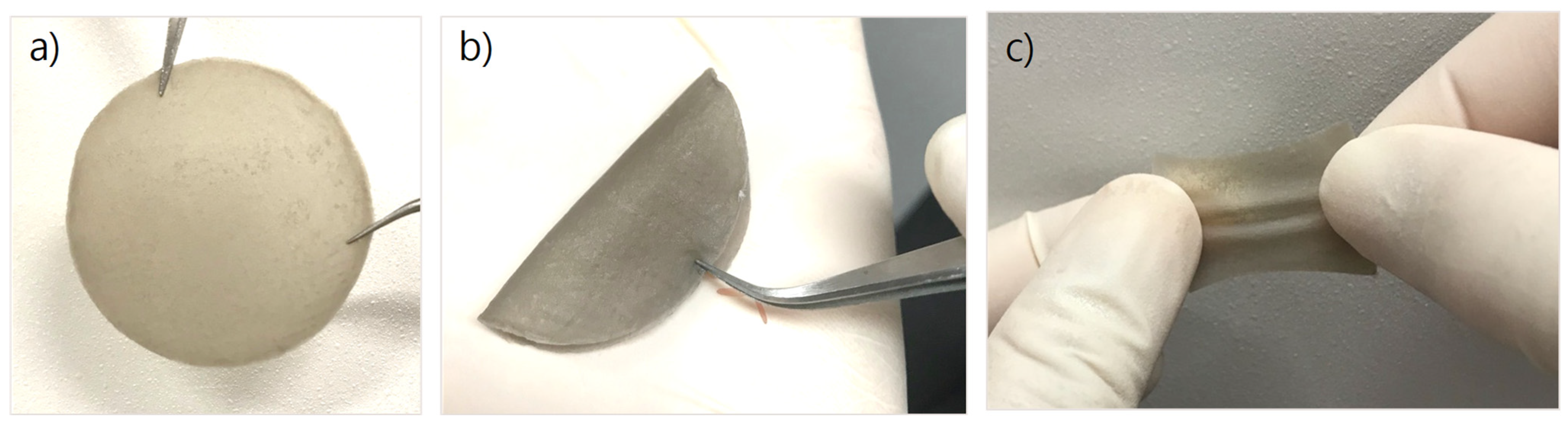
3.1.1. Physical Properties of CS/PVA/Pd Dressing

The synthesized membranes have good physical properties. They can be blended or stretched (Figure 1). It is suitable for wound dressing applications.

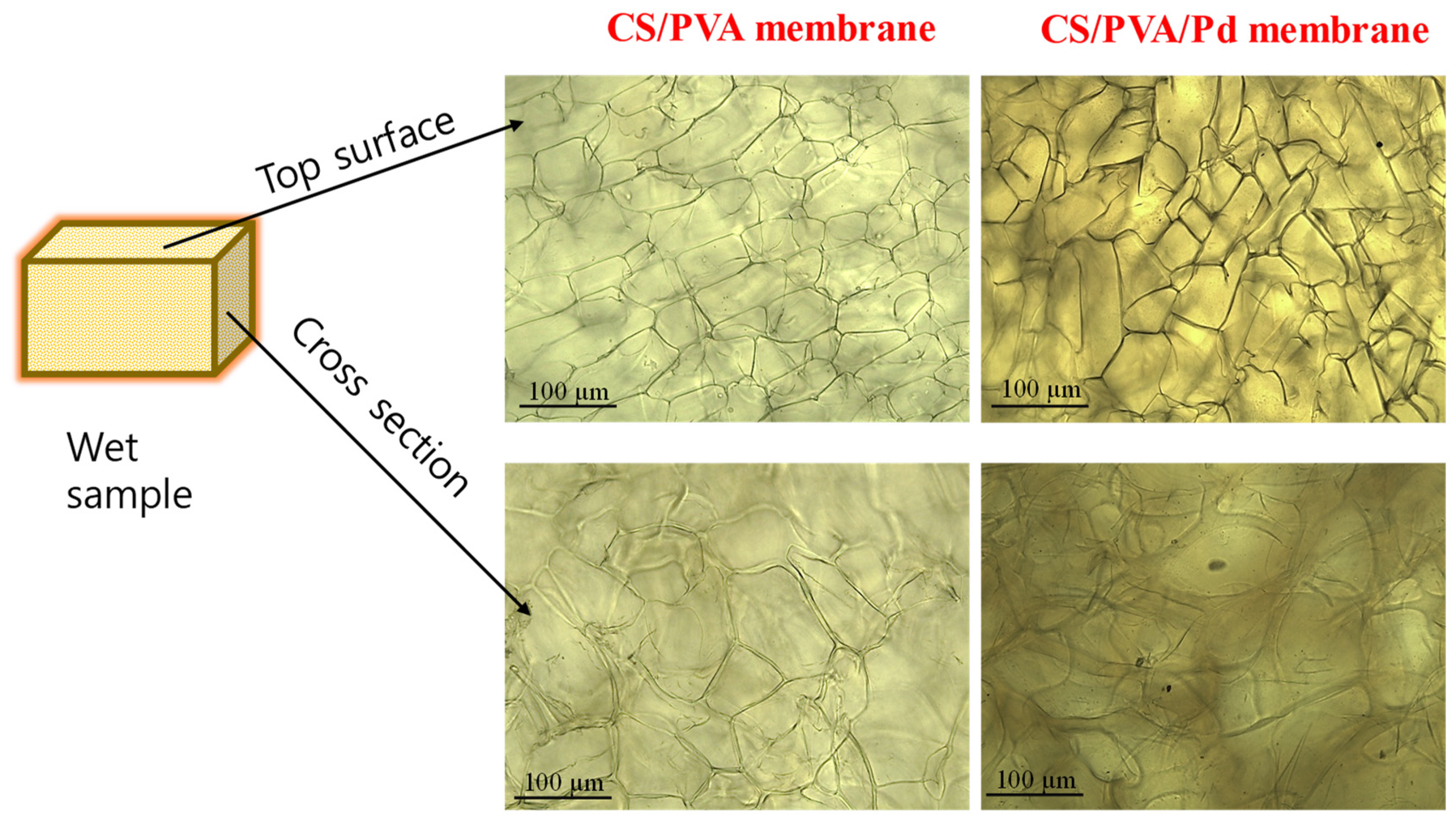
3.1.2. Surface and Morphology of CS/PVA/Pd Dressing

The bright-field images of the dressings were shown in Figure 2. The membrane has a high porosity and average pore diameters of about 80–100 µm. Not much different in the pore diameters between the two groups. That indicates that the PdNPs do not affect the porosity and pore size of the membrane. The High porosity and large surface area of porous membranes are similar to the extracellular matrix that facilitates vascularization and cell migration [3].

3.2. Anti-Bacterial Performance of CS/PVA/Pd Dressing

The live and dead assays were conducted to evaluate the in vitro PTT effect of CS/PVA and CS/PVA/Pd dressings. The membranes were directly put on the surface of the 1 × 10⁸ CFU/mL E. coli bacterial suspension on the 6-wells plate. Then, each well was continuously irradiated by 808 nm laser at 1 W/cm2 for 10 min. Then, the AO/PI staining was performed to evaluate the bacteria viability. As shown in Figure 3, most of the bacteria in the CS/PVA/Pd treated group was emitted red fluorescence, indicating all bacteria were dead. The results evidenced that CS/PVA/Pd dressing is effectively killing the bacteria when being exposed to NIR irradiation.

4. Conclusions

The CS/PVA/Pd membrane was successfully prepared by the gelation method. The in vitro experiment proved the excellent antibacterial ability of the prepared membrane. The photothermal responsive membrane is very promising for the treatment of an infected wound in the future.

Funding

This research received no external funding.

Institutional Review Board Statement

Not applicable.

Informed Consent Statement

Not applicable.

Data Availability Statement

Not applicable.

Conflicts of Interest

The authors declare no conflict of interest.

References

  1. Bowler, P.G.; Duerden, B.I.; Armstrong, D.G. Wound Microbiology and Associated Approaches to Wound Management. Clin. Microbiol. Rev. 2001, 14, 244–269. [Google Scholar] [CrossRef] [PubMed] [Green Version]
  2. Ou, K.-L.; Hosseinkhani, H. Development of 3D In Vitro Technology for Medical Applications. Int. J. Mol. Sci. 2014, 15, 17938–17962. [Google Scholar] [CrossRef] [PubMed] [Green Version]
  3. Loh, Q.L.; Choong, C. Three-Dimensional Scaffolds for Tissue Engineering Applications: Role of Porosity and Pore Size. Tissue Eng. Part B Rev. 2013, 19, 485–502. [Google Scholar] [CrossRef] [PubMed] [Green Version]
  4. Phan, T.T.V.; Hoang, G.; Nguyen, V.T.; Nguyen, T.P.; Kim, H.H.; Mondal, S.; Manivasagan, P.; Moorthy, M.S.; Lee, K.D.; Junghwan, O. Chitosan as a stabilizer and size-control agent for synthesis of porous flower-shaped palladium nanoparticles and their applications on photo-based therapies. Carbohydr. Polym. 2019, 205, 340–352. [Google Scholar] [CrossRef] [PubMed]
  5. Madihally, S.; Matthew, H. Porous chitosan scafolds for tissue engineering. Biomaterials 1999, 20, 1133–1142. [Google Scholar] [CrossRef]
Figure 1. (a) The CS/PVA/Pd membrane, (b) Blending the membrane, and (c) stretching the membrane.
Blsf 07 00009 g001
Figure 2. The bright-field images of the membrane under microscopy.
Blsf 07 00009 g002
Figure 3. Anti-bacterial performance of CS/PVA and CS/PVA/Pd membranes.
Blsf 07 00009 g003
Publisher’s Note: MDPI stays neutral with regard to jurisdictional claims in published maps and institutional affiliations.

