Communication

Interaction of Chicken Heterophils and Eimeria tenella Results in Different Phenotypes of Heterophil Extracellular Traps (HETs)

by
Zaida Rentería-Solís
1,2,*,†,
Liliana M. R. Silva
3,4,5,*,†,
Thomas Grochow
1,6,
Runhui Zhang
1,7,
Tran Nguyen-Ho-Bao
1,8,
Arwid Daugschies
1,2,
Anja Taubert
3,
Iván Conejeros
3 and
Carlos Hermosilla
3
1
Institute of Parasitology, Centre for Infection Medicine, Faculty of Veterinary Medicine, Leipzig University, An den Tierkliniken 35, 04103 Leipzig, Germany
2
Albrecht-Daniel-Thaer Institute, Rudolf-Breitscheid-Str. 38, 04463 Groesspoesna, Germany
3
Institute of Parasitology, Justus Liebig University Giessen, Schubertstr. 81, 35392 Giessen, Germany
4
Egas Moniz Center for Interdisciplinary Research (CiiEM), Egas Moniz School of Health & Science, Caparica, 2829-511 Almada, Portugal
5
MED—Mediterranean Institute for Agriculture, Environment and Development & CHANGE—Global Change and Sustainability Institute, Universidade de Évora, 7002-554 Évora, Portugal
6
Institute for Veterinary Anatomy, Faculty of Veterinary Medicine, Leipzig University, An den Tierkliniken 43, 04103 Leipzig, Germany
7
College of Animal and Veterinary Sciences, Southwest Minzu University, Chengdu 610041, China
8
Faculty of Veterinary Medicine, College of Agriculture, Can Tho University, Can Tho 900000, Vietnam
*
Authors to whom correspondence should be addressed.
These authors contributed equally to this work.
Poultry 2024, 3(3), 318-329; https://doi.org/10.3390/poultry3030024
Submission received: 23 May 2024 / Revised: 12 August 2024 / Accepted: 23 August 2024 / Published: 9 September 2024
(This article belongs to the Special Issue Current Research and Key Issues in Poultry Immunology)

Abstract

:
Chicken coccidiosis causes annual losses exceeding GBP 10 billion globally. The most pathogenic species for domestic fowls including Eimeria tenella, E. acervulina, and E. maxima, can lead to gastrointestinal issues ranging from mild to fatal. In this study, stages of E. tenella and freshly isolated chicken heterophils were co-cultured for 180 min. These interactions were analyzed using live 3D holotomographic and confocal microscopy. We observed that E. tenella stages were entrapped by heterophils and heterophil extracellular traps (HETs). Notably, different HET phenotypes, specifically sprHETs and aggHETs, were induced regardless of the stage. Furthermore, the quantification of extracellular DNA release from co-cultures of heterophils and sporozoites (ratio 1:1) for 180 min demonstrated a significantly higher release (p = 0.04) compared to negative controls. In conclusion, research on the chicken innate immune system, particularly fowl-derived HETs, remains limited. More detailed investigations are needed, such as exploring the time-dependent triggering of HETs, to establish a standard incubation time for this pathogen defense mechanism. This will enhance our understanding of its role in parasite survival or death during HET confrontation.

1. Introduction

Chicken coccidiosis in an economically important malady for the poultry industry worldwide, causing global annual losses of more than GBP 10 billion [1]. Coccidiosis in domestic fowls can produce mild to fatal gastrointestinal disorders caused by the apicomplexan monoxenous parasite genus Eimeria [2]. Eimeria spp. are monoxenic parasites with exogenous developmental stages. In naïve hosts, such as young chicks, one single sporulated Eimeria oocyst is sufficient to cause disease [3]. After mechanical and enzymatic activation within the gut lumen (i.e., CO2 concentration, bile and trypsin presence, and adequate temperatures), both the oocyst and sporocyst walls degrade. Consequently, released Eimeria sporozoites infect epithelial cells, mostly enterocytes. After active sporozoite host cell invasion, several merogonies (two to four) occur within a parasitophorous vacuole (PV) followed by sexual reproduction, known as gamogony, which culminates with the production of newly formed unsporulated oocysts. Unsporulated oocysts are shed within the feces and become sporulated in the presence of oxygen, humidity, and temperature in the environment within approximately 2 days [2].
Seven Eimeria species are mostly associated with chicken eimeriosis: E. acervulina, E. brunetti, E. maxima, E. mitis, E. necatrix, E. praecox, and E. tenella. Variations in pathogenicity, parasite location in the intestinal tract, lesions, and oocysts morphology can be found between species [4]. Additionally, three cryptic operational taxonomic units (OTUs) have been recently described in the southern hemisphere [5]. Eimeria spp. prevalence is higher in intensive farming systems than in free-range or backyard chickens [6]. Other factors that can increase the presence of Eimeria spp. are poor hygiene and husbandry, high flock density, and high humidity [7]. Followed by E. maxima and E. acervulina, E. tenella is considered the most common Eimeria species worldwide [6,7] with a global estimated prevalence of around 44% [8] E. tenella infects the caeca epithelium, causing mild to fatal hemorrhagic typhlitis with the erosion of the mucosa, weight loss, and low egg production. Additionally, E. tenella is one of the most pathogenic species and it is associated with high morbidity and mortality [9].
Eimeria spp. oocysts are resilient to environmental challenges; thus, chicken eimeriosis (coccidiosis) control has always been problematic [10] and a challenge to the international poultry industry [11]. Control measures consist mainly of husbandry management, diet supplements, vaccination, and anticoccidial drug therapy [10]. The misuse of the latter can lead to anti-coccidial resistance development [12], whereas vaccination with formulations of live wild type or attenuated parasites, when unevenly applied, could lead to clinical disease [10]. An alternative approach to the above-mentioned measures is to breed birds for increased natural genetic resistance [13] and increased vaccine response to coccidiosis, since there is evidence for relevant host genetic variation [11]. For instance, the Fayoumi chicken line is more resistant to Eimeria spp. infections than the Leghorn line [13].
Granulocytic phagocytes, as part of the host innate immune system, present several effector mechanisms to fight invasive microorganisms, including parasites [14]. In addition to the classical bactericidal and parasiticidal mechanisms, such as oxidative burst, degranulation, and phagocytosis, the release of extracellular traps (ETs) has also been added recently as a relevant microbicidal mechanism [14]. Granulocytes also modulate the development of an acquired immune response, which starts with the detection of molecules unique to invading pathogens, i.e., the so-called pathogen-associated molecular patterns (PAMPs) [15]. Therefore, both polymorphonuclear neutrophils (PMNs, in mammals) and heterophils (in avians and reptiles) are pivotal in initiating the host innate immune response, being the first cell type to arrive to the site of infection [15].
Extruded ETs consist of fine filaments composed of a DNA backbone decorated with histones, elastase, and other granule proteins such as myeloperoxidase (MPO), pentraxin, lactoferrin, cathepsin G, and calprotein, among others [16]. ETs are considered an evolutionary conserved defense mechanism and has been reported in a variety of animal taxa [16,17]. In mammals, ETosis was identified from different phagocytic cells (PMN, macrophages, monocytes, eosinophils, mast cells, and basophils) which are capable of extruding ETs in response to a variety of stimuli [16,17]. ETosis has been described to efficiently kill or entrap bacteria, fungi, and parasites, including Eimeria species [16]. Over the years, neutrophil extracellular traps (NETs) were intensively studied as an early host innate immune reaction, not only against protozoans, but also against large helminths [18]. Through all this previous research, different phenotypes of NETs have been discovered, as well as triggering mechanisms [16,19,20]. Currently, the following NET phenotypes have been described in the literature: diffuse (diffNETs), spread (sprNETs), aggregated (aggNETs), cell free-, and anchored NETs [16,21]. DiffNETs consist of an extracellular chromatin mesh with antimicrobial proteins and have a globular and compact form with a 15–20 µm diameter. AggNETs are agglomerations of a high number of neutrophils (size > 50 µm in diameter) that have underwent NETosis, while sprNETs are characterized as smooth, elongated, thin, and web-like, constituted by DNA and decorated by elastase and histones [14,16,21]. However, ETs have been scarcely studied in the avian counterpart of neutrophils, namely heterophils [14,22,23,24,25]. ETs were described for the first time in chicken-derived heterophils (HETs) over a decade ago [14]. Different stimuli have been reported to induce HET formation, e.g., phorbol-12-myristat-13-acetate (PMA), hydrogen peroxide (H2O2) [14], Salmonella [22], mycotoxins [23,25], and more recently E. tenella sporozoites [24].
The objective of this study was to analyze in vitro E. tenella interactions with chicken heterophils and to assess the possible induction of different HETs phenotypes, as reported for mammalian PMN [diffused HETs (diffHETs), aggregated HETs (aggHETs), spread HETs (sprHETs)], after exposure to the first E. tenella parasitic stages (i.e., oocysts, sporocysts, sporozoites), to be encountered by intestinal heterophils in vivo after oral infection. Extracellular DNA quantification was conducted after the stimulation of avian heterophils with vital E. tenella sporozoites. Finally, live cell 3D-holotomographic microscopy was performed not only to unveil but also to better understand early avian heterophil–parasite interactions.

2. Materials and Methods

2.1. Parasite Maintenance and Excystation

Eimeria tenella, Houghton strain [kindly provided by Prof. Damer Blake, Royal Veterinary College (RVC), University of London, London, UK], was used for this study. Sporulated oocysts were passaged in 10-day old chicks and purified from the feces according to a modified method [2]. E. tenella sporulated oocysts were kept in 4% potassium dichromate at 4 °C until further use. Sporozoite excystation and purification was conducted according to the method of Rentería-Solís et al. [26].

2.2. Purification of Poultry Heterophils

The isolation of chicken heterophils was performed following published methods [14,27], with some modifications. Briefly, peripheral blood was collected in 10% EDTA from the wing vein (Vena cutanea ulnaris) of adult healthy chickens (n = 3). One volume of blood was diluted with one volume of 1% methylcellulose, viscosity: 25cP (Sigma-Aldrich, Steinheim am Albuch, Germany). The mix was centrifuged at 20× g for 15 min. After centrifugation, the upper clear layer was transferred to a separate tube. The erythrocyte layer was then washed twice with HBSS without calcium, magnesium, and phenol red (all Thermo Fisher Scientific, Dreieich, Germany), and enriched with 0.01% fetal bovine serum (HBSS-S). After each wash, the upper clear layer was collected. Subsequently, all washes were centrifuged together at 370× g for 10 min and the pellet was re-suspended in 2 mL of HBSS-S. For heterophil separation, the suspended pellet was carefully placed over a Ficoll-Hypaque® (Sigma-Aldrich, Steinheim am Albuch, Germany) double gradient (specific gravity of 1.077 over 1.117g/mL) and centrifuged at 500× g for 30 min without break function. After centrifugation, the 1.077/1.119 g/mL and 1.119 g/mL layers were taken gently, avoiding contamination with the bottom layer (erythrocytes). Thereafter, the layers were washed two times with HBSS-S and centrifuged at 370× g for 5 min. Finally, heterophils were resuspended in HBSS-S, counted, and kept at 41 °C until further use.

2.3. Live Cell Imaging of Eimeria tenella Sporozoite-Heterophil Interactions Using 3D-Holotomographic Microscopy

Freshly excysted E. tenella sporozoites were seeded in an Ibidi® plastic cell plate (35 mm low imaging dish) inside a top stage incubation chamber (Ibidi®, Gräfelfing, Germany; 41 °C, 5% CO2) and allowed to settle for 10–15 min. Freshly isolated heterophils (2 × 10⁵ cells) were suspended in an imaging medium [0.1% BSA (Sigma-Aldrich), 2 μM 1,5-bis[2-(di-methylamino) ethyl]amino-4,8-dihydroxyanthracene-9,10-dione (DRAQ5) (Thermo Fisher Scientific), and 0.5 μM SytoxTM Green (Life Technologies, Darmstadt, Germany)], and added to the plastic cell plate. Over 180 min, cell interactions were followed using a live 3D Cell Explorer-fluo® (Nanolive, Tolochenaz, Switzerland) (60× magnification and depth of field 30 μm) every 30 s to explore instant live cell interactions. Images were analyzed using STEVE software v.2.6 (Nanolive) to obtain a refractive index (RI)-based z-stack. Images were also digitally stained based on the cell physical RI using STEVE Software v.2.6 (Nanolive). Image processing was performed with Fiji ImageJ v.1.7 using Z-projection, being restricted to an overall adjustment of brightness and contrast.

2.4. Characterization of Eimeria tenella-Triggered HET Formation

The in vitro confrontation of chicken heterophils with E. tenella sporozoites was conducted following previous protocols, with some modifications [14]. Coverslips (14 mm diameter) (Thermo Fisher Scientific) were coated with poly-L-lysine (0.01%) (Sigma-Aldrich, Steinheim am Albuch, Germany) and placed into 24 well-plates (Greiner Bio-one, Kremsmünster, Austria). Chicken heterophils (n = 3) (2.5 × 10⁵/well) and sporozoites were placed on the coverslips at a 1:1 (heterophil–sporozoite) infection ratio, and incubated for 3 h in RPMI-1640 medium (Thermo Fisher Scientific) enriched with 5% fetal bovine serum (FBS) (Gibco, Schwerte, Germany), 5% chicken serum, penicillin (100 U/mL), streptomycin (0.1 mg/mL), and amphotericin B (0.0025 mg/mL) (all Sigma-Aldrich, Steinheim am Albuch, Germany). Incubation conditions were 41 °C and 5% CO2 in a humidified environment. After incubation, cells were fixed with 4% paraformaldehyde (Merck, Darmstadt, Germany) for 10 min.
The visualization of HETs was achieved by the staining of DNA using 4′,6-diaminide-2′-phenylindole (DAPI, Thermo Fisher Scientific) and HETs-specific structures: histone (anti-histone H2B antibody, 1:500, AB52484, Abcam, Berlin, Germany) and heterophil elastase (anti-elastase antibody, 1:500, AB21593, Abcam). For this, fixed samples were washed thrice with 1X PBS and incubated for 10 min with a permeabilization solution (0.1% Triton X-100 in 1X PBS) at room temperature (RT). After washing with 1X PBS (3×, 5 min each), the samples were blocked for 30 min with 10% of goat serum (Abcam) in 1X PBS with 0.1% Tween 20 (PBST). HETs were then washed again with 1X PBS (3×) and incubated for 60 min at RT in a humidified chamber with primary antibodies in PBST with 1% of goat serum. Secondary antibodies (goat anti-mouse IgG antibody with Alexa fluor® 488 conjugate, 1:500, AB150113, Abcam, and goat anti-rabbit IgG with DyLight 594 conjugate, 1:1000, Invitrogen, Darmstadt, Germany) were diluted in PBST with 1% goat serum and incubated with the samples for 1 h at RT in a dark chamber. Afterwards, cells were washed thrice with 1X PBS (5 min each time) and mounted in DAPI-containing anti-fade mountant (ProLong™ Gold Antifade Mountant, Thermo Fisher Scientific). The determination of HETs was conducted using confocal laser scanning microscopy (Leica TCS SP8). The identification of different types of HET phenotypes was conducted following previously published morphological descriptions [19,20,21]. Briefly, aggregated NETs (aggNETs) are roundish clusters of HETs structures with a diameter no larger than 20 µm; diffuse HETs (diffHETs) are characterized by a decondensed chromatin mesh morphology between 15 to 20 µm in size; and spread HETs (sprHETs) are smooth, thin, and elongated with a 15–17 µm diameter. The fluorescence range of emission definition, co-localization, and sequential acquisition of stacks (every 0.3 µm) was performed using the Leica Application Suite X Software version 3.5.5 (Leica, Wetzlar, Germany). Imaris® Software version 9.7 (Bitplane, Abingdon, UK) was used to generate a 3D model.

2.5. Quantification of Extracellular Heterophil DNA

The quantification of extracellular heterophil DNA was conducted as previously described [14]. Briefly, poultry heterophils (2.5 × 10⁵ cells) were confronted with different concentrations of E. tenella sporozoites (1:1, 1:2 and 1:3, heterophil–sporozoite ratio) and incubated in RPMI-1640 medium, enriched as described above for 3 h at 41 °C. Positive controls consisted of zymosan (Sigma-Aldrich) (1 mg/mL). Additionally, non-stimulated heterophils were used as negative controls. After incubation, HETs were disrupted with micrococcal nuclease (New England Biolabs, Frankfurt am Main, Germany) (0.1 U/µL) and incubated for 15 min at 37 °C. Consequently, the samples were centrifuged, and the supernatant was placed in 96-well plates in triplicates (100 µL of supernatant/triplicate), and 50 µL of PicoGreen (Quant-iT™ PicoGreen™ dsDNA reagent, Invitrogen) was added to each triplicate. HETs were quantified with a hybrid multi-mode microplate reader (Synergy H1™, BioTek, Agilent, Waldbronn, Germany).

2.6. Statistical Analysis

A one-way analysis of variance (ANOVA) and Tukey’s multiple comparison tests were performed to analyze the difference between groups. All tests were performed using GraphPad Prism version 8.4.3 (San Diego, CA, USA). The significance level was set at p < 0.05.

3. Results

3.1. Visualization of Eimeria tenella–Heterophils Interactions and HET Formation

The live cell imaging of the interactions between freshly isolated chicken heterophils (2 × 10⁵ cells) and vital E. tenella sporozoites were recorded for 3 h with a live 3D Cell Explorer-fluo® (Nanolive). At the end of the experiment, some heterophils were still viable (Figure 1) and entrapping E. tenella sporozoites. Sporozoites were observed frequently entrapped by heterophils by their basal end (Figure 1) and not by the apical end, indicating that this interaction was not a result of an attempt of active sporozoite invasion. Moreover, the sporozoite membrane (Figure 1) was not visible at its basal end, confirming a firm attachment of the heterophil to the sporozoite, also visible in the 3D rendering image (Figure 1).
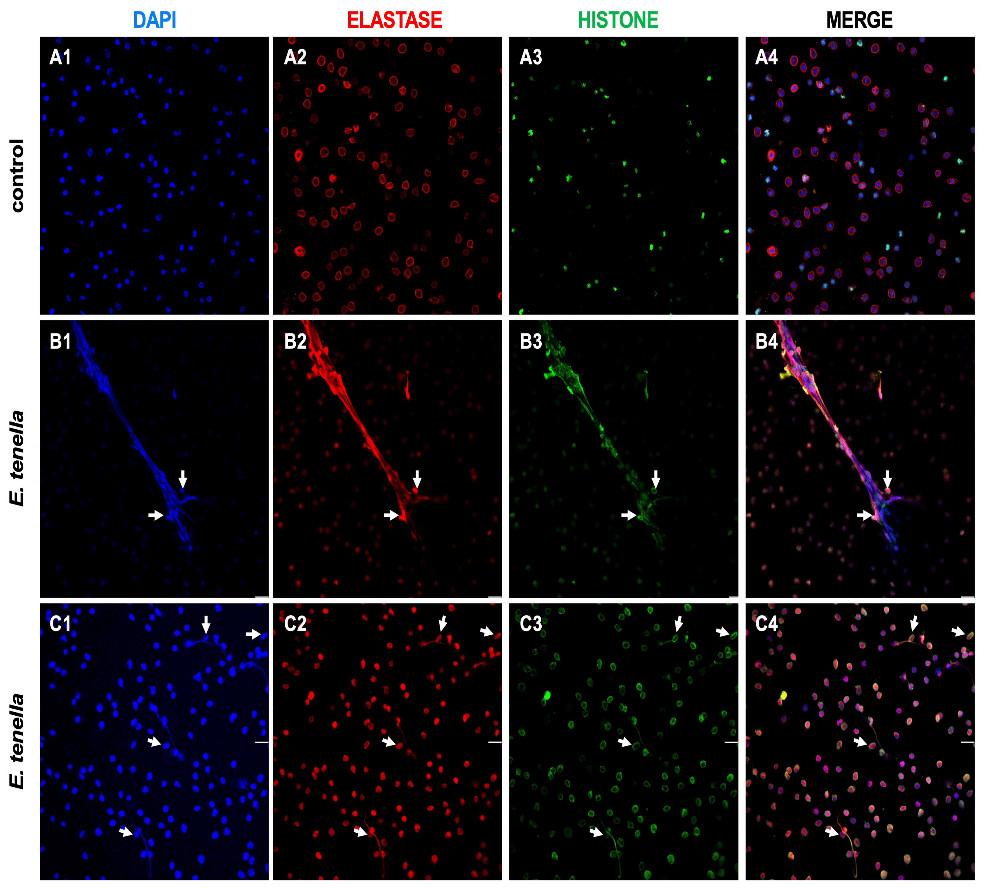
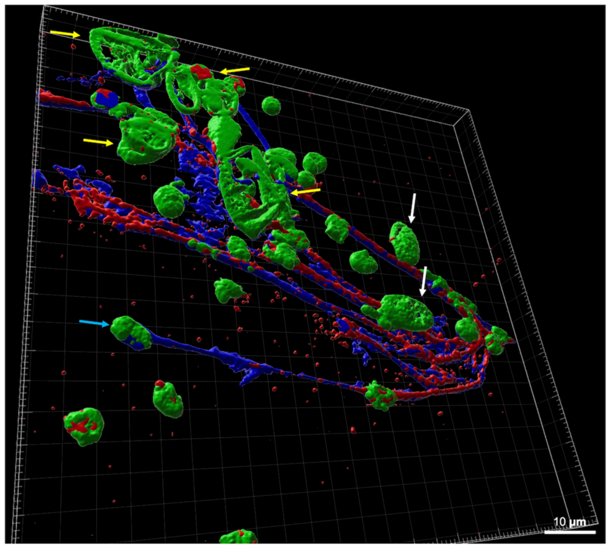
To further characterize interactions between chicken heterophils and E. tenella sporozoites, an immunofluorescence analysis was performed with confocal microscopy. HET-like structures were observed after 3 h of incubation of freshly isolated heterophils exposed to vital E. tenella sporozoites. The immunofluorescence imaging revealed a DNA backbone (Figure 2(B1,C1)), co-localized with elastase (Figure 2(B2,C2)) and histone (Figure 2(B3,C3)) as structural components of the released HETs. E. tenella sporozoites were entrapped within HETs structures (Figure 2), which presented two different phenotypes: spread HETs (sprHETs) and aggregated HETs (aggHETs). E. tenella-induced aggHETs were observed as an agglomerate of several heterophils (Figure 2(B1–B4)) that underwent HETosis entrapping sporozoites, while sprHETs were characterized by delicate filigree filaments (Figure 2(C1–C4)) also capable of entrapping sporozoites. Given that during the excystation process in the intestine in vivo, a mixture of E. tenella stages can be present (i.e., oocysts, sporocysts and sporozoites), co-cultures of E. tenella stages and chicken heterophils were also performed (Figure 3), showing that HETosis was a stage-independent process. After the 3D reconstruction of the confocal microscopy immunofluorescence analysis, sprHETs constituted by DNA filaments (in blue), decorated by elastase granules and histone granules, were observed entrapping oocysts, sporocysts, and sporozoites. Interestingly, several sprHETs seemed to originate from aggHETs, as previously observed (Figure 2(B1–B4)).
Additionally, the percentage of cells undergoing HETosis, at 1:1 ratio (heterophils–sporozoites) was calculated, considering more than 100 cells per animals (n = 4), by the observation of confocal microscopy images. In total, 6.14% ± 1.35% (mean ± standard deviation) of the heterophils stimulated with E. tenella sporozoites were releasing HETs, while no events were observed in non-stimulated heterophils.

3.2. Quantification of Eimeria tenella-Triggered HETs

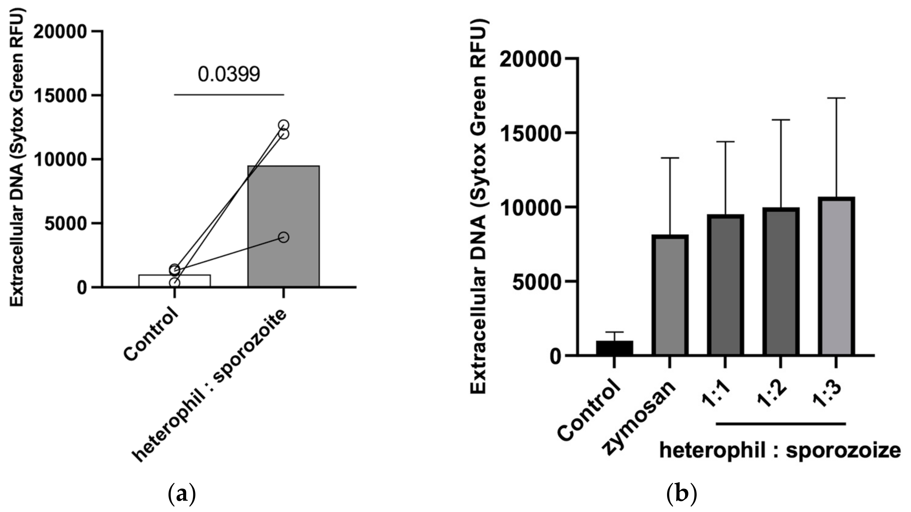
The HETosis quantification after E. tenella sporozoite stimulation was further confirmed with the fluorescence quantification of extracellular DNA. Confrontations of heterophils with E. tenella sporozoites significantly increased the fluorescence intensity in comparison to the negative control (p = 0.04) (Figure 4). However, high donor variation was observed. While heterophils of two donors clearly extruded a high amount of DNA after sporozoite stimulation, being considered high responders, the third donor was revealed to be a low responder since the amount of extracellular DNA detected after the sporozoite co-culture was much lower than the amount observed for the other donors.
Furthermore, HET-derived DNA release was not dependent on the sporozoite–heterophil ratio. Additionally, zymosan stimulations were used as positive controls and seemed to work in the chicken innate immune system (Figure 4b).

4. Discussion

In this work, we studied early interactions between freshly isolated E. tenella sporozoites and chicken-derived heterophils in vitro. Similarly to their mammalian counterpart, the PMN, avian heterophils are able to rapidly migrate to the site of infection, localize, remove, and kill microbial pathogens [28] by applying several effector mechanisms, such as oxidative burst, which generate reactive oxygen species (ROS) [29], degranulation, and phagocytosis to fight pathogens [28].
The capability of chicken heterophils to extrude HETs was reported for the first time by Chuammitri et al. [14] more than ten years ago. Chicken heterophils stimulated with PMA (1 µg/mL) or H2O2 (0.15 mM) successfully triggered chicken HETosis, and H2O2 induction was significantly higher than PMA induction. Since then, some studies reported on chicken-derived HET formation, both in vitro and in vivo, in response to bacteria [22], rare metals [30], mycotoxins [25], and parasites [24]. In the same way, over the last years many reports have focused on the ability of diverse immunocompetent leukocytes (i.e., PMN, monocytes, eosinophils, macrophages, hemocytes, mast cells, and basophils) of different hosts (e.g., humans, cattle, sheep, goats, water buffalo, gastropods, dogs, dolphins, seals, murine, cockroaches, fish) to cast ETs in response to several pathogens [31]. Moreover, highly motile nematode and trematode stages also trigger strong NETosis not only in vitro but also in vivo [16]. Some helminths [32,33] have been reported to be firmly entrapped by ET structures. A broad range of protozoan species can trigger ETosis as well, e.g., Toxoplasma gondii [34], Leishmania spp. [35], and different Eimeria species [16]. The induction of ruminant Eimeria-triggered NETosis is based on the key role of NOX (NADPH oxidase), NE (neutrophil elastase), MPO, CD11b, ERK1/2, and p38 MAP kinase signaling pathways, Ca2+ influx, and TLR 2 and TLR4 expression [16].
Similar to ruminant Eimeria species, the first report of E. tenella-induced HETosis focused on the different signaling pathways involved in HET release [24]. The authors described that E. tenella-triggered HETosis was dependent on NADPH, p38, and Rac1 signaling pathways. In the present work, detailed morphological studies of early parasite–heterophil interactions via a live cell 3D holotomographic microscopy analysis showed chicken heterophils being able to entrap E. tenella sporozoites by their basal end while maintaining their typical spherical morphology (Figure 1, white arrows). Contrary to what was observed here, E. bovis sporozoites were also capable of actively invading bovine PMN but were then efficiently trapped inside PMN [16]. The fact that chicken heterophils are apt to trap Eimeria sporozoites by the basal end, could be associated with a form that prevents active heterophil invasion and avoids contact with the sporozoite protein complex, composed by microneme- and rhoptry-derived molecules, which are mainly secreted during the active host cell invasion of sporozoites via secretion through their apical ends [36,37]. At 3 h of co-culture, heterophils confining E. tenella sporozoites were still viable (negative to Sytox™ Green staining), showing that this close interaction seems to occur without the death of exposed heterophils, in contrast to what occurs during suicidal HETosis. Furthermore, immunofluorescence studies via confocal microscopy analyses after the exposure of chicken heterophils with freshly isolated E. tenella sporozoites allowed for the morphological characterization of E. tenella-induced HET formation. Here, for the first time, different phenotypes of chicken HETs were described, i.e., both aggHETs and sprHETs entrapping E. tenella sporozoites. Over the last years, different forms of ETs have been described, such as vital or suicidal ETosis, and several morphological phenotypes have also been described: diffuse (diffNETs), spread (sprNETs), aggregated (aggNETs), cell-free, and anchored NETs [19,20]. Most of the studies referring to different ET phenotypes have been performed with neutrophils, the mammalian counter part of heterophils. Here, aggHETs were identified as agglomerations of a high number of heterophils (size > 50 µm in diameter) that underwent HETosis after marked attraction and activation, fitting well with previous reports on parasite-triggered aggNETs [19,20]. E. tenella sporozoites were observed to be entrapped in aggHETs, i.e., thicker fibers constituted by DNA and decorated by elastase and histones; however, a low percentage was observed. AggNETs have been suggested to promote the resolution of neutrophilic inflammation by degrading cytokines and chemokines via serine proteases and disrupting neutrophil recruitment and activation [19], and to further limit inflammation via the sequestration and detoxification of histones [38], whilst the other type of HETs identified here, the sprHETs, were associated with proinflammation at the early phase of the innate immune response [39]. SprHETs are characterized by smooth and elongated extremely thin web-like structures [20]. Although the majority of sprHETs induced by E. tenella sporozoites were short, several sporozoites were entrapped by the filaments. Nevertheless, longer sprNETs casted by neutrophils of different mammals were induced not only by larger and highly motile nematodes but also by other ruminant Eimeria species [16]. Interestingly, no other types of HETs were detected after E. tenella stages confrontation. Therefore, it remains to be elucidated if poultry heterophils are unable to present other HET phenotypes or if E. tenella stages simply do not induce such types of HETosis. Similarly, Branzk et al. [32] described that mammalian neutrophils can sense the pathogen size and selectively react through one effector mechanism, such as ETs, with a detriment to phagocytosis or degranulation. The same mechanism could be related to the ability of heterophils to select which ET phenotype to cast. Another hypothesis might be that granulocytes are driven by the motility of certain pathogens.
After the oral uptake of E. tenella sporulated oocysts, excystation takes place within the gut lumen and at this moment, oocysts, sporocysts, and sporozoites are simultaneously present. Since heterophils migrate from the mucosal epithelium to the intestinal lumen of chickens after experimental infection with Salmonella enteriditis [40], it is likely to assume that they might interact with other pathogens as well. Hence, after the co-culture of all three E. tenella stages (ooycysts, sporocysts, sporozoites) with heterophils, all of them were found entrapped in HETs, supporting E. tenella-induced HETosis as a stage-independent process. For this analysis, a 3D model of the HET-trapping parasitic stages was used. DNA-backbone filaments decorated with proteins such as elastase and histones clearly entrapped sporozoites, sporocysts, and oocysts within the same structure. Accordingly, such “decoration” was reproduced by the applied 3D model from this study, where the granular structure of elastases and histones became visible on extruded HETs. Clear visible variations in the concentration of such enzymes and the thickness of the filaments could even be related to some anti-parasitic effects or pro-/anti-inflammatory responses. For this reason, 3D models derived from the immunofluorescence analyses could represent a helpful tool to further elucidate HET phenotypes and the mechanical pathogen entrapment process. Remarkably, it seems that all observed sprHETs have an origin in the same aggHET formation, even though no remaining intact heterophils were present. Some stages were “grabbed” by sprHET filaments. This is particularly important, since pathogen entanglement is a main consequence of ET release. More likely than killing parasites, entrapment appears to be the most common mechanism used by granulocytes of different hosts in response to distinct parasites, being much larger and motile than bacterial, viral, and/or fungi pathogens [16,41].
Extracellular DNA quantification was determined at the ratios of 1:1, 1:2, and 1:3, and only the 1:1 ratio was significantly higher than non-exposed controls. Although two of the heterophil donors responded strongly to E. tenella sporozoite confrontation, with more than a six-fold amount than the control, the third animal only presented increases in the order of two to three-fold. Therefore, a clear increase in the amount of extracellular DNA released after the co-culture with E. tenella sporozoites was noted, and no significant differences were appreciated. In a previous work, lower parasite ratios were used to quantify extracellular DNA release. The authors showed that a significant increase in HETosis was only observed with a 1:1 ratio [24]. Comparing the two studies, the incubation time used in this study (180 min) was longer than the one used by Wei et al. [24] (120 min), which could justify the differences found. Chuammitri et al. [14] reported 180 min as the optimal incubation time for HET production based on their preliminary studies. However, these data have not been published. Nonetheless, it has been shown that apicomplexan-derived NETs can increase over time [42].
Altogether, our results show that E. tenella stages are able to trigger the formation of different phenotypes of extracellular traps in avian hosts, just as their mammalian counterparts. Fowl-derived HET research is still in its infancy, and further detailed investigations are needed, as reported for mammalians. So far, only heterophils have been tested for their ability to extrude HETs. Several other points should be considered in future studies, such as the time-dependent triggering of HETs, in order to stablish a standard incubation time for chicken innate immune systems.

Author Contributions

Conceptualization, Z.R.-S., L.M.R.S., I.C. and C.H.; methodology, Z.R.-S., L.M.R.S., T.G., R.Z. and I.C.; formal analysis, Z.R.-S., L.M.R.S., T.G., R.Z. and I.C.; investigation, Z.R.-S., L.M.R.S., T.G., R.Z., T.N.-H.-B. and I.C.; resources, Z.R.-S., L.M.R.S., I.C., A.D. and C.H.; data curation, Z.R.-S., L.M.R.S. and I.C.; writing—original draft preparation, Z.R.-S. and L.M.R.S.; writing—review and editing, Z.R.-S., L.M.R.S., I.C., A.D., A.T. and C.H.; visualization, Z.R.-S., L.M.R.S., T.G. and I.C.; supervision, Z.R.-S., L.M.R.S., I.C. and C.H.; project administration, Z.R.-S., L.M.R.S., I.C. and C.H.; funding acquisition, Z.R.-S., L.M.R.S. and C.H. All authors have read and agreed to the published version of the manuscript.

Funding

This research was partially funded by the Leipzig University Flexible Funds Program for Junior Researchers (Project no. 43700871 for Z.R.-S.). Article processing charges were covered thanks to the support of the Open-Access Publishing Fund Program from Leipzig University. The funding institutions had no role in this study.

Institutional Review Board Statement

All procedures performed involving live animals were in accordance with the ethical standards of the local authorities (Landesdirektion Sachsen, permits no. A04/19: parasite maintenance; V04/14: blood sample collection), and in accordance with prevalent European Animal Welfare Legislation (ART13TFEU) and current applicable German Animal Protection Laws.

Informed Consent Statement

Not applicable.

Data Availability Statement

The data that support the findings of this study are available from the corresponding author, [Z.R.-S.], upon reasonable request.

Acknowledgments

The authors are grateful to R. Schumacher and S. Fritsche (Institute of Parasitology, Faculty of Veterinary Medicine, Leipzig University) for their excellent work as animal keepers. Many thanks also to L. Flores (Clinic for Ruminants, Faculty of Veterinary Medicine, Leipzig University) for his help during heterophil purification. We are in debt to R. Bergmann (Institute of Bacteriology and Mycology, Faculty of Veterinary Medicine, Leipzig University), J. Kacza (Saxon Incubator for Clinical Translation; Leipzig University), and the Bioimaging Core Unit Facility, Leipzig University for facilitating access to equipment. We are also thankful to R. Schmäschke (Institute of Parasitology, Faculty of Veterinary Medicine, Leipzig University) for his valuable work during the preparation of animal permits.

Conflicts of Interest

The authors declare no conflicts of interest.

References

  1. Blake, D.P.; Knox, J.; Dehaeck, B.; Huntington, B.; Rathinam, T.; Ravipati, V.; Ayoade, S.; Gilbert, W.; Adebambo, A.O.; Jatau, I.D.; et al. Re-Calculating the Cost of Coccidiosis in Chickens. Vet. Res. 2020, 51, 115. [Google Scholar] [CrossRef]
  2. Shirley, M.W.; Smith, A.L.; Tomley, F.M. The Biology of Avian Eimeria with an Emphasis on Their Control by Vaccination. In Advances in Parasitology; Elsevier: Amsterdam, The Netherlands, 2005; Volume 60, pp. 285–330. ISBN 978-0-12-031760-8. [Google Scholar]
  3. Deplazes, P.; Eckert, J.; Mathis, A.; von Samson-Himmelstjerna, G.; Zahner, H. Parasitology in Veterinary Medicine; Wageningen Academic Publishers: Wageningen, The Netherlands, 2016. [Google Scholar]
  4. López-Osorio, S.; Chaparro-Gutiérrez, J.J.; Gómez-Osorio, L.M. Overview of Poultry Eimeria Life Cycle and Host-Parasite Interactions. Front. Vet. Sci. 2020, 7, 384. [Google Scholar] [CrossRef]
  5. Blake, D.P.; Vrba, V.; Xia, D.; Jatau, I.D.; Spiro, S.; Nolan, M.J.; Underwood, G.; Tomley, F.M. Genetic and Biological Characterisation of Three Cryptic Eimeria Operational Taxonomic Units That Infect Chickens (Gallus gallus Domesticus). Int. J. Parasitol. 2021, 51, 621–634. [Google Scholar] [CrossRef]
  6. Tadesse, C.; Feyissa, B.D. Poultry Coccidiosis: Prevalence and Associated Risk Factors in Extensive and Intensive Farming Systems in Jimma Town, Jimma, Ethiopia. J. Vet. Med. Anim. Health 2016, 8, 223–227. [Google Scholar] [CrossRef]
  7. Blake, D.P.; Marugan-Hernandez, V.; Tomley, F.M. Spotlight on Avian Pathology: Eimeria and the Disease Coccidiosis. Avian Pathol. 2021, 50, 209–213. [Google Scholar] [CrossRef]
  8. Badri, M.; Olfatifar, M.; Hayati, A.; Bijani, B.; Samimi, R.; Abdoli, A.; Nowak, O.; Diaz, D.; Eslahi, A.V. The Global Prevalence and Associated Risk Factors of Eimeria Infection in Domestic Chickens: A Systematic Review and Meta-analysis. Vet. Med. Sci. 2024, 10, e1469. [Google Scholar] [CrossRef]
  9. Macdonald, S.E.; Nolan, M.J.; Harman, K.; Boulton, K.; Hume, D.A.; Tomley, F.M.; Stabler, R.A.; Blake, D.P. Effects of Eimeria tenella Infection on Chicken Caecal Microbiome Diversity, Exploring Variation Associated with Severity of Pathology. PLoS ONE 2017, 12, e0184890. [Google Scholar] [CrossRef] [PubMed]
  10. Blake, D.P.; Tomley, F.M. Securing Poultry Production from the Ever-Present Eimeria Challenge. Trends Parasitol. 2014, 30, 12–19. [Google Scholar] [CrossRef]
  11. Boulton, K.; Nolan, M.J.; Wu, Z.; Riggio, V.; Matika, O.; Harman, K.; Hocking, P.M.; Bumstead, N.; Hesketh, P.; Archer, A.; et al. Dissecting the Genomic Architecture of Resistance to Eimeria Maxima Parasitism in the Chicken. Front. Genet. 2018, 9, 528. [Google Scholar] [CrossRef]
  12. Chapman, H.D. Biochemical, Genetic and Applied Aspects of Drug Resistance in Eimeria Parasites of the Fowl. Avian Pathol. 1997, 26, 221–244. [Google Scholar] [CrossRef] [PubMed]
  13. Heams, T.; Bed’hom, B.; Rebours, E.; Jaffrezic, F.; Pinard-van Der Laan, M.-H. Insights into Gene Expression Profiling of Natural Resistance to Coccidiosis in Contrasting Chicken Lines. BMC Proc. 2011, 5, S26. [Google Scholar] [CrossRef]
  14. Chuammitri, P.; Ostojić, J.; Andreasen, C.B.; Redmond, S.B.; Lamont, S.J.; Palić, D. Chicken Heterophil Extracellular Traps (HETs): Novel Defense Mechanism of Chicken Heterophils. Vet. Immunol. Immunopathol. 2009, 129, 126–131. [Google Scholar] [CrossRef]
  15. Swaggerty, C.L.; Ferro, P.J.; Pevzner, I.Y.; Kogut, M.H. Heterophils Are Associated with Resistance to Systemic Salmonella enteritidis Infections in Genetically Distinct Chicken Lines. FEMS Immunol. Med. Microbiol. 2005, 43, 149–154. [Google Scholar] [CrossRef]
  16. Silva, L.M.R.; Muñoz-Caro, T.; Burgos, R.A.; Hidalgo, M.A.; Taubert, A.; Hermosilla, C. Far beyond Phagocytosis: Phagocyte-Derived Extracellular Traps Act Efficiently against Protozoan Parasites In Vitro and In Vivo. Mediat. Inflamm. 2016, 2016, 1–13. [Google Scholar] [CrossRef]
  17. Ramos-Martínez, E.; Hernández-González, L.; Ramos-Martínez, I.; Pérez-Campos Mayoral, L.; López-Cortés, G.I.; Pérez-Campos, E.; Mayoral Andrade, G.; Hernández-Huerta, M.T.; José, M.V. Multiple Origins of Extracellular DNA Traps. Front. Immunol. 2021, 12, 621311. [Google Scholar] [CrossRef]
  18. Omar, M.; Abdelal, H. NETosis in Parasitic Infections: A Puzzle That Remains Unsolved. Int. J. Mol. Sci. 2023, 24, 8975. [Google Scholar] [CrossRef]
  19. Schauer, C.; Janko, C.; Munoz, L.E.; Zhao, Y.; Kienhöfer, D.; Frey, B.; Lell, M.; Manger, B.; Rech, J.; Naschberger, E.; et al. Aggregated Neutrophil Extracellular Traps Limit Inflammation by Degrading Cytokines and Chemokines. Nat. Med. 2014, 20, 511–517. [Google Scholar] [CrossRef]
  20. Salinas, C.; Barriga, K.; Albornoz, A.; Alarcon, P.; Quiroga, J.; Uberti, B.; Sarmiento, J.; Henriquez, C.; Ehrenfeld, P.; Burgos, R.A.; et al. Tamoxifen Triggers the In Vitro Release of Neutrophil Extracellular Traps in Healthy Horses. Front. Vet. Sci. 2023, 9, 1025249. [Google Scholar] [CrossRef]
  21. Muñoz-Caro, T.; Conejeros, I.; Zhou, E.; Pikhovych, A.; Gärtner, U.; Hermosilla, C.; Kulke, D.; Taubert, A. Dirofilaria Immitis Microfilariae and Third-Stage Larvae Induce Canine NETosis Resulting in Different Types of Neutrophil Extracellular Traps. Front. Immunol. 2018, 9, 968. [Google Scholar] [CrossRef]
  22. Pieper, J.; Locke, M.; Ruzaike, G.; Voigt, S.; Methner, U.; Berndt, A. In Vitro and in Vivo Generation of Heterophil Extracellular Traps after Salmonella Exposure. Vet. Immunol. Immunopathol. 2017, 188, 1–11. [Google Scholar] [CrossRef]
  23. Wu, D.; Li, S.; Li, P.; Jiang, A.; Liu, Z.; Zhang, Y.; Wang, J.; Yang, Z.; Wei, Z. Diacetoxyscirpenol-Induced Heterophil Extracellular Traps Contribute to the Immune Toxicity of Liver Injury in Chickens. Food Chem. Toxicol. 2021, 148, 111926. [Google Scholar] [CrossRef]
  24. Wei, Z.; Zhao, Y.; Zhang, N.; Han, Z.; Liu, X.; Jiang, A.; Zhang, Y.; Wang, C.; Gong, P.; Li, J.; et al. Eimeria tenella Induces the Release of Chicken Heterophil Extracellular Traps. Vet. Parasitol. 2019, 275, 108931. [Google Scholar] [CrossRef]
  25. Han, Z.; Zhang, Y.; Wang, C.; Liu, X.; Jiang, A.; Liu, Z.; Wang, J.; Yang, Z.; Wei, Z. Ochratoxin A-Triggered Chicken Heterophil Extracellular Traps Release through Reactive Oxygen Species Production Dependent on Activation of NADPH Oxidase, ERK, and P38 MAPK Signaling Pathways. J. Agric. Food Chem. 2019, 67, 11230–11235. [Google Scholar] [CrossRef]
  26. Rentería-Solís, Z.; Zhang, R.; Taha, S.; Daugschies, A. A Modified Method for Purification of Eimeria tenella Sporozoites. Parasitol. Res. 2020, 119, 1429–1432. [Google Scholar] [CrossRef]
  27. Andreasen, C.B.; Frank, D.E. The Effects of Ascorbic Acid on in Vitro Heterophil Function. Avian Dis. 1999, 43, 656–663. [Google Scholar] [CrossRef]
  28. Kogut, M.H.; Genovese, K.J.; Lowry, V.K. Differential Activation of Signal Transduction Pathways Mediating Phagocytosis, Oxidative Burst, and Degranulation by Chicken Heterophils in Response to Stimulation with Opsonized Salmonella enteritidis. Inflammation 2001, 25, 7–15. [Google Scholar] [CrossRef]
  29. He, H.; Farnell, M.B.; Kogut, M.H. Inflammatory Agonist Stimulation and Signal Pathway of Oxidative Burst in Neonatal Chicken Heterophils. Comp. Biochem. Physiol. Part A Mol. Integr. Physiol. 2003, 135, 177–184. [Google Scholar] [CrossRef]
  30. Jiang, A.; Zhang, Y.; Wu, D.; Li, S.; Liu, Z.; Yang, Z.; Wei, Z. Sodium Molybdate Induces Heterophil Extracellular Traps Formation in Chicken. Ecotoxicol. Environ. Saf. 2021, 210, 111886. [Google Scholar] [CrossRef]
  31. Nascimento, M.T.C.; Silva, K.P.; Garcia, M.C.F.; Medeiros, M.N.; Machado, E.A.; Nascimento, S.B.; Saraiva, E.M. DNA Extracellular Traps Are Part of the Immune Repertoire of Periplaneta americana. Dev. Comp. Immunol. 2018, 84, 62–70. [Google Scholar] [CrossRef]
  32. Branzk, N.; Lubojemska, A.; Hardison, S.E.; Wang, Q.; Gutierrez, M.G.; Brown, G.D.; Papayannopoulos, V. Neutrophils Sense Microbe Size and Selectively Release Neutrophil Extracellular Traps in Response to Large Pathogens. Nat. Immunol. 2014, 15, 1017–1025. [Google Scholar] [CrossRef]
  33. Ehrens, A.; Lenz, B.; Neumann, A.-L.; Giarrizzo, S.; Reichwald, J.J.; Frohberger, S.J.; Stamminger, W.; Buerfent, B.C.; Fercoq, F.; Martin, C.; et al. Microfilariae Trigger Eosinophil Extracellular DNA Traps in a Dectin-1-Dependent Manner. Cell Rep. 2021, 34, 108621. [Google Scholar] [CrossRef] [PubMed]
  34. Abi Abdallah, D.S.; Lin, C.; Ball, C.J.; King, M.R.; Duhamel, G.E.; Denkers, E.Y. Toxoplasma gondii Triggers Release of Human and Mouse Neutrophil Extracellular Traps. Infect. Immun. 2012, 80, 768–777. [Google Scholar] [CrossRef] [PubMed]
  35. Gabriel, C.; McMaster, W.R.; Girard, D.; Descoteaux, A. Leishmania donovani Promastigotes Evade the Antimicrobial Activity of Neutrophil Extracellular Traps. J. Immunol. 2010, 185, 4319–4327. [Google Scholar] [CrossRef]
  36. Morrissette, N.S.; Sibley, L.D. Cytoskeleton of Apicomplexan Parasites. Microbiol. Mol. Biol. Rev. 2002, 66, 21–38. [Google Scholar] [CrossRef]
  37. Striepen, B.; Jordan, C.N.; Reiff, S.; Van Dooren, G.G. Building the Perfect Parasite: Cell Division in Apicomplexa. PLoS Pathog. 2007, 3, e78. [Google Scholar] [CrossRef]
  38. Knopf, J.; Leppkes, M.; Schett, G.; Herrmann, M.; Muñoz, L.E. Aggregated NETs Sequester and Detoxify Extracellular Histones. Front. Immunol. 2019, 10, 2176. [Google Scholar] [CrossRef] [PubMed]
  39. Neumann, A.; Brogden, G.; Von Köckritz-Blickwede, M. Extracellular Traps: An Ancient Weapon of Multiple Kingdoms. Biology 2020, 9, 34. [Google Scholar] [CrossRef] [PubMed]
  40. Ševčíková, Z.; Asheg, A.A.; Kolodzieyski, L.; Cigánková, V.; Komorová, T.; Levkut, M. Heterophils and Macrophage-like Cells in the Caeca Chicks’ Caeca after Experimental Infection with Salmonella enteritidis PT4. Acta Vet. Brno 2003, 72, 565–570. [Google Scholar] [CrossRef]
  41. Rada, B. Neutrophil Extracellular Traps. In NADPH Oxidases; Knaus, U.G., Leto, T.L., Eds.; Methods in Molecular Biology; Springer: New York, NY, USA, 2019; Volume 1982, pp. 517–528. ISBN 978-1-4939-9423-6. [Google Scholar]
  42. Yildiz, K.; Gokpinar, S.; Gazyagci, A.N.; Babur, C.; Sursal, N.; Azkur, A.K. Role of NETs in the Difference in Host Susceptibility to Toxoplasma gondii between Sheep and Cattle. Vet. Immunol. Immunopathol. 2017, 189, 1–10. [Google Scholar] [CrossRef] [PubMed]
Figure 1. Live cell 3D-holotomographic microscopy unveiling interactions of chicken heterophils with E. tenella sporozoites. For 180 min, vital E. tenella sporozoites were co-cultured with freshly isolated heterophils. At the end of the experiment, some cells were still viable (green arrow), and some of them were entrapping sporozoites (zoom, 3D rendering, entrapment: black and white arrows). Yellow arrow: sporozoite membrane, which was not visible at the entrapment point (white and black arrows). Scale bars: 20 and 5 µm.
Poultry 03 00024 g001
Figure 2. Eimeria tenella-triggered different types of HETs. Confocal microscopy analysis of non-confronted heterophils ((A1A4) control panels) and heterophils confronted with E. tenella sporozoites for 180 min ((B1B4,C1C4) E. tenella panels). Structures were stained for DNA (DAPI, blue), elastase (red), and histones (green). Co-localization of the stains proves the nature of avian HETs. (B1B4) aggHETs with the involvement of multiple heterophils and the entrapment of E. tenella sporozoites (white arrows). (C1C4) Several short sprHETs are observed with ensnared E. tenella sporozoites (white arrows) in these thin web-like structures. Percentage of HETosis was 6.14% ± 1.35% (Mean ± standard deviation). Scale bar: 10 µm.
Poultry 03 00024 g002
Figure 3. Eimeria tenella-triggered HETosis is a stage-independent process. E. tenella stages were co-cultured for 180 min (1:1). Sporozoites (light blue arrows), sporocysts (white arrows), and oocysts (yellow arrows) were entrapped within HET-derived filaments formed by DNA backbone (blue) decorated with histones (green) and elastase (red) granules. A sporozoite (light blue arrow) was firmly grabbed by a fine DNA web-like structure. Scale bar: 10 µm.
Poultry 03 00024 g003
Figure 4. Extracellular DNA quantification. (a) Confrontation of heterophils and E. tenella sporozoites (1:1) conducted to a higher extracellular DNA release than non-stimulated heterophils (p = 0.0399). Individual variation is observed: two animals responded highly to the stimulus, while the third animal reacted mildly to the confrontation of E. tenella sporozoites. (b) Extracellular DNA quantification showed no dose dependency.
Poultry 03 00024 g004
Disclaimer/Publisher’s Note: The statements, opinions and data contained in all publications are solely those of the individual author(s) and contributor(s) and not of MDPI and/or the editor(s). MDPI and/or the editor(s) disclaim responsibility for any injury to people or property resulting from any ideas, methods, instructions or products referred to in the content.