


Abstract
Engineered brain models, including brain organoids and brain-on-a-chip systems, are generally assessed in terms of their physiological relevance. Although this language is useful for emphasizing the need to better approximate human biology, it can also obscure important differences among context of use, required validation strategy and ethical considerations. In this Opinion, we argue that physiological relevance should not be treated as a universal measure of model quality. Instead, its meaning should be defined relative to its application domain, including animal-model comparison, interpretation of single-cell atlases, clinical translation, donor representation, and emerging functional applications such as synthetic biological intelligence. For some applications, particularly patient-specific disease modeling and therapeutic screening, greater human physiological relevance may be required. For others, including biohybrid computing, controllable neural interfaces, interpretability, and ethical considerations may be more important. Moving beyond simplistic terminology will help improve scientific interpretation, prevent overstating findings, and support more responsible development of engineered brain models.
1. Introduction
It has never been easier to control in vitro and in vivo brain models, thanks to brain organoids, gene editing tools, and microfluidic platforms. The use of engineered brain models is a promising tool for studying fundamental neuroscience, reducing reliance on animal models, and serving as a potential platform for biocomputing [1]. However, vague definitions of physiological relevance hamper their applicability, with unique ethical and regulatory concerns. Alongside a shift away from restrictive views on these tools, a clearer categorization of physiological relevance within specific domains is urgently needed.
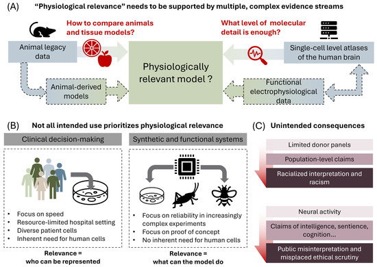
A common argument in drug discovery is that animal models incorrectly predict the efficacy of drug candidates in clinical trials, resulting in a low success rate. Conversely, organoid models and the microfluidic apparatus to maintain them are becoming increasingly sophisticated, accessible and better understood [1,2,3]. In this context, the term ‘physiological relevance’ has become ubiquitous as a quality label for organoid models, where a model is often perceived as superior when it closely mimics the function of a human tissue or organ. There are, however, very few tools to accurately compare the heterogeneous landscape of models; physiological relevance is an all-encompassing vague term with no standard measure of quantification. As single-cell datasets increasingly guide the design and interpretation of engineered brain models, they provide a powerful but incomplete template for defining this relevance. Their relationship to legacy animal data remains unclear, limiting how directly engineered models can be used to compare with, translate from, or reinterpret animal-derived findings (Figure 1A). Beyond this comparison, the value of physiological relevance also depends on the intended use of the model, with clinical applications constrained by donor representation and practical scalability, while functional biohybrid systems may depend more on controllability than on biological mimicry (Figure 1B).
Figure 1.
Physiological relevance as a context-dependent criterion for engineered brain models. (A) Animal legacy data, animal-derived models, human single-cell atlases and functional electrophysiological data provide distinct but incompletely connected reference points. Dashed paths indicate indirect or underexplored relationships. (B) The required physiological relevance depends on the intended application. Clinical decision-making requires representative donor cells and clinically useful timescales, whereas synthetic and functional systems may prioritize controllability and functional output. (C) Limited donor panels can lead to racialized interpretation, while conflating neural activity with intelligence, sentience or cognition can promote public misinterpretation and misplaced ethical scrutiny.
We therefore do not argue that pursuing some level of physiological relevance is unnecessary. Rather, this criterion is multidimensional and context-dependent, with a different meaning across applications. For many applications targeting fundamental biophysical processes, such as the effect of chemical [4], thermal [5], and physical stimulation [6] of individual neurons, or localized phenomena such as saltatory conduction due to myelination [7], the model is always necessarily simple as individual cells need to be accessible. In principle, findings are more easily comparable and limitations apparent. As 3D stem cell models become more adopted, their biophysical development is increasingly compared to the developing brain or nervous system of humans and animals [8,9]. Comparisons and interpretation of the findings, as well as their limitations, are often challenging. Nonetheless, for an increasing number of pharmaceutical and tissue engineering applications, these more complex models are preferred [1]. The use of terminology such as ‘physiological relevance’ is in those cases common but risks becoming meaningless or even counterproductive.
In the following sections, we first discuss how physiological relevance is difficult to define in practice when these engineered models are compared with animal data and single-cell atlases. In these cases, physiologically mimicking human biology might be more constrained by current technology than conceptually. We will then provide several examples of how clinical translation requiring diverse donors, public perception, and biohybrid applications demand partly different criteria (Figure 1C). For biohybrid applications in particular, several conceptual and ethical challenges arise that might render physiological relevance less useful, or even counterproductive, for the development of the technology. Engineered brain models are often valued as both simplified systems for isolating developmental or functional principles and as increasingly complex models intended to approximate human physiology. This dual role creates tension, where the features that make a model experimentally useful are not always the same features that make it more anatomically or physiologically complete.
2. Model Choice: Animal or Engineered
For decades, substantial public and private biomedical R&D investment has relied on mouse, primate, and other animal models. In most fields of neuroscience, researchers remain skeptical about the ability of brain organoids to replace animal models [10,11]. Animal models retain organism-level features that engineered brain models will lack, such as behavior. In addition, even more fundamental physiological processes will not be readily integrated into cellular or tissue models within the near future, such as vascular regulation, immune interactions, endocrine signaling, and long-range connectivity across (adult) brain regions. In contrast, animal models introduce species-specific processes during development, causing anatomical and ultimately pharmacological differences that can limit their direct relevance to human biology. Nonetheless, the direct use or modeling of human organs is prohibitive, such that practical considerations demand a compromise.
This compromise is not limited to a binary choice between animal models and human-derived tissue models. Several alternatives are available, such as large-mammal perfused models, human organotypic slices, or even computational approaches. For example, BrainEx restored microcirculation and selected cellular functions in isolated porcine brains several hours post-mortem, whereas OrganEx later extended related perfusion principles to the porcine whole body after an hour of warm ischaemia [12,13]. While this approach might allow for more intense use of animal brains or even their entire physiology, the throughput is prohibitively low for major pharmaceutical or even most fundamental research studies. In addition, many questions remain for this technology. Human organotypic slices can only be retrieved post-mortem or after specific surgery and are therefore not readily available. They can preserve native human cytoarchitecture, cellular diversity, and aspects of vascular structure [14]. Adult murine hippocampal vitrification studies further show that ex vivo preparations can preserve electrophysiological activity, synaptic transmission, and long-term potentiation, making functional recovery measurable outside the living animal [15]. While this could slightly improve the efficient use of animal models, cost, ethical and regulatory issues related to animal use largely remain.
In theory, computational approaches could provide the most controllable and well-characterized models. Their relevance depends on whether the biological constraints encoded in the model are appropriate for the question being asked. Large-scale endeavors such as the Human Brain Project, which fell short of their promises, have shown the considerable expenses needed to fully model every aspect of the brain from basic building blocks [16]. More recent biomimetic models make a narrower claim. For example, Pathak et al. modeled corticostriatal microassemblies across cellular, circuit, electrophysiological, and behavioral levels, and then compared the model with macaque data rather than treating the simulation as self-validating [17]. While these attempts can give some insight, they are still technologically challenging and not readily scalable for the pharmaceutical or biomedical research sector. In essence, tissue and animal models will have to provide a ground truth to which computational models have to correspond.
While there is a wide range of models, for a large portion of pharmaceutical and biomedical applications, gold-standard animal models and stem cell-derived engineered brain models will be the only viable options for the foreseeable future. Since there is a significant amount of legacy data on animal models, it might therefore be economically more efficient to maximize the use of this data. Consequently, further work is needed to characterize the distinctions between animal model species and advance the development of engineered brain models based on these species [18]. For example, Kanton et al. (2019) compared macaque, chimpanzee and human brain organoids, finding comparatively slower human neuronal development [19]. While there is a significant focus on human-relevant engineered brain models, animal-derived brain models might provide a missing link. Nevertheless, the development of computational and physical tools to make comparisons at scale remains largely underexplored in the grand scheme of things. As these capabilities mature, we have the potential to gain a deeper understanding of how in vivo and in vitro findings are related. In addition to benefiting basic science, interspecies comparisons can also be useful in reinterpreting legacy data.
3. The Limits of Single-Cell Data and Model Comparison
If engineered stem cell or tissue models of the brain are pursued for their human physiological relevance, the next question is what should guide that relevance. Animal models alone cannot be used to benchmark the human physiological relevance of an engineered brain model. An obvious alternative is to compare engineered models with single-cell genomic, transcriptomic, or epigenomic atlases of the human brain, several of which have been generated by large consortia. Nevertheless, although these efforts provide valuable resources for determining research directions, these alone are not necessarily appropriate for assessing the physiological relevance of engineered models, as there are still technical limitations in these atlases and substantial decision-making is required to represent and interpret data [20,21,22,23,24,25]. Alternatively, a variety of genetic manipulation tools, such as CRISPR-engineered models with knock-out/-in genes, can be used to test interpretations based on cell cultures [26] and potentially in vivo single-cell data [27]. Nevertheless, single-cell studies use classification schemes that differ from traditional terminology in classical neuroscience and more applied fields (such as in vitro models) [28,29]. Single-cell studies sometimes claim to have discovered thousands of new cell types, a prohibitive number to physically model. Moreover, molecular or morphological resemblance does not necessarily imply functional physiological relevance. Animal and human electrophysiological activity and its role in brain functioning should therefore be replicated, rather than exclusively relying on morphological features and potentially over-granular clustering of single-cell data. Several studies have begun to explore this elusive aspect in organoids. Trujillo et al. (2019) showed that cortical organoids develop oscillatory network activity over months, with electrophysiological features that can be compared with aspects of preterm human electroencephalography [30]. Sharf et al. (2022) used high-density microelectrode arrays to resolve single-unit spiking, functional connectivity, local field potentials, theta oscillations, and pharmacological modulation in human brain organoids [31]. Van der Molen et al. (2026) further showed that human and murine brain organoids can generate structured firing sequences resembling those observed in neonatal cortical slices, whereas two-dimensional primary cultures did not sustain comparable sequential patterns [32]. For more advanced applications, e.g., the development of therapeutic approaches for rare genetic disorders impacting the heart and nervous system, in vitro electrophysiology data are critical [33]. It is therefore clear that human physiological relevance cannot be boiled down to one or even several metrics very easily.
4. Bias, Representation, and Clinical Relevance
For most applications, the central dogma of increasing (human) physiological relevance is nonetheless logical. Increasing clinical relevance of brain organoids in particular, however, requires considerations of usability, interpretability and consistency. These challenges are compounded for useful patient-specific models, including those for applications such as in-clinic personalized drug screening. Antón-Bolaños et al. achieved the robust generation of patient-derived brain organoid models by generating multi-patient chimeroids [34]. While examining cellular interactions from different donors makes data analysis more complex [34,35], this model achieved a major milestone, namely relevance in the clinic.
Additionally, for engineered brain models to be broadly clinically relevant, a diverse group of people must be represented in their development. It is at this point that bold or simplified claims about physiological relevance could potentially pose unintended challenges. For example, racial, ethnic and gender bias are prevalent across medicine and drug discovery, including in neuroscience- and brain-related fields [36,37,38]. For instance, most modern in vitro models rely on stem cell models, yet major repositories overwhelmingly contain cells from donors of European or Asian ancestry [39]. Even studies that explicitly frame electrophysiological phenotyping as robust often depend on limited donor panels. Mossink et al. (2021), for example, benchmarked MEA-derived neuronal network activity across ten healthy control lines and provided valuable standardization recommendations, but this remains a narrow basis for claims about population-level physiological relevance [40]. As a consequence, any claim made using subsequent models and protocols remains tied to the donor lines on which those systems were developed and validated, unless broader applicability is demonstrated.
Besides the challenge of lacking a clear technical capacity to easily quantify physiological relevance, or making claims about a clinically relevant diverse set, the physiologically relevant modeling of the human brain can have a societal impact. Misrepresentation and misinterpretation of results can have severe consequences. At present, many engineers and biomedical researchers making claims about these stem cell models such as organoids are insufficiently trained on the potential ramifications of certain claims. To contextualize this argument, similar longstanding challenges in genetics can be observed. For example, even large consortia with experience can still fail at communicating diversity within their research adequately. In 2024, the All of Us Research Program, a longitudinal cohort study aiming to enroll at least one million individuals across the USA, was criticized after a landmark publication represented the diversity of its cohort in a way that had the potential for misinterpretation among those supporting pseudoscientific racial beliefs [41,42,43]. Follow-up studies indicated the value in mapping this diversity [44], though questions remain about the appropriate approach to characterizing participants [45], adding to the complexity of the discussion.
As a more poignant example, the misrepresentation or misinterpretation of scientific findings can directly endanger the marginalized communities discussed in a study. In 2022, an 18-year-old gunman included figures from genetics papers in his manifesto to justify the killing of 10 Black people [46]. Analysis of preprints in biological research indicates neuroscience and genetics attract above-average attention from white nationalist social media users [47]. The problem has long been flagged by geneticists, yet experts emphasize that guidance and training remain too vague [46]. The modeling of the human brain using an increasingly diverse set of donors but nonetheless small sample sizes and few replicates, especially in MEA studies, is a cause for concern. This becomes particularly sensitive when functional readouts, such as electrophysiological activity, are linked to socially loaded donor categories. For example, when experiments are based on small sample numbers, how should one discuss the findings of several brain-on-a-chip models using cells taken from a handful of participants from different racial groups? In this hypothetical interpretive problem, a substantial part of the differential activity of cortical neurons arising from samples of various ethnic groups would most likely be indicative of a long history of optimizing media for specific (typically European-derived) cell lines, interactions with the model setup we do not understand, or illustrate a limited understanding of how cortical activity translates to cellular activity in vivo. We note, just as with the abovementioned examples, that even if the intention is not to make racial comparisons, poor data representation can lead to abuse. The concern is therefore not only whether such findings are technically correct, but whether claims about physiological relevance exceed what individual, technically constrained experiments can support. Such complex challenges and questions are beyond the scope of many engineers, biotechnologists, and even medical professionals, evidencing the need for an improved regulatory and training framework.
5. Functional Applications Demand Different Criteria
Engineered brain models will further play a vital role in the upcoming field of synthetic biological intelligence, biocomputing and biohybrid systems. Here, biohybrid systems refer to engineered systems in which living (neural) tissue is functionally coupled to artificial components, including robotic bodies, sensors, actuators, or closed-loop electronic interfaces. The human brain effectively serves as a biological ground truth of efficient computing. Access to the enabling hardware is improving, although unevenly. Additive and castable fabrication now allow complex microfluidic devices to be prototyped beyond traditional cleanroom workflows [48,49], whereas establishing long-term closed-loop MEA experiments remains technically demanding [2]. Integrated platforms such as Cortical Labs’ CL1 and FinalSpark’s Neuroplatform aim to lower this barrier [3]. Nonetheless, as using highly human-relevant models poses logistical, regulatory, and ethical challenges, these can be partially addressed by prioritizing defined functional outputs over maximal human physiological mimicry.
The field is still in its infancy, so the question remains as to whether nonhuman or even non-primate species can be used. For instance, controllable stimulation of insect brains has not presented the same challenges or public response as human-relevant models in biohybrid systems [50,51]. Recently, Sheng et al. (2025) succeeded in integrating mesh electrodes into a developing Xenopus embryo brain to track its development [52]. Furthermore, there is little evidence indicating that human-mimicking neurons are the only viable approach for biocomputing, where limiting genetic manipulation to physiological relevance may prevent the development of more controllable and better-understood systems. Multimodal approaches to controlling engineered brain models could further be provided by highly CRISPR-engineered neurons with optogenetic or sonogenetic receptors. Moreover, synthetic genetic circuitry can provide a better understanding of chaotic systems.
In addition, the use of terminology such as organoid intelligence or synthetic biological intelligence opens up the possibility of misinformation and misleading conclusions about human intelligence. With the introduction of terms such as “organoid intelligence” [53] and the interest in investigating the basis of cognition in in vitro brain models derived from stem cells [2], the potential for misunderstandings with the public is dramatically increased. The term organoid intelligence (OI), just as its computational counterpart, artificial intelligence (AI), should at the moment be interpreted as aspirational rather than descriptive of an established property. Claims of sentience, intelligence, or cognition in these biological models are even more precarious at the moment [54]. Recent criticism surrounding the use of the word “sentience” within the field of synthetic biological intelligence spurred initiatives to find a common nomenclature [55,56]. Beyond semantics, ethical reviews of human brain organoids continue to identify consciousness, moral status, welfare, ownership, commercialization, donor consent, and public communication as unresolved concerns [57,58]. Defining and detecting consciousness in human brain organoids remains particularly difficult, with existing theories offering conflicting predictions and no generally accepted procedure for determining whether such systems are conscious [58]. For example, it is unclear whether consciousness requires embodiment and, if so, what forms of sensory input would be necessary. Long before these questions can be resolved, however, the field must contend with the challenges of perceived consciousness.
Empirical work shows that public attitudes towards embodied brain organoids and biocomputers vary based on perceived consciousness, perceived application, and prior beliefs about the boundary between human and non-human systems [59,60]. Japanese survey data similarly show broad support for human brain organoid research, but substantial concern about unanticipated risks, commercialization, consciousness, and leakage of personal information [61]. These concerns extend to consent. In a related Japanese survey, only a minority of participants were willing to donate cells under broad consent once informed that donated cells could be used to generate human brain organoids, while many opposed broad consent or stated that their answer depended on the specific research use [62]. This issue may need to be approached with additional care for certain applications until regulators and the general public become more familiar with the field. Otherwise, there is the potential for a politically imposed research freeze similar to that surrounding embryoid models.
Ultimately, overemphasis on physiological relevance constrains engineered brain models, creates unnecessary ethical hurdles and limits the applicability of new technologies. When developing these models, we should not only use more precise language but also state explicitly which criteria are most relevant for the intended application, including biological mimicry, controllability, reproducibility, interpretability, scalability, and functional output. Synthetic biological intelligence, biocomputing and biohybrid systems require performance rather than physiological relevance. Within neuroscience, claims of modeling higher-order brain processes or abilities, such as sentience or intelligence, should be thoroughly tested and held to high ethical standards. Clinical and drug screening applications may value a model that works reliably over one that broadly reproduces human physiology. Regardless, physiological relevance often functions as a qualitative label, or even a marketing term, and offers little value for comparing model quality.
Author Contributions
Conceptualization, B.S.; writing—original draft preparation, B.S.; writing—review and editing, D.J.C. and D.R.N. All authors have read and agreed to the published version of the manuscript.
Funding
The authors would like to acknowledge support for this work from an Australian Research Council (ARC) Future Fellowship (FT230100220, D.R.N.), Australian Research Council (ARC) Discovery Project (DP230102550, D.J.C.) and National Health and Medical Research Council (NHMRC) Ideas Grant (APP2003446, D.J.C.).
Institutional Review Board Statement
Not applicable.
Informed Consent Statement
Not applicable.
Data Availability Statement
No new data were created or analyzed in this study. Data sharing is not applicable to this article.
Conflicts of Interest
The authors declare no conflicts of interest.
References
- Servais, B.; Mahmoudi, N.; Gautam, V.; Tong, W.; Ibbotson, M.R.; Nisbet, D.R.; Collins, D. Engineering Brain-on-a-Chip Platforms. Nat. Rev. Bioeng. 2024, 2, 691–709. [Google Scholar] [CrossRef] [Scilit]
- Kagan, B.J.; Habibollahi, F.; Watmuff, B.; Azadi, A.; Doensen, F.; Loeffler, A.; Byun, S.H.; Servais, B.; Desouza, C.; Abu-Bonsrah, K.D.; et al. Harnessing Intelligence from Brain Cells In Vitro. Neurosci. 2025, 31, 536–555. [Google Scholar] [CrossRef] [Scilit] [PubMed]
- Servais, B. How Scientists Are Growing Computers from Human Brain Cells—And Why They Want to Keep Doing It. Conversat 2025. [Google Scholar] [CrossRef] [Scilit]
- Aebersold, M.J.; Dermutz, H.; Demkó, L.; Cogollo, J.F.S.; Lin, S.C.; Burchert, C.; Schneider, M.; Ling, D.; Forró, C.; Han, H.; et al. Local Chemical Stimulation of Neurons with the Fluidic Force Microscope (FluidFM). ChemPhysChem 2018, 19, 1234–1244. [Google Scholar] [CrossRef] [Scilit] [PubMed]
- Kim, J.; Seo, E.; Kim, N.Y.; Chung, B.G.; Lee, J.; Kim, T.; Cho, S.-W.; Kim, G.-H.; Kim, S.S.; Park, J. Microfluidics-Guided Localized Low-Temperature Modulation of Axonal Signal Propagation. Lab Chip 2026, 26, 991–999. [Google Scholar] [CrossRef] [Scilit] [PubMed]
- Kasuba, K.C.; Buccino, A.P.; Bartram, J.; Gaub, B.M.; Fauser, F.J.; Ronchi, S.; Kumar, S.S.; Geissler, S.; Nava, M.M.; Hierlemann, A.; et al. Mechanical Stimulation and Electrophysiological Monitoring at Subcellular Resolution Reveals Differential Mechanosensation of Neurons within Networks. Nat. Nanotechnol. 2024, 19, 825–833. [Google Scholar] [CrossRef] [Scilit] [PubMed]
- Ahuja, K.; Clément, B.F.; Amos, G.; Küchler, J.; Wierda, K.; Chai, Y.C.; Moons, L.; Verfaillie, C. MyeliMAP: A Microfluidic-Multielectrode Array Hybrid Platform to Investigate Oligodendrocyte Function in Human IPSC Derived Brain-like Networks. Lab Chip 2026, 26, 1915–1929. [Google Scholar] [CrossRef] [Scilit] [PubMed]
- Abdel Fattah, A.R.; Kolaitis, N.; Van Daele, K.; Daza, B.; Rustandi, A.G.; Ranga, A. Targeted Mechanical Stimulation via Magnetic Nanoparticles Guides in Vitro Tissue Development. Nat. Commun. 2023, 14, 5281. [Google Scholar] [CrossRef] [Scilit] [PubMed]
- Karzbrun, E.; Kshirsagar, A.; Cohen, S.R.; Hanna, J.H.; Reiner, O. Human Brain Organoids on a Chip Reveal the Physics of Folding. Nat. Phys. 2018, 14, 515–522. [Google Scholar] [CrossRef] [Scilit] [PubMed]
- Aerts, L.; Miccoli, B.; Delahanty, A.; Witters, H.; Verstraelen, S.; De Strooper, B.; Braeken, D.; Verstreken, P. Do We Still Need Animals? Surveying the Role of Animal-free Models in Alzheimer’s and Parkinson’s Disease Research. EMBO J. 2022, 41, EMBJ2021110002. [Google Scholar] [CrossRef] [Scilit] [PubMed]
- Padmanabhan, P.; Götz, J. Clinical Relevance of Animal Models in Aging-Related Dementia Research. Nat. Aging 2023, 3, 481–493. [Google Scholar] [CrossRef] [Scilit] [PubMed]
- Vrselja, Z.; Daniele, S.G.; Silbereis, J.; Talpo, F.; Morozov, Y.M.; Sousa, A.M.M.; Tanaka, B.S.; Skarica, M.; Pletikos, M.; Kaur, N.; et al. Restoration of Brain Circulation and Cellular Functions Hours Post-Mortem. Nature 2019, 568, 336–343. [Google Scholar] [CrossRef] [Scilit] [PubMed]
- Andrijevic, D.; Vrselja, Z.; Lysyy, T.; Zhang, S.; Skarica, M.; Spajic, A.; Dellal, D.; Thorn, S.L.; Duckrow, R.B.; Ma, S.; et al. Cellular Recovery after Prolonged Warm Ischaemia of the Whole Body. Nature 2022, 608, 405–412. [Google Scholar] [CrossRef] [Scilit] [PubMed]
- Joseph, K.; Vasilikos, I.; Grauvogel, J.; Shah, M.J.; Reinacher, P.C.; Nakagawa, J.M.; Häussler, U.; Straehle, J.; Neidert, N.N.; Fistouris, P.; et al. Human Brain Slice Cultures: Translational Applications and Ethical Considerations. Life Sci. Alliance 2025, 8, e202403160. [Google Scholar] [CrossRef] [Scilit] [PubMed]
- German, A.; Akdaş, E.Y.; Flügel-Koch, C.; Erterek, E.; Frischknecht, R.; Fejtova, A.; Winkler, J.; Alzheimer, C.; Zheng, F. Functional Recovery of the Adult Murine Hippocampus after Cryopreservation by Vitrification. Proc. Natl. Acad. Sci. USA 2026, 123, e2516848123. [Google Scholar] [CrossRef] [Scilit] [PubMed]
- Naddaf, M. Europe Spent €600 Million to Recreate the Human Brain in a Computer. How Did It Go? Nature 2023, 620, 718–720. [Google Scholar] [CrossRef] [Scilit]
- Pathak, A.; Brincat, S.L.; Organtzidis, H.; Strey, H.H.; Senneff, S.; Antzoulatos, E.G.; Mujica-Parodi, L.R.; Miller, E.K.; Granger, R. Biomimetic Model of Corticostriatal Micro-Assemblies Discovers a Neural Code. Nat. Commun. 2025, 17, 390. [Google Scholar] [CrossRef] [Scilit] [PubMed]
- Devine, P.J.; Ekert, J.; Kopec, A.K.; Stressed, D.M.; Ramsden, D.; Szilagyi, J.; Hewitt, P.; Hardwick, R.N.; Laing, S.; Van Vleet, T.R.; et al. Pharmaceutical Industry Perspective on the Utility of Animal Cell-Based Microphysiological Systems to Support Human Drug Development. ALTEX 2025, 42, 700–713. [Google Scholar] [CrossRef] [Scilit] [PubMed]
- Kanton, S.; Boyle, M.J.; He, Z.; Santel, M.; Weigert, A.; Sanchís-Calleja, F.; Guijarro, P.; Sidow, L.; Fleck, J.S.; Han, D.; et al. Organoid Single-Cell Genomic Atlas Uncovers Human-Specific Features of Brain Development. Nature 2019, 574, 418–422. [Google Scholar] [CrossRef] [Scilit] [PubMed]
- Chari, T.; Pachter, L. The Specious Art of Single-Cell Genomics. PLoS Comput. Biol. 2023, 19, e1011288. [Google Scholar] [CrossRef] [Scilit] [PubMed]
- Li, B.; Zhang, W.; Guo, C.; Xu, H.; Li, L.; Fang, M.; Hu, Y.; Zhang, X.; Yao, X.; Tang, M.; et al. Benchmarking Spatial and Single-Cell Transcriptomics Integration Methods for Transcript Distribution Prediction and Cell Type Deconvolution. Nat. Methods 2022, 19, 662–670. [Google Scholar] [CrossRef] [Scilit] [PubMed]
- Mereu, E.; Lafzi, A.; Moutinho, C.; Ziegenhain, C.; McCarthy, D.J.; Álvarez-Varela, A.; Batlle, E.; Sagar; Grün, D.; Lau, J.K.; et al. Benchmarking Single-Cell RNA-Sequencing Protocols for Cell Atlas Projects. Nat. Biotechnol. 2020, 38, 747–755. [Google Scholar] [CrossRef] [Scilit] [PubMed]
- Li, H.; Zhou, J.; Li, Z.; Chen, S.; Liao, X.; Zhang, B.; Zhang, R.; Wang, Y.; Sun, S.; Gao, X. A Comprehensive Benchmarking with Practical Guidelines for Cellular Deconvolution of Spatial Transcriptomics. Nat. Commun. 2023, 14, 1548. [Google Scholar] [CrossRef] [Scilit] [PubMed]
- Massoni-Badosa, R.; Iacono, G.; Moutinho, C.; Kulis, M.; Palau, N.; Marchese, D.; Rodríguez-Ubreva, J.; Ballestar, E.; Rodriguez-Esteban, G.; Marsal, S.; et al. Sampling Time-Dependent Artifacts in Single-Cell Genomics Studies. Genome Biol. 2020, 21, 112. [Google Scholar] [CrossRef] [Scilit] [PubMed]
- De Rop, F.V.; Hulselmans, G.; Flerin, C.; Soler-Vila, P.; Rafels, A.; Christiaens, V.; González-Blas, C.B.; Marchese, D.; Caratù, G.; Poovathingal, S.; et al. Systematic Benchmarking of Single-Cell ATAC-Sequencing Protocols. Nat. Biotechnol. 2024, 42, 916–926. [Google Scholar] [CrossRef] [Scilit] [PubMed]
- Camp, J.G.; Platt, R.; Treutlein, B. Mapping Human Cell Phenotypes to Genotypes with Single-Cell Genomics. Science 2019, 365, 1401–1405. [Google Scholar] [CrossRef] [Scilit] [PubMed]
- Weninger, A.; Arlotta, P. A Family Portrait of Human Brain Cells: A Cell Census Provides Information on the Source of Human Brain Specialization. Science 2023, 382, 168–169. [Google Scholar] [CrossRef] [Scilit] [PubMed]
- Zeng, H.; Sanes, J.R. Neuronal Cell-Type Classification: Challenges, Opportunities and the Path Forward. Nat. Rev. Neurosci. 2017, 18, 530–546. [Google Scholar] [CrossRef] [Scilit] [PubMed]
- Fleck, J.S.; Camp, J.G.; Treutlein, B. What Is a Cell Type? Science 2023, 381, 733–734. [Google Scholar] [CrossRef] [Scilit] [PubMed]
- Trujillo, C.A.; Gao, R.; Negraes, P.D.; Gu, J.; Buchanan, J.; Preissl, S.; Wang, A.; Wu, W.; Haddad, G.G.; Chaim, I.A.; et al. Complex Oscillatory Waves Emerging from Cortical Organoids Model Early Human Brain Network Development. Cell Stem Cell 2019, 25, 558–569.e7. [Google Scholar] [CrossRef] [Scilit] [PubMed]
- Sharf, T.; van der Molen, T.; Glasauer, S.M.K.; Guzman, E.; Buccino, A.P.; Luna, G.; Cheng, Z.; Audouard, M.; Ranasinghe, K.G.; Kudo, K.; et al. Functional Neuronal Circuitry and Oscillatory Dynamics in Human Brain Organoids. Nat. Commun. 2022, 13, 4403. [Google Scholar] [CrossRef] [Scilit] [PubMed]
- van der Molen, T.; Spaeth, A.; Chini, M.; Hernandez, S.; Kaurala, G.A.; Schweiger, H.E.; Duncan, C.; McKenna, S.; Geng, J.; Lim, M.; et al. Preconfigured Neuronal Firing Sequences in Human Brain Organoids. Nat. Neurosci. 2026, 29, 123–135. [Google Scholar] [CrossRef] [Scilit] [PubMed]
- Chen, X.; Birey, F.; Li, M.Y.; Revah, O.; Levy, R.; Thete, M.V.; Reis, N.; Kaganovsky, K.; Onesto, M.; Sakai, N.; et al. Antisense Oligonucleotide Therapeutic Approach for Timothy Syndrome. Nature 2024, 628, 818–825. [Google Scholar] [CrossRef] [Scilit] [PubMed]
- Antón-Bolaños, N.; Faravelli, I.; Faits, T.; Andreadis, S.; Kastli, R.; Trattaro, S.; Adiconis, X.; Wei, A.; Sampath Kumar, A.; Di Bella, D.J.; et al. Brain Chimeroids Reveal Individual Susceptibility to Neurotoxic Triggers. Nature 2024, 631, 142–149. [Google Scholar] [CrossRef] [Scilit] [PubMed]
- Caporale, N.; Castaldi, D.; Rigoli, M.T.; Cheroni, C.; Valenti, A.; Stucchi, S.; Lessi, M.; Bulgheresi, D.; Trattaro, S.; Pezzali, M.; et al. Multiplexing Cortical Brain Organoids for the Longitudinal Dissection of Developmental Traits at Single-Cell Resolution. Nat. Methods 2025, 22, 358–370. [Google Scholar] [CrossRef] [Scilit] [PubMed]
- Webb, E.K.; Etter, J.A.; Kwasa, J.A. Addressing Racial and Phenotypic Bias in Human Neuroscience Methods. Nat. Neurosci. 2022, 25, 410–414. [Google Scholar] [CrossRef] [Scilit] [PubMed]
- Rutten-Jacobs, L.; McIver, T.; Reyes, A.; Pereira, M.; Rosenthal, R.; Parusel, C.T.; Wagner, K.R.; Doody, R. Racial and Ethnic Diversity in Global Neuroscience Clinical Trials. Contemp. Clin. Trials Commun. 2024, 37, 101255. [Google Scholar] [CrossRef] [Scilit] [PubMed]
- Thirkill, M.; Childers, E.; Norris, S.M.P. (Eds.) National Academies of Sciences, Engineering, and Medicine; Health and Medicine Division; Board on Health Sciences Policy; Forum on Neuroscience and Nervous System Disorders. Mitigating Health Disparities in Brain Disorders Starting with Basic Science; National Academies Press: Washington, DC, USA, 2024. [Google Scholar]
- Ghosh, S.; Nehme, R.; Barrett, L.E. Greater Genetic Diversity Is Needed in Human Pluripotent Stem Cell Models. Nat. Commun. 2022, 13, 7301. [Google Scholar] [CrossRef] [Scilit] [PubMed]
- Mossink, B.; Verboven, A.H.A.; van Hugte, E.J.H.; Klein Gunnewiek, T.M.; Parodi, G.; Linda, K.; Schoenmaker, C.; Kleefstra, T.; Kozicz, T.; van Bokhoven, H.; et al. Human Neuronal Networks on Micro-Electrode Arrays Are a Highly Robust Tool to Study Disease-Specific Genotype-Phenotype Correlations in Vitro. Stem Cell Rep. 2021, 16, 2182–2196. [Google Scholar] [CrossRef] [Scilit] [PubMed]
- Pachter, L. All of Us Failed. Available online: https://liorpachter.wordpress.com/2024/02/26/all-of-us-failed/ (accessed on 14 October 2025).
- Kozlov, M. ‘All of Us’ Genetics Chart Stirs Unease over Controversial Depiction of Race. Nature 2024. [Google Scholar] [CrossRef] [Scilit] [PubMed]
- Bick, A.G.; Metcalf, G.A.; Mayo, K.R.; Lichtenstein, L.; Rura, S.; Carroll, R.J.; Musick, A.; Linder, J.E.; Jordan, I.K.; Nagar, S.D.; et al. Genomic Data in the All of Us Research Program. Nature 2024, 627, 340–346. [Google Scholar] [CrossRef] [Scilit] [PubMed]
- Venner, E.; Patterson, K.; Kalra, D.; Wheeler, M.M.; Chen, Y.-J.; Kalla, S.E.; Yuan, B.; Karnes, J.H.; Walker, K.; Smith, J.D.; et al. The Frequency of Pathogenic Variation in the All of Us Cohort Reveals Ancestry-Driven Disparities. Commun. Biol. 2024, 7, 174. [Google Scholar] [CrossRef] [Scilit] [PubMed]
- Gouveia, M.H.; Meeks, K.A.C.; Borda, V.; Leal, T.P.; Kehdy, F.S.G.; Mogire, R.; Doumatey, A.P.; Tarazona-Santos, E.; Kittles, R.A.; Mata, I.F.; et al. Subcontinental Genetic Variation in the All of Us Research Program: Implications for Biomedical Research. Am. J. Hum. Genet. 2025, 112, 1286–1301. [Google Scholar] [CrossRef] [Scilit] [PubMed]
- Carlson, J.; Henn, B.M.; Al-Hindi, D.R.; Ramachandran, S. Counter the Weaponization of Genetics Research by Extremists. Nature 2022, 610, 444–447. [Google Scholar] [CrossRef] [Scilit] [PubMed]
- Carlson, J.; Harris, K. Quantifying and Contextualizing the Impact of BioRxiv Preprints through Automated Social Media Audience Segmentation. PLoS Biol. 2020, 18, e3000860. [Google Scholar] [CrossRef] [Scilit] [PubMed]
- Servais, B.; Nisbet, D.R.; Collins, D.J. Castable 3D Monolithic Microfluidic Devices. Lab Chip 2026. newly published. [Google Scholar] [CrossRef] [Scilit] [PubMed]
- Su, R.; Wang, F.; McAlpine, M.C. 3D Printed Microfluidics: Advances in Strategies, Integration, and Applications. Lab Chip 2023, 23, 1279–1299. [Google Scholar] [CrossRef] [Scilit] [PubMed]
- Huang, X.; Sun, K.; Yang, B. Neural Probe for Integrated Insect Locomotion Control and Olfactory Neural Signal Recording. Adv. Mater. Technol. 2025, 10, e01158. [Google Scholar] [CrossRef] [Scilit]
- Yu, L.; Zhao, J.; Ma, Z.; Wang, W.; Yan, S.; Jin, Y.; Fang, Y. Experimental Verification on Steering Flight of Honeybee by Electrical Stimulation. Cyborg Bionic Syst. 2022, 2022, 9895837. [Google Scholar] [CrossRef] [Scilit] [PubMed]
- Sheng, H.; Liu, R.; Li, Q.; Lin, Z.; He, Y.; Blum, T.S.; Zhao, H.; Tang, X.; Wang, W.; Jin, L.; et al. Brain Implantation of Soft Bioelectronics via Embryonic Development. Nature 2025, 642, 954–964. [Google Scholar] [CrossRef] [Scilit] [PubMed]
- Smirnova, L.; Caffo, B.S.; Gracias, D.H.; Huang, Q.; Morales Pantoja, I.E.; Tang, B.; Zack, D.J.; Berlinicke, C.A.; Boyd, J.L.; Harris, T.D.; et al. Organoid Intelligence (OI): The New Frontier in Biocomputing and Intelligence-in-a-Dish. Front. Sci. 2023, 1, 1017235. [Google Scholar] [CrossRef] [Scilit]
- Nestor, M.W.; Wilson, R.L. Assessing the Utility of Organoid Intelligence: Scientific and Ethical Perspectives. Organoids 2025, 4, 9. [Google Scholar] [CrossRef] [Scilit]
- Kagan, B.J.; Mahlis, M.; Bhat, A.; Bongard, J.; Cole, V.M.; Corlett, P.; Gyngell, C.; Hartung, T.; Jupp, B.; Levin, M.; et al. Toward a Nomenclature Consensus for Diverse Intelligent Systems: Call for Collaboration. Innovation 2024, 5, 100658. [Google Scholar] [CrossRef] [Scilit] [PubMed]
- Montoya, D. Organoid Intelligence: Can We Separate Intelligent Behavior from an Intelligent Being? Organoids 2025, 4, 29. [Google Scholar] [CrossRef] [Scilit]
- Presley, A.; Samsa, L.A.; Dubljević, V. Media Portrayal of Ethical and Social Issues in Brain Organoid Research. Philos. Ethics Humanit. Med. 2022, 17, 8. [Google Scholar] [CrossRef] [Scilit] [PubMed]
- Van Gyseghem, A.; Dierickx, K.; Barnhart, A.J. Consciousness and Human Brain Organoids: A Conceptual Mapping of Ethical and Philosophical Literature. AJOB Neurosci. 2026, 17, 78–92. [Google Scholar] [CrossRef] [Scilit] [PubMed]
- Boyd, J.L.; Jensen, E.A.; Jensen, A.M.; Lipshitz, N. Ethical Concerns about Embodied Brain Organoids Shaped by Foundational Distinctions and Perceptions of Consciousness. Sci. Rep. 2026, 16, 10885. [Google Scholar] [CrossRef] [Scilit] [PubMed]
- Boyd, J.L.; Jensen, E.A.; Jensen, A.M.; Lipshitz, N. Views on the Distribution of Consciousness Influence Ethical Judgements toward Brain Organoids as Biological Computers: An Exploratory Study. Neurosci. Conscious. 2026, 2026, niag023. [Google Scholar] [CrossRef] [Scilit] [PubMed]
- Sawai, T.; Koike, M.; Kataoka, M. Japanese Attitudes toward Human Brain Organoid Research: Broad Support amid Ethical Concerns. Front. Genet. 2026, 17, 1788161. [Google Scholar] [CrossRef] [Scilit] [PubMed]
- Kataoka, M.; Koike, M.; Sawai, T. Japanese Attitudes toward Cell Donation in Human Brain Organoid Research: Many Oppose Broad Consent. Front. Genet. 2025, 16, 1606923. [Google Scholar] [CrossRef] [Scilit] [PubMed]
Disclaimer/Publisher’s Note: The statements, opinions and data contained in all publications are solely those of the individual author(s) and contributor(s) and not of MDPI and/or the editor(s). MDPI and/or the editor(s) disclaim responsibility for any injury to people or property resulting from any ideas, methods, instructions or products referred to in the content. |
© 2026 by the authors. Licensee MDPI, Basel, Switzerland. This article is an open access article distributed under the terms and conditions of the Creative Commons Attribution (CC BY) license.