Systematic Review

Rapamycin’s Impact on Age-Related Macular Degeneration—A Systematic Review and Hormesis Perspective

by
Knut Sandok Wigestrand
1,*,
Santosh Gupta
1,*,
Kulbhushan Sharma
2 and
Goran Petrovski
1,3,4,5
1
Center for Eye Research and Innovative Diagnostics, Department of Ophthalmology, Institute for Clinical Medicine, Faculty of Medicine, University of Oslo, 0450 Oslo, Norway
2
Department of Molecular Medicine, Institute of Basic Medical Sciences, University of Oslo, 0372 Oslo, Norway
3
Department of Ophthalmology, Oslo University Hospital, 0450 Oslo, Norway
4
Department of Ophthalmology, University of Split School of Medicine and University Hospital Centre, 21000 Split, Croatia
5
UKLONetwork, University St. Kliment Ohridski–Bitola, 7000 Bitola, North Macedonia
*
Authors to whom correspondence should be addressed.
J. Clin. Transl. Ophthalmol. 2024, 2(3), 99-112; https://doi.org/10.3390/jcto2030009
Submission received: 11 January 2024 / Revised: 16 July 2024 / Accepted: 17 July 2024 / Published: 17 September 2024

Abstract

:
Background: Pre-clinical studies related to the use of rapamycin (Sirolimus®), a mammalian target of rapamycin (mTOR) inhibitors, for age-related macular degeneration (AMD) have shown improved therapeutic outcomes. However, knowledge of its dose–effect relationship in humans with AMD has been limited and requires further investigation. Objective: The aim of this study is to assess the safety and efficacy of Sirolimus® for treatment of AMD in humans and determine the dose range for its application in the eye. Methods: A systematic literature review was conducted following the PRISMA guidelines. The MEDLINE, Embase, CINAHL, Scopus and Cochrane Central Registry of Controlled Trials databases were searched for original clinical studies examining the effects of Sirolimus® on outcomes linked to AMD in humans. This review has been registered in the PROSPERO database. Results: Only four studies were found to satisfy the inclusion and exclusion criteria and were analyzed in this systematic review in a narrative way. The dose range of rapamycin in the limited number of studies appears to be toxic to the retina. Conclusion: Future studies should focus on establishing the optimal low-dose range of Sirolimus® that effectively induces autophagy without causing retinal toxicity, as current data indicate a potential therapeutic window that remains underexplored. Specifically, longitudinal, controlled studies with larger, heterogeneous patient populations are necessary to determine the precise dosing that balances efficacy and safety in treating AMD.

1. Introduction

Age-related macular degeneration (AMD) is a leading cause of visual impairment among older people in developed countries. In 2019, an estimated 11.64% of US citizens over 40 years old were affected by dry (early-stage) AMD, and 0.94% were living with wet (late-stage) AMD [1]. In Europe, the prevalence is similar, and the number of people with AMD is projected to almost double in the coming two decades [2].
In AMD, the central area of the retina known as the macula is affected; the macula is responsible for providing sharp vision, covering about 10% of the entire visual field. Damages or lesions in this region of the retina can, therefore, have severe consequences for patients’ vision and quality of life [3]. AMD also carries significant healthcare costs: a 2021 Spanish study estimated a mean annual cost of EUR ~5000 per AMD patient in routine clinical practice, not including other societal costs of impaired vision [4].
Dry AMD constitutes 85–90% of the cases and usually does not cause severe vision loss, while wet AMD constitutes 10–15% of the cases and is the major cause of severe vision loss [3].
While there are no approved therapies that significantly impact the progression of dry AMD, several therapies are being used in wet AMD: laser photocoagulation, photodynamic therapy (PDT), and most importantly, intraocular injections of anti-vascular endothelial growth factor (anti-VEGF) drugs such as ranibizumab, aflibercept and bevacizumab [5]. The limited availability of efficacious therapies, along with the fact that approximately 10% of patients with dry AMD will develop choroidal neovascularizations (NVs), the hallmark of wet AMD [6], warrants investigation into possible novel clinical interventions that can stop or slow down the progression of dry AMD.
AMD is closely related to the aging processes—the occurrence of advanced AMD rises progressively with each decade after the age of 50 years, with the highest prevalence observed in individuals aged 80 and above [7]. The condition is characterized by degradation of retinal pigment epithelial (RPE) cells, photoreceptors, and choriocapillaris. The mechanism by which these cells die is not completely understood, but various cell death modes have been linked to alterations in the autophagic function of the RPE. Impairment of autophagy, which is vital for maintaining the homeostasis of the RPE, can result in the build-up of deposits of cellular debris and of non-functional or harmful proteins like lipofuscin. This process contributes to the development of drusen—another hallmark of dry AMD [8].
This systematic review aims to explore the potential of autophagy-enhancing molecules, specifically rapamycin and rapalogs (analogues of rapamycin), as means to prevent or delay the progression of AMD. Rapamycin, most widely known as an immunosuppressive drug, can modulate autophagy through inhibition of the mammalian target of the rapamycin (mTOR) pathway. It has shown conceptual promise in preclinical studies targeting potentially mechanistic processes involved in the pathogenesis of AMD [9].
Considering this, upregulating autophagy through inhibition of mTORC1 with rapamycin, or rapalogs, is a potentially attractive prophylactic or therapeutic opportunity in the early stages of the disease.
This systematic review aims to benefit the scientific community in exploring a much-needed novel approach to hinder and, potentially, treat the increasing burden of AMD for the good of patients. In order to develop novel therapeutic treatments, an overview of the currently existing knowledge and clinical data is essential.
The connection between the age-related phenomenon of impaired autophagy and its significance in AMD warrants further exploration into how regulation of autophagy can impact AMD therapeutic strategies. Hence, we identify and analyze clinical studies investigating the effects of rapamycin and rapalogs on AMD, with the aim of gaining a deeper understanding of their impact and exploring their potential therapeutic value.

2. Materials and Methods

This systematic review was conducted in accordance with the Preferred Reporting Items for Systematic Reviews and Meta-Analyses (PRISMA) checklist, providing a systematic and comprehensible structure [10]. The review has been registered in the PROSPERO database, with the registration ID: CRD42023493986. It was registered on or around 27 December 2023.
A search was performed on 12 June 2023, by experienced librarians at the Hospital Library at Rikshospitalet, Oslo, Norway, using the following digital databases: MEDLINE, Embase, CINAHL, Cochrane and Scopus. The search was performed in line with the objective of identifying clinical studies linking rapamycin (or rapalog) therapy with outcomes associated with AMD. Keywords for interventions included: rapamycin, Sirolimus, rapamune, as well as the Unique Ingredient Identifiers (UIIs) of relevant molecules. The search did not filter out results based on publication type, study design, publication date or language. A complete search strategy is shown in Appendix A. In this systematic review, the study time varied between 3 to 24 months.
After importing the results stemming from the electronic database search process to EndNote 20, two reviewers (SG, KW) independently examined titles and abstracts in order to determine which articles were relevant and within the purview of the review. The following inclusion and exclusion criteria were used:
Inclusion criteria: Clinical studies linking rapamycin (or rapalogs) therapy with AMD.
Exclusion criteria: Review studies, pilot studies, case series, case reports, animal studies, studies involving pediatric patients, and studies written in languages other than English. Subsequently, a thorough review of relevant articles was performed after which a consensus was reached among the two reviewers (SG, KW) as to which articles could be included in the review. The analysis was performed solely on available published information, meaning unpublished data from relevant clinical trials were excluded. The level of evidence was evaluated using the Oxford Centre for Evidence-Based Medicine (OCEBM) 2011 guidelines, which provide a framework for evaluating the strength and quality of medical research [11]. The OCEBM level of evidence was assessed by the reviewers independently for each of the includable records—they were all deemed to satisfy the criteria for Level 2 evidence. The quality of evidence was evaluated using the Grading of Recommendations Assessment, Development and Evaluation (GRADE) system, which is a transparent framework for developing and presenting summaries of evidence and provides a systematic approach to subjective evaluation of evidence. All four records were evaluated for risk of bias, imprecision, inconsistency, indirectness, and publication bias as well as magnitude of effect and dose–response gradient according to the system. Independent assessment by the reviewers (SG, KW) resulted in a GRADE level of Moderate for all four included records.

3. Results

A total of 538 records were identified in the database search (Figure 1). The number of retrieved articles from each database was as follows: MEDLINE (Ovid): 96, Embase: 153, Cochrane: 15, CINAHL: 12, and Scopus: 262. The keywords and extraction processes are specified in Appendix A. After duplicates were removed, 317 records were screened by the reviewers (KW, SG), and 14 articles remained for full-text analysis. Finally, four studies were deemed includable, satisfying the inclusion and exclusion criteria used in the study (Table 1).
The characteristics of the included studies are shown in Table 1.
Table 1. Study characteristics including quality, type of intervention and level of evidence.
Author
(et al.)
YearStudy DesignDosingSample Size (Eyes)InterventionGRADELevel (OCEBM)
Gensler [12]2018Randomized440 µg monthly IVT27Intravitreal Sirolimus®Moderate2
Petrou [13]2014Randomized440 µg bimonthly IVT5Intravitreal Sirolimus®Moderate2
Wong [14]2013Randomized440 µg trimonthly SCJ8Subconjunctival Sirolimus®Moderate2
Dugel [15]2012RandomizedVarious doses **50IVT and SCJ Sirolimus® *Moderate2
* IVT = Intravitreal; SCJ = Subconjunctival; GRADE = Grading of Recommendations, Assessment, Development, and Evaluations; OCEBM = Oxford Centre for Evidence-Based Medicine; SD = Standard Deviation; MPS DA = Macular Photocoagulation Study Disc Area; BCVA = Best-Corrected Visual Acuity. ** Single dose, either SCJ (220, 440, 880, 1320 or 1760 µg) or IVT (44, 110, 176, 264 or 352 µg).
Dugel et al. (2012), set out to evaluate the safety and tolerability of a single subconjunctival (SCJ) or intravitreal (IVT) injection of an ophthalmic Sirolimus® formulation in eyes with diabetic macular edema (DME) [15]. The study was designed as a randomized, open-label, dose-escalating phase I clinical study, where 50 patients with DME were recruited. No dose-limiting toxicities or serious ocular events were observed, and ocular adverse events, such as conjunctival hemorrhage and hyperemia, were primarily related to the injection procedure itself. The group found the study intervention to be well tolerated and the results supportive of advancing the Sirolimus® formulation into further clinical studies. The mean change in BCVA showed a gain of 1.3 ± 7.1 letters after IVT Sirolimus®, while the mean change in subfoveal thickness decreased by 71.8 + 60.4 µm (Table 2).
Wong et al. (2013) examined the safety and efficacy of SCJ Sirolimus® for the treatment of geographic atrophy (GA) [14]. The study was a single-center, open-label phase II trial, enrolling 11 patients with bilateral GA. The group found the study drug to be well tolerated with few symptoms and related adverse events. Furthermore, the study group found no statistically significant structural or functional benefits relative to the control fellow eyes. The authors did, however, find a significant decrease in visual acuity in the study eyes (−21.0 letters) compared to the untreated fellow eyes (−3.0 letters) at 24 months (p < 0.03). Substantial differences were not detected in the mean changes in drusen area, central retinal thickness and macular sensitivity. The mean change in BCVA showed a loss of 21 ± 21.5 letters after SCJ Sirolimus® and no change in the mean drusen area (Table 2).
Petrou et al. (2014) aimed to assess the safety and efficacy of intravitreal Sirolimus® for the potential treatment of GA [13]. The study was a single-center, open-label, phase I/II trial enrolling six participants with bilateral GA. Recruitment and treatment were suspended after two participants showed signs of accelerated retinal thinning. This was considered to possibly be related to the study drug. Comparisons of treated and untreated eyes showed no evidence of treatment benefit and generally favored the untreated eyes. The group found that intravitreal Sirolimus® was not associated with systemic safety issues in patients with GA. The mean change in BCVA showed a loss of 15.6 ± 7.23 letters after IVT Sirolimus® and no change in the mean drusen area (Table 2).
Gensler et al. (2018) sought to evaluate the safety and efficacy of monthly intravitreal Sirolimus® for the treatment of AMD-associated geographic atrophy [12]. The study was designed as a randomized, controlled, single-masked multi-center phase II clinical trial of intravitreal Sirolimus® vs. sham therapy. Fifty-two participants were enrolled with 27 study eyes assigned to Sirolimus®. The group reported no statistically significant difference in the GA growth rates (p = 0.33) and changes in visual acuity (p = 0.19) between the two treatment groups. The intervention was stopped early due to three cases of sterile endophthalmitis in the Sirolimus® group. Interestingly, the authors stated that, due to the irreversible and cumulative cellular damage that occurred at the late stage of AMD or GA stage, a better therapeutic approach could, instead, be used for the prevention of progression at the earlier stages of AMD. The mean change in BCVA was a loss of 3.7 ± 8.7 letters after IVT Sirolimus®; however, the sham group also exhibited a loss of 7.3 ± 16.8 letters at both 12- and 24-month observation periods. The mean change in foveal thickness revealed a decrease of 16.7 ± 20.3 and 31.8 + 32.8 µm at the 12- and 24-month checkpoints, respectively, and there was no change in the mean drusen area (Table 2).
Table 2. Study outcomes of best-corrected visual acuity (BCVA), subfoveal thickness and absolute change in drusen area.
Mean Change in BCVA (No. letters (SD))Mean Change in
Subfoveal Thickness µm (SD)
Mean Absolute Change in
Drusen Area (MPS DA (SD))
AuthorYearNo. MonthsIntervention
for Sirolimus
N (Sirolimus, Control)IVT SirolimusShamUntreated EyeSCJ SirolimusN (Sirolimus, Control)IVT SirolimusShamSCJ SirolimusN (Sirolimus, Control)IVT SirolimusUntreated EyeSCJ Sirolimus
Gensler et al. [12]201812IVT22, 20−3.7 (8.7)−7.3 (16.8)--20, 20−16.7 (20.3)−12.8 (20.5)-----
Gensler et al. [12]201824IVT7, 5−3.7 (8.6)−10.6 (16.2)--11, 10−31.8
(32.8)
−25 (25.6)-----
Petrou et al. [13]201512IVT5, 5−15.6 (7.23)-0 (13.47)-----3, 30.02 (0.19)0.29 (0.78)-
Wong et al. [14]201324SCJ8, 8--−3 (8.1) *−21 (21.5) *----8, 8-0.08 (0.36)0.04 (0.58)
Dugel et al. [15]20123IVT25, 01.3 (7.1)---25, 0−71.8
(60.4)
------
Dugel et al. [15]20123SCJ25, 0---4.0 (7.6)25, 0---11.1 (118.6)----
Statistically significant difference (p < 0.03). Legend: IVT = Intravitreal; SCJ = Subconjunctival; SD = Standard Deviation.

4. Discussion

In general, rapamycin is often described to be well tolerated and have a favorable safety profile. Several studies have reported on the safety and tolerability of rapamycin, highlighting the finding that the intervention and control groups generally have similar adverse events, with stomatitis being an exception with its significantly higher incidence in rapamycin-treated patients [16,17,18]. Some studies, such as Bruss et al. [19], report other conditions including anemia, thrombocytopenia, leukopenia, hyperglycemia and dyslipidemia, as well as dermatological conditions including acneiform dermatitis, folliculitis and rashes, to be associated with the use of Sirolimus®. Ocular administration of Sirolimus®, however, has been shown to lead to the drug residing primarily within the eye, with systemic concentrations being well below the threshold for systemic immunosuppression, hence limiting the consequential adverse effects of Sirolimus® treatment [20]. Therefore, it is reasonable to assume that the side-effect profile of Sirolimus® in the local ocular administration setting will be mainly centered around ocular adverse effects.
This is the first systematic review evaluating the effect of Sirolimus® (rapamycin) on AMD in humans. Although the scientific databases appear to have extensive studies pertaining to the use of this drug for various diseases in humans, including its use in children and various types of diseases like age-related musculoskeletal diseases [21], only a limited number of detailed studies on AMD patients exist [12,13,14,15].
Sirolimus® acts by inhibiting the mTOR pathway, thus inducing autophagy—the cellular physiological process that removes debris from within cells [9]. Throughout aging, autophagy is thought to be dysregulated, thus leading to the manifestation of age-related changes. Use of autophagy-inducing drugs could, thus, potentially be an alternative strategy to reduce the effect of these degenerative processes in cells and tissues, which, if left untreated, lead to the manifestation of diseases.
Kolosova et al. (2012) showed that rapamycin reduced the incidence and severity of retinopathy and attenuated AMD disease progression in a dose-dependent manner [22]. Some histological abnormalities associated with retinopathy were notably reduced: rapamycin decreased nucleus heterogeneity and normalized the intervals between the nuclei of RPE cell layers; prevented nuclear and cellular pyknosis in photoreceptor cells; and prevented the destruction of ganglion cells and neurons in the retina. Another study by Salas et al. (2023) showed significant reduction in the NV in both choroidal and retinal NV models induced in adult mice with laser photocoagulation and hyperoxia/hypoxia, respectively [23].
Preclinical and clinical studies regarding the use of Sirolimus® in AMD have, overall, been positive, but in order to assess the broader positive or negative effect in a cell and tissue type-dependent manner, further experiments are warranted. Study of Sirolimus® in eye-associated, age-related degeneration like AMD has not been performed extensively in clinical settings, mostly due to its improper dose selection and hormetic action.
Hormesis is a phenomenon in which a substance that is harmful at high doses or concentrations has a beneficial effect at low doses [24]. This concept has been observed in various fields, including toxicology and pharmacology [25]. Drug hormesis specifically refers to the application of hormetic principles in the development and use of pharmaceutical agents. The mechanism of drug hormesis is complex and can vary depending on the specific substance and the biological system involved. The hallmark of hormesis is a biphasic dose–response curve, where low doses of a substance have a stimulatory or beneficial effect, while high doses have inhibitory or toxic effects. At low doses, a substance may induce a mild stress response in cells. This stress response can activate various cellular defense mechanisms, such as antioxidant pathways, DNA repair mechanisms and cellular detoxification processes. These responses contribute to the overall adaptive and protective effects observed in hormesis. The adaptive response induced by low doses of a substance can result in enhanced resilience and resistance to subsequent stressors. This adaptive response is thought to be a form of preconditioning, where exposure to a mild stressor prepares the organism to better handle more severe stressors.
Hormetic substances may have anti-aging effects by promoting cellular repair and resilience. This is particularly relevant in age-related disorders like AMD, where cellular damage and dysfunction play a significant role, and the role of autophagy appears to diminish [26]. Hormetic agents have shown promise in protecting the nervous system against Alzheimer’s and Parkinson’s diseases [27]. The mild stress induced by hormetic substances may activate protective mechanisms in neurons. Some hormetic compounds have been studied for their potential to protect the cardiovascular system as well. The adaptive responses induced by hormesis may contribute to improved cardiovascular function and reduced risk of age-related cardiovascular diseases [24]. Furthermore, in the context of cancer prevention, low doses of certain substances may stimulate cellular defenses that help prevent the development of cancer or slow its progression [28]. Hormetic agents may also impact metabolic health, potentially offering protection against age-related metabolic disorders such as diabetes. The adaptive responses induced by hormesis may improve insulin sensitivity and mitigate metabolic dysfunction [29]. The biological mechanisms of Sirolimus’s dose-dependent effects involve the following: (1) mTOR pathway inhibition, which leads to the induction of autophagy, crucial for maintaining cellular homeostasis and preventing the accumulation of toxic cellular components associated with AMD pathogenesis; and (2) autophagy induction, the dysregulation of which may be implicated in the progression of AMD, where impaired clearance of cellular debris contributes to disease manifestation. Sirolimus’s ability to induce autophagy presents a potential therapeutic strategy for enhancing cellular clearance mechanisms and mitigating the degenerative processes underlying AMD.
Sirolimus has a broad spectrum of therapeutic actions, including inhibition of inflammation, proliferation, angiogenesis, fibrosis and hyperpermeability [30]. Additionally, Sirolimus inhibits the translation and activity of hypoxia-inducible factor-1 alpha (HIF-1α), a protein involved in angiogenesis and hyperpermeability [30]. This mechanism of action makes Sirolimus a promising candidate for diseases with a neovascular component, such as neovascular AMD.
The optimal therapeutic dose of Sirolimus highlights the importance of selecting the dose that maximizes beneficial outcomes while minimizing potential toxic effects, thereby achieving favorable treatment responses in AMD patients. Understanding the dose–response relationship of Sirolimus can facilitate the implementation of personalized medicine approaches in AMD treatment. Tailoring the dosage based on individual patient characteristics and disease severity can optimize treatment efficacy and minimize adverse effects, ultimately improving patient outcomes.
Few clinical trials have shown Sirolimus to have an anti-angiogenic effect on wet AMD [31,32,33,34]. In a pilot study by Minturn et al. (2021), it was observed that the Sirolimus-treated group showed anatomical effectiveness of the drug with a statistically significant decline in central subfield thickness (CST) over a 6-month period [35]. However, there are limited clinical trials exploring the effect of rapamycin upon cellular autophagy and its overall effect on AMD. This could be substantiated by our systematic review, where only four studies fulfilled the inclusion and exclusion criteria.
The experimental design used in all four studies were different and the outcome was heterogeneous. The group differences in the outcome data of the experiments were not statistically significant, except for Wong et al. (2013), who showed a significant negative change in BCVA in patients treated with subconjunctival sirolimus [14].
The studies included in the review provide limited insight concerning the safety and efficacy of Sirolimus in the context of treating ocular conditions of the retina. While the initial findings from Dugel et al. [15] indicated some promising results with tolerable adverse events, subsequent investigations by Petrou et al. [13] and Gensler et al. [12] raise concerns regarding the potential toxicity of Sirolimus®, particularly when administered intravitreally. Petrou et al. [13] halted their study due to the accelerated retinal thinning observed in some participants, suggesting a possible adverse effect of the drug on retinal health. Similarly, Gensler et al. [12] noted cases of sterile endophthalmitis in the Sirolimus® group, prompting an early termination of their trial. The consistent trend of worsening visual acuity in the Sirolimus®-treated eyes across these studies, as evidenced by the significant decrease in BCVA reported by Wong et al. [14], supports the argument that the doses of rapamycin used may have been too high and toxic to the retina. Moreover, the lack of structural or functional benefits relative to the control eyes in several studies underscores the need for caution in the administration of Sirolimus®, suggesting that lower doses or alternative delivery methods may be warranted to mitigate potential retinal toxicity while still harnessing the therapeutic potential of this drug.
With respect to the hormetic action of Sirolimus®, it is difficult to derive any inferential or conclusive information from the studies used in this systematic review. For example, Deugel et al. used low to high doses of Sirolimus® by administering it IVT and SCJ. They did not elaborate on the effect of a low dose on the end-point parameters like mean change in BCVA, foveal thickness or change in drusen area. Similar studies need to be undertaken with either a single route or multiple routes of administration for a low dose of Sirolimus® and explore any possible change in clinical parameters. Furthermore, other studies using a high dose of Sirolimus® included in this review did not report any information related to drusen.
The limitations of this review mainly relate to the small number of clinical studies linking rapamycin treatment with AMD. Additionally, the studies themselves were consistently underpowered to show significant differences in outcomes. A lack of differentiated dosing also limits any potential inferences that can be made from the studies.
Future direction pertaining to the use of Sirolimus® in AMD management with a focus on the hormesis concept should certainly be explored. The studies included in this systematic review used high and low doses of Sirolimus®. To advance our understanding of the application of Sirolimus® in treating AMD, future research should specifically aim to conduct longitudinal studies to observe the long-term effects of Sirolimus® on AMD progression. In addition, controlled trials that compare different dosages and administration routes (e.g., intravitreal vs. subconjunctival) should be implemented to identify the safest and most effective methods. Utilization of larger sample sizes and inclusion of diverse patient demographics to ensure the findings are generalizable across different populations is needed, as well as a focus on early-stage AMD patients to explore the potential of Sirolimus® in preventing disease progression; the latter may offer more substantial benefits given the cumulative damage seen in late-stage AMD.

5. Conclusions

In conclusion, this review of 538 records led to the inclusion of four studies that investigated the safety and efficacy of Sirolimus®, specifically its SCJ or IVT administration, in the treatment of various ocular conditions. The safety and tolerability of IVT Sirolimus® in DME could be demonstrated, with promising visual and anatomical outcomes. SCJ Sirolimus® for GA had no significant structural or functional benefits, and a decrease in visual acuity was noted, while safety concerns regarding intravitreal Sirolimus® for GA led to the suspension of recruitment and treatment. Monthly intravitreal Sirolimus® for AMD-associated GA was halted due to several cases of sterile endophthalmitis. Despite the inconclusive results in some studies, the potential for Sirolimus® in treating earlier stages of AMD was suggested. These findings highlight the complexity and challenges in developing effective treatments for ocular conditions. Further research is needed to establish the optimal low-dose range of Sirolimus® that effectively induces autophagy without causing retinal toxicity in the treatment of AMD, as well as establishing administration routes and heterogenous patient selection for Sirolimus® interventions.

Author Contributions

Conceptualization, All; methodology, All; validation, All; formal analysis, K.S.W. and S.G.; resources, K.S. and G.P.; data curation, K.S.W. and S.G.; writing—original draft preparation, K.S.W. and S.G.; writing—review and editing, All; visualization, K.S.W. and S.G.; supervision, K.S. and G.P. All authors have read and agreed to the published version of the manuscript.

Funding

This research received no external funding.

Conflicts of Interest

The authors declare no conflict of interest.

Appendix A

Table A1. Comprehensive documentation of the literature search performed.
DatabaseNumber of Retrieved References
MEDLINE (Ovid)96
Embase153
Cochrane15
CINAHL12
Scopus262
Number of references before deduplication538
Number of references after deduplication317
Table A2. Search syntax.
Ovid Databases
exp/Exploded index term
/ After an index term indicates a subject heading was selected.
.ti,ab,kf. Search for a term in the title, abstract, and author keywords.
.kw. =keyword heading
* At the end of a term indicates that this term has been truncated, e.g., diet* retrieves both diet, diets, dietary.
Adj3 Search for two terms next to each other, in any order, up to 3 words in between.
Cochrane Library
ti,ab,kw Search for a word in the title, abstract, or keyword.
NEAR/3 Search for two terms next to each other, in any order, up to 3 words in between.
Database(s): Ovid MEDLINE(R) ALL 1946 to 9 June 2023.
Table A3. Search Strategy.
#SearchesResults
1Sirolimus/20,663
2(rapamune or rapamycin or sirolimus or w36zg6ft64 or abi009 or ay22989 or de109 or
drgt182 or fyarro or hyftor or nabrapamycin or nabsirolimus or npc12 or opsiria or
pascomer or perceiva or ptx001 or rapalimus or rapammune).tw,kw,kf.
43,895
31 or 248,752
4exp Macular Degeneration/29,853
5(tay adj3 choroiditis).tw,kw,kf.1
6(age adj3 related adj3 (maculopathies or maculopathy)).tw,kw,kf.894
7(age adj3 related adj3 (macular or macula) adj3 (degeneration or degenerations or dystrophies or dystrophy)).tw,kw,kf.22,546
8(senile adj3 (macula or macular) adj3 degeneration).tw,kw,kf.388
9(central adj3 areolar adj3 choroidal adj3 (atrophy or sclerosis)).tw,kw,kf.9
10or/4–938,382
113 and 1096
Database(s): Embase Classic + Embase 1947 to 9 June 2023.
Table A4. Search Strategy.
#SearchesResults
1sirolimus/ or rapamycin/5022
2(rapamune or rapamycin or sirolimus or w36zg6ft64 or abi009 or ay22989 or de109 or
drgt182 or fyarro or hyftor or nabrapamycin or nabsirolimus or npc12 or opsiria or pascomer or perceiva or ptx001 or rapalimus or rapammune).tw,kw,kf.
65,065
31 or 267,301
4exp age related macular degeneration/15,071
5(tay adj3 choroiditis).tw,kw,kf.1
6(age adj3 related adj3 (maculopathies or maculopathy)).tw,kw,kf.1091
7(age adj3 related adj3 (macular or macula) adj3 (degeneration or degenerations or dystrophies or dystrophy)).tw,kw,kf.31,821
8(senile adj3 (macula or macular) adj3 degeneration).tw,kw,kf.586
9(central adj3 areolar adj3 choroidal adj3 (atrophy or sclerosis)).tw,kw,kf.12
10or/4–935,712
113 and 10153
Database(s): Cochrane.
Table A5. Date Run: 12 June 2023 14:02:46.
#SearchesResults
1(rapamune or rapamycin or sirolimus or w36zg6ft64 or
abi009 or ay22989 or de109 or
drgt182 or fyarro or hyftor or
nabrapamycin or nabsirolimus
or npc12 or opsiria or
pascomer or perceiva or
ptx001 or rapalimus or
rapammune):ti,ab,kw
5094
2(age) NEAR/3 (related) NEAR/3
(maculopathies OR
maculopathy):ti,ab,kw
112
3(senile) NEAR/3 (macula OR
macular) NEAR/3
(degeneration):ti,ab,kw
47
4senile macula degeneration OR
senile macular degeneration
OR central areolar choroidal
atrophy OR central areolar
choroidal sclerosis or age
related macular degeneration
3574
5#2 or #3 or #43603
6#1 AND #515
Table A6. Database(s): CINAHL Monday, 12 June 2023, 12:50:36 PM.
#SearchesResults
1(MH ‘Sirolimus’) OR
(rapamune or rapamycin or
sirolimus or w36zg6ft64 or
abi009 or ay22989 or de109 or
drgt182 or fyarro or hyftor or
nabrapamycin or nabsirolimus
or npc12 or opsiria or
pascomer or perceiva or
ptx001 or rapalimus or
rapammune)
6841
2age related macular
degeneration OR ((age) N3
(related) N3 (maculopathies
OR maculopathy)) OR ((age)
N3 (related) N3 (macular OR
macula) N3 (degeneration OR
degenerations OR dystrophies
OR dystrophy)) OR ((senile)
N3 (macula OR macular) N3
(degeneration)) OR ((central)
N3 (areolar) N3 (choroidal) N3
(atrophy OR sclerosis))
6862
3S1 AND S212
Table A7. Database(s): Scopus Monday, 12 June 2023, 12:50:36 PM.
#SearchesResults
1rapamune OR rapamycin OR
sirolimus OR w36zg6ft64 OR
abi009 OR ay22989 OR de109
OR drgt182 OR fyarro OR
hyftor OR nabrapamycin OR
nabsirolimus OR npc12 OR
opsiria OR pascomer OR
perceiva OR ptx001 OR
rapalimus OR rapammune
112,807
2‘age related macular
degeneration’ OR ‘tay
choroiditis’ OR ‘age related
maculopathies’ OR ‘age
related maculopathy’ OR ‘age
related macular degeneration’
OR‘age related macular
degenerations’ OR ‘age
related macula degeneration’
OR ‘age related macular
dystrophies’ OR ‘age related
macula dystrophy’
30,407
31 AND 2262

References

  1. Rein, D.B.; Wittenborn, J.S.; Burke-Conte, Z.; Gulia, R.; Robalik, T.; Ehrlich, J.R.; Lundeen, E.A.; Flaxman, A.D. Prevalence of Age-Related Macular Degeneration in the US in 2019. JAMA Ophthalmol. 2022, 140, 1202–1208. [Google Scholar] [CrossRef] [PubMed]
  2. Colijn, J.M.; Buitendijk, G.H.S.; Prokofyeva, E.; Alves, D.; Cachulo, M.L.; Khawaja, A.P.; Cougnard-Gregoire, A.; Merle, B.M.J.; Korb, C.; Erke, M.G.; et al. European Eye Epidemiology, Prevalence of Age-Related Macular Degeneration in Europe: The Past and the Future. Ophthalmology 2017, 124, 1753–1763. [Google Scholar] [CrossRef] [PubMed]
  3. Gehrs, K.M.; Anderson, D.H.; Johnson, L.V.; Hageman, G.S. Age-related macular degeneration--emerging pathogenetic and therapeutic concepts. Ann. Med. 2006, 38, 450–471. [Google Scholar] [CrossRef] [PubMed]
  4. Ruiz-Moreno, J.M.; Arias, L.; Abraldes, M.J.; Montero, J.; Udaondo, P.; RAMDEBURS Study Group. Economic burden of age-related macular degeneration in routine clinical practice: The RAMDEBURS study. Int. Ophthalmol. 2021, 41, 3427–3436. [Google Scholar] [CrossRef] [PubMed]
  5. Jimenez-Gomez, Y.; Alba-Molina, D.; Blanco-Blanco, M.; Perez-Fajardo, L.; Reyes-Ortega, F.; Ortega-Llamas, L.; Villalba-Gonzalez, M.; Fernandez-Choquet de Isla, I.; Pugliese, F.; Stoikow, I.; et al. Novel Treatments for Age-Related Macular Degeneration: A Review of Clinical Advances in Sustained Drug Delivery Systems. Pharmaceutics 2022, 14, 1473. [Google Scholar] [CrossRef]
  6. Hobbs, S.D.; Pierce, K. Wet Age-Related Macular Degeneration (Wet AMD). In StatPearls; StatPearls Publishing: Treasure Island, FL, USA, 2023. [Google Scholar]
  7. Gheorghe, A.; Mahdi, L.; Musat, O. Age-Related Macular Degeneration. Rom. J. Ophthalmol. 2015, 59, 74–77. [Google Scholar]
  8. Kaarniranta, K.; Tokarz, P.; Koskela, A.; Paterno, J.; Blasiak, J. Autophagy regulates death of retinal pigment epithelium cells in age-related macular degeneration. Cell Biol. Toxicol. 2017, 33, 113–128. [Google Scholar] [CrossRef]
  9. Mitter, S.K.; Song, C.; Qi, X.; Mao, H.; Rao, H.; Akin, D.; Lewin, A.; Grant, M.; Dunn, W., Jr.; Ding, J.; et al. Dysregulated autophagy in the RPE is associated with increased susceptibility to oxidative stress and AMD. Autophagy 2014, 10, 1989–2005. [Google Scholar] [CrossRef]
  10. Page, M.J.; McKenzie, J.E.; Bossuyt, P.M.; Boutron, I.; Hoffmann, T.C.; Mulrow, C.D.; Shamseer, L.; Tetzlaff, J.M.; Akl, E.A.; Brennan, S.E.; et al. The PRISMA 2020 statement: An updated guideline for reporting systematic reviews. BMJ 2021, 372, n71. [Google Scholar] [CrossRef]
  11. Durieux, N.; Vandenput, S.; Pasleau, F. OCEBM levels of evidence system. Rev. Med. Liege 2013, 68, 644–649. [Google Scholar]
  12. Gensler, G.; Clemons, T.E.; Domalpally, A.; Danis, R.P.; Blodi, B.; Wells, J., 3rd; Rauser, M.; Hoskins, J.; Hubbard, G.B.; Elman, M.J.; et al. Treatment of Geographic Atrophy with Intravitreal Sirolimus: The Age-Related Eye Disease Study 2 Ancillary Study. Ophthalmol. Retin. 2018, 2, 441–450. [Google Scholar] [CrossRef] [PubMed]
  13. Petrou, P.A.; Cunningham, D.; Shimel, K.; Harrington, M.; Hammel, K.; Cukras, C.A.; Ferris, F.L.; Chew, E.Y.; Wong, W.T. Intravitreal sirolimus for the treatment of geographic atrophy: Results of a phase I/II clinical trial. Investig. Ophthalmol. Vis. Sci. 2014, 56, 330–338. [Google Scholar] [CrossRef]
  14. Wong, W.T.; Dresner, S.; Forooghian, F.; Glaser, T.; Doss, L.; Zhou, M.; Cunningham, D.; Shimel, K.; Harrington, M.; Hammel, K.; et al. Treatment of geographic atrophy with subconjunctival sirolimus: Results of a phase I/II clinical trial. Investig. Ophthalmol. Vis. Sci. 2013, 54, 2941–2950. [Google Scholar] [CrossRef]
  15. Dugel, P.U.; Blumenkranz, M.S.; Haller, J.A.; Williams, G.A.; Solley, W.A.; Kleinman, D.M.; Naor, J. A randomized, dose-escalation study of subconjunctival and intravitreal injections of sirolimus in patients with diabetic macular edema. Ophthalmology 2012, 119, 124–131. [Google Scholar] [CrossRef] [PubMed]
  16. Adams, D.M.; Trenor, C.C., 3rd; Hammill, A.M.; Vinks, A.A.; Patel, M.N.; Chaudry, G.; Wentzel, M.S.; Mobberley-Schuman, P.S.; Campbell, L.M.; Brookbank, C.; et al. Efficacy and Safety of Sirolimus in the Treatment of Complicated Vascular Anomalies. Pediatrics 2016, 137, e20153257. [Google Scholar] [CrossRef]
  17. Li, M.; Zhou, Y.; Chen, C.; Yang, T.; Zhou, S.; Chen, S.; Wu, Y.; Cui, Y. Efficacy and safety of mTOR inhibitors (rapamycin and its analogues) for tuberous sclerosis complex: A meta-analysis. Orphanet J. Rare Dis. 2019, 14, 39. [Google Scholar] [CrossRef]
  18. Boers-Doets, C.B.; Raber-Durlacher, J.E.; Treister, N.S.; Epstein, J.B.; Arends, A.B.; Wiersma, D.R.; Lalla, R.V.; Logan, R.M.; van Erp, N.P.; Gelderblom, H. Mammalian target of rapamycin inhibitor-associated stomatitis. Future Oncol. 2013, 9, 1883–1892. [Google Scholar] [CrossRef]
  19. Bruss, Z.; Farook, S.; Shah, S.; Baker Sheikh, A. Sirolimus-Induced Rash in a Kidney Transplant Patient. Eur. J. Case Rep. Intern. Med. 2022, 9, 003565. [Google Scholar] [CrossRef]
  20. Nguyen, Q.D.; Merrill, P.T.; Sepah, Y.J.; Ibrahim, M.A.; Banker, A.; Leonardi, A.; Chernock, M.; Mudumba, S.; Do, D.V. Intravitreal Sirolimus for the Treatment of Noninfectious Uveitis: Evolution through Preclinical and Clinical Studies. Ophthalmology 2018, 125, 1984–1993. [Google Scholar] [CrossRef]
  21. Lin, H.; Salech, F.; Lim, A.; Vogrin, S.; Duque, G. The effect of rapamycin and its analogues on age-related musculoskeletal diseases: A systematic review. Aging Clin. Exp. Res. 2022, 34, 2317–2333. [Google Scholar] [CrossRef]
  22. Kolosova, N.G.; Muraleva, N.A.; Zhdankina, A.A.; Stefanova, N.A.; Fursova, A.Z.; Blagosklonny, M.V. Prevention of age-related macular degeneration-like retinopathy by rapamycin in rats. Am. J. Pathol. 2012, 181, 472–477. [Google Scholar] [CrossRef] [PubMed]
  23. Salas, A.; Badia, A.; Fontrodona, L.; Zapata, M.; Garcia-Arumi, J.; Duarri, A. Neovascular Progression and Retinal Dysfunction in the Laser-Induced Choroidal Neovascularization Mouse Model. Biomedicines 2023, 11, 2445. [Google Scholar] [CrossRef] [PubMed]
  24. Calabrese, E.J.; Mattson, M.P. How does hormesis impact biology, toxicology, and medicine? NPJ Aging Mech. Dis. 2017, 3, 13. [Google Scholar] [CrossRef]
  25. Li, X.; Yang, T.; Sun, Z. Hormesis in Health and Chronic Diseases. Trends Endocrinol. Metab. 2019, 30, 944–958. [Google Scholar] [CrossRef] [PubMed]
  26. Rattan, S.I. Hormesis in aging. Ageing Res. Rev. 2008, 7, 63–78. [Google Scholar] [CrossRef]
  27. Kyriazis, M.; Swas, L.; Orlova, T. The Impact of Hormesis, Neuronal Stress Response, and Reproduction, upon Clinical Aging: A Narrative Review. J. Clin. Med. 2023, 12, 5433. [Google Scholar] [CrossRef]
  28. Gaya, A.; Akle, C.A.; Mudan, S.; Grange, J. The Concept of Hormesis in Cancer Therapy-Is Less More? Cureus 2015, 7, e261. [Google Scholar] [CrossRef]
  29. Panfoli, I.; Puddu, A.; Bertola, N.; Ravera, S.; Maggi, D. The Hormetic Effect of Metformin: “Less Is More”? Int. J. Mol. Sci. 2021, 22, 6297. [Google Scholar] [CrossRef]
  30. Pravin, M.; Dugel, U. Sirolimus in the Treatment of Retinal Diseases: mTOR Inhibitors: A New Class of Therapeutics. 2009. Available online: https://retinatoday.com/articles/2009-oct/1009_07-php (accessed on 22 March 2024).
  31. Asani, B.; Siedlecki, J.; Wertheimer, C.; Liegl, R.; Wolf, A.; Ohlmann, A.; Priglinger, S.; Priglinger, C. Anti-angiogenic properties of rapamycin on human retinal pericytes in an in vitro model of neovascular AMD via inhibition of the mTOR pathway. BMC Ophthalmol. 2022, 22, 138. [Google Scholar] [CrossRef]
  32. Seront, E.; Van Damme, A.; Legrand, C.; Bisdorff-Bresson, A.; Orcel, P.; Funck-Brentano, T.; Sevestre, M.A.; Dompmartin, A.; Quere, I.; Brouillard, P.; et al. Preliminary results of the European multicentric phase III trial regarding sirolimus in slow-flow vascular malformations. JCI Insight 2023, 8, e173095. [Google Scholar] [CrossRef]
  33. Keogh, A.; Richardson, M.; Ruygrok, P.; Spratt, P.; Galbraith, A.; O’Driscoll, G.; Macdonald, P.; Esmore, D.; Muller, D.; Faddy, S. Sirolimus in de novo heart transplant recipients reduces acute rejection and prevents coronary artery disease at 2 years: A randomized clinical trial. Circulation 2004, 110, 2694–2700. [Google Scholar] [CrossRef] [PubMed]
  34. Weiss, B.; Widemann, B.C.; Wolters, P.; Dombi, E.; Vinks, A.; Cantor, A.; Perentesis, J.; Schorry, E.; Ullrich, N.; Gutmann, D.H.; et al. Sirolimus for progressive neurofibromatosis type 1-associated plexiform neurofibromas: A neurofibromatosis Clinical Trials Consortium phase II study. Neuro Oncol. 2015, 17, 596–603. [Google Scholar] [CrossRef] [PubMed]
  35. Minturn, R.J.; Bracha, P.; Klein, M.J.; Chhablani, J.; Harless, A.M.; Maturi, R.K. Intravitreal sirolimus for persistent, exudative age-related macular degeneration: A Pilot Study. Int. J. Retin. Vitr. 2021, 7, 11. [Google Scholar] [CrossRef] [PubMed]
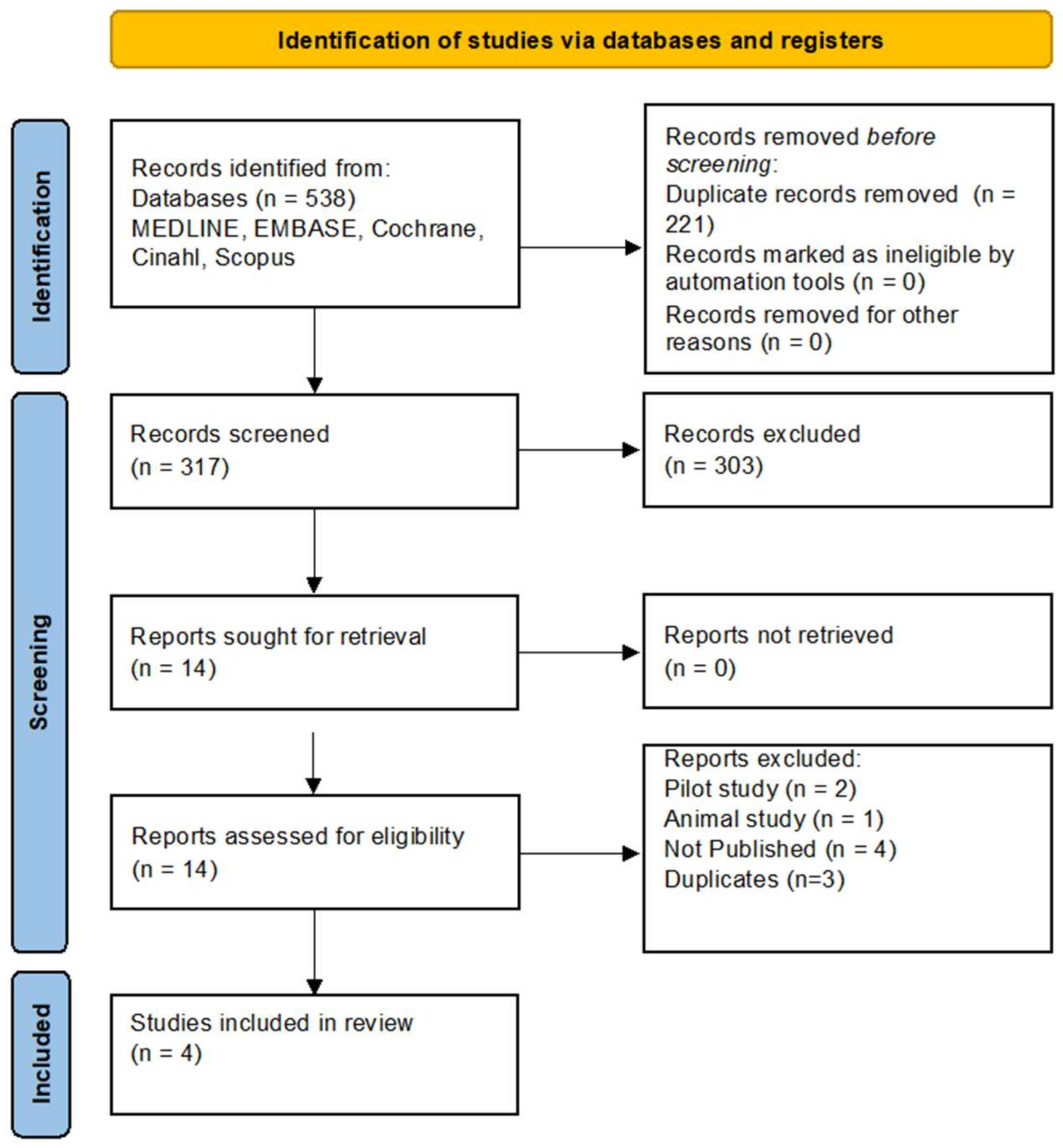
Figure 1. Flow diagram illustrating the search and screening process in line with the Preferred Reporting Items for Systematic Reviews and Meta-Analyses (PRISMA) guidelines [10].
Jcto 02 00009 g001
Disclaimer/Publisher’s Note: The statements, opinions and data contained in all publications are solely those of the individual author(s) and contributor(s) and not of MDPI and/or the editor(s). MDPI and/or the editor(s) disclaim responsibility for any injury to people or property resulting from any ideas, methods, instructions or products referred to in the content.