Review

Three-Dimensional Printing for Accessible and Personalized Ophthalmic Care: A Review

1
Cumming School of Medicine, University of Calgary, Calgary, AB T2N 1N4, Canada
2
Sprott School of Business, Carleton University, Ottawa, ON K1S 5B6, Canada
3
School of Medicine and Dentistry, Griffith University, Gold Coast, QLD 4215, Australia
4
Max Rady College of Medicine, University of Manitoba, Winnipeg, MB R3E 0W2, Canada
5
Section of Ophthalmology, Department of Surgery, University of Calgary, Calgary, AB T2N 1N4, Canada
*
Author to whom correspondence should be addressed.
J. Clin. Transl. Ophthalmol. 2025, 3(2), 6; https://doi.org/10.3390/jcto3020006
Submission received: 27 August 2024 / Revised: 17 December 2024 / Accepted: 17 March 2025 / Published: 26 March 2025

Abstract

:
Over 2.2 billion people across the globe face significant barriers to accessing essential ophthalmic care, with elderly, rural, and refugee populations being disproportionately affected, deepening existing disparities in eye care. Three-dimensional printing is a novel technology that has the potential to transform the field and improve access by alleviating many patient-specific barriers. This article delves into the evolution of 3D printing within ophthalmology, highlighting its current applications and future potential. It explores various 3D printing techniques and numerous biomaterials discussing their effectiveness in creating advanced solutions such as bioengineered corneas, ocular prosthetics, and innovative treatments for dry eye syndrome, from punctal plugs to lacrimal gland models. Additionally, 3D printing has revolutionized drug delivery systems for conditions like glaucoma, retinal diseases, and ocular brachytherapy. Whether through 3D printed contact lens-based drug delivery systems or polycaprolactone implants that biodegrade and provide sustained drug release without adverse effects, these systems hold immense potential in the field. Despite its promise, the integration of 3D printing into clinical practice presents challenges, which the article addresses alongside strategies for overcoming them. By mapping out the technological advancements and challenges, this review offers a roadmap for enhancing global eye care accessibility and improving patient outcomes on a global scale.

1. Introduction

In 2013, for the first time, physicians received the approval of the Food and Drug Administration (FDA) to create and place a customized, bioresorbable 3D printed tracheal splint in a newborn with life-threatening tracheobronchomalacia [1,2]. This revolutionary invention, approved through the emergency-use exemption, set a precedent for 3D printing in medicine. Since then, 3D printing has been increasingly gaining traction in medicine, including in ophthalmology leading to substantial developments over the past decade. Among many notable inventions, recent advancements include the completion of the first clinical trials for fully automated, 3D printed custom ocular prostheses (NCT05093348, https://clinicaltrials.gov, accessed on 5 July 2024) [3] as well as the bioprinting of the first artificial corneal tissue in vitro [4].
Three-dimensional printing continues to offer solutions to increasingly complex problems in ophthalmology. At its simplest level, it has allowed for the printing of non-biocompatible objects including customizable eyewear [5,6], adapters that enable smartphone-based retinal imaging [7,8], and surgical tools [9,10,11] (Figure 1A–C). Increasing in complexity, 3D printing uses biocompatible materials for the design of medical devices such as punctal plugs, ocular and facial prostheses, and contact lenses that minimize the immunogenic response following implantation (Figure 1D–F). Finally, in its most advanced form to date, 3D printing involves using both cellular and acellular biomaterials to generate scaffolds that promote tissue regeneration [12] (Figure 1G).
The recent advancements in 3D printing technologies and their implementation in ophthalmology have facilitated growth in the field and provided a prospect for alleviating some barriers to accessing eye care. Even in its simplest form, where 3D printing is limited to creating inanimate, non-biocompatible objects such as a smartphone fundus imaging adapter, it can create a dramatic difference to low-resource regions that do not have access to traditional desktop fundus cameras. The difference is stark—an open source 3D printed adapter may be manufactured for USD 13, while traditional clinic fundus cameras typically cost tens of thousands of dollars [13]. Another prominent example of how 3D printing can drastically improve access to eye care is in corneal transplantation. Corneal transplants can restore sight to those living with corneal blindness from various underlying etiologies including infection, inflammation, trauma, or congenital conditions. However, in a recent international survey of corneal transplantation and eye banking, there was a significant shortage of corneal graft tissue worldwide [14]. Globally, the authors estimated that only 1 cornea is available for every 70 needed, leaving 12.7 million people awaiting corneal transplants [14,15]. Unfortunately, these shortages are most severe in the least developed countries [16] due to a lack of eye banking infrastructure and a lack of donations possibly due to cultural attitudes or religious beliefs [14,15]. Further, natural corneal tissue carries inherent risks, including a high rejection rate, which can limit access to those with a history of unsuccessful keratoplasty. Graft failure due to the surgical procedure can also contribute to the shortages. Corneal shortages are a complex multi-factorial problem with economic, political, technical, cultural, and ethical barriers [17,18]. Regardless of the underlying causes, this shortage demands a multifaceted solution. In addition to encouraging donations and creating the necessary infrastructure, complementary solutions such as corneal bioengineering and bioprinting have been suggested as a potential means to alleviate the shortages [14].
In the following sections, this review examines recent advancements in 3D printing technologies in ophthalmology, with a focus on innovations that have significant clinical potential. The paper provides a comprehensive overview of how 3D printing can help address disparities in eye care across ophthalmic subspecialties, including cornea, oculoplastics, retina, glaucoma, and ocular oncology. It highlights the latest advances in the field while discussing the challenges and future directions that may limit clinical translation.

2. Brief Review of the Mechanisms of 3D Printing

Three-dimensional printing is a method of additive manufacturing where structures are created through layer-by-layer deposition of materials. Additive manufacturing is differentiated from traditional subtractive manufacturing in that the latter takes away material from a preset block to create the desired structure [19]. Regardless, both techniques rely on computer-aided design (CAD) to generate and guide the sequential manufacturing process.
The first step of 3D printing is to generate a digital model of the product [20]. This model is either designed through CAD or obtained by scanning the object. Typically, creating models through CAD is time-consuming and requires an experienced designer with knowledge of the software. Additionally, it can be difficult to replicate designs of natural anatomical structures, with their nuanced geometry. Consequently, to overcome these challenges, 3D data are now often generated by obtaining 3D scans through imaging modalities including CT or MRI [21]. Through these methods, it is possible to obtain accurate data on the topography of the scanned structure; however, these scans typically require further segmentation or refinement to obtain the final model. Once the model is created, the next step is to generate a toolpath for the printer to follow. This toolpath is a point-by-point instruction guiding the printer to create the desired model. It takes into consideration many input parameters including the printing speed, the desired quality, and the printing material. Most printers have an associated software that can generate these instructions in the form of a “GCODE” file that is uploaded to the 3D printer. At this stage, the printer is set up with the appropriate material, and the print is started. Upon completion, the final product undergoes some postprocessing, which is highly dependent on the type of printer used. Many 3D printers have been applied in medicine, these have been detailed in Table 1 highlighting the advantages and disadvantages of each [22,23].
Three-dimensional bioprinting is a subset of 3D printing that involves the layer-by-layer deposition of bioinks, which contain living cells and biomaterials, to create complex, three-dimensional biological structures. This process is akin to traditional 3D printing, but it incorporates biological elements, enabling the fabrication of tissue-like structures and organs for use in medical and research applications. Bioprinting typically utilizes various printing techniques, such as extrusion-based printing, inkjet printing, and laser-assisted printing, to precisely place cells, growth factors, and scaffolding materials in patterns that mimic the natural architecture of human tissues.
The primary goal of 3D bioprinting is to create functional tissues that can be used for regenerative medicine with the aim of producing viable scaffolds for transplantation. By allowing for the precise control of cell placement, bioprinting enables the creation of highly structured tissues with specific cell types and extracellular matrices, making it a powerful tool in the development of personalized medical treatments. As the technology continues to evolve, 3D bioprinting holds great promise in overcoming challenges in organ donation, tissue repair, and other areas of healthcare.

3. Three-Dimensional Printing in Ophthalmology

In the following sections, we present the current progress and applications of 3D printing in various subspecialties and areas within ophthalmology.

3.1. Cornea

As discussed, 3D bioprinting is an innovative process for creating artificial grafts and tissues by using additive manufacturing to provide precise and intricate arrangements of cells and tissue structures. Years of research and development have led to the current advancements in bioprinting corneal tissue. Corneal bioprinting aims to manufacture a biomimetic scaffold that supports and integrates seeded corneal cells to regenerate [12,29]. Manufacturing an artificial cornea requires a deep understanding of the basic anatomy, fundamental physiological properties, and cellular components of this unique and intricate structure of a natural cornea.

3.1.1. Biological Factors for Corneal Transplantation

The cornea, though small, is remarkably complex. Its morphology is dome-shaped, extending 11–12 mm in diameter and having a central thickness of approximately 560 μm [30]. The cornea is composed of cellular and acellular components. Corneal keratocytes are the most abundant of the cellular components and collagen is the most abundant acellular component, which comprises 70% of the entire corneal mass. Other cellular elements include epithelial cells, limbal stem cells, Langerhans cells, and dendritic cells [12]. Similarly, beyond collagen, other acellular elements include proteoglycans [30]. The cornea consists of six distinct layers—epithelium, Bowman’s membrane, stroma, pre-Descemet or Duas layer [31,32], Descemet’s layer, and endothelium. These structural elements and layers provide the cornea with its biomechanical properties and give rise to its functional qualities. For instance, the distinct arrangement of collagen lamellae in the corneal stroma and the complex composition of the extracellular matrix (ECM) with its glycosaminoglycans are what provide the cornea with clarity and mechanical strength. A deviation in the number of these ECM glycosaminoglycans and distortion of the collagen fibers is the underlying pathophysiology associated with keratoconus—the condition that in itself is the leading cause of corneal transplantation [12]. Similarly, the acellular biomolecule keratocan is also fundamental to the cornea’s symmetric curvature, which allows it to effectively transmit light and contribute to approximately two-thirds of the total eye refractive power [33,34]. By the same token, the cornea’s well-developed sensitivity to stimuli, immune regulation, and ability to self-regulate electrolyte and fluid balance are all dependent on cellular and acellular elements of the cornea that harmoniously function together [12].
From this simplified overview of the corneal microstructure and physiology, one can begin to appreciate the complexity of the cornea and learn of the intricacies that arise when attempting to bioprinting corneal tissue. The creation of a biocompatible, mechanically stable, optically transparent scaffold that is capable of encapsulating corneal cells and promoting their growth, migration, proliferation, and functional expression, is undeniably difficult. Nevertheless, over the years, the field has advanced, overcoming many of these challenges; one main area of advancement is bioink development.

3.1.2. Bioink in Corneal Applications

A key component of the bioprinting process that dictates the functional and biomechanical properties of the manufactured cornea is the bioink. A bioink is a combination of biomaterial and living cells which, when deposited or crosslinked in the process of 3D bioprinting, creates the desired design [35]. Common biomaterials, which have been utilized in corneal bioprinting include both natural and synthetic materials. The natural biomaterials collagen, gelatin, chitosan, decellularized extracellular matrix (dECM), alginate, and hyaluronic acid (HA) are the most commonly studied [16]. These materials, with their advantages and disadvantages, and relevant studies relating them to corneal bioprinting have been summarized in Table 2.
As can be seen in Table 2, often bioinks are formed through a combination of several complementary materials to overcome the disadvantages of one biomaterial. In a study by Wu et al., the authors bioprinted human corneal epithelial cells using a combination of collagen/gelatin/alginate incubated in a medium of sodium citrate [40]. While such significant modifications and mixtures of materials may lead one to conclude that the design is too complex for real-life manufacturing and clinical translation, some industries have demonstrated otherwise. For instance, in the clothing industry, textiles have evolved over the past decades to consist mostly of blended fabrics that combine several materials. These blends have accommodated a variety of functional considerations including improved antibacterial properties, durability, UV resistance, hydrophobicity, and flame-resistant properties [56]. Undoubtedly, such significant strides require time, research, and extensive trial-and-error, which, are currently underway for corneal bioprinting bioinks.
Optimizing the bioink, in the context of manufacturing artificial corneas, involves accounting for physical properties, rheological characteristics, and biological factors.
In terms of the physical properties, the material ought to provide sufficient mechanical strength and optical transparency. Specifically, the structure should be capable of withstanding an intraocular pressure, at least up to 21 mmHg, and ideally have a tolerance for higher pressures. In studies, these mechanical properties are typically assessed by comparing the stress–strain curves of the material with that of the cornea [12]. Often, with the naturally occurring materials mentioned in Table 2, one of the common disadvantages is their weak mechanical properties. To overcome this, and thereby allow scaffolds to support themselves, researchers have identified that adding sodium alginate imparts mechanical strength when combined with other materials [39]. However, while alginate provides mechanical support, and is generally non-immunogenic, among other advantages, it does have a low degradation rate, and a limited cell adhesion capacity [12]. When studying the optical properties, usually three tests are performed to evaluate the suitability of the material, namely the refractive index, transmittance assessments at various wavelengths, and qualitative analyses [12]. These properties have been thoroughly researched in the field, and combinations of materials have been used to optimize transparency, including the use of GelMA to optimize the optical properties of dECM [49]. Nevertheless, while the field has made significant strides, bioink optimization remains a challenge in manufacturing synthetic corneas.
Concerning the rheological characteristics of scaffolds, the bioink must be adjusted for gelation time and shear thinning. Both gelation and shear thinning viscosity properties are essential variables to control to ensure that the bioink is suitable for the printing process. Not only do the intrinsic properties of the material matter, but the selection of the 3D printing method and specific printer is equally important to ensure the desired outcomes.
Finally, from a biological perspective, the bioink must be biocompatible, biodegradable, and have sufficient permeability to oxygen and nutrients [16]. Biocompatibility involves ensuring that the material does not provoke an immunologic reaction and that it is nontoxic for the laden cells. Further, the material should encourage high cell viability consequently allowing the expression of proteins; often, to achieve high viability materials ought to be permeable. With every material used, biocompatibility is tested with the seedings of cells and serial assessments of cell viability.

3.1.3. Recent Advancements in Bioinks and Fabrication Strategies

Among the recent advancements in the field of corneal bioprinting have been the development of mechanically robust multi-material bioinks [57], and new fabrication strategies that combine different methods such as electrospinning with 3D bioprinting [58].
Firstly, in a recent, novel study by Ghosh et al. (2024), the authors enhanced the mechanical robustness of decellularized cornea matrix (DCM) hydrogel through a hybrid blend with silk fibroin, resulting in a stronger, more durable material [57]. Unlike previous approaches, which focused exclusively on mechanical properties, this innovative study combined two distinct bioinks to recreate the natural anisotropy of the cornea. The outer peripheral corneal rim was bioprinted using the robust polymeric blend of DCM and silk fibroin, while the central optic zone was printed with pure DCM to preserve its transparency [57]. This dual-material strategy successfully maintained both structural integrity and optical clarity. The authors encapsulated human limbus-derived mesenchymal stem cells in both materials and bioprinted them in concentric and parallel patterns to enhance the native-like organization of the tissue. The resulting bilayer corneal construct supported high cell viability and expressed keratocyte core proteins, indicating optimal functionality [57]. It is also important to note that as with this example, many recent studies in the literature have started incorporating stem cells within bioprinted corneal scaffolds to understand the impact that the materials can have on differentiation, viability, and adhesion of cells [59,60].
Secondly, in a novel study by Zhang et al. (2024), the authors presented an innovative approach to creating bioartificial corneas by combining electrospun micro-nanofibrous decellularized extracellular matrix (dECM) with digital light processing (DLP) 3D bioprinting [58]. The electrospinning technology is a scalable process that fabricates micro-nanofibrous scaffolds allowing for a high degree of control over the thickness and alignment of the fibers [58]. Through electrospinning, the authors were able to shape the dECM while preserving its native biochemical components, which more closely mimic the natural corneal microenvironment. This method significantly improved the mechanical properties of the artificial cornea, showing a five-fold increase in mechanical properties, superior cell adhesion, organization, and maintenance of keratocyte phenotype [58]. In animal studies, transplantation of the bioartificial cornea promoted faster corneal epithelialization and maintained transparency [58]. This approach offers a promising alternative for the future of corneal graft manufacturing.
Even with these recent advancements, to overcome the ongoing challenges with corneal bioprinting, bioinks must be optimized to include a wide range of mechanical properties and layer cohesion considerations. To do this, artificial intelligence (AI) is now being included in 3D bioprinting to optimize bioinks and streamline the production process [61]. In the pre-printing phase, AI processes tissue imaging and CAD models to predict bioink suitability, reducing trial-and-error experimentation. By training neural networks with printing parameters, AI fine-tunes algorithms for better bioink performance [61]. This integration accelerates bioink optimization, reduces reliance on manual adjustments, and enables smarter, more efficient bioprinting workflows, giving hope to an optimized bioink solution [61].

3.1.4. Challenges and Future Prospects in Corneal Bioprinting
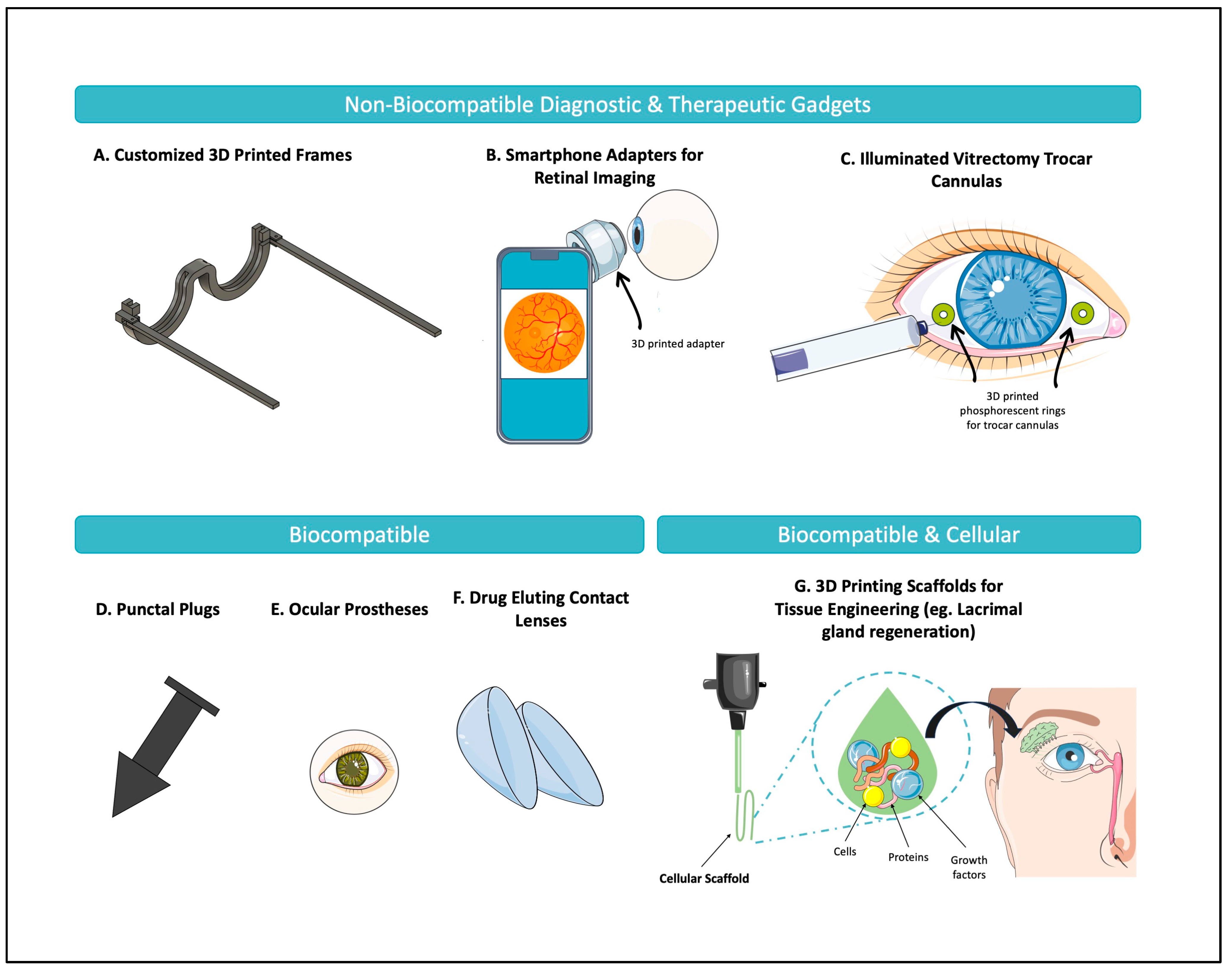
Clinical translation of biomimetic corneal tissue requires overcoming a few challenges and traversing through the regulatory pathways. One of the challenges encountered by researchers thus far is the bioprinting of several layers of the cornea. Most of the studies in the field have focused, unsurprisingly, on bioprinting the stromal layer, given that it is the thickest of the corneal layers. Some studies have examined epithelial bioprinting, but only a limited number of studies have assessed the potential of endothelial bioprinting [62,63,64]. Unfortunately, since the endothelium remains a clinical challenge given its lack of regenerative capacity, further research in this area is essential and should be pursued. Additionally, it would be beneficial for researchers to continue examining the potential of bioprinting bilayers, such as the epithelium and stroma, which have been previously studied in a few instances [29,36,65]. While current bioprinting would not meet the needs of penetrating keratoplasty (full-thickness graft), having the capacity to bioprint one of the bilayers would allow us to supply tissue for DALK (Deep Anterior Lamellar Keratoplasty), which transplants all but the inner layers.
Focusing on the corneal stroma, one of the main challenges encountered in the bioprinting process is replicating the stroma’s highly ordered structure, which is crucial for its biomechanical and optical properties [66]. Often, when cultured under standard conditions, corneal cells can lose their original phenotype and take on a fibroblastic phenotype, leading to a disorganized ECM that is similarly seen with corneal scarring. Further, when corneal cells are cultured, they often lose their ability to produce cornea-like ECM after the cell population doubles [67]. In a study by Gouveia et al., the authors demonstrated that by regulating topographical cues in vitro, corneal stromal fibroblasts can be guided to align in a manner like their native structure [68]. This study provides a possible path forward, and further research should focus on expanding the field’s understanding of these topographical features. Along with understanding the organization of the ECM, it is important to continue studying the incorporation of other cells and acellular components in the corneal scaffold. For instance, the cornea includes many immune cells and serves an important protective role, which should be explored further in the future. Additional work is also still necessary to ensure that while replicating the cornea, all its fundamental functions are considered.
Finally, one of the main challenges to clinical translation is ensuring that the final product is compatible with regulatory pathways [12]. These have been further described in Section 4 of this paper. Further, some of the previously reported challenges including the printing time associated with 3D bioprinting have already been addressed, at least partially. For instance, in an experimental study, scientists developed a support scaffold that facilitates the printing of 6–12 corneas at a time, by using a combination of stereolithography printing, extrusion-based printing, and micro-transfer molding techniques [39]. The field has demonstrated clear growth over the past decade and should continue to be driven with a purpose—to alleviate the shortages of corneal transplants worldwide, and thus, enable restoration of vision to those who are currently living with corneal blindness.

3.2. Oculoplastics

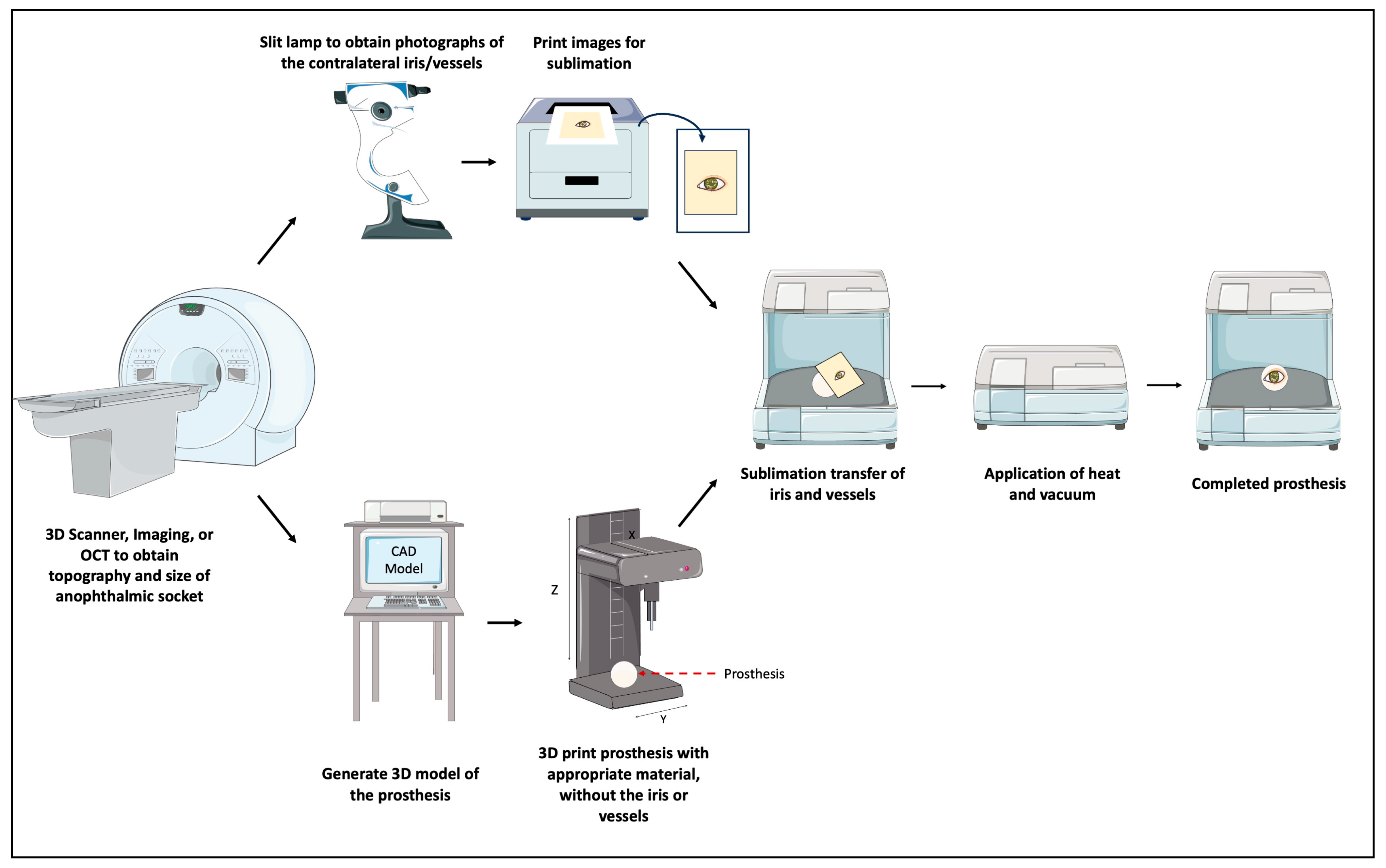
3.2.1. Ocular Prosthetics

An ocular prosthesis is an artificial substitute for an enucleated eyeball. Ocular prosthetics are an opportunity to restore function and symmetry to anophthalmic patients. The most common etiologies of enucleation and evisceration are traumatic in nature, thus, making young people often the affected demographic [69]. Reproducing the pigmented human iris with its intricacies and manufacturing an implant that replaces the orbital volume is challenging [70]. Historically, prostheses have been produced through two main methods: (1) ready-made stock shells or (2) custom design. Of the two methods, custom ocular prostheses are better fitting, increase the movement of the eyeball, and provide better cosmetic outcomes [71]. Unfortunately, the fabrication of custom prostheses is time-consuming, labor-intensive, costly, and involves reliance on the experience of the ocularist [3,72]. These challenges undoubtedly form barriers to those living in developing countries where the health care system may already be fragile. To overcome some of these barriers, 3D printing has been implemented to create a streamlined, automated, and affordable option. An example of the success of using 3D printing for creating personalized medical aids has been its use in creating hand and arm prosthetics for patients in a rural area of Sierra Leone, a country that ranked 184 of 187 on the UN Development Index [73,74]. Through international collaboration and by harvesting the most advanced technology from around the globe, a feasibility study demonstrated that 3D printing is suitable for creating customized, 3D printed arm prostheses in low-resource areas [73]. A follow-up study demonstrated an increase in the health-related quality of life with the use of these prostheses [74].
The first documented case of a 3D printed ocular prosthetic being designed and fitted to a patient occurred in 2016 [75]. Since then, with the increasing emphasis on research [70,76,77,78], the field has made significant developments. First, researchers created pathways that incorporated 3D printing of prostheses while maintaining a dependence on manual painting by an ocularist [79]. Then, the process evolved to a semi-automated one, where, after obtaining an impression of the anophthalmic socket, a 3D scanner is used to obtain 3D model data of the shape of the eye, which is 3D printed without the iris or vessels. In conjunction, a slit lamp biomicroscope is used to obtain photographs of the contralateral iris/vessels, and the mirrored images were transferred onto the 3D printed model via a sublimation process [80]. This process is demonstrated in Figure 2. Finally, and most recently, researchers developed a fully automated digital end-to-end 3D printing pathway for creating a customized prosthesis [3], a process that was recently studied in clinical trials (NCT05093348, https://clinicaltrials.gov, access on 5 July 2024). In this process, image data from the contralateral eye is obtained by using an anterior segment optical coherence tomography device (OCT). Then, using automatic data-driven design software, an appropriate prosthesis shape is determined, and a textured 3D model is generated. Finally, a multi-material full-color 3D printer is used to create the prosthetic [3].
The latest 3D printed devices have demonstrated several advantages. Firstly, the material used for manufacturing these prostheses has been assessed for biocompatibility and revealed no evidence of toxicity, per the stringent criteria of ISO 10993 [3,81,82]. Secondly, 3D printing, when implemented through an automated process, can accurately replicate the color and anatomy of the adjacent eye, providing patients with the hope of restoring symmetry and promoting comfort [3]. Thirdly, the full automation of the process minimizes labor time and is always reproducible. Given that OCT images are now being introduced into the process, it eliminates the need for impressions [3]. This is important because the impressions needed in the traditional customized method are time-consuming and uncomfortable for the patient. All these advantages can serve to reduce the barriers to access including the cost associated with the customization and ocularist labor and the multiple visits needed to appropriately fit a prosthesis.
With clear advantages, the novelty of 3D printed ocular prosthetics raises concerns about their durability, maintenance, and safety. The advancements in 3D printing materials, including the development of biocompatible and high durable resins, are steadily addressing these issues. The materials used in modern 3D printing can withstand typical wear and tear associated with daily use, such as the physical impact of removing and inserting the prosthetic. In a recent systematic review and meta-analysis, Valenti et al. (2024) evaluated the mechanical properties of 3D printed prosthetics as compared to conventional milling or subtractive manufacturing [83]. Their results demonstrate that 3D printing is comparable to conventional subtractive methods in terms of mechanical properties [83]. While their systematic review was specifically for prosthetics in dentistry, it is reasonable to extrapolate these results for ocular prosthetics, though certainly, studies should carefully examine and compare the mechanical properties of traditional ocular prosthetics to 3D printed ones. Regarding the potential colonization of opportunistic pathogens through ocular prostheses, a recent randomized controlled trial (2023) compared 3D printed acrylic resin ocular prostheses to conventional poly-methyl methacrylate ones assessing their impact on the biofilm and microbial flora of the anophthalmic socket [84]. Their results demonstrate no statistically significant difference in anophthalmic flora when comparing both fabrication techniques [84]. However, the 3D printed ocular prosthetics demonstrate a degree of anti-colonization ability against pathogenic bacteria [84].
While weighing these significant advantages, the main limitation of this design and process is affordability [3]. To truly make this product affordable for patients, these prostheses must be manufactured in larger quantities—a problem that is common to many industries. Scaling the product allows companies to find areas of optimization and reduce their overall costs, making such products affordable for the end user. Further, to address the concern of scaling and cost reduction for prosthetics in underserved regions, one potential solution lies in partnerships with local organizations, healthcare providers, and non-profits. These collaborations could facilitate the establishment of production facilities in regions with high demand for prosthetics, reducing transportation costs and improving accessibility. Large-scale partnerships with international NGOs or government health programs could also support the distribution of prosthetics to underserved populations, further driving down the per-unit cost through economies of scale. Such alliances and programs have been previously effective. A prominent example was projects aimed at eliminating trachoma, including the Alliance for Global Elimination of Trachoma, which was established by the World Health Organization, and the World Health Assembly’s SAFE (Surgery, Antibiotics, Facial Cleanliness, and Environmental Change) strategy [85]. Through such projects, trachoma has been eliminated in many areas where the condition was endemic [85].

3.2.2. Facial and Orbital Implants

Restoring the anatomical structure of the face and orbit after a fracture is notably difficult and can unfortunately lead to problems such as diplopia and enophthalmos. While ocular prostheses are limited to mimicking the shape and function of the globe, orbital prostheses typically involve the reconstruction of the surrounding bone structure(s) and can be more complex. The application for 3D printing in orbital prostheses has seen significant growth over the past few years, with ongoing clinical translational efforts [86,87,88,89,90,91]. The results have been promising thus far, but have been demonstrated predominantly in case reports [92,93,94]. An overall review of the literature reveals that the use of 3D printing in orbital fracture surgeries has demonstrated several benefits, including reducing the duration of the procedure, lowering operating room costs, and achieving consistent orbital volume outcomes [95]. Undeniably, these benefits make it possible to alleviate many barriers to care. For instance, many of the elderly with comorbidities may only have surgical interventions if operative time is reduced. However, while continuously growing, one of the areas requiring further research is the reliance on using the adjacent eye anatomy to create most of these prostheses. While it is often possible to rely on the contralateral orbit for a 3D model, this is not always possible in cases of severe trauma where both orbits may be impacted or when a systemic disease causes a need for bilateral implants. Additionally, it should be noted that facial anatomy is not perfectly symmetric, and direct mirroring can sometimes result in functional defects [96]. As such, other approaches should continue to be researched, including reconstruction software that takes the contralateral eye as an input and uses spline interpolation for further refinement [97].

3.2.3. Eyelid Crutches

Another use of 3D printing in oculoplastics has been its application to manufacture eyelid crutches for patients with blepharoptosis [98]. Blepharoptosis, or ptosis, is an abnormally low-positioned upper eyelid that can often occlude the visual axis and dramatically impact the quality of life. Often, for visually significant ptosis, operative intervention is the treatment of choice. However, in many cases such as patient refusal, high operative risk due to comorbidities, or a lack of medical and surgical infrastructure, surgical approaches may not be possible. In these cases, where certain barriers limit access, ptosis crutches may be considered [99]. Conventional crutches have drawbacks because they are inflexible and must be tailored individually, necessitating the expertise of a specialized optician, increasing their cost to around USD 80–USD 100 [98]. Three-dimensional printing offers a cost-effective alternative for producing eyelid crutches, especially with the widespread adoption of the technology. In a case report from the United States, the authors successfully printed a set of personalized eyelid crutches for a patient with recurrent ptosis, who was reluctant to pursue any further surgical intervention [98]. The designed crutches were customized to the patient’s eyelid dimensions and their existing eyeglasses. The final printed device was produced with a biocompatible material and provided significant visual improvement to the patient. Specifically, the patient’s MRD-1 measurement, which was at −2 mm before the crutches improved to an MRD-1 of 1 mm following the application of the crutches. Further, the patient was also able to achieve adequate eye closure while wearing the crutches [98]. This case study demonstrates the application of eyelid crutches to provide the best quality care to a patient with evident barriers to eye care.

3.2.4. Dry Eye Syndrome: Lacrimal Gland Regeneration and Punctal Plugs

Keratoconjunctivitis sicca, also known as dry eye syndrome (DES), is an ocular disease that is highly prevalent, impacting more than 50% of the population in some groups [100]. DES is characterized by inflammation and damage of the ocular surface due to tear lake hyperosmolarity secondary to a decrease in tear production or an increase in tear evaporation. Unsurprisingly, DES has significant economic burdens and impacts on quality of life, which can have disproportionate impacts on those with limited access to resources [101]. As a significant challenge in ophthalmology, researchers have leaned on 3D printing as a possible tool for addressing the disease.
Interestingly, one of the solutions described in the literature to understand more about DES involves creating models of the lacrimal gland (LG) [102,103]. Magnetic 3D bioprinting has been tried as a method of engineering both the lacrimal gland, as well as other secretory epithelial organoids, like the salivary glands [104]. In magnetic 3D bioprinting, biocompatible magnetic nanoparticles are employed to label cells for printing into a specific 3D arrangement [102]. Magnetic 3D bioprinting has been successful in producing salivary gland organoids with printed cells having 90% viability three days following differentiation [104]. Similarly, in a recent study by Ferreira et al. (2024), the authors used a magnetic 3D bioprinting platform to successfully assemble two LG in vitro models to subsequently use them to understand senescence-associated ocular pathogenesis of DES [105]. Further, as published in a recent conference abstract, researchers developed a bioink combining decellularized porcine LG ECM with 2% alginate to create a printable, stable scaffold for bioprinting [106]. LG bioink demonstrated shear-thinning properties, improved mechanical strength, and enhanced cell distribution, outperforming 2% alginate in cell viability and sedimentation [106]. Printed structures had mechanical properties like native LG tissue, and lacrimal epithelial cells printed in LG bioink showed significantly higher viability [106]. The results suggest that LG bioink is a promising approach to combining the benefits of ECM hydrogels with bioprinting, enabling better tissue models and therapeutic applications [106]. While still evidently in the research stages, these results are promising for the utilization of bioprinting in gland regeneration aimed at potentially managing DES in the future, and reducing some of the barriers associated with the economic burdens of DES. To achieve this however, the field will have to overcome a variety of challenges including regulatory challenges, like the manufacturing of corneal tissues, described earlier in the paper. A further discussion regarding the regulatory considerations of clinical translation is reviewed in Section 4.
Another area of research addressing dry eye disease is 3D printed punctal plugs [107,108]. Punctal plugs are a widely used non-invasive treatment approach to alleviate symptoms of DES. By blocking the canaliculi, which usually drains the tear fluid to the nasopharynx, these plugs prevent tear drainage [109]. Consequently, these plugs reduce the ocular tear lake hyperosmolarity, which is a major contributing factor in the pathophysiology of DES [110]. Further, it would be advantageous to use these plugs, which can stay in the canaliculi for several weeks or months, to administer medications to patients who may be unable to adhere to frequent dosages. For instance, this can be applied to those who are elderly, or to those with limitations to their mobility. In a European experimental study, the authors developed 3D printed punctal plugs that were loaded with dexamethasone, a commonly used corticosteroid [107]. Their results demonstrated that punctal plugs made with 100% polyethylene glycol diacrylate exhibited prolonged drug release of more than three weeks [107]. The application of drug-loaded punctal plugs across other subspecialties of ophthalmology is yet to be exploited. For example, drug-loaded punctal plugs are still being researched for glaucoma therapy [111], where strict medication regimens and adherence are both necessary, but also often difficult to comply with [112].

3.3. Drug Delivery Systems—Glaucoma, Retina, and Uveal Melanoma

Glaucoma is one of the primary causes of irreversible blindness worldwide. Due to the aging population and growing prevalence, there is an urgent need for effective treatments to alleviate the global burden of the disease. Unfortunately, glaucoma management is often demanding of patients and noncompliance is prevalent [113]. Financial hardship, older age, advanced glaucoma, and lengthier frequency of follow-up have been associated with non-adherence [113]. These factors exacerbate the necessity of an appropriate solution.
One of the management techniques for glaucoma, namely trabeculectomy, involves surgically creating a permanent fistula between the anterior chamber and the subconjunctival space, allowing the drainage of aqueous humor. While trabeculectomy is one of the suitable interventions in glaucoma treatment [114], management with (antifibrotic) antifrotic agents like 5-fluorouracil (5-FU) is often necessary to prevent failure secondary to scarring following surgery [115]. 5-FU requires frequent administration through subconjunctival injections, causing barriers for those with limited mobility to clinics, those at increased risk for infections, and anyone without the financial means. Consequently, sustained drug delivery systems (DDS), which can gradually release antifibrotic agents can then potentially alleviate some of these barriers to care, since they will allow patients to receive the drug at home.
In a recent international, experimental study by Ioannou et al. (2023), the authors used 3D printing to design a long-acting drug-eluting scaffold made of polycaprolactone and chitosan, infused with 5-FU [116]. The scaffold was evaluated with cultured human conjunctival fibroblasts and demonstrated excellent biocompatibility with no significant effect on cell viability. Further, the authors evaluated the implant’s ability to suppress conjunctival fibrosis, by measuring the expression of a key fibrotic regulator, the myocardin-related transcription factor (MRTF). Their results demonstrated that the implant effectively downregulated the MRTF pathway that contributes to conjunctival fibrosis [116]. In another international, experimental study, Mohamdeen et al. fabricated a drug-eluting contact lens that provides sustained release of timolol, a glaucoma medication [117]. In their study, the 3D printed contact lenses designed from biocompatible medical-grade polymers provided a sustained release of timolol over 3 days. Similar studies have evaluated the use of 3D printed contact lenses for a variety of applications [117,118,119,120,121,122].
Depending on the application, the appropriate material is selected for 3D printing, based on technical considerations. For instance, in the realm of drug-eluting contact lenses, hydrophilic silicone-based 3D printed hydrogels are particularly common [25]. Yet, even though it is a common material, its hydrophobicity, slow drying time, and low viscosity make it challenging to 3D print [25]. Only recently was the first soft biomaterial based on hydrophilic silicone functioning as a controlled DDS designed and 3D printed [25]. To delay the drug release, the authors manipulated the content of one of the compositional ingredients, the aminosilicone content, allowing for a straight-line release of the drug for the first 8 h [25].
When considering other applications of drug delivery systems, retinal vascular diseases are at the forefront, given the requirement for repeated intravitreal drug injections. These requirements lead to significant health and economic burdens. To address these challenges, Won et al. developed a new drug delivery system that relies on a 3D printed drug rod to deliver two drugs—bevacizumab (BEV) and dexamethasone (DEX) from a single implant [123]. Through this design, the rod’s size and drug concentration can be optimized for drug release. The rod can be injected into the vitreous using a small-gauge needle, making the procedure less invasive. Further, the specific material used to manufacture the rod shell in this study was polycaprolactone (PCL) [123]. PCL is an FDA-approved material that degrades under in vivo conditions within 2 years, without any adverse effects caused by degradation and removes any need for implant removal from the patient’s body [123]. Furthermore, by employing degradation-based release control, drug rods with tailored release stabilities can be produced by selecting materials that degrade at different rates in vivo [123]. The efficacy of these systems has been demonstrated in animal models. There, the drug rod demonstrated superior efficacy in reducing inflammation and providing long-term suppression of neovascularization compared to traditional BEV injections, suggesting it is a promising alternative for treating retinal vascular diseases [102].
In the realm of drug delivery, 3D printing has also been studied for brachytherapy, a type of treatment also known as internal radiation therapy [124]. Ocular brachytherapy is often implemented in the management of uveal melanoma. In it, episcleral plaques (EPs), which are hemisphere-shaped metal objects that carry the radioactive seeds, are implanted to administer radiation. EP brachytherapy is an effective treatment but is unfortunately associated with significant side effects due to a lack of precision in dose delivery. In a Canadian study, to improve treatment precision, Lescot et al. designed and 3D printed a tumor-specific implant with a biocompatible material called Polyetheretherketone [124]. In their study, the authors demonstrated that through the custom implant, they were able to modulate the dose profile of the radioactive material, which can be used to target the contours of cancerous tissues [124]. Similarly, in a 2016 Polish study with 11 patients, the authors described the application of 3D printing technology to produce customized boluses for patients undergoing electron beam therapy for skin lesions near the eye canthi [125]. The study compared the effectiveness of these 3D printed boluses to manually fabricated paraffin boluses. CT scans of the 3D printed and paraffin boluses were obtained and overlaid onto patient CT images to assess fit, bolus uniformity, and dose distribution to the underlying tissues. The results demonstrated that 3D printing is a viable method for creating boluses for small eye lesions, offering superior performance compared to boluses manually crafted from paraffin sheets. These studies have implications for the future of personalized medicine—from reducing the side effect profile to reducing the costs to the patient and the healthcare system, 3D printing has significant potential in the field.
Three-dimensionally printed drug delivery devices offer several advantages over traditional methods, particularly in terms of efficacy, customization, and patient outcomes. Unlike conventional delivery systems, which often have fixed dosages and limited designs such as with manually crafted paraffin boluses, 3D printing allows for precise control over the shape, size, and drug delivery profile. This level of customization can lead to more targeted therapies, improving the therapeutic effectiveness of drugs. Additionally, 3D printing enables the creation of personalized drug delivery systems tailored to individual patient needs, including anatomical requirements. This personalized approach is likely to improve patient compliance and overall treatment outcomes.

3.4. Medical Education

To enhance clinical practice, 3D printing has been increasingly integrated into medical training. This includes the use of 3D printed eye models for teaching ophthalmoscopy to undergraduate students [126], as well as printed replicas of orbital dissections [127]. Numerous other applications have also been explored [128,129,130,131,132,133]. A prominent example is a randomized study by Wu et al. that explored the educational benefits of using a 3D printed ocular model for teaching ophthalmoscopy to medical students [126]. In this study, 92 students were randomly divided into two groups: a model-assisted training group, where students practiced using both a simulated eye and their peers, and a traditional training group, where the practice was limited to peer interactions. Both groups received equal training time before being evaluated on their ability to visualize the fundus and accurately determine the cup-to-disc ratio in patients. Among the model-assisted group, 93.48% (43/46) successfully identified the ratio, compared to only 45.65% (21/46) in the traditional group, which demonstrated the potential of 3D printed simulators in ophthalmology to enhance learning before transitioning to a clinical setting. Similarly, in a 2023 pre-test/post-test study from the United States that used 3D printed models for orbital fracture education for 20 medical residents. The study found that over 90% of the interviewed residents reported that the 3D model was useful for conceptualizing the anatomy and was a useful training tool [128]. It is valuable to note that 3D printed simulators, or 3D printed orbital dissection reproductions, may alleviate barriers and improve clinical training. It may be particularly beneficial where access to cadaveric specimens may be limited, whether due to the expenses associated with cadaver donation programs, the limited time available in contemporary curricula, or for students from geographically isolated areas without access to cadaveric anatomy labs [127].
In surgical planning, 3D printed models of an eye with uveal melanoma were manufactured for planning the optimal scheme for linear accelerator-based stereotactic radiosurgery [134]. Similarly, Dorbandt et al. used 3D printing in cases of orbital and peri-orbital masses for dogs, and identified their exceptional potential in improving surgical planning of veterinarians, and communication with owners [135]. From creating patient-specific models for optimizing treatment strategies in ocular tumors to aiding in the management of complex orbital conditions, 3D printing technology empowers clinicians with tools for better visualization and collaboration, ultimately advancing patient outcomes, and reducing possible communication barriers.

4. Future Directions

It is evident from this review that the application of 3D printing in ophthalmology is underway. While some applications, including ocular prostheses, are in clinical trials and are likely to be commercialized by companies soon, other areas still require significant research before clinical translation, including the use of 3D printing for drug delivery.
It is interesting to envision some of the possibilities 3D printing can have on ophthalmology. One interesting consideration is the design of custom 3D printed intraocular lenses (IOL) for managing color blindness [136]. In a study by Alam et al., the authors used a wavelength-selective Atto 565 filtering dye and combined it with an in-house prepared printing resin made of highly transparent in situ synthesized poly-2-hydroxyethyl methacrylate (pHEMA) and polyethylene glycol diacrylate (PEGDA) resin. Through this, they successfully created an IOL that successfully blocks 50% of the unwanted wavelength (565 nm) which is responsible for red–green color blindness [136]. Similarly, it would be interesting to see the application of 3D printing with other technological advancements being studied in ophthalmology, including intraocular pressure biosensors [137] and retinal prostheses [138]. There is a possibility in the future to incorporate in situ bioprinting, that is, direct printing onto the patient when the technology is ready. This could be applied in trauma settings, when, for instance, there may be a need for urgent orbital floor fracture repair. Additionally, the concept of 4D printing, where a time-based variable allows printed materials to change in response to a stimulus, may be possible.

4.1. Regulatory Considerations

Navigating the regulatory landscape is an important step for the commercialization and clinical translation of research projects. A variety of regulatory considerations exist for the different products described in this paper. The most stringent considerations are certainly those relating to the transplantation of bioengineered tissues, which in this paper includes both the cornea and the lacrimal gland tissue.
Developing an Advanced Therapy Medicinal Product (ATMP), such as corneal tissue or lacrimal gland scaffolds, requires parallel clinical and regulatory activities, along with meticulous documentation of the design, testing, and compliance processes. The development begins with the acquisition and processing of living tissues and cells, adhering to stringent quality and safety standards. Preclinical development follows, focusing on safety and toxicity studies conducted under Good Laboratory Practices (GLP) [12]. Establishing a robust manufacturing process that complies with Good Manufacturing Practices (GMP) is crucial, including rigorous validation of equipment and software [12]. Risk analysis is another key component, involving dynamic assessments to identify and address potential hazards associated with the clinical implementation of these technologies [12]. Clinical trials are conducted in phased stages to evaluate safety, efficacy, and therapeutic impact. Regulatory approval is obtained through centralized or national procedures, supported by scientific evaluations and collaboration with regulatory bodies [12]. Post-market surveillance ensures the maintenance of a positive benefit–risk profile through pharmacovigilance studies and continued monitoring of product use in patients. While regulatory frameworks vary globally, with oversight provided by entities such as the EMA in Europe, the FDA in the United States, and the PMDA in Japan, challenges persist in transitioning from research to clinical applications, meeting stringent manufacturing standards, and achieving international regulatory harmonization [12].
The regulatory requirements for clinical translation are intentionally set to ensure a holistic product that is safe and effective. In the case of bioengineered cornea, the requirements extend to the determination of effective sterilization techniques, optimal storage parameters, and shelf life [12]. Efforts to ensure that bioengineered corneas meet these requirements have been established. For instance, Gamma irradiation is a sterilization method that has been used to eliminate microbial contaminants, including bacteria, fungi, viruses, and prion agents [139]. Tissue Banks International has employed this technique to produce Vision Graft Sterile Cornea, a decellularized stromal collagen matrix [139]. Since these matrices are classified as HCT/P (human cells, tissues, and cellular and tissue-based products), they follow the same regulatory and distribution standards as standard corneal tissue [139].

4.2. Accessibility

To enable the widespread adoption of 3D printing technologies in ophthalmology, particularly in low-resource settings, several infrastructure challenges must be addressed. These include ensuring access to affordable 3D printing equipment, as well as the necessary software and materials for manufacturing ophthalmic devices and prosthetics. By selecting the appropriate low-cost printers such as extrusion-based devices and by targeted efforts through international and national programs, as with those previously targeted at eliminating trachoma, the authors believe that it is possible to incorporate 3D printing in clinical settings even in resource-constrained areas. Strengthening healthcare infrastructure through targeted investments in training, resource allocation, and regulatory development will also be essential. By addressing these challenges, 3D printing technologies could become a powerful tool for improving access to quality ophthalmic care across the globe, starting first with those innovations that are near-ready for clinical translation such as ocular prostheses.

5. Conclusions

Three-dimensional printing has shown tremendous growth in ophthalmology over the past decade, including significant strides, particularly in material development, bioink optimization, and the automation of the manufacturing process of ocular prostheses. It is evident from this review that 3D bioprinting technology has the potential to meet the needs of patients in underserved communities. The technology has great implications for reducing global health disparities through improving access, lowering costs, and enabling personalized medical treatments. There are also great implications for improving patient outcomes, reducing wait times, and improving the overall quality of care. Whether as demonstrated in Sierra Leone, or through the numerous examples noted in the paper, 3D bioprinting can be used to personalize healthcare, increasing affordability and accessibility to underserved populations. Even in areas with a well-established health care system, it could be possible to bring 3D printing technology to further eliminate barriers to ophthalmic care access. For the successful integration of 3D printing in ophthalmology, several areas of future research warrant attention. First, there is a need for continued exploration of advanced materials and biocompatible scaffolds that can continue expanding on the biomimetic functionality of bioengineered tissue. Research should also focus on the development of scalable and cost-effective 3D printing technologies that can be easily adopted in both high- and low-resource settings. Additionally, expanding clinical trials to evaluate the safety, efficacy, and long-term outcomes of 3D printed devices and treatments will be crucial for regulatory approval and widespread adoption.
In terms of clinical applications, addressing the training and education of healthcare professionals in 3D printing technologies will be important for ensuring their successful implementation in the future. These efforts will help pave the way for 3D printing to become a transformative tool in ophthalmology, providing more accessible, personalized, and effective solutions for patients worldwide.

Author Contributions

Conceptualization, M.M. and N.G.; writing—original draft preparation, M.M. and A.K.G.; writing—review and editing, F.M. and D.G.; visualization, M.M.; supervision, N.G. All authors have read and agreed to the published version of the manuscript.

Funding

This research received no external funding.

Institutional Review Board Statement

Not applicable.

Informed Consent Statement

Not applicable.

Data Availability Statement

No new data were created or analyzed in this study. Data sharing is not applicable to this article.

Conflicts of Interest

The authors declare no conflicts of interest.

References

  1. Zopf, D.A.; Hollister, S.J.; Nelson, M.E.; Ohye, R.G.; Green, G.E. Bioresorbable Airway Splint Created with a Three-Dimensional Printer. N. Engl. J. Med. 2013, 368, 2043–2045. [Google Scholar] [CrossRef] [PubMed]
  2. Huang, W.; Zhang, X. 3D Printing: Print the Future of Ophthalmology. Investig. Ophthalmol. Vis. Sci. 2014, 55, 5380. [Google Scholar] [CrossRef]
  3. Reinhard, J.; Urban, P.; Bell, S.; Carpenter, D.; Sagoo, M.S. Automatic data-driven design and 3D printing of custom ocular prostheses. Nat. Commun. 2024, 15, 1360. [Google Scholar] [CrossRef] [PubMed]
  4. Isaacson, A.; Swioklo, S.; Connon, C.J. 3D bioprinting of a corneal stroma equivalent. Exp. Eye Res. 2018, 173, 188–193. [Google Scholar] [CrossRef]
  5. Lee, L.; Burnett, A.M.; Panos, J.G.; Paudel, P.; Keys, D.; Ansari, H.M.; Yu, M. 3-D printed spectacles: Potential, challenges and the future. Clin. Exp. Optom. 2020, 103, 590–596. [Google Scholar] [CrossRef]
  6. Tian, Y.; Li, L.; Ball, R. A Qualitative Study for Parametric Designed Custom-Fit Eyewear Frames: Fit Test Evaluation and User Insights. In Design, User Experience, and Usability; In Lecture Notes in Computer Science; Marcus, A., Rosenzweig, E., Soares, M.M., Eds.; Springer Nature Switzerland: Cham, Switzerland, 2024; Volume 14712, pp. 354–370. [Google Scholar] [CrossRef]
  7. Latip, A.A.A.; Kipli, K.; Kamaruddin, A.M.N.A.; Sapawi, R.; Lias, K.; Jalil, M.A.; Tamrin, K.F.; Tajudin, N.M.A.; Ong, H.Y.; Mahmood, M.H.; et al. Development of 3D-printed universal adapter in enhancing retinal imaging accessibility. 3D Print. Med. 2024, 10, 23. [Google Scholar] [CrossRef]
  8. Rubegni, G.; Cartocci, A.; Tognetti, L.; Tosi, G.; Salfi, M.; Caruso, A.; Castellino, N.; Orione, M.; Cappellani, F.; Fallico, M.; et al. Design of a new 3D printed all-in-one magnetic smartphone adapter for fundus and anterior segment imaging. Eur. J. Ophthalmol. 2024, 35, 119–125. [Google Scholar] [CrossRef] [PubMed]
  9. Ruzza, A.; Parekh, M.; Ferrari, S.; Salvalaio, G.; Nahum, Y.; Bovone, C.; Ponzin, D.; Busin, M. Preloaded donor corneal lenticules in a new validated 3D printed smart storage glide for Descemet stripping automated endothelial keratoplasty. Br. J. Ophthalmol. 2015, 99, 1388–1395. [Google Scholar] [CrossRef]
  10. Chandrakanth, P.; Verghese, S.; Chandrakanth, K.; Basaiawmoit, P.; Joseph, V. The Glowport—Illuminated vitrectomy trocar cannulas. Indian J. Ophthalmol. 2024, 73, S169–S171. [Google Scholar] [CrossRef]
  11. Navajas, E.V.; Hove, M.T. Three-Dimensional Printing of a Transconjunctival Vitrectomy Trocar-Cannula System. Ophthalmologica 2017, 237, 119–122. [Google Scholar] [CrossRef]
  12. Gómez-Fernández, H.; Alhakim-Khalak, F.; Ruiz-Alonso, S.; Díaz, A.; Tamayo, J.; Ramalingam, M.; Larra, E.; Pedraz, J.L. Comprehensive review of the state-of-the-art in corneal 3D bioprinting, including regulatory aspects. Int. J. Pharm. 2024, 662, 124510. [Google Scholar] [CrossRef] [PubMed]
  13. Hu, A.; Damji, K.F. New open source 3-dimensional printed smartphone fundus imaging adaptor. Can. J. Ophthalmol. 2019, 54, 399–400. [Google Scholar] [CrossRef] [PubMed]
  14. Gain, P.; Jullienne, R.; He, Z.; Aldossary, M.; Acquart, S.; Cognasse, F.; Thuret, G. Global Survey of Corneal Transplantation and Eye Banking. JAMA Ophthalmol. 2016, 134, 167. [Google Scholar] [CrossRef]
  15. Jeng, B.H.; Ahmad, S. In Pursuit of the Elimination of Corneal Blindness: Is Establishing Eye Banks and Training Surgeons Enough? Ophthalmology 2021, 128, 813–815. [Google Scholar] [CrossRef]
  16. Balters, L.; Reichl, S. 3D bioprinting of corneal models: A review of the current state and future outlook. J. Tissue Eng. 2023, 14, 20417314231197793. [Google Scholar] [CrossRef] [PubMed]
  17. Mathews, P.M.; Lindsley, K.; Aldave, A.J.; Akpek, E.K. Etiology of Global Corneal Blindness and Current Practices of Corneal Transplantation: A Focused Review. Cornea 2018, 37, 1198–1203. [Google Scholar] [CrossRef]
  18. Oliva, M.; Schottman, T.; Gulati, M. Turning the tide of corneal blindness. Indian. J. Ophthalmol. 2012, 60, 423. [Google Scholar] [CrossRef]
  19. Moraru, E.; Dontu, G.O.; Cananau, S.; Stanescu, V.-A. Approaches and Processing Technologies for Medical Devices: Considerations from Micro- and Macroscale Perspectives. In International Conference on Reliable Systems Engineering (ICoRSE)—2023; In Lecture Notes in Networks and Systems; Cioboată, D.D., Ed.; Springer Nature Switzerland: Cham, Switzerland, 2023; Volume 762, pp. 345–362. [Google Scholar] [CrossRef]
  20. Mobarak, M.H.; Islam, M.A.; Hossain, N.; Al Mahmud, M.Z.; Rayhan, M.T.; Nishi, N.J.; Chowdhury, M.A. Recent advances of additive manufacturing in implant fabrication—A review. Appl. Surf. Sci. Adv. 2023, 18, 100462. [Google Scholar] [CrossRef]
  21. Haleem, A.; Javaid, M. Role of CT and MRI in the design and development of orthopaedic model using additive manufacturing. J. Clin. Orthop. Trauma. 2018, 9, 213–217. [Google Scholar] [CrossRef]
  22. Dong, C.; Petrovic, M.; Davies, I.J. Applications of 3D printing in medicine: A review. Ann. 3D Print. Med. 2024, 14, 100149. [Google Scholar] [CrossRef]
  23. Lin, N.; Gagnon, M.; Wu, K.Y. The Third Dimension of Eye Care: A Comprehensive Review of 3D Printing in Ophthalmology. Hardware 2024, 2, 1–32. [Google Scholar] [CrossRef]
  24. Ng, W.L.; An, J.; Chua, C.K. Process, Material, and Regulatory Considerations for 3D Printed Medical Devices and Tissue Constructs. Engineering 2024, 36, 146–166. [Google Scholar] [CrossRef]
  25. Ganguly, S.; Wulff, D.; Phan, C.-M.; Jones, L.W.; Tang, X.S. Injectable and 3D Extrusion Printable Hydrophilic Silicone-Based Hydrogels for Controlled Ocular Delivery of Ophthalmic Drugs. ACS Appl. Bio Mater. 2024, 7, 6286–6296. [Google Scholar] [CrossRef] [PubMed]
  26. Murphy, S.V.; Atala, A. 3D bioprinting of tissues and organs. Nat. Biotechnol. 2014, 32, 773–785. [Google Scholar] [CrossRef]
  27. Aimar, A.; Palermo, A.; Innocenti, B. The Role of 3D Printing in Medical Applications: A State of the Art. J. Healthc. Eng. 2019, 2019, 5340616. [Google Scholar] [CrossRef] [PubMed]
  28. Ligon, S.C.; Liska, R.; Stampfl, J.; Gurr, M.; Mülhaupt, R. Polymers for 3D Printing and Customized Additive Manufacturing. Chem. Rev. 2017, 117, 10212–10290. [Google Scholar] [CrossRef]
  29. Nie, X.; Tang, Y.; Wu, T.; Zhao, X.; Xu, Z.; Yang, R.; Sun, Y.; Wu, B.; Han, Q.; Hui, J.; et al. 3D printing sequentially strengthening high-strength natural polymer hydrogel bilayer scaffold for cornea regeneration. Regen. Biomater. 2024, 11, rbae012. [Google Scholar] [CrossRef]
  30. Sridhar, M. Anatomy of cornea and ocular surface. Indian J. Ophthalmol. 2018, 66, 190. [Google Scholar] [CrossRef]
  31. Dua, H.S.; Faraj, L.A.; Said, D.G.; Gray, T.; Lowe, J. Human Corneal Anatomy Redefined. Ophthalmology 2013, 120, 1778–1785. [Google Scholar] [CrossRef]
  32. Downie, L.E.; Bandlitz, S.; Bergmanson, J.P.; Craig, J.P.; Dutta, D.; Maldonado-Codina, C.; Ngo, W.; Siddireddy, J.S.; Wolffsohn, J.S. BCLA CLEAR—Anatomy and physiology of the anterior eye. Contact Lens Anterior Eye 2021, 44, 132–156. [Google Scholar] [CrossRef]
  33. Ruberti, J.W.; Zieske, J.D. Prelude to corneal tissue engineering—Gaining control of collagen organization. Prog. Retin. Eye Res. 2008, 27, 549–577. [Google Scholar] [CrossRef] [PubMed]
  34. Muller, L.J. The specific architecture of the anterior stroma accounts for maintenance of corneal curvature. Br. J. Ophthalmol. 2001, 85, 437–443. [Google Scholar] [CrossRef] [PubMed]
  35. Gungor-Ozkerim, P.S.; Inci, I.; Zhang, Y.S.; Khademhosseini, A.; Dokmeci, M.R. Bioinks for 3D bioprinting: An overview. Biomater. Sci. 2018, 6, 915–946. [Google Scholar] [CrossRef] [PubMed]
  36. Sorkio, A.; Koch, L.; Koivusalo, L.; Deiwick, A.; Miettinen, S.; Chichkov, B.; Skottman, H. Human stem cell based corneal tissue mimicking structures using laser-assisted 3D bioprinting and functional bioinks. Biomaterials 2018, 171, 57–71. [Google Scholar] [CrossRef]
  37. Campos, D.F.D.; Zhang, S.; Kreimendahl, F.; Köpf, M.; Fischer, H.; Vogt, M.; Blaeser, A.; Apel, C.; Esteves-Oliveira, M. Hand-held bioprinting for de novo vascular formation applicable to dental pulp regeneration. Connect. Tissue Res. 2020, 61, 205–215. [Google Scholar] [CrossRef]
  38. Liu, Y.; Ren, L.; Wang, Y. Crosslinked collagen–gelatin–hyaluronic acid biomimetic film for cornea tissue engineering applications. Mater. Sci. Eng. C 2013, 33, 196–201. [Google Scholar] [CrossRef]
  39. Kutlehria, S.; Dinh, T.C.; Bagde, A.; Patel, N.; Gebeyehu, A.; Singh, M. High-throughput 3D bioprinting of corneal stromal equivalents. J. Biomed. Mater. Res. 2020, 108, 2981–2994. [Google Scholar] [CrossRef]
  40. Wu, Z.; Su, X.; Xu, Y.; Kong, B.; Sun, W.; Mi, S. Bioprinting three-dimensional cell-laden tissue constructs with controllable degradation. Sci. Rep. 2016, 6, 24474. [Google Scholar] [CrossRef]
  41. Nikkhah, M.; Akbari, M.; Paul, A.; Memic, A.; Dolatshahi-Pirouz, A.; Khademhosseini, A. Gelatin-Based Biomaterials For Tissue Engineering And Stem Cell Bioengineering. In Biomaterials from Nature for Advanced Devices and Therapies, 1st ed.; Neves, N.M., Reis, R.L., Eds.; Wiley: Hoboken, NJ, USA, 2016; pp. 37–62. [Google Scholar] [CrossRef]
  42. Tonsomboon, K.; Oyen, M.L. Composite electrospun gelatin fiber-alginate gel scaffolds for mechanically robust tissue engineered cornea. J. Mech. Behav. Biomed. Mater. 2013, 21, 185–194. [Google Scholar] [CrossRef]
  43. Bektas, C.K.; Hasirci, V. Cell loaded 3D bioprinted GelMA hydrogels for corneal stroma engineering. Biomater. Sci. 2020, 8, 438–449. [Google Scholar] [CrossRef]
  44. Mahdavi, S.S.; Abdekhodaie, M.J.; Kumar, H.; Mashayekhan, S.; Baradaran-Rafii, A.; Kim, K. Stereolithography 3D Bioprinting Method for Fabrication of Human Corneal Stroma Equivalent. Ann. Biomed. Eng. 2020, 48, 1955–1970. [Google Scholar] [CrossRef] [PubMed]
  45. Vijayaraghavan, R.; Loganathan, S.; Valapa, R.B. Fabrication of GelMA—Agarose Based 3D Bioprinted Photocurable Hydrogel with In Vitro Cytocompatibility and Cells Mirroring Natural Keratocytes for Corneal Stromal Regeneration. Macromol. Biosci. 2024, 24, 2400136. [Google Scholar] [CrossRef]
  46. Bhutani, U.; Dey, N.; Chowdhury, S.K.; Waghmare, N.; Das Mahapatra, R.; Selvakumar, K.; Chandru, A.; Bhowmick, T.; Agrawal, P. Biopolymeric corneal lenticules by digital light processing based bioprinting: A dynamic substitute for corneal transplant. Biomed. Mater. 2024, 19, 035017. [Google Scholar] [CrossRef]
  47. Ulag, S.; Ilhan, E.; Sahin, A.; Yilmaz, B.K.; Kalaskar, D.M.; Ekren, N.; Kilic, O.; Oktar, F.N.; Gunduz, O. 3D printed artificial cornea for corneal stromal transplantation. Eur. Polym. J. 2020, 133, 109744. [Google Scholar] [CrossRef]
  48. Kim, H.; Park, M.-N.; Kim, J.; Jang, J.; Kim, H.-K.; Cho, D.-W. Characterization of cornea-specific bioink: High transparency, improved in vivo safety. J. Tissue Eng. 2019, 10, 204173141882338. [Google Scholar] [CrossRef]
  49. Zhang, M.; Yang, F.; Han, D.; Zhang, S.-Y.; Dong, Y.; Li, X.; Ling, L.; Deng, Z.; Cao, X.; Tian, J.; et al. 3D bioprinting of corneal decellularized extracellular matrix: GelMA composite hydrogel for corneal stroma engineering. Int. J. Bioprinting 2023, 9, 774. [Google Scholar] [CrossRef]
  50. Uyanıklar, M.; Günal, G.; Tevlek, A.; Hosseinian, P.; Aydin, H.M. Hybrid Cornea: Cell Laden Hydrogel Incorporated Decellularized Matrix. ACS Biomater. Sci. Eng. 2020, 6, 122–133. [Google Scholar] [CrossRef] [PubMed]
  51. Kostenko, A.; Swioklo, S.; Connon, C.J. Alginate in corneal tissue engineering. Biomed. Mater. 2022, 17, 022004. [Google Scholar] [CrossRef]
  52. Zhang, B.; Xue, Q.; Hu, H.-Y.; Yu, M.-F.; Gao, L.; Luo, Y.-C.; Li, Y.; Li, J.-T.; Ma, L.; Yao, Y.-F.; et al. Integrated 3D bioprinting-based geometry-control strategy for fabricating corneal substitutes. J. Zhejiang Univ. Sci. B 2019, 20, 945–959. [Google Scholar] [CrossRef]
  53. Sun, M.; Puri, S.; Mutoji, K.N.; Coulson-Thomas, Y.M.; Hascall, V.C.; Jackson, D.G.; Gesteira, T.F.; Coulson-Thomas, V.J. Hyaluronan Derived From the Limbus is a Key Regulator of Corneal Lymphangiogenesis. Investig. Ophthalmol. Vis. Sci. 2019, 60, 1050. [Google Scholar] [CrossRef]
  54. Mörö, A.; Samanta, S.; Honkamäki, L.; Rangasami, V.K.; Puistola, P.; Kauppila, M.; Narkilahti, S.; Miettinen, S.; Oommen, O.; Skottman, H. Hyaluronic acid based next generation bioink for 3D bioprinting of human stem cell derived corneal stromal model with innervation. Biofabrication 2023, 15, 015020. [Google Scholar] [CrossRef]
  55. Zhong, Z.; Balayan, A.; Tian, J.; Xiang, Y.; Hwang, H.H.; Wu, X.; Deng, X.; Schimelman, J.; Sun, Y.; Ma, C.; et al. Bioprinting of dual ECM scaffolds encapsulating limbal stem/progenitor cells in active and quiescent statuses. Biofabrication 2021, 13, 044101. [Google Scholar] [CrossRef]
  56. Singh, M.K. Textiles Functionalization—A Review of Materials, Processes, and Assessment. In Textiles for Functional Applications; Kumar, B., Ed.; IntechOpen: Rijeka, Croatia, 2021. [Google Scholar] [CrossRef]
  57. Ghosh, A.; Bera, A.K.; Singh, V.; Basu, S.; Pati, F. Bioprinting of anisotropic functional corneal stroma using mechanically robust multi-material bioink based on decellularized cornea matrix. Biomater. Adv. 2024, 165, 214007. [Google Scholar] [CrossRef] [PubMed]
  58. Zhang, S.-Y.; Zhang, M.; Li, X.-Y.; Huang, S.; Han, D.; Chang, L.; Ling, L.; Huo, Y.; Alzogool, M.; Yang, N.; et al. Development of a novel bioartificial cornea using 3D bioprinting based on electrospun micro-nanofibrous decellularized extracellular matrix. Biofabrication 2024, 16, 025039. [Google Scholar] [CrossRef] [PubMed]
  59. Xie, Z.; Yuan, B.; Chi, M.; Hong, J. Focus on seed cells: Stem cells in 3D bioprinting of corneal grafts. Front. Bioeng. Biotechnol. 2024, 12, 1423864. [Google Scholar] [CrossRef]
  60. Kong, B.; Chen, Y.; Liu, R.; Liu, X.; Liu, C.; Shao, Z.; Xiong, L.; Liu, X.; Sun, W.; Mi, S. Fiber reinforced GelMA hydrogel to induce the regeneration of corneal stroma. Nat. Commun. 2020, 11, 1435. [Google Scholar] [CrossRef] [PubMed]
  61. Ma, L.; Yu, S.; Xu, X.; Amadi, S.M.; Zhang, J.; Wang, Z. Application of artificial intelligence in 3D printing physical organ models. Mater. Today Bio 2023, 23, 100792. [Google Scholar] [CrossRef]
  62. Grönroos, P.; Mörö, A.; Puistola, P.; Hopia, K.; Huuskonen, M.; Viheriälä, T.; Ilmarinen, T.; Skottman, H. Bioprinting of human pluripotent stem cell derived corneal endothelial cells with hydrazone crosslinked hyaluronic acid bioink. Stem Cell Res. Ther. 2024, 15, 81. [Google Scholar] [CrossRef]
  63. Kim, K.W.; Lee, S.J.; Park, S.H.; Kim, J.C. Ex Vivo Functionality of 3D Bioprinted Corneal Endothelium Engineered with Ribonuclease 5-Overexpressing Human Corneal Endothelial Cells. Adv. Healthc. Mater. 2018, 7, 1800398. [Google Scholar] [CrossRef]
  64. Duffy, G.L.; Liang, H.; Williams, R.L.; Wellings, D.A.; Black, K. 3D reactive inkjet printing of poly-ɛ-lysine/gellan gum hydrogels for potential corneal constructs. Mater. Sci. Eng. C 2021, 131, 112476. [Google Scholar] [CrossRef]
  65. He, B.; Wang, J.; Xie, M.; Xu, M.; Zhang, Y.; Hao, H.; Xing, X.; Lu, W.; Han, Q.; Liu, W. 3D printed biomimetic epithelium/stroma bilayer hydrogel implant for corneal regeneration. Bioact. Mater. 2022, 17, 234–247. [Google Scholar] [CrossRef] [PubMed]
  66. De Araujo, A.L. Corneal stem cells and tissue engineering: Current advances and future perspectives. World J. Stem Cells 2015, 7, 806. [Google Scholar] [CrossRef]
  67. Karamichos, D.; Funderburgh, M.L.; Hutcheon, A.E.K.; Zieske, J.D.; Du, Y.; Wu, J.; Funderburgh, J.L. A Role for Topographic Cues in the Organization of Collagenous Matrix by Corneal Fibroblasts and Stem Cells. PLoS ONE 2014, 9, e86260. [Google Scholar] [CrossRef] [PubMed]
  68. Gouveia, R.M.; Lepert, G.; Gupta, S.; Mohan, R.R.; Paterson, C.; Connon, C.J. Assessment of corneal substrate biomechanics and its effect on epithelial stem cell maintenance and differentiation. Nat. Commun. 2019, 10, 1496. [Google Scholar] [CrossRef]
  69. Modugno, A.; Mantelli, F.; Sposato, S.; Moretti, C.; Lambiase, A.; Bonini, S. Ocular prostheses in the last century: A retrospective analysis of 8018 patients. Eye 2013, 27, 865–870. [Google Scholar] [CrossRef]
  70. Groot, A.L.W.; Remmers, J.S.; Hartong, D.T. Three-Dimensional Computer-Aided Design of a Full-Color Ocular Prosthesis with Textured Iris and Sclera Manufactured in One Single Print Job. 3D Print. Addit. Manuf. 2021, 8, 343–348. [Google Scholar] [CrossRef]
  71. Gunaseelaraj, R.; Karthikeyan, S.; Kumar, M.; Balamurugan, T.; Jagadeeshwaran, A. Custom-made ocular prosthesis. J. Pharm. Bioall Sci. 2012, 4, 177. [Google Scholar] [CrossRef]
  72. Goiato, M.C.; Bannwart, L.C.; Haddad, M.F.; Santos, D.M.D.; Pesqueira, A.A.; Miyahara, G.I. Fabrication Techniques for Ocular Prostheses—An Overview. Orbit 2014, 33, 229–233. [Google Scholar] [CrossRef]
  73. van der Stelt, M.; Verhulst, A.C.; Nunes, J.H.V.; Koroma, T.A.R.; Nolet, W.W.E.; Slump, C.H.; Grobusch, M.P.; Maal, T.J.J.; Brouwers, L. Improving Lives in Three Dimensions: The Feasibility of 3D Printing for Creating Personalized Medical Aids in a Rural Area of Sierra Leone. Am. J. Trop. Med. Hyg. 2020, 102, 905–909. [Google Scholar] [CrossRef]
  74. Sterkenburg, A.; Van der Stelt, M.; Koroma, A.; Gaalen, V.; Van der Pols, M.; Grobusch, M.; Slump, C.; Maal, T.; Brouwers, L. Quality of life of patients with 3D-printed arm prostheses in a rural area of Sierra Leone. Heliyon 2021, 7, e07447. [Google Scholar] [CrossRef]
  75. Ruiters, S.; Sun, Y.; De Jong, S.; Politis, C.; Mombaerts, I. Computer-aided design and three-dimensional printing in the manufacturing of an ocular prosthesis. Br. J. Ophthalmol. 2016, 100, 879–881. [Google Scholar] [CrossRef] [PubMed]
  76. Puls, N.; Carluccio, D.; Batstone, M.D.; Novak, J.I. The rise of additive manufacturing for ocular and orbital prostheses: A systematic literature review. Ann. 3D Print. Med. 2021, 4, 100036. [Google Scholar] [CrossRef]
  77. Kim, B.R.; Kim, S.H.; Ko, J.; Baek, S.W.; Park, Y.K.; Kim, Y.J.; Yoon, J.S. A Pilot Clinical Study of Ocular Prosthesis Fabricated by Three-dimensional Printing and Sublimation Technique. Korean J. Ophthalmol. 2021, 35, 37–43. [Google Scholar] [CrossRef]
  78. Park, S.-Y.; An, J.-H.; Kwon, H.; Choi, S.-Y.; Lim, K.-Y.; Kwak, H.-H.; Hussein, K.H.; Woo, H.-M.; Park, K.-M. Custom-made artificial eyes using 3D printing for dogs: A preliminary study. PLoS ONE 2020, 15, e0242274. [Google Scholar] [CrossRef]
  79. Alam, M.S.; Sugavaneswaran, M.; Arumaikkannu, G.; Mukherjee, B. An innovative method of ocular prosthesis fabrication by bio-CAD and rapid 3-D printing technology: A pilot study. Orbit 2017, 36, 223–227. [Google Scholar] [CrossRef]
  80. Ko, J.; Kim, S.H.; Baek, S.W.; Chae, M.K.; Yoon, J.S. Semi-automated fabrication of customized ocular prosthesis with three–dimensional printing and sublimation transfer printing technology. Sci. Rep. 2019, 9, 2968. [Google Scholar] [CrossRef]
  81. Kormann, R.B.; Mörschbächer, R.; Moreira, H.; Akaishi, P. A three-dimensional printed photopolymer resin implant for orbital rehabilitation for evisceration. Arq. Bras. Oftalmol. 2019, 82, 471–475. [Google Scholar] [CrossRef]
  82. ISO 10993-1:2018; Biological Evaluation of Medical Devices—Part 1: Evaluation and Testing Within a Risk Management Process. 5th ed. International Organization for Standardization: Geneva, Switzerland, 2018; 41p. Available online: https://www.iso.org/standard/68936.html (accessed on 2 July 2024).
  83. Valenti, C.; Federici, M.I.; Masciotti, F.; Marinucci, L.; Xhimitiku, I.; Cianetti, S.; Pagano, S. Mechanical properties of 3D printed prosthetic materials compared with milled and conventional processing: A systematic review and meta-analysis of in vitro studies. J. Prosthet. Dent. 2024, 132, 381–391. [Google Scholar] [CrossRef]
  84. Tahmawy, Y.A.; Mohamed, F.S.; Elfeki, S.; Abd-ELLAH, M.E. Microbiological evaluation of conjunctival anopthalmic flora after using digital 3D-printed ocular prosthesis compared to conventional one: A randomized clinical trial. BMC Oral Health 2023, 23, 1012. [Google Scholar] [CrossRef]
  85. Cicinelli, M.; Marmamula, S.; Khanna, R. Comprehensive eye care—Issues, challenges, and way forward. Indian. J. Ophthalmol. 2020, 68, 316. [Google Scholar] [CrossRef]
  86. Kim, Y.C.; Jeong, W.S.; Park, T.; Choi, J.W.; Koh, K.S.; Oh, T.S. The accuracy of patient specific implant prebented with 3D-printed rapid prototype model for orbital wall reconstruction. J. Cranio-Maxillofac. Surg. 2017, 45, 928–936. [Google Scholar] [CrossRef] [PubMed]
  87. Kang, S.; Kwon, J.; Ahn, C.J.; Esmaeli, B.; Kim, G.B.; Kim, N.; Sa, H.-S. Generation of customized orbital implant templates using 3-dimensional printing for orbital wall reconstruction. Eye 2018, 32, 1864–1870. [Google Scholar] [CrossRef]
  88. Murray-Douglass, A.; Snoswell, C.; Winter, C.; Harris, R. Three-dimensional (3D) printing for post-traumatic orbital reconstruction, a systematic review and meta-analysis. Br. J. Oral Maxillofac. Surg. 2022, 60, 1176–1183. [Google Scholar] [CrossRef]
  89. Mukai, S.; Tsuge, T.; Akaishi, S.; Ogawa, R.; Kuwahara, H. Utilizing 3D Printing for the Surgical Management of Orbital Floor Fractures. Plast. Reconstr. Surg. Glob. Open 2023, 11, e5433. [Google Scholar] [CrossRef]
  90. Callahan, A.B.; Campbell, A.A.; Petris, C.; Kazim, M. Low-Cost 3D Printing Orbital Implant Templates in Secondary Orbital Reconstructions. Ophthalmic Plast. Reconstr. Surg. 2017, 33, 376–380. [Google Scholar] [CrossRef] [PubMed]
  91. Weisson, E.H.; Fittipaldi, M.; Concepcion, C.A.; Pelaez, D.; Grace, L.; Tse, D.T. Automated Noncontact Facial Topography Mapping, 3-Dimensional Printing, and Silicone Casting of Orbital Prosthesis. Am. J. Ophthalmol. 2020, 220, 27–36. [Google Scholar] [CrossRef]
  92. Oh, T.S.; Jeong, W.S.; Chang, T.J.; Koh, K.S.; Choi, J.-W. Customized Orbital Wall Reconstruction Using Three-Dimensionally Printed Rapid Prototype Model in Patients With Orbital Wall Fracture. J. Craniofacial Surg. 2016, 27, 2020–2024. [Google Scholar] [CrossRef] [PubMed]
  93. Mourits, D.L.; Wolff, J.; Forouzanfar, T.; Ridwan-Pramana, A.; Moll, A.C.; De Graaf, P.; Remmers, J.S.; Kraal-Biezen, E.; Hartong, D.T. 3D Orbital Reconstruction in a Patient with Microphthalmos and a Large Orbital Cyst—A Case Report. Ophthalmic Genet. 2016, 37, 233–237. [Google Scholar] [CrossRef]
  94. Vehmeijer, M.; Van Eijnatten, M.; Liberton, N.; Wolff, J. A Novel Method of Orbital Floor Reconstruction Using Virtual Planning, 3-Dimensional Printing, and Autologous Bone. J. Oral Maxillofac. Surg. 2016, 74, 1608–1612. [Google Scholar] [CrossRef]
  95. Amin, D.; Nguyen, N.; Manhan, A.J.; Kim, J.H.; Roser, S.M.; Bouloux, G.F. Does a Point-of-Care 3-Dimensional Printer Result in a Decreased Length of Surgery for Orbital Fractures? J. Oral Maxillofac. Surg. 2024, 82, 1275–1284. [Google Scholar] [CrossRef]
  96. Tel, A.; Sembronio, S.; Costa, F.; Stenico, A.S.; Bagatto, D.; D’Agostini, S.; Robiony, M. Endoscopically assisted computer-guided repair of internal orbital floor fractures: An updated protocol for minimally invasive management. J. Cranio-Maxillofac. Surg. 2019, 47, 1943–1951. [Google Scholar] [CrossRef]
  97. Jamayet, N.B.; Abdullah, Y.J.; Rajion, Z.A.; Husein, A.; Alam, M.K. New Approach to 3D Printing of Facial Prostheses Using Combination of Open Source Software and Conventional Techniques: A Case Report. Bull. Tokyo Dent. Coll. 2017, 58, 117–124. [Google Scholar] [CrossRef]
  98. Sun, M.G.; Rojdamrongratana, D.; Rosenblatt, M.I.; Aakalu, V.K.; Yu, C.Q. 3D printing for low cost, rapid prototyping of eyelid crutches. Orbit 2019, 38, 342–346. [Google Scholar] [CrossRef]
  99. Lapid, O. Eyelid Crutches for Ptosis: A Forgotten Solution. Plast. Reconstr. Surg. 2000, 106, 1213–1214. [Google Scholar] [PubMed]
  100. Wróbel-Dudzińska, D.; Osial, N.; Stępień, P.W.; Gorecka, A.; Żarnowski, T. Prevalence of Dry Eye Symptoms and Associated Risk Factors among University Students in Poland. Int. J. Environ. Res. Public Health 2023, 20, 1313. [Google Scholar] [CrossRef] [PubMed]
  101. Chan, C.; Ziai, S.; Myageri, V.; Burns, J.G.; Prokopich, C.L. Economic burden and loss of quality of life from dry eye disease in Canada. BMJ Open Ophthalmol. 2021, 6, e000709. [Google Scholar] [CrossRef]
  102. Lieu, A.C.; Shoji, M.K.; Aakalu, V.K.; Liu, C.Y. Approaches to Restoring Lacrimal Gland Function: From stem Cells to Tissue Engineering. Curr. Ophthalmol. Rep. 2024, 12, 55–62. [Google Scholar] [CrossRef]
  103. Rodboon, T.; Yodmuang, S.; Chaisuparat, R.; Ferreira, J.N. Development of high-throughput lacrimal gland organoid platforms for drug discovery in dry eye disease. SLAS Discov. 2022, 27, 151–158. [Google Scholar] [CrossRef]
  104. Adine, C.; Ng, K.K.; Rungarunlert, S.; Souza, G.R.; Ferreira, J.N. Engineering innervated secretory epithelial organoids by magnetic three-dimensional bioprinting for stimulating epithelial growth in salivary glands. Biomaterials 2018, 180, 52–66. [Google Scholar] [CrossRef]
  105. Ferreira, J.N.; Bhummaphan, N.; Chaisuparat, R.; Van Phan, T.; Oo, Y.; Jaru-Ampornpan, P.; Matangkasombut, O.; Mutirangura, A. Unveiling senescence-associated ocular pathogenesis via lacrimal gland organoid magnetic bioassembly platform and HMGB1-Box A gene therapy. Sci. Rep. 2024, 14, 21784. [Google Scholar] [CrossRef]
  106. Grumm, L.; Zakour, K.E.W.-B.; Kaya, S.; Groeber-Becker, F.; Geerling, G.; Witt, J. Designing a hybrid hydrogel of lacrimal gland extracellular matrix and alginate for 3D bioprinting. Investig. Ophthalmol. Vis. Sci. 2023, 64, 3291. [Google Scholar]
  107. Xu, X.; Awwad, S.; Diaz-Gomez, L.; Alvarez-Lorenzo, C.; Brocchini, S.; Gaisford, S.; Goyanes, A.; Basit, A.W. 3D Printed Punctal Plugs for Controlled Ocular Drug Delivery. Pharmaceutics 2021, 13, 1421. [Google Scholar] [CrossRef]
  108. Khanna, T.; Akkara, J.; Bawa, V.; Sargunam, E. Designing and making an open source, 3D-printed, punctal plug with drug delivery system. Indian. J. Ophthalmol. 2023, 71, 297. [Google Scholar] [CrossRef] [PubMed]
  109. Marcet, M.M.; Shtein, R.M.; Bradley, E.A.; Deng, S.X.; Meyer, D.R.; Bilyk, J.R.; Yen, M.T.; Lee, W.B.; Mawn, L.A. Safety and Efficacy of Lacrimal Drainage System Plugs for Dry Eye Syndrome. Ophthalmology 2015, 122, 1681–1687. [Google Scholar] [CrossRef]
  110. Gayton, J. Etiology, prevalence, and treatment of dry eye disease. Clin. Ophthalmol. 2009, 3, 405–412. [Google Scholar] [CrossRef]
  111. Singh, R.B.; Ichhpujani, P.; Thakur, S.; Jindal, S. Promising therapeutic drug delivery systems for glaucoma: A comprehensive review. Ophthalmol. Eye Dis. 2020, 12, 251584142090574. [Google Scholar] [CrossRef] [PubMed]
  112. Quaranta, L.; Novella, A.; Tettamanti, M.; Pasina, L.; Weinreb, R.N.; Nobili, A. Adherence and Persistence to Medical Therapy in Glaucoma: An Overview. Ophthalmol. Ther. 2023, 12, 2227–2240. [Google Scholar] [CrossRef] [PubMed]
  113. Tamrat, L.; Gessesse, G.; Gelaw, Y. Adherence to topical glaucoma medications in Ethiopian patients. Middle East. Afr. J. Ophthalmol. 2015, 22, 59. [Google Scholar] [CrossRef]
  114. Wagner, F.M.; Schuster, A.K.; Kianusch, K.; Stingl, J.; Pfeiffer, N.; Hoffmann, E.M. Long-term success after trabeculectomy in open-angle glaucoma: Results of a retrospective cohort study. BMJ Open 2023, 13, e068403. [Google Scholar] [CrossRef]
  115. Gedde, S.J.; Schiffman, J.C.; Feuer, W.J.; Herndon, L.W.; Brandt, J.D.; Budenz, D.L. Treatment Outcomes in the Tube Versus Trabeculectomy (TVT) Study After Five Years of Follow-up. Am. J. Ophthalmol. 2012, 153, 789–803.e2. [Google Scholar] [CrossRef]
  116. Ioannou, N.; Luo, J.; Qin, M.; Di Luca, M.; Mathew, E.; Tagalakis, A.D.; Lamprou, D.A.; Yu-Wai-Man, C. 3D-printed long-acting 5-fluorouracil implant to prevent conjunctival fibrosis in glaucoma. J. Pharm. Pharmacol. 2023, 75, 276–286. [Google Scholar] [CrossRef] [PubMed]
  117. Mohamdeen, Y.M.G.; Tabriz, A.G.; Tighsazzadeh, M.; Nandi, U.; Khalaj, R.; Andreadis, I.; Boateng, J.S.; Douroumis, D. Development of 3D printed drug-eluting contact lenses. J. Pharm. Pharmacol. 2022, 74, 1467–1476. [Google Scholar] [CrossRef]
  118. Alam, F.; Elsherif, M.; AlQattan, B.; Salih, A.; Lee, S.M.; Yetisen, A.K.; Park, S.; Butt, H. 3D Printed Contact Lenses. ACS Biomater. Sci. Eng. 2021, 7, 794–803. [Google Scholar] [CrossRef] [PubMed]
  119. Hisham, M.; Salih, A.E.; Butt, H. 3D Printing of Multimaterial Contact Lenses. ACS Biomater. Sci. Eng. 2023, 9, 4381–4391. [Google Scholar] [CrossRef]
  120. Zhao, F.; Wang, J.; Wang, L.; Chen, L. An approach for simulating the fitting of rigid gas-permeable contact lenses using 3D printing technology. Contact Lens Anterior Eye 2019, 42, 165–169. [Google Scholar] [CrossRef]
  121. Hittini, S.; Salih, A.E.; Alam, F.; Shanti, A.; Lee, S.; Polychronopoulou, K.; AlSafar, H.; Almaskari, F.; Butt, H. Fabrication of 3D-Printed Contact Lenses and Their Potential as Color Blindness Ocular Aids. Macro Mater. Amp. Eng. 2023, 308, 2200601. [Google Scholar] [CrossRef]
  122. Alam, F.; Elsherif, M.; AlQattan, B.; Ali, M.; Ahmed, I.M.G.; Salih, A.; Antonysamy, D.S.; Yetisen, A.K.; Park, S.; Butt, H. Prospects for Additive Manufacturing in Contact Lens Devices. Adv. Eng. Mater. 2021, 23, 2000941. [Google Scholar] [CrossRef]
  123. Won, J.Y.; Kim, J.; Gao, G.; Kim, J.; Jang, J.; Park, Y.-H.; Cho, D.-W. 3D printing of drug-loaded multi-shell rods for local delivery of bevacizumab and dexamethasone: A synergetic therapy for retinal vascular diseases. Acta Biomater. 2020, 116, 174–185. [Google Scholar] [CrossRef]
  124. Lescot, T.; Lebel-Cormier, M.; Seniwal, B.; Gros-Louis, P.; Bellerive, C.; Landreville, S.; Beaulieu, L.; Fortin, M. Tumor Shape-Specific Brachytherapy Implants by 3D-Printing, Precision Radioactivity Painting, and Biomedical Imaging. Adv. Healthc. Mater. 2023, 12, 2300528. [Google Scholar] [CrossRef]
  125. Łukowiak, M.; Jezierska, K.; Boehlke, M.; Więcko, M.; Łukowiak, A.; Podraza, W.; Lewocki, M.; Masojć, B.; Falco, M. Utilization of a 3D printer to fabricate boluses used for electron therapy of skin lesions of the eye canthi. J. Appl. Clin. Med. Phys. 2017, 18, 76–81. [Google Scholar] [CrossRef]
  126. Wu, C.; Luo, M.; Liu, Y.; Dai, R.; Zhang, M.; Zhong, Y.; Chen, Y. Application of a 3D-printed eye model for teaching direct ophthalmoscopy to undergraduates. Graefes Arch. Clin. Exp. Ophthalmol. 2022, 260, 2361–2368. [Google Scholar] [CrossRef] [PubMed]
  127. Adams, J.W.; Paxton, L.; Dawes, K.; Burlak, K.; Quayle, M.; McMenamin, P.G. 3D printed reproductions of orbital dissections: A novel mode of visualizing anatomy for trainees in ophthalmology or optometry. Br. J. Ophthalmol. 2015, 99, 1162–1167. [Google Scholar] [CrossRef] [PubMed]
  128. Rama, M.; Schlegel, L.; Wisner, D.; Pugliese, R.; Ramesh, S.; Penne, R.; Watson, A. Using three-dimensional printed models for trainee orbital fracture education. BMC Med. Educ. 2023, 23, 467. [Google Scholar] [CrossRef]
  129. Tsui, J.K.S.; Bell, S.; Cruz, L.D.; Dick, A.D.; Sagoo, M.S. Applications of three-dimensional printing in ophthalmology. Surv. Ophthalmol. 2022, 67, 1287–1310. [Google Scholar] [CrossRef] [PubMed]
  130. Maloca, P.M.; Tufail, A.; Hasler, P.W.; Rothenbuehler, S.; Egan, C.; de Carvalho, J.E.R.; Spaide, R.F. 3D printing of the choroidal vessels and tumours based on optical coherence tomography. Acta Ophthalmol. 2017, 97, e313–e316. [Google Scholar] [CrossRef]
  131. Lichtenberger, J.P.; Tatum, P.S.; Gada, S.; Wyn, M.; Ho, V.B.; Liacouras, P. Using 3D Printing (Additive Manufacturing) to Produce Low-Cost Simulation Models for Medical Training. Mil. Med. 2018, 183 (Suppl. S1), 73–77. [Google Scholar] [CrossRef]
  132. Famery, N.; Abdelmassih, Y.; El-Khoury, S.; Guindolet, D.; Cochereau, I.; Gabison, E.E. Artificial chamber and 3D printed iris: A new wet lab model for teaching Descemet’s membrane endothelial keratoplasty. Acta Ophthalmol. 2019, 97, E179–E183. [Google Scholar] [CrossRef]
  133. Pugalendhi, A.; Ranganathan, R.; Venkatapathy, N.; Narendran, K.; Shah, P.K. Design and development of model eye for retina laser by using additive manufacturing. Proc. Inst. Mech. Eng. H 2021, 235, 89–98. [Google Scholar] [CrossRef]
  134. Furdová, A.; Sramka, M.; Thurzo, A.; Furdová, A. Early experiences of planning stereotactic radiosurgery using 3D printed models of eyes with uveal melanomas. Clin. Ophthalmol. 2017, 11, 267–271. [Google Scholar] [CrossRef]
  135. Dorbandt, D.M.; Joslyn, S.K.; Hamor, R.E. Three-dimensional printing of orbital and peri-orbital masses in three dogs and its potential applications in veterinary ophthalmology. Vet. Ophthalmol. 2017, 20, 58–64. [Google Scholar] [CrossRef]
  136. Alam, F.; Ali, M.; Elsherif, M.; Salih, A.E.; El-Atab, N.; Butt, H. 3D printed intraocular lens for managing the color blindness. Addit. Manuf. Lett. 2023, 5, 100129. [Google Scholar] [CrossRef]
  137. Raveendran, R.; Prabakaran, L.; Senthil, R.; Yesudhason, B.V.; Dharmalingam, S.; Sathyaraj, W.V.; Atchudan, R. Current Innovations in Intraocular Pressure Monitoring Biosensors for Diagnosis and Treatment of Glaucoma—Novel Strategies and Future Perspectives. Biosensors 2023, 13, 663. [Google Scholar] [CrossRef] [PubMed]
  138. Ramirez, K.A.; Drew-Bear, L.E.; Vega-Garces, M.; Betancourt-Belandria, H.; Arevalo, J.F. An update on visual prosthesis. Int. J. Retin. Vitr. 2023, 9, 73. [Google Scholar] [CrossRef]
  139. Chaurasia, S.; Das, S.; Roy, A. A review of long-term corneal preservation techniques: Relevance and renewed interests in the COVID-19 era. Indian. J. Ophthalmol. 2020, 68, 1357. [Google Scholar] [CrossRef] [PubMed]
Figure 1. Overview of 3D printing in ophthalmology. This figure was partly generated, with changes, using images from Servier Medical Art. Servier Medical Art by Servieris licensed under a Creative Commons Attribution 4.0 Unported License (CC BY) (https://smart.servier.com/citation-sharing/ accessed on 5 July 2024) (https://creativecommons.org/licenses/by/4.0/ accessed on 5 July 2024).
Jcto 03 00006 g001
Figure 2. Semi-automated approach to manufacturing ocular prostheses. This figure was partly generated, with changes, using images from Servier Medical Art. Servier Medical Art by Servieris licensed under a Creative Commons Attribution 4.0 Unported License (CC BY) (https://smart.servier.com/citation-sharing/, access on 2 July 2024) (https://creativecommons.org/licenses/by/4.0/, access on 2 July 2024).
Jcto 03 00006 g002
Table 1. Common 3D printing techniques with their costs, advantages, and disadvantages.
Bioprinting MethodMaterial ExtrusionPowder Bed FusionVat PolymerizationMaterial Jetting
TechniqueDispenses material through a
nozzle
Uses thermal energy from a laser source to sinter powderCrosslinks liquid photopolymer resin using a light sourceDispenses inkjet droplets
ExamplesFDMSLSSLA, DLPMJM
Machine Cost [23]Low–MediumHighLow–HighMedium–High
Material Cost [23]Low–MediumHighMedium–HighHigh
Resolution [24]200–1200 μm (depending on the size of the nozzle)50–100 μm20–50 μm15–30 μm
AdvantagesMain 3D printing method for tissue constructs, (53.98% of cases) [25]Produces porous and dense complex structures with high mechanical strength [22]
Material variety [23]
Good surface finish quality [23]
Can be used for complex structures with fine details [23]
Allows multi-material and multi-color printing
Enables relatively high cell viability (>85% cell viability) [26]
DisadvantagesThermal and shear stresses from the extruder nozzle can impact cell viability (40–80% cell viability) [26]
Requires a moderate to high viscosity ink
High cost can limit accessibility
Relatively lower speed [27]
Limited print sizes [27]
Difficult to print in multi-color, multi-materials [28]
The product remains UV sensitive even following curation [23]
Requires low-viscosity inks
Requires a completely dense support structure, necessitating more material, and thus, making this method less economical than other methods [28]
FDM = Fused Deposition Modeling; SLS = Selective Laser Sintering; SLA = Stereolithography; DLP = Digital Light Processing; MJM = Multijet Modeling.
Table 2. Common biomaterials adopted in corneal tissue bioprinting with their advantages and drawbacks.
BiomaterialAdvantagesDisadvantagesReferences
Collagen
  • Collagen is the most abundant protein in the ECM of corneas.
  • Causes low immunological reactions.
  • Promotes increased cell growth, adhesion, and attachment.
  • Transparent.
  • Low mechanical strength.
  • Slow gelation time.
  • Acidic pH.
  • Lacks biosimilarity to the cornea in terms of the other components including cytokines and growth factors.
[33]: Collagen–alginate
[36]: Collagen–laminin
[37]: Collagen–agarose
[38]: Collagen–gelatin–hyaluronic acid
[39,40]: Collagen–alginate–gelatin
Gelatin
  • Manufactured through the partial hydrolysis of collagen and thus retains transparency, biocompatibility, and non-toxicity [41].
  • More economical than collagen.
  • Evokes a low immune response.
  • Lacks mechanical stability: however, modification by attaching methacrylate to create GelMA has often been implemented to improve structural strength.
[42]: Electrospun gelatin nanofibers + infiltrated alginate
[43,44]: GelMA
[45]: GelMA-agarose
[46]: GelMA-HAMA
Chitosan
  • Low toxicity including degradation metabolites.
  • Biocompatible.
  • Antimicrobial activity.
  • Poor mechanical properties.
  • Rapid degradation.
[47]: Chitosan + PVA
dECM
  • Consists of many components which are naturally occurring in the cornea including collagen, growth factors, and glycosaminoglycans (GAGs).
  • Transparent.
  • Supports differentiation of human turbinate-derived mesenchymal stem cells (hTMSCs) to a keratocyte lineage.
  • Biocompatible with no cytotoxic effects on cells.
  • Low transparency.
  • Non-printability.
[48]: dECM
[49,50]: dECM + GelMA
Alginate
  • Biocompatible, non-immunogenic.
  • Mucoadhesive [51].
  • Transparent.
  • Contains a hydrogel water content like native corneal tissue [51].
  • Inexpensive
  • Poor cellular adhesion, proliferation, and differentiation.
  • Slow degradation rates.
[33]: Collagen–alginate
[39,40]: Collagen–alginate–gelatin
[52]: Alginate–gelatin
HA
  • Excellent shear thinning properties.
  • High viscosity.
  • Good printability with extrusion-based bioprinting.
  • Self-healing properties.
  • High cytocompatibility.
  • Not naturally occurring in corneal tissue.
  • Associated with lymphangiogenesis in the limbus [53].
[54]: HA-carbodihydrazide + HA-aldehyde + collagen and HA-carbodihydrazide-dopamine + HA-aldehyde + collagen
[55]: HA glycidyl methacrylate + GelMA
GelMA = gelatin methacrylate; HAMA = methacrylated hyaluronic acid; PVA = polyvinyl-alcohol; dECM = decellularized extracellular matrix; HA = hyaluronic acid.
Disclaimer/Publisher’s Note: The statements, opinions and data contained in all publications are solely those of the individual author(s) and contributor(s) and not of MDPI and/or the editor(s). MDPI and/or the editor(s) disclaim responsibility for any injury to people or property resulting from any ideas, methods, instructions or products referred to in the content.

