Review

The Dark Side of Cardiac and Aortic Interventions: Unveiling Cerebral Microbleeds with Susceptibility-Weighted Imaging

1
Neuroradiology Unit, Department of Radiology, Careggi University Hospital, 50134 Florence, Italy
2
Diagnostic Imaging Unit, Department of Experimental and Clinical Biomedical Sciences, University of Florence, 50134 Florence, Italy
3
Vascular Surgery Unit, Department of Experimental and Clinical Biomedical Sciences, University of Florence, 50134 Florence, Italy
4
Neuroradiology Unit, Department of Experimental and Clinical Biomedical Sciences, University of Florence, 50134 Florence, Italy
*
Author to whom correspondence should be addressed.
These authors equally contributed to this work.
J. Vasc. Dis. 2025, 4(2), 16; https://doi.org/10.3390/jvd4020016
Submission received: 17 February 2025 / Revised: 20 March 2025 / Accepted: 1 April 2025 / Published: 7 April 2025
(This article belongs to the Section Neurovascular Diseases)

Abstract

:
Cerebral microbleeds (CMBs) are increasingly detected in patients with aortic and cardiac diseases following transcatheter aortic valve implantation (TAVI), thoracic endovascular aortic repair (TEVAR), or cardiac surgery. CMBs can be observed in magnetic resonance imaging (MRI) when susceptibility-weighted imaging (SWI) or T2*-Gradient-Echo (GRE) sequences are used. Differential diagnosis of CMBs from other causes, such as cerebral amyloid angiopathy (CAA), is crucial because of its clinical implications, particularly for anticoagulation management. A literature search was conducted using publicly available online databases to identify relevant studies for this review. The selection criteria focused on publications utilizing MRI with T2*-GRE or SWI sequences to detect CMBs in patients following cardiac or endovascular procedures. The extracted data included study characteristics, lesion distribution, and associated clinical factors. Ten studies were included in this review, with 50% analyzing a prospective cohort. Cerebral T2*-GRE or SWI hypointensities after cardiac and vascular procedures often showed a lobar distribution, thus complicating the differential diagnosis with “probable” CAA. However, CMBs seem predominantly located in subcortical white matter (SWM), unlike CAA, and commonly not associated with other alterations. Furthermore, CMBs seem to correlate with prolonged procedural duration, especially in the case of cardiopulmonary bypass, and anticoagulation therapy. Regarding etiology, various hypotheses have been proposed, with the most widely accepted being microhemorrhagic. CMBs are a common finding following cardiac procedures, either surgical or endovascular. Their distribution patterns may aid in differentiating from CAA-related lesions, with important implications for anticoagulation strategies. Identifying and characterizing these lesions is essential for optimizing postoperative management.

1. Introduction

1.1. Background and Rationale

Susceptibility-weighted imaging (SWI) and T2*-gradient-echo (GRE) magnetic resonance imaging (MRI) are highly sensitive techniques for detecting cerebral microbleeds (CMBs). These lesions typically appear as small, round hypointense (“dark”) foci, often without corresponding abnormalities on other imaging sequences.
Although CMBs are mostly associated with dementia [1,2], cerebral small vessel diseases [3,4], and diffuse axonal injury [5], in addition to normal aging [1], they have been described after cardiac surgical interventions, such as valve replacement with or without cardiopulmonary bypass (CPB) and coronary artery bypass grafting (CABG), and endovascular treatments such as thoracic endovascular aortic repair (TEVAR) or transcatheter aortic valve implantation (TAVI).
The presence of cerebral microbleeds (CMBs) seems to be associated with compromised white matter integrity [6], thus contributing to cognitive decline. This association correlates with alterations observed in diffusion tensor imaging (DTI) metrics, which indicate the microstructural deterioration of white matter tracts [6]. Such changes have been documented across various pathological conditions, including cerebral small vessel disease [7] and traumatic brain injury [8,9].
From a neuroradiologic point of view, regardless of the causal factor, the distribution pattern of CMBs is the most important element in determining differential diagnosis with small vessel diseases, which are associated with a higher risk of cerebral hemorrhage. These findings appear crucial for undertaking therapeutic decisions on antithrombotic treatments and for assessing the risks associated with thrombolysis in patients with ischemic stroke.
Concerning cardiac or aortic interventions, either surgical or endovascular, CMBs often assume a “lobar” distribution, so resembling cerebral amyloid angiopathy (CAA) pattern, thus making challenging the differential diagnosis with “probable” CAA [10], especially if not associated with other typical findings of CAA such as cortical hemosiderosis. This could result in the inappropriate discontinuation of antithrombotic therapy or avoidance of reperfusion treatments in patients whose microbleeds are secondary to cardiac surgery rather than an underlying CAA.

1.2. Objectives

This review aimed to investigate the incidence and distribution pattern of CMBs in patients with a history of cardiac or endovascular treatment trying to delineate possible pathogenesis and differences with patients with “probable CAA”. To achieve this, a scoping review was conducted to systematically map CMBs and provide an overview of the most recent and emerging perspectives about their pathogenesis and risk factors.

2. Materials and Methods

This review was conducted in accordance with the Preferred Reporting Items for Systematic Reviews and Meta-Analyses (PRISMA) guidelines [11].
To conduct this review, we searched the online PubMed database (http://pubmed.ncbi.nlm.nih.gov, accessed on 15 December 2024), compiling a list of English-language papers regardless of their publication date. The search terms included a combination of medical subject headings and keywords related to the inquiry: “cerebral microbleeds”, “Thoracic Endovascular Aortic Repair” or “TEVAR”, “Transcatheter Aortic Valve Replacement” or “TAVR”, “Transcatheter Aortic Valve Implantation” or “TAVI”, “cardiac surgery”, “brain”, “magnetic resonance imaging”, “susceptibility-weighted imaging”, and “gradient echo imaging”. The publication records of authors and the reference lists of the identified papers were systematically reviewed for additional related resources. Duplicate citations were removed.

2.1. Eligibility Criteria and Data Items

We abstracted data on article characteristics, and inclusion and exclusion criteria were developed based on the research aim. The studies were included in the review on the following basis:
  • Participants were identified with heart or aortic disease requiring mandatory surgery;
  • Studies had to investigate specific surgical and/or endovascular procedures, such as cardiac surgery, TEVAR, and TAVR;
  • Post-surgery MRI was performed using a 1.5 or 3 Tesla scanner with a protocol including at least one sequence between T2*-GRE or SWI; pre-surgery MRI was not mandatory;
  • “Cerebral microbleeds” was either a primary or secondary outcome variable;
  • The paper was the result of primary research;
  • The paper had to be written in English.
The studies that did not align with the conceptual framework like the presence of CMBs without surgery, or other cerebral manifestations after cardiac or endovascular procedures than CMBs, and papers without full-text availability were excluded from this review. No additional inclusion or exclusion criteria related to patients, types of intervention, or follow-up period were considered.
The following patient characteristics were extracted from the studies: target population, type of intervention (either surgical or endovascular), number of patients, mean or median age, incidence, and location of CMBs. The duration of follow-up was reported for each estimate.
The outcome of interest included new CMBs after the intervention, differential diagnosis with other conditions, in particular with CAA, and possible neurological impairment.

2.2. Selection of Sources of Evidence, Data Charting and Synthesis of Results
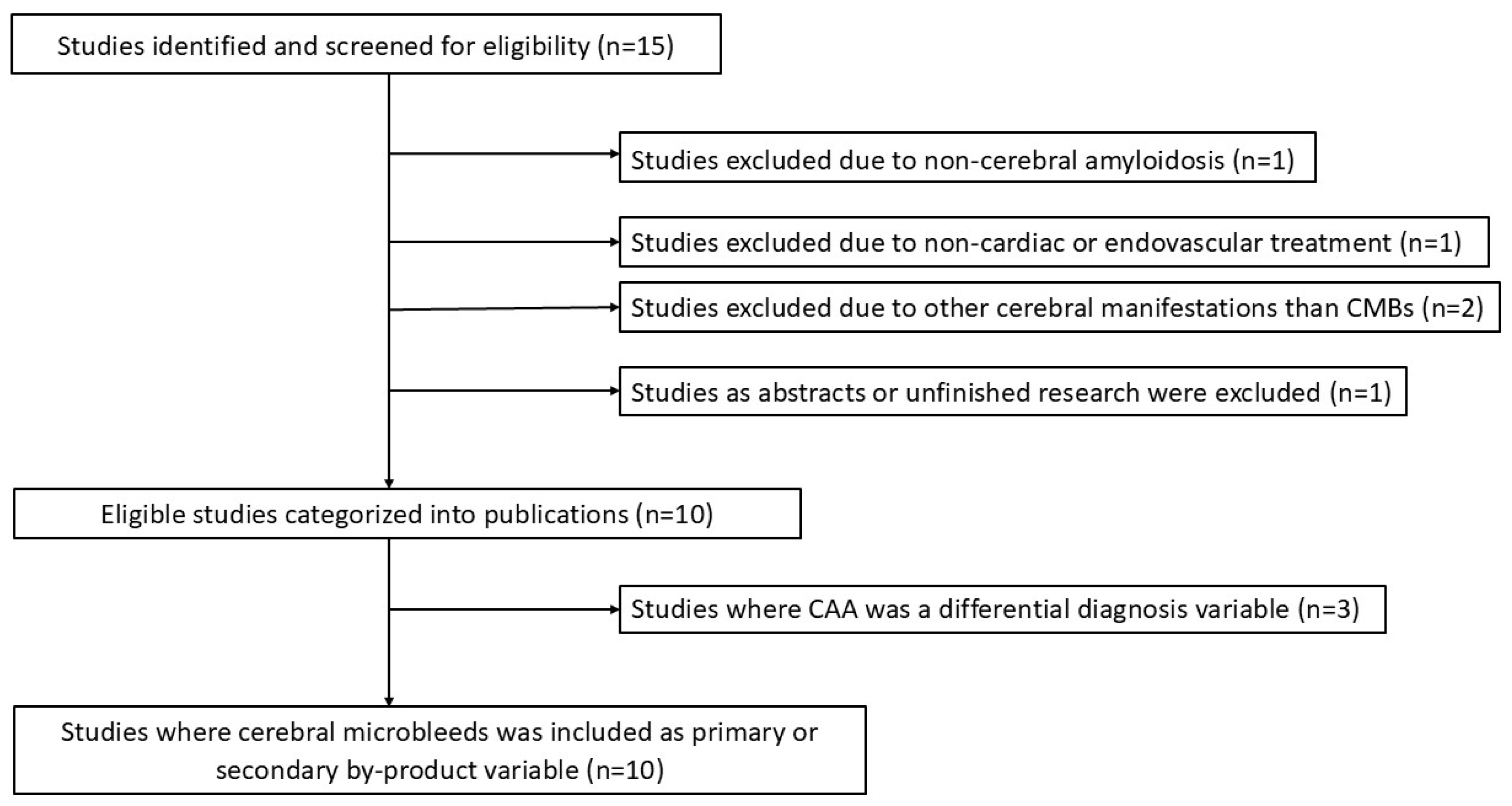
To increase consistency, all the reviewers screened the same 15 publications, discussed the results, and amended the screening and data extraction manually. Reviewers working solo sequentially evaluated the titles, abstracts, and then the full text of all the publications identified by our searches for potentially relevant information and which one to exclude. We resolved disagreements on study selection and data extraction by consensus and discussion with other reviewers if needed.
A data-charting form was jointly developed by two reviewers (M.G.M. and T.C.) to determine which variables to extract. The two reviewers independently charted the data, discussed the results, and continuously updated the data-charting form in an iterative process.
We grouped the studies by incidence of post-procedural CMBs, and summarized the patients’ cohorts, type of surgeries, and CMB location for each group, along with the measures used and broad findings. The study selection process for the review is described in the flowchart (Figure 1).

3. Results

The included studies were published during the 14-year period spanning 2010–2023. The countries of origin were mainly Europe, United States, and Australia and the study design was 50% retrospective and 50% prospective. This review evaluated 10 papers and investigated many cohorts of patients who underwent cardiac or endovascular surgery during a period between 2001 and 2020.
The results of individual sources of evidence are summarized in Table S1 (Supplementary Materials).
All the patients who underwent multiple combined surgery were always excluded from all papers. All the patients were operated on using standardized anesthetic procedures. The preoperative and postoperative MRI protocol included a T2*-GRE or SWI sequence performed with a 1.5 or 3 Tesla Magnetic Resonance (MR) scanner. Only in three studies [12,13,14], preoperative MR was not reported; when performed, pre-procedural imaging was obtained from a maximum of one year to a minimum of one day before surgery. At the same time, post-procedural MR was scheduled from 1 day to 4–6 years after surgery according to the patient’s clinical condition. The majority of the dedicated images were interpreted by two or three investigators, while only in four papers [14,15,16,17] imaging was observed by one neuroradiologist with at least one year of experience.
The majority of CMBs were characterized as small, rounded, or circular, well-defined hypointense lesions within brain parenchyma with clear margins and size between 1 mm and 10 mm on the SWI or T2*-GRE images, while two papers defined them as lesions measuring less than 5 mm [12,17].
All these studies seemed to lean towards the idea of two main differential diagnoses: calcific or metallic material emboli and microhemorrhages, even though the majority of them predominantly support the hemorrhagic nature. Moreover, De Sciscio et al. [14] and Breiding et al. [18] stated no significant relevance of CMBs between mechanical or biological heart valve implantation. Therefore, the hypothesis of microembolization from metallic abrasion was gradually being abandoned.
Indeed, another relevant predictor of CMBs seems to be prolonged surgical times, especially in the case of CPB. Hence, all the papers focusing on CPB confirmed a strong association between CPB time and SWI or T2*-GRE lesion genesis. For example, in Patel et al. [15] and Kim et al. [12], CPB increased the percentage of new microbleeds by about 97% and 91%, respectively.
A key factor in assessing the differential diagnosis of CMBs seems to be their location and distribution in brain parenchyma.
Only six studies [14,16,17,18,19,20] reported CMBs located in the deep white matter and/or basal ganglia following cardiac or endovascular interventions, ensuring that differential diagnoses were made to distinguish them from calcifications. However, the occurrence of CMBs in deep locations was not commonly observed, less than 33%, with most CMBs showing a mixed distribution (both lobar and deep) [14,16]. Moreover, none of the papers deepens the possible correlation with other known risk factors such as hypertension, diabetes, or cardiac and small vessel diseases.
Meanwhile, seven [13,14,15,16,17,20,21] papers describe the majority of CMBs located in the lobar subcortical white matter (SWM), typically with a bilateral distribution (Figure 2).
Notably, De Sciscio et al. [14] and Xiong et al. [17] reported a predilection at the gray–white matter junction in 76% and 72.5% of the cases, respectively, thus performing a significant trial in the differential diagnosis with CAA. In this framework, three researchers [14,15,18] specifically analyzed MRI scans of all the patients, trying to assess the differential diagnosis with CAA using the Modified Boston Criteria for “probable CAA”.
In the De Sciscio et al. study [14], 39% of the patients who had undergone cardiac surgery met the Modified Boston Criteria for “probable CAA” based on both clinical and radiological assessments. Although data on target INR or anticoagulation status were not always available, the burden of CMBs remained comparable between anticoagulated and non-anticoagulated patients, suggesting its independence from anticoagulation therapy.
Since they found out the proportion of CMBs in white matter was higher in cardiac surgery patients compared to the CAA patients, they evaluated that the ratio of SWM to cortical CMBs was significantly lower in the latter group; however, no significant differences were found among different surgical procedures, especially valve replacement and CABG.
In Patel et al. [15], only 12% of the patients were diagnosed with probable CAA with the Evolution of Boston Criteria for Diagnosis of Cerebral Amyloid Angiopathy [10]. All the patients were reported to have developed new CMBs after cardiac surgery pointing also up the vulnerability of patients with probable CAA to cerebral hemorrhagic damage when undergoing cardiac surgery.
Breiding et al. [18] observed that the criteria of “possible” and “probable” CAA were fulfilled in 6.1% and 16.4% of the patients, respectively, on the first post-interventional scan, with only 3.6% of the patients showing superficial siderosis. They also noted that patients with biological heart valves were more likely to fulfill CAA criteria versus patients with mechanical heart valves.
From a clinical point of view, however, the majority of the studies did not show a significant link between cognitive impairment and post-surgical CMBs, although their association with long-term neurological dysfunction remains unknown.
Only De Sciscio et al. [14] found a sample of patients presenting transient neurological symptoms, although they underlined that this association may not be representative of the broader population with a history of either cardiac surgery or CAA. Indeed, all the papers stated that the presence of CMBs in the general population is known to be associated with cognitive decline, so further investigation of larger cohorts with long-term follow-up, including neurological and neuropsychiatric assessment, could help to clarify a potential association between CMBs, cognitive function, and dementia in patients undergoing endovascular or open surgeries.

4. Discussion

4.1. Summary of Evidence

Our study analyzed 10 papers published over a 14-year period from 2010 to 2023 and included a cohort of more than 500 patients who underwent various cardiac or endovascular interventions between 2001 and 2020. The primary focus was the detection of new cerebral microbleeds after treatment, analyzed with MRI using GRE or SWI techniques. CMBs were mostly small (usually <10 mm) rounded lesions, and the main assumption about their hemorrhagic nature is strongly linked to prolonged procedural times, particularly after CPB, which is associated with significant platelet consumption. Another key finding was the distribution of CMBs, primarily observed in the SWM. This highlights the importance of accurately assessing the exact location of CMBs to differentiate from hypertension and CAA. In the first case, CMBs are often found in deep regions of the brain while in CAA these are typically found in the cortical layers of the brain, as defined by the Modified Boston Criteria, and are often associated with cortical hemosiderosis.
Among the included studies, only Van Belle et al. [19] found an association between CMBs and the presence of hypertension, diabetes, or TIA (p-values, respectively, 0.04, 0.05 and 0.02). In addition, hypertension-related CMBs, which are typically localized in deep brain regions such as the basal ganglia and cerebellar dentate nuclei, differ from the lobar distribution commonly observed in CAA. This distinct pattern facilitates a more straightforward differential diagnosis between the two conditions [1].
CMBs are emerging as key imaging markers of small vessel pathology and have been robustly linked to cognitive decline and dementia [22]. Different studies have shown that patients with CMBs tend to perform worse on global cognitive assessments [22,23]. Furthermore, the spatial distribution of these lesions, especially the difference between strictly lobar and deep microbleeds, seems to modulate both the severity of cognitive impairment and the specific domain mostly affected [24].
Diffusion tensor imaging (DTI) has revealed significant microstructural changes in white matter tracts, such as the internal capsule and corpus callosum [6], in patients with CMBs. These findings suggest that CMBs may indicate widespread brain connectivity disruption, underscoring their potential role in the early detection and prognostication of cognitive decline [7].
Although the relationship between CMBs and cognitive impairment currently remains inconclusive, it represents a critical area for future research, particularly in studies with long-term follow-up.

4.2. The Accuracy of MR Imaging—A SWI Technical Note

The accurate identification and quantification of CMBs vary significantly depending on magnetic field strength and the imaging technique used, such as SWI or T2*-GRE sequences. Indeed, SWI is a magnetic resonance sequence that is highly sensitive to the presence of hemosiderin, and it has been shown to be 3–6 times more sensitive than conventional T2*-GRE sequences to detect microhemorrhages [12] (Figure 3).
Therefore, the lower rate of postoperative CMB lesions might be linked to the lower sensitivity of T2*-GRE sequences compared with SWI [18]. In our review, three papers [16,19,20] used only T2*-GRE sequences to detect microhemorrhages while three others used both techniques (when available).
SWI is an advanced MRI technique that, like conventional T2*-GRE weighted imaging, is sensitive to substances that distort the local magnetic field (either paramagnetic or diamagnetic compounds). However, SWI, unlike T2*-GRE, can be considered the evolution of standard GRE sequences since that combines high spatial resolution (being a three-dimensional sequence) with specific post-processing techniques such as phase filtering and masking to highlight these substances. In SWI images, like in T2*-GRE images, calcifications and hemosiderin deposits appear both as small, round foci of hypointensity. For this reason, a critical technical aspect of SWI is the “handedness” of the MRI system, which influences the interpretation of phase shifts in the image, thus allowing the differentiation between paramagnetic (e.g., hemosiderin or deoxygenated blood) or diamagnetic substances (e.g., calcium). In fact, as already said, the accuracy in detecting the nature (i.e., calcium oh hemosiderin) of a SWI-hypointense focus might be challenging without looking at phase images. Depending on their positive and negative phase shifts, we can distinguish right- and left-handed systems in which paramagnetic and diamagnetic substances’ appearance changes. Indeed, it was described that right-handed systems, in which microbleeds appear hypointense and calcification hyperintense on phase images, seem the most effective in enhancing their differentiation [25] (Figure 4). However, the choice of the handedness of an MRI system depends on the vendor. Clinicians and radiologists should always be aware of the handedness of their system, even though there are some ‘tips’ to assess it when it is not explicitly specified [25].
To date, the SWI sequence is widely available but requires MR scanners with high field strength (at least 1.5 Tesla). Although SWI offers greater sensitivity than T2* GRE, it may yield a higher number of false-positive CMBs [26] and its longer acquisition times make it more prone to motion artifacts [27]. Moreover, SWI might lose quality near air–tissue interfaces—particularly around the temporal bone—due to susceptibility artifacts [28].

4.3. “Probable” or “Possible” CAA—Differential Diagnosis and Clinical Implications

Among the included papers, only three [14,15,18] highlight the challenging differential diagnosis between post-surgery CMBs and CAA, identifying “probable” or “possible” diagnosis of cerebral amyloidosis based on Boston Modified Criteria (Table S2 in Supplementary Materials).
Therefore, a reliable statement from a “definite” CAA has not yet been identified primarily due to the use of invasive methods to have an accurate diagnosis. The diagnosis of CAA is a four-tiered system using a combination of clinical and neuroradiological parameters confirmed with cerebral biopsy as assessed by Boston Criteria 2.0 [29]. Therefore, a reliable, non-invasive method for diagnosing CAA would facilitate both clinical decisions and the inclusion of patients in drug trials for this untreatable disorder [30]. In fact, in the study by Knudsen et al. [30], which used post-mortem specimens, cortical biopsies, or evacuated hematomas (diagnosed by CT or MRI), all 13 patients clinically diagnosed with “probable” CAA were also found to have CAA pathologically. Among the remaining 26 patients with “possible” CAA, 16 were pathologically diagnosed with CAA. No alternative pathological causes of hemorrhage were identified in the 10 samples without CAA, and none of the pathological specimens showed severe hypertensive vascular changes, vascular malformations, or saccular aneurysms [30]. So, although neuropathologic examination remains the gold standard for “definite” CAA, it is suggested that a reliable diagnosis can be reached from the combination of clinical information and radiological findings [30]. Not only that, recently it has been underlined that an increased sensitivity and specificity in diagnosing amyloidosis depend on whether CMBs are found in symptomatic patients and if they are associated with intraparenchymal hematomas [31] or/and with cortical superficial siderosis [10]. Indeed, superficial siderosis, either focal or disseminated and mainly with a supratentorial location, seems most commonly associated with CAA but not with other causes of lobar intracerebral macro hemorrhage [32].
Therefore, in this setting, MRI, especially SWI sequences, assumes a crucial role not only in detecting the location of CMBs but also in ruling out other findings that may aid in differential diagnosis.
In De Sciscio et al. [14], a curve analysis of a ‘location-based’ ratio was obtained by calculating SWM/(SWM + strictly cortical CMBs) for patients who underwent CABG or valve replacement and for CAA patients. Indeed, most patients in both valve replacement and CABG groups had a lobar-restricted pattern (with a smaller proportion exhibiting a mixed pattern). A significant percentage of these patients mainly showed cortical/juxtacortical microbleeds, especially in the valve replacement group, while fewer microbleeds were found in the subcortical white matter. No significant differences regarding microbleed location or frequency were observed. When comparing the CMB burden in patients with CAA to those with post-surgical microbleeds, the CAA group exhibited a significantly higher load, particularly in cortical regions. For this reason, the authors conclude that the optimal “location-based ratio” to differentiate post-treatment CMBs from CAA might be approximately 0.45. Additionally, superficial siderosis was found in a significant proportion of CAA patients, while it was absent in those who underwent cardiac surgery [14] (Figure 5).
In summary, CAA typically presents in older patients with a history of lobar hemorrhages and cognitive decline, with MRI findings showing lobar microbleeds and cortical superficial siderosis. In contrast, perioperative microbleeds occur in the context of surgery, often associated with hemodynamic instability or microembolization. Radiologically, CAA exhibits a characteristic lobar distribution, while perioperative CMBs tend to show a more diffuse or non-specific pattern. Clinical history and timing relative to surgery are crucial for distinguishing between the two conditions.
From a more pragmatic point of view, it might be suggested to perform a preoperative MRI with SWI or T2*-GRE sequences, especially in elderly patients or in those with familiarity with brain small vessel disease or CAA, to determine the presence of CMBs and thus make the differential diagnosis easier.

4.4. Pathophysiological Mechanisms and Insights

It is well known that there are various manifestations of perioperative cerebral injury, including ischemic or, less commonly, hemorrhagic stroke, encephalopathy, and neurocognitive dysfunction after surgery [33]. Still, the underlying pathophysiological mechanisms remain a topic of ongoing debate, particularly in the field of cardiac surgery.
Cardiac surgery, especially when associated with CPB, induces an acute phase reaction that seems implicated in the pathogenesis of several postoperative complications [34] and systemic inflammatory response syndrome (SIRS) [35]. A complex sequence of events leads to the activation of leukocytes and endothelial cells, responsible for cell dysfunction in different organs. Activation of the contact system, endotoxemia, ischemia, reperfusion injury, and surgical trauma are potential inflammation triggers following CPB, where different pro- and anti-inflammatory mediators (cytokines and adhesion molecules) are involved [34]. Therefore, a state of inflammatory reaction contributes to general postoperative complications with multiple organ failure (MOF), with cerebral injury often presenting as multiple embolic infarcts detected via diffusion-weighted imaging (DWI) [20].
Kazuo et al.’s [36] research deepened the pathological mechanism for ischemic stroke after cardiac surgery with CPB and found the serum levels of S-100B gradually increased after CPB, with high levels at 24 h. These results suggest that S-100B may be a useful marker in the prediction of cerebral infarction after cardiac surgery. This protein, part of the calcium-binding S-100 family, leaks from damaged nerve cells into the cerebrospinal fluid and crosses into serum when blood–brain barrier (BBB) permeability increases.
CMBs are another form of brain injury following surgery, though their exact pathogenesis remains unclear. Unlike ischemic lesions, new GRE or SWI lesions seem to be mostly asymptomatic and not associated with ischemia [16,17,20]; thus, we can assume the pathogenesis to be different.
Shasha et al.’s [37] study found a strong link between inflammatory cytokines and the increased risk of CMBs in cerebral small vessel disease. Indeed, they assumed that high circulation levels of cytokine FGF-21 exhibited a potential reverse and significant correlation with the risk of CMBs. FGF-21 belongs to the fibroblast growth factor (FGF) family and seems to be protective against inflammatory cerebral vessel diseases.
It is well established that the BBB prevents peripheral harmful substances from entering the central nervous system, but in some pathological conditions, an increased BBB permeability is observed. Indeed, they suggest that FGF-21 promotes angiogenesis by up-regulating vascular endothelial growth factor (VEGF) expression, thereby preventing BBB leakage and reducing neuroinflammation. In addition, it seems VEGF to be a crucial indicator of endothelial dysfunction and is strongly associated with CMBs.
These processes—both systemic inflammation and vascular permeability—may facilitate CMB origin.
However, while it has been suggested that inflammation and increased BBB leakage are associated with CMBs [38], a recent paper [4] studying cerebral small vessel disease patients found no association between CMBs and BBB leakage or microglial activation.
Cardiac and endovascular surgeries, particularly those involving CPB, often generate embolic particles such as calcific debris, thrombotic material, and gaseous emboli. These emboli can reach small cerebral vessels, causing localized endothelial damage and subsequent microhemorrhages. Furthermore, mechanical manipulation during procedures like TAVI and TEVAR may amplify these risks due to the physical stress exerted on calcified structures and vessel walls. Indeed, CMB lesions are located in multiple vascular territories, so an embolic and secondary hemorrhagic origin may be hypothesized mainly referring to embolic microcalcifications from large vessels, such as the aorta [13].
In almost all the papers, a common assumption consists of CMBs being the consequence of haemodilution in CPB leading to platelet consumption and plasma coagulation factor depletion [20]. These processes lead to an impairment of the coagulation cascades and increase vascular permeability. Moreover, prolonged bypass times may exacerbate platelet activation and consumption, further compromising BBB integrity. It should also be emphasized that patients with pre-existing cerebrovascular disease, such as CAA or small vessel disease, are particularly susceptible to CMB formation due to their structurally weakened vessel walls [14]. In fact, the presence of amyloid deposits weakens vessel walls, making them more prone to damage under procedural stress; for these reasons, these patients might be at increased risk of intracerebral hemorrhage or more prone to developing cognitive decline.
In conclusion, the formation of CMBs following cardiac and endovascular procedures may result from a complex interplay of hemodynamic, hematological, and procedural factors where the integrity of the cerebral microvasculature seems to be significantly affected, serving as a central element in this process. Shear stress from altered hemodynamic conditions during these procedures can also damage endothelial cells, increasing capillary fragility. The neurovascular unit, composed of endothelial cells, astrocytes, pericytes, and neurons, also plays a crucial role in maintaining BBB integrity. Procedural stress and systemic inflammation compromise this unit, facilitating blood component leakage into brain parenchyma and leading to CMBs. A schematic overview of the main pathophysiological mechanisms leading to CMBs is provided in Table S3 (in Supplementary Materials).

5. Limitations

Our scoping review has several limitations. To enhance feasibility, we restricted our analysis to a sample of papers sourced from a single public website. Consequently, our results are likely representative only of the publicly available literature. Additionally, the intervals between surgery and MRI scans were highly heterogeneous, and pre-procedural scans were conducted in only six studies. The indications for MRI scanning and the intervals between scans also lacked standardization, as well as the imaging protocol used (SWI or GRE).
Our findings suggest that other factors beyond CPB and prolonged procedural times may contribute to SWI lesion formation. Moreover, CPB might reflect the complexity of procedures and other confounding factors rather than serving as a definitive pathophysiological explanation for SWI lesions.

6. Conclusions and Future Directions

This review confirmed that CMBs are a common occurrence following cardiac, TAVR, and TEVAR surgeries, with the majority of patients exhibiting new CMBs in post-treatment imaging. Advances in imaging techniques such as SWI sequences have further highlighted the prevalence of subtle procedural injuries previously undetectable with conventional methods, offering new insights into the microvascular changes that occur post-surgery. These CMBs were predominantly located in the lobar SWM, a distribution pattern that may be useful in differential diagnosis with CAA-related CMBs. Among all the factors analyzed, only surgical duration and CPB time emerged as significant predictors of new CMBs. Although anticoagulation use and neurological impairment were not significantly associated with new CMBs, these trends warrant further investigation in larger and multicenter studies, with standardized imaging protocols, to clarify their potential implications (Table 1).
Long-term follow-up studies are essential to fully assess the cumulative impact of CMBs on cognitive decline and dementia progression. Moreover, advanced techniques such as diffusion tensor imaging (DTI) could help detect microstructural alterations in white matter. When combined with comprehensive neurological evaluation, these approaches would provide a deeper understanding of the underlying mechanisms and allow for more effective patient management strategies.

Supplementary Materials

The following supporting information can be downloaded at https://www.mdpi.com/article/10.3390/jvd4020016/s1, Table S1: Major findings and characteristics of sources of evidence; Table S2: Main and common neurological presentations between cerebral amyloid angiopathy (CAA) and post-cardiac and endovascular procedures events. Table S3. Main pathophysiological mechanisms leading to CMBs.

Author Contributions

Conceptualization: M.G.M., T.C. and I.L.; data collection: M.G.M., T.C. and R.T.; data and imaging analysis: M.G.M., T.C. and I.L.; original draft preparation: M.G.M.; writing, review and editing: T.C.; critical review: I.L., A.B., A.G., G.B., S.S. and W.D.; supervision: E.F. All authors have read and agreed to the published version of the manuscript.

Funding

This research received no external funding.

Data Availability Statement

The original data presented in the study are openly available in the online PubMed database (https://pubmed.ncbi.nlm.nih.gov/, accessed on 15 December 2024).

Conflicts of Interest

The authors declare no conflicts of interest.

Abbreviations

The following abbreviations are used in this manuscript:
SWIsusceptibility-weighted imaging
GREgradient-echo
MRmagnetic resonance
MRImagnetic resonance imaging
CMBscerebral microbleeds
CPBcardiopulmonary bypass
CABGcoronary artery bypass grafting
TEVARthoracic endovascular aortic repair
TAVItranscatheter aortic valve implantation
CAAcerebral amyloid angiopathy
TAVRTranscatheter Aortic Valve Replacement
SWMsubcortical white matter
SIRSsystemic inflammatory response syndrome
MOFMultiorgan Failure
DWIdiffusion-weighted imaging
FGFfibroblast growth factor
BBBblood–brain barrier
VEGFvascular endothelial growth factor
vWFVon Willebrand Factor

References

  1. Greenberg, S.M.; Vernooij, M.W.; Cordonnier, C.; Viswanathan, A.; Al-Shahi Salman, R.; Warach, S.; Launer, L.J.; Van Buchem, M.A.; Breteler, M.M. Cerebral Microbleeds: A Guide to Detection and Interpretation. Lancet Neurol. 2009, 8, 165–174. [Google Scholar] [CrossRef] [PubMed]
  2. Akoudad, S.; Wolters, F.J.; Viswanathan, A.; de Bruijn, R.F.; van der Lugt, A.; Hofman, A.; Koudstaal, P.J.; Ikram, M.A.; Vernooij, M.W. Association of Cerebral Microbleeds With Cognitive Decline and Dementia. JAMA Neurol. 2016, 73, 934. [Google Scholar] [CrossRef] [PubMed]
  3. Agarwal, A.; Ajmera, P.; Sharma, P.; Kanekar, S. Cerebral Microbleeds: Causes, Clinical Relevance, and Imaging Approach—A Narrative Review. J. Neurosci. Rural Pract. 2024, 15, 169–181. [Google Scholar] [CrossRef]
  4. Cai, L.; Tozer, D.J.; Markus, H.S. Cerebral Microbleeds and Their Association With Inflammation and Blood-Brain Barrier Leakage in Small Vessel Disease. Stroke 2025, 56, 427–436. [Google Scholar] [CrossRef]
  5. Liu, J.; Kou, Z.; Tian, Y. Diffuse Axonal Injury after Traumatic Cerebral Microbleeds: An Evaluation of Imaging Techniques. Neural Regen. Res. 2014, 9, 1222. [Google Scholar] [CrossRef] [PubMed]
  6. Liu, J.Y.; Zhou, Y.J.; Zhai, F.F.; Han, F.; Zhou, L.X.; Ni, J.; Yao, M.; Zhang, S.; Jin, Z.; Cui, L.; et al. Cerebral Microbleeds Are Associated with Loss of White Matter Integrity. Am. J. Neuroradiol. 2020, 41, 1397–1404. [Google Scholar] [CrossRef]
  7. Sui, C.; Wen, H.; Wang, S.; Feng, M.; Xin, H.; Gao, Y.; Li, J.; Guo, L.; Liang, C. Characterization of White Matter Microstructural Abnormalities Associated with Cognitive Dysfunction in Cerebral Small Vessel Disease with Cerebral Microbleeds. J. Affect. Disord. 2023, 324, 259–269. [Google Scholar] [CrossRef]
  8. Studerus-Germann, A.M.; Gautschi, O.P.; Bontempi, P.; Thiran, J.-P.; Daducci, A.; Romascano, D.; von Ow, D.; Hildebrandt, G.; von Hessling, A.; Engel, D.C. Central Nervous System Microbleeds in the Acute Phase Are Associated with Structural Integrity by DTI One Year after Mild Traumatic Brain Injury: A Longitudinal Study. Neurol. Neurochir. Pol. 2018, 52, 710–719. [Google Scholar] [CrossRef]
  9. Dahl, J.; Tenovuo, O.; Posti, J.P.; Hirvonen, J.; Katila, A.J.; Frantzén, J.; Maanpää, H.-R.; Takala, R.; Löyttyniemi, E.; Tallus, J.; et al. Cerebral Microbleeds and Structural White Matter Integrity in Patients With Traumatic Brain Injury—A Diffusion Tensor Imaging Study. Front. Neurol. 2022, 13, 888815. [Google Scholar] [CrossRef]
  10. Greenberg, S.M.; Charidimou, A. Diagnosis of Cerebral Amyloid Angiopathy Evolution of the Boston Criteria. Stroke 2018, 49, 491–497. [Google Scholar] [CrossRef]
  11. Tricco, A.C.; Lillie, E.; Zarin, W.; O’Brien, K.K.; Colquhoun, H.; Levac, D.; Moher, D.; Peters, M.D.J.; Horsley, T.; Weeks, L.; et al. PRISMA Extension for Scoping Reviews (PRISMA-ScR): Checklist and Explanation. Ann. Intern. Med. 2018, 169, 467–473. [Google Scholar] [CrossRef] [PubMed]
  12. Kim, P.P.C.; Nasman, B.W.; Kinne, E.L.; Oyoyo, U.E.; Kido, D.K.; Jacobson, J.P. Cerebral Microhemorrhage: A Frequent Magnetic Resonance Imaging Finding in Pediatric Patients after Cardiopulmonary Bypass. J. Clin. Imaging Sci. 2017, 7, 27. [Google Scholar] [CrossRef]
  13. Eilenberg, W.; Bechstein, M.; Charbonneau, P.; Rohlffs, F.; Eleshra, A.; Panuccio, G.; Bhangu, J.S.; Fiehler, J.; Greenhalgh, R.M.; Haulon, S.; et al. Cerebral Microbleeds Following Thoracic Endovascular Aortic Repair. Br. J. Surg. 2022, 109, 46–52. [Google Scholar] [CrossRef] [PubMed]
  14. De Sciscio, M.; De Sciscio, P.; Vallat, W.; Kleinig, T. Cerebral Microbleed Distribution Following Cardiac Surgery Can Mimic Cerebral Amyloid Angiopathy. BMJ Neurol. Open 2021, 3, e000166. [Google Scholar] [CrossRef] [PubMed]
  15. Patel, N.; Banahan, C.; Janus, J.; Horsfield, M.A.; Cox, A.; Li, X.; Cappellugola, L.; Colman, J.; Egan, V.; Garrard, P.; et al. Perioperative Cerebral Microbleeds after Adult Cardiac Surgery. Stroke 2019, 50, 336–343. [Google Scholar] [CrossRef]
  16. Braemswig, T.B.; Kusserow, M.; Bellmann, B.; Beckhoff, F.; Reinthaler, M.; von Rennenberg, R.; Erdur, H.; Scheitz, J.F.; Galinovic, I.; Villringer, K.; et al. New Cerebral Microbleeds After Catheter-Based Structural Heart Interventions: An Exploratory Analysis. J. Am. Heart Assoc. 2023, 12, e8189. [Google Scholar] [CrossRef]
  17. Xiong, T.Y.; Luo, C.Y.; Tang, H.H.; Søndergaard, L.; Prendergast, B.; Lui, S.; Chen, M. Novel Neuroimaging Evidence of Brain Lesions Following Transcatheter Aortic Valve Replacement. J. Am. Heart Assoc. 2021, 10, e023395. [Google Scholar] [CrossRef]
  18. Breiding, P.S.; Duerrenmatt, J.T.; Meinel, F.G.; Carrel, T.; Schönhoff, F.; Zibold, F.; Kaesmacher, J.; Gralla, J.; Pilgrim, T.; Jung, S.; et al. Prevalence and Evolution of Susceptibility-Weighted Imaging Lesions in Patients With Artificial Heart Valves. J. Am. Heart Assoc. 2019, 8, e012814. [Google Scholar] [CrossRef]
  19. Van Belle, E.; Debry, N.; Vincent, F.; Kuchcinski, G.; Cordonnier, C.; Rauch, A.; Robin, E.; Lassalle, F.; Pontana, F.; Delhaye, C.; et al. Cerebral Microbleeds during Transcatheter Aortic Valve Replacement: A Prospective Magnetic Resonance Imaging Cohort. Circulation 2022, 146, 383–397. [Google Scholar] [CrossRef]
  20. Jeon, S.B.; Lee, J.W.; Kim, S.J.; Chung, C.H.; Kwon, S.U.; Choi, C.G.; Choo, S.J.; Nah, H.W.; Kim, J.S.; Kang, D.W. New Cerebral Lesions on T2*-Weighted Gradient-Echo Imaging after Cardiac Valve Surgery. Cerebrovasc. Dis. 2010, 30, 194–199. [Google Scholar] [CrossRef]
  21. Michałowska, I.; Furmanek, M.I.; Smaga, E.; Juraszyński, Z.; Zieliński, T.; Chełstowska, S.; Kuśmierczyk, M.; Szpakowski, E.; Mierzyńska, A.; Walecki, J.M. Evaluation of Brain Lesions in Patients after Coronary Artery Bypass Grafting Using MRI with the Emphasis on Susceptibility-Weighted Imaging. Kardiochirurgia I Torakochirurgia Pol. 2015, 12, 1–7. [Google Scholar] [CrossRef]
  22. Wu, R.; Feng, C.; Zhao, Y.; Jin, A.P.; Fang, M.; Liu, X. A Meta-Analysis of Association between Cerebral Microbleeds and Cognitive Impairment. Med. Sci. Monit. 2014, 20, 2189–2198. [Google Scholar] [CrossRef] [PubMed]
  23. Cui, Y.; Zhao, T.; Zhang, W.; Wang, R.; Hu, M.; He, X.; Wang, Y.; Xie, H. Risk Factor Analysis in Patients Exhibiting Cerebral Microbleeds and the Correlation with Cognitive Impairment. Gerontol. Geriatr. Med. 2024, 10, 1–12. [Google Scholar] [CrossRef] [PubMed]
  24. Chung, C.P.; Chou, K.H.; Chen, W.T.; Liu, L.K.; Lee, W.J.; Chen, L.K.; Lin, C.P.; Wang, P.N. Strictly Lobar Cerebral Microbleeds Are Associated with Cognitive Impairment. Stroke 2016, 47, 2497–2502. [Google Scholar] [CrossRef]
  25. Haller, S.; Haacke, E.M.; Thurnher, M.M.; Barkhof, F. Susceptibility-Weighted Imaging: Technical Essentials and Clinical Neurologic Applications. Radiology 2021, 299, 3–26. [Google Scholar] [CrossRef]
  26. Shams, S.; Martola, J.; Cavallin, L.; Granberg, T.; Shams, M.; Aspelin, P.; Wahlund, L.O.; Kristoffersen-Wiberg, M. SWI or T2*: Which MRI Sequence to Use in the Detection of Cerebral Microbleeds? The Karolinska Imaging Dementia Study. Am. J. Neuroradiol. 2015, 36, 1089–1095. [Google Scholar] [CrossRef]
  27. Rubin, A.; Waszczuk, Ł.; Trybek, G.; Kapetanakis, S.; Bladowska, J. Application of Susceptibility Weighted Imaging (SWI) in Diagnostic Imaging of Brain Pathologies—A Practical Approach. Clin. Neurol. Neurosurg. 2022, 221, 107368. [Google Scholar] [CrossRef] [PubMed]
  28. Halefoglu, A.M.; Yousem, D.M. Susceptibility Weighted Imaging: Clinical Applications and Future Directions. World J. Radiol. 2018, 10, 30–45. [Google Scholar] [CrossRef]
  29. Jensen-Kondering, U.; Heß, K.; Neumann, A.; Margraf, N.G. Neuroradiological Findings in Cerebral Amyloid Angiopathy with a Particular Consideration of the Boston Criteria 2.0: An Imaging Review. Biomolecules 2024, 14, 1459. [Google Scholar] [CrossRef]
  30. Knudsen, K.A.; Rosand, J.; Karluk, D.; Greenberg, S.M. Clinical Diagnosis of Cerebral Amyloid Angiopathy: Validation of the Boston Criteria. Neurology 2001, 56, 537–539. [Google Scholar] [CrossRef]
  31. van Rooden, S.; van der Grond, J.; van den Boom, R.; Haan, J.; Linn, J.; Greenberg, S.M.; van Buchem, M.A. Descriptive Analysis of the Boston Criteria Applied to a Dutch-Type Cerebral Amyloid Angiopathy Population. Stroke 2009, 40, 3022–3027. [Google Scholar] [CrossRef] [PubMed]
  32. Linn, J.; Halpin, A.; Demaerel, P.; Ruhland, J.; Giese, A.D.; Dichgans, M.; van Buchem, M.A.; Bruckmann, H.; Greenberg, S.M. Prevalence of Superficial Siderosis in Patients with Cerebral Amyloid Angiopathy. Neurology 2010, 74, 1346–1350. [Google Scholar] [CrossRef]
  33. Hogue, C.W.; Gottesman, R.F.; Stearns, J. Mechanisms of Cerebral Injury from Cardiac Surgery. Crit. Care Clin. 2008, 24, 83–98, viii–ix. [Google Scholar] [CrossRef]
  34. Paparella, D.; Yau, T.M.; Young, E. Cardiopulmonary Bypass Induced Inflammation: Pathophysiology and Treatment. An Update. Eur. J. Cardiothorac. Surg. 2002, 21, 232–244. [Google Scholar] [CrossRef] [PubMed]
  35. Hohn, A.; Malewicz-Oeck, N.M.; Buchwald, D.; Annecke, T.; Zahn, P.K.; Baumann, A. REmoval of Cytokines during CArdiac Surgery (RECCAS): A Randomised Controlled Trial. Crit. Care 2024, 28, 406. [Google Scholar] [CrossRef] [PubMed]
  36. Nakamura, K.; Ueno, T.; Yamamoto, H.; Iguro, Y.; Yamada, K.; Sakata, R. Relationship between Cerebral Injury and Inflammatory Responses in Patients Undergoing Cardiac Surgery with Cardiopulmonary Bypass. Cytokine 2005, 29, 95–104. [Google Scholar] [CrossRef]
  37. Han, S.; Chen, Q.; Zhu, Q.; Han, W. Circulating Inflammatory Cytokines and the Risk of Cerebral Small Vessel Disease: A Bidirectional Mendelian Randomization Analysis. J. Stroke Cerebrovasc. Dis. 2025, 34, 108163. [Google Scholar] [CrossRef]
  38. Rudilosso, S.; Stringer, M.S.; Thrippleton, M.; Chappell, F.; Blair, G.W.; Jaime Garcia, D.; Doubal, F.; Hamilton, I.; Janssen, E.; Kopczak, A.; et al. Blood-Brain Barrier Leakage Hotspots Collocating with Brain Lesions Due to Sporadic and Monogenic Small Vessel Disease. J. Cereb. Blood Flow Metab. 2023, 43, 1490–1502. [Google Scholar] [CrossRef]
Figure 1. Flowchart describing study selection process.
Jvd 04 00016 g001
Figure 2. Susceptibility-weighted imaging (SWI) of a patient after cardiac valve replacement demonstrating lobar and subcortical white matter microbleeds in the frontal and parietal lobes.
Jvd 04 00016 g002
Figure 3. Different accuracy in detection of CMBs burden in gradient-echo (GRE; (A)) and susceptibility-weighted imaging (SWI; (B)) sequences (same patient). Red circles: microbleeds.
Jvd 04 00016 g003
Figure 4. Comparison between the Minimum Intensity Projection (mIP) of susceptibility-weighted imaging, right-handed Phase Imaging, and non-contrast CT scan (same patient). Phase Imaging aids in the differential diagnosis between diamagnetic substances (e.g., calcium, orange arrow in (A,B)) and paramagnetic substances (e.g., CMB, orange arrowhead in (D,E)). The diagnosis is confirmed in a non-contrast CT scan, which clearly demonstrates the presence of a microcalcification in the brain parenchyma (arrow in (C)) and no findings in the site of the microhemorrhage (orange circle in (F)).
Jvd 04 00016 g004
Figure 5. Different CMB distributions (green arrows) in a patient who underwent endovascular surgery (A) versus a patient with CAA (B). Note the location of the microbleed in a lobar-restricted pattern in (A) while a prevalent cortical distribution with superficial hemosiderosis (yellow arrow) in the CAA patient.
Jvd 04 00016 g005
Table 1. Helpful key points of our review.
Key Points
Neuroimaging technique for CMBsImaging sensitivity is crucial: SWI is more sensitive than T2*-weighted gradient echo for CMB detection.
Differential DiagnosisThe distribution of CMBs after cardiac surgery can mimic CAA, highlighting the importance of considering this condition in the differential diagnosis.
Pathophysiological analysisThe mechanisms remain partially understood, with potential contributors including the application of CPB, embolization, issues in anticoagulation management, and inflammatory processes associated with surgery.
Future directionsResearch priorities include assessing the long-term impact of CMBs, as well as the development of targeted interventions to minimize neurological complications in the post-surgical cohort.
Disclaimer/Publisher’s Note: The statements, opinions and data contained in all publications are solely those of the individual author(s) and contributor(s) and not of MDPI and/or the editor(s). MDPI and/or the editor(s) disclaim responsibility for any injury to people or property resulting from any ideas, methods, instructions or products referred to in the content.

