Review

Advancements in Multiple Myeloma Therapies: A Comprehensive Review by Disease Stage

by
Hager Hisham El Khatib
,
Kanz Abdulla
,
Layla Khaled Nassar
,
Mariam Gouda Ellabban
and
Andreas Kakarougkas
*
Department of Biology, The American University in Cairo, Cairo 11835, Egypt
*
Author to whom correspondence should be addressed.
Lymphatics 2025, 3(1), 2; https://doi.org/10.3390/lymphatics3010002
Submission received: 7 October 2024 / Revised: 16 December 2024 / Accepted: 7 January 2025 / Published: 22 January 2025

Abstract

:
Multiple myeloma is an incurable hematologic malignancy arising from plasma cells. The uncontrolled growth of monoclonal plasma cells leads to an abnormal overproduction of immunoglobulins. The recommended course of treatment for MM is according to disease progression and responses to therapeutic intervention, highlighting the necessity for multiple treatment options that alleviate different parts of MM. This comprehensive review provides insights into the current treatments and how to take preventative and prognostic measures. In advanced MM, osteoporosis is a common symptom that originates from a lack of regulation in osteoclast activity and bone resorption. Bisphosphonates such as zoledronic acid and pamidronate along with monoclonal antibodies such as denosumab hinder osteoclast function and aid in reducing the risk of fractures in patients with advanced MM. For targeted therapy approaches, proteasome inhibitors impede protein degradation pathways that cause an accumulation of misfolded proteins promoting cancer cell proliferation in patients with MM. CAR-T is another targeted therapy that can utilize T cells to target and isolate MM cells. Overall, this review highlights the frontrunners of treatments for those diagnosed with MM.

1. Introduction

Multiple myeloma (MM) is a hematological malignancy marked by the uncontrolled clonal expansion of plasma cells in the bone marrow. It presents a significant challenge in oncology due to its intricate pathogenesis and progression. The acronym CRAB (hypercalcemia, renal insufficiency, anemia, and bone lesions) encapsulates the key clinical manifestations of myeloma, reflecting the diverse systemic effects of the disease [1]. The aggregation of aberrant monoclonal plasma cells and immunoglobulins has several complications that are not limited to susceptibility to infections, bone lesions, osteoporosis, and renal failure [2]. MM typically evolves from an initial asymptomatic stage known as monoclonal gammopathy of unknown significance (MGUS) to a transitional phase called smoldering multiple myeloma (SMM), before advancing to active multiple myeloma. The risk of progression from MGUS to multiple myeloma is relatively low, occurring at a rate of about 1% per year. In contrast, patients with SMM face a significantly higher risk of progression, with a rate of around 10% per year [3]. As the disease advances, it can lead to debilitating complications and organ damage, adversely impacting the quality of life and survival of affected individuals. Accurate staging, using systems such as the International Staging System (ISS) and Revised International Staging System (R-ISS), is crucial in determining the extent of myeloma involvement and guiding treatment decisions [1]. In the development of multiple myeloma, the interaction between plasma cells and their surrounding environment is just as crucial as the genetic features of the cancer cells themselves. Angiogenesis, the process of new blood vessel formation, also plays a critical role in the progression of multiple myeloma (MM). In the bone marrow microenvironment, myeloma cells secrete pro-angiogenic factors such as vascular endothelial growth factor (VEGF) and fibroblast growth factor (FGF), which stimulate endothelial cell proliferation and promote tumor growth and survival [4]. Multiple myeloma is genetically very diverse, with nearly all patients exhibiting chromosomal abnormalities and common intraclonal heterogeneity, both of which significantly impact the course of the disease. In reality, multiple myeloma may soon be recognized as a collection of related but distinct diseases rather than a single entity [5]. Treatment options for MM depend on the stage, specific characteristics of the disease, as well as the patient’s age and overall health. There are also cases of relapse that are influenced by factors such as the frequency of relapse, previous treatments, responses to prior therapies, and the patient’s ability to tolerate potential side effects [6]. The management of myeloma involves a multifaceted approach, incorporating various therapeutic modalities, including observational therapy, bi-specific antibodies, bone-modifying drugs, targeted therapies, CAR-T therapy, immunomodulatory drugs, and supportive care measures, aimed at directly targeting the malignant plasma cells or indirectly addressing associated symptoms and complications [1]. The increasing therapeutic options have significantly enhanced the number of multiple myeloma patients achieving a complete response to treatment and have also led to improved progression-free survival. Despite these advancements, a persistent low level of disease or minimal residual disease (MRD) remains, which ultimately contributes to the relapse of the disease in patients with multiple myeloma [7]. This review will focus on the treatment options most commonly administered in the different stages of multiple myeloma.

2. Early-Stage Myeloma

2.1. Smoldering Multiple Myeloma

Monoclonal gammopathy of undetermined significance (MGUS) is characterized by the presence of a serum M-protein less than 3 g/dL, less than 10% clonal plasma cells in the bone marrow, and the absence of plasma cell myeloma (PCM)-related end-organ damage (CRAB symptoms: hypercalcemia, renal insufficiency, anemia, and bone lesions) and absence of B cell lymphoma or other disease known to produce an M-protein [8]. Smoldering multiple myeloma (SMM) is similarly a plasma cell neoplasm that indicates a more advanced stage than MGUS [9]. It is characterized by the presence of ≥3 g/dL serum monoclonal proteins (IgG or IgA) or urinary monoclonal proteins ≥ 5 mg per 24 h and/or 10–60% bone marrow plasma cell infiltration with no myeloma-defining event [10]. The word “smoldering” indicates a “metastable” state that could rapidly evolve into multiple myeloma [9]. While the same can be said for MGUS, patients with SMM are at a significantly higher risk of MM progression [9]. However, it is important to distinguish that it is not a biological intermediate stage between MGUS and MM [11]. In 2014, the International Myeloma Working Group drafted a new set of criteria that would allow those at risk of MM development from SMM to start therapy (Table 1) [12]. According to these criteria, SMM came to include some patients with biological pre-malignancy (biological MGUS) and some with biologic malignancy (multiple myeloma) [12]. In particular, the subset of SMM patients with 70% to 80% risk of progression at 2 years (ultra-high risk SMM) became categorized as active MM and recommended for treatment before the development of end-organ damage. After the exclusion of this group, patients with approximately a 50% risk of progression in 2 years became considered high-risk SMM [13]. Understanding the variability in progression risks within SMM is essential for determining the appropriate treatment approach, as patients with low-risk profiles can often be monitored without intervention, while high-risk patients may benefit from earlier therapeutic strategies.

2.2. Low-Risk SMM

Patients with SMM biomarkers and the absence of high-risk criteria are considered to have low-risk SMM [12]. Biomarkers include a bone marrow plasma cell (BMPC) percentage of at least 60%, CRAB features, lytic lesions, and mild anemia [12]. Patients with low-risk SMM are recommended to adhere to observation as the standard of care with the monitoring of serum M proteins, serum FLC levels, complete blood count, serum calcium, and serum creatinine every 3–4 months [1]. Observation without therapy should take place every 3–4 months. After the first five years, the interval for follow-up can be reduced to once every 6 months [14]. Beyond 6 months, a radiographic examination is also recommended [15]. Data have identified that patients exhibiting 20% or more bone marrow involvement, a more than 10% increase in monoclonal protein level, as well as a more than 0.5 g/dL decrease in blood hemoglobin since diagnosis have a >90% risk of progression within 2 years [16]. If, during follow-up, high-risk SMM criteria are identified, early intervention should be implemented.

2.3. High-Risk SMM

While there are patients with high-risk SMM who can remain stable without therapy for several years, clinical trial data indicate that early treatment delays disease progression and increases overall survival compared to just observation [10]. In particular, the data validate the effectiveness of using lenalidomide plus dexamethasone (Rd) for two years after diagnosis followed by maintenance therapy with lenalidomide [17].
Another proposed treatment option for patients with high-risk SMM is the administration of bisphosphonates. Bisphosphonates are a generally effective class of agents used to fight multiple myeloma; however, clinical trials have failed to prove their role in delaying SMM-associated bone events. One randomized trial of 177 patients compared disease outcomes while administering pamidronate compared to observation alone. Experimental groups were administered doses of both 60 and 90 mg once a month for 12 months and were compared to a group that only underwent observation. While the frequency of skeletal-related events (SREs) in the experimental groups was reduced to once a month for 12 months, no improvement in survival or time to disease progression was recorded [18]. Zoledronic acid was similarly assessed in another randomized trial of 163 participants. A dose of 4 mg once a month compared to just observation was noted for 12 months. Similar results to the pamidronate study were produced with a reduction in SREs but no notable delay in progression or increased survival. Based on this data, Rajkumar et al. [19] have recommended an annual bisphosphonate only for patients with SMM who have osteopenia or osteoporosis.
Recently, more aggressive approaches have been considered for tackling SMM to achieve a minimal residual disease (MRD). A pilot study on 12 high-risk SMM patients involved the administration of 28 cycles of second-generation proteasome inhibitor carfilzomib in combination with lenalidomide and dexamethasone (KRD) [20]. All 12 patients achieved at least a near CR rate, with an MRD negativity rate of 92% by multiparametric flow cytometry and 75% by next-generation sequencing [20]. A larger subset in the same study included patients with Newly Diagnosed Multiple Myeloma (NDMM). It was found that patients with NDMM did not achieve responses as deep as the subset with high-risk SMM [20]. However, further studies are essential to definitively claim that deeper responses in high-risk SMM translate to a clinical benefit of early treatment as opposed to waiting after symptomatic development [20]. While high-risk SMM patients are primarily managed with therapies aimed at delaying progression, another unique aspect of myeloma involves its non-secretory subtype, which presents distinct challenges in diagnosis and monitoring due to the absence of monoclonal proteins.

2.4. Non-Secretory Multiple Myeloma

A subtype of multiple myeloma known as non-secretory multiple myeloma (NSMM) is distinguished by the lack of synthesis of monoclonal proteins in the urine or blood [21]. Compared to secretory multiple myeloma, which is defined by the synthesis of monoclonal proteins that can produce a variety of symptoms such as kidney damage, anemia, and hypercalcemia, this kind of myeloma is frequently linked with a more indolent clinical history. The diagnosis and monitoring of non-secretory multiple myeloma (NSMM) are both rare and difficult. Due to the absence of M-protein secretion, NSMM is commonly diagnosed using different techniques including bone marrow biopsy, imaging tests, and flow cytometry or immunohistochemistry to identify plasma cell types [21]. Treatment approaches for non-small cell myopathy (NSMM) are essentially the same as with secretory types of the illness, ranging from asymptomatic (smoldering) to advanced stages [22]. Depending on the disease’s stage and severity, therapeutic strategies may involve the use of autologous stem cell transplantation, proteasome inhibitors, immunomodulatory medicines (IMiDs), and monoclonal antibodies [22]. Because NSMM does not produce monoclonal proteins, it can be difficult to track the course of the illness; and therefore, it is essential to consider the unique features of NSMM when developing treatment strategies and monitoring disease progression in these patients (Table 2) [23].

3. Advanced-Stage Myeloma

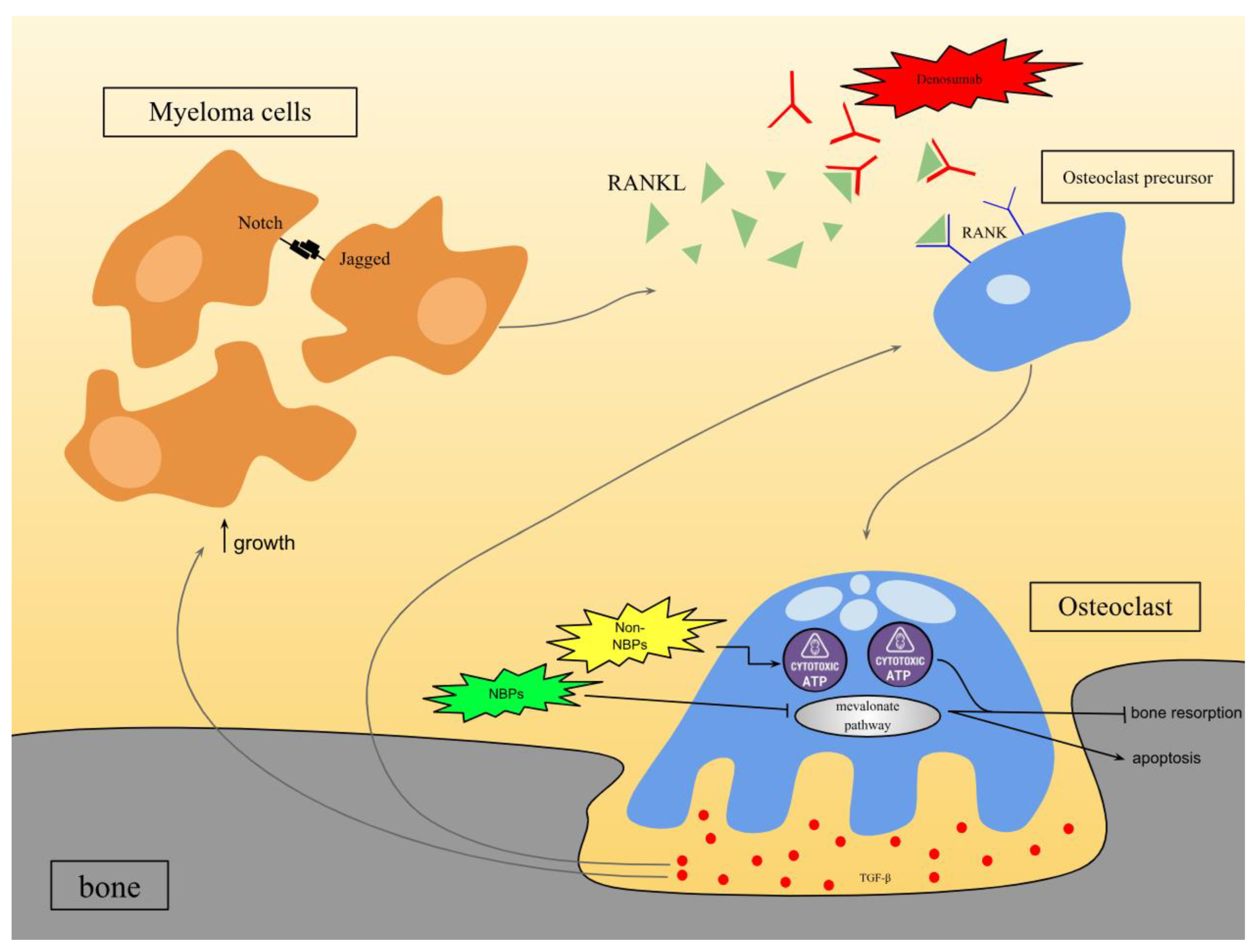
3.1. Bone-Modifying Drugs

One of the biggest symptoms of advanced-stage multiple myeloma (MM) is osteoporosis or weakened bones. This is due to the activity of osteoclasts that dissolve the bone tissue and cause resorption [24]. The mechanism of action is described in Figure 1, and it involves the interaction between receptors Notch and Jagged that stimulate the release of RANKL, a membrane-bound protein. Transforming growth factor beta (TGF-β) is eventually released by the osteoclasts, which further stimulate osteoclastogenesis and myeloma cell growth. This is termed the “vicious cycle” of tumor-related bone diseases [7,25]. The two most common treatment options are bisphosphonates and denosumab.

3.1.1. Bisphosphonates

Bisphosphonates are pyrophosphate analogs that are utilized for antiresorptive therapies [26,27]. They are classified into two classes: the nitrogen-containing bisphosphonates (such as zoledronic acid, ibandronate, and pamidronate) and the non-nitrogen-containing bisphosphonates (such as clodronate and etidronate) [27]. Bisphosphonates bind to hydroxyapatite crystals in the bone matrix where they are absorbed into the osteoclast cells [26,27]. Both classes lead to the loss of osteoclast function and eventual apoptosis; however, they differ in their mechanism of action (illustrated in Figure 1). Nitrogen-containing bisphosphonates (NBPs) function by inhibiting the farnesyl pyrophosphate synthase (FPPS), which is a key regulatory enzyme in the mevalonate pathway. The inhibition of FPPS leads to the inhibition of the mevalonate pathway, which impairs protein prenylation. This induces loss of osteoclast function and prevents bone resorption [28,29]. The non-nitrogen-containing bisphosphonates (non-NBPs) produce cytotoxic metabolites, which lead to the production of cytotoxic ATP-analogs. These cytotoxic ATP analogs compete with the functional ATPs and accumulate intracellularly in osteoclasts, thus interfering with osteoclast function through the inhibition of ATP-dependent enzymes. This similarly causes the loss of osteoclast function and prevents bone resorption [30]. This, in turn, disrupts the tumor cell–osteoclast communication and indirectly influences tumor cell proliferation by preventing the release of the growth factor products of osteoclastogenesis [25]. Nitrogen-containing bisphosphonates are more potent than non-nitro due to their mode of action, which causes a more prominent cellular effect on osteoclasts [30]. Zoledronic acid, for example, reduced skeletal-related fracture risk in 75% of patients in a clinical trial [31]. Pamidronate also reduced vertebral fractures and non-vertebral fractures in 82% and 87% of patients, respectively [31]. Some bisphosphonates have also been reported to work directly on the tumor cells, in vitro. Incadronate (also referred to as YM175) was discovered to cause a decrease in the cell number of human myeloma cell lines JJN-3 and HSSultan in a dose-dependent manner through decreased proliferation or increased cell death. It also inhibits cell cycle progression and causes DNA fragmentation [32]. However, there are not any recent studies that further investigated this mode of action. However, while bisphosphonates proved to be effective at treating bone diseases caused by MM cells, they also have side effects. Several studies have documented cases of bisphosphonate-induced hypocalcemia in multiple myeloma patients. For example, a case report highlighted a patient with multiple myeloma who developed severe hypocalcemia after receiving zoledronic acid, which was refractory to treatment until vitamin D levels were found to be low and intravenous paricalcitol was initiated [33]. Bisphosphonates can also cause renal toxicity in myeloma patients, especially those with reduced kidney functions. Studies have shown that zoledronic acid, a type of bisphosphonate, can lead to renal impairment in patients with multiple myeloma [34]. While hypocalcemia and renal toxicity are considered common side effects of bisphosphonates, a rare but severe side effect is osteonecrosis of the jaw (ONJ). A case study presented a 57-year-old male multiple myeloma patient who had been receiving intravenous zoledronic acid every 4 weeks for 1 year. He developed an ulcerated lesion with purulent secretion in his lower jaw 15 days after invasive dental treatment and after radiological evaluation; it was revealed that osteonecrosis with bone rarefaction in the jaw had occurred [35].

3.1.2. Denosumab

Despite the presence of bisphosphonates like zoledronic acid and pamidronate to address skeletal issues linked to multiple myeloma or bone metastases, there is a critical need for a more convenient and safe treatment option in the medical field. Denosumab is a fully human monoclonal antibody of the IgG2 class that binds to RANKL and has high affinity and specificity for RANKL as shown in Figure 1. This prevents RANKL from binding to RANK receptors on osteoclasts and inhibits bone resorption. Results from two similar clinical studies with denosumab showed that it was not statistically superior to zoledronic acid in delaying time to first on-study skeletal-related events (SREs) or time to multiple SRE [36,37]. However, another clinical study presented results of denosumab being effective in decreasing bone resorption rapidly and for a sustained period in patients with multiple myeloma. Higher doses of denosumab were associated with a longer duration of bone turnover suppression compared to pamidronate. Although the standard turnover rate was 3 to 4 weeks after pamidronate administration, the inhibitory effects of denosumab lasted for at least 3 months. Denosumab exhibits a longer half-life compared to osteoprotegerin-Fc, which showed similar efficacy to pamidronate in influencing bone turnover markers; this extended half-life of denosumab is attributed to processes like recycling through the neonatal Fc receptor, glycosylation for proteolytic degradation prevention, and immune system evasion as a fully human immunoglobulin. Along with denosumab’s potent and specific inhibition of osteoclast formation, factors such as its activity and survival likely contribute to its superior effectiveness as this study suggested [38]. Thus, while some studies suggest that denosumab has comparable efficacy and safety with zoledronic acid in the management of myeloma bone disease and may even replace bisphosphonates in the future, other studies suggest that denosumab could be an additional option for the standard of care for patients with multiple myeloma with bone disease [36,38].

3.2. Targeted Therapy

Targeted cancer therapy is a sophisticated treatment modality that focuses on identifying and eradicating cancer cells while sparing healthy tissues from damage. This precision approach involves pinpointing specific molecular targets within cancer cells that are essential for their growth, division, and spread. By disrupting these critical pathways, the primary objective is to trigger programmed cell death in cancer cells while safeguarding the normal cells in the body. There are a few main strategies commonly utilized in targeted cancer therapy. One of them involves proteasome inhibitors hindering the enzymes or proteins necessary for the growth and survival of cancer cells by disrupting proteasome function. Another strategy is through monoclonal antibodies targeting surface proteins or receptors on cancer cells, either inducing cell death directly or delivering cytotoxic agents to the cancer cells [39]. Moving beyond bone-targeting treatments, the field of MM therapy also embraces more systemic approaches, such as targeted therapies, which focus on directly attacking the molecular mechanisms driving tumor growth and progression.

3.2.1. Proteasome Inhibitors

The ubiquitin-proteasome pathway (UPP) drives protein degradation in eukaryotic cells. Cancer cells, with heightened proteasome activity, are more sensitive to proteasome inhibition, making it a key therapeutic target in oncology [40]. Structurally, the 20 S proteasome comprises several subunits, primarily including chymotrypsin-like (β5), caspase-like (β1), and trypsin-like (β2) catalytic subunits responsible for proteolytic activity [41].
Inhibition of proteasome activity results in the accumulation of misfolded proteins within the endoplasmic reticulum (ER), triggering ER stress [42]. This stress initiates signaling pathways that can either promote or inhibit cell proliferation, disrupt cell cycle regulation, activate apoptotic pathways, and ultimately lead to cell death (Figure 2). Another mechanism of cellular toxicity induced by proteasome inhibitors involves the direct induction of apoptosis through the activation of c-Jun NH2-terminal kinase (JNK) and p53. JNK activation triggers apoptosis by upregulating Fas and activating caspase-8 and caspase-3. Furthermore, proteasome inhibition leads to the accumulation and phosphorylation of p53, inducing pro-apoptotic proteins such as the NADPH oxidase activator (NOXA) and Bcl-2-associated X protein (Bax), ultimately culminating in apoptosis through mitochondrial dysfunction as seen in Figure 2 [42,43]. Recent studies have shown that dual β1/β2 and β5 inhibition effectively suppresses proteasome function and induces cytotoxicity in multiple myeloma cells. Bortezomib and carfilzomib are two proteasome inhibitors approved by the US Food and Drug Administration (FDA). Bortezomib, a well-established proteasome inhibitor, primarily exerts its action on MM cells by predominantly inhibiting β5/chymotrypsin-like activity and to a lesser extent β1/caspase-like activity [44]. Additionally, bortezomib inhibits the nuclear factor-κB (NF-κB) pathway, which is crucial for the proliferation of myeloma cells. NF-κB activation stimulates the production of growth factors like interleukin-6 (IL-6) and vascular endothelial growth factor (VEGF), cell cycle regulators such as c-Myc and cyclin D1 and enhances MM cell adhesion to stromal cells. Bortezomib also reduces the expression of several cell adhesion molecules, including very late antigen (VLA)-4, thereby sensitizing adhesion-mediated drug resistance in MM cells [42,43]. However, recent research suggests that bortezomib may paradoxically promote constitutive NF-κB activity in MM cells, indicating potential variability in its effects on the NF-κB pathway across different cell types and NF-κB pathways in MM cells.
Despite being a potent antineoplastic agent targeting the proteasome, bortezomib’s significant toxicities and resistance in certain cancer cells have limited its clinical utility. Consequently, a second-generation proteasome inhibitor, carfilzomib, was developed with enhanced efficacy and safety profiles. In contrast to bortezomib, which forms a reversible complex with the proteasome, carfilzomib irreversibly binds to the proteasome, inhibiting its chymotrypsin-like activity. At therapeutic concentrations, carfilzomib does not significantly inhibit trypsin- or caspase-like activity but markedly reduces chymotrypsin-like activity by over 80% through the inhibition of the β5 subunit of the proteasome [45]. Moreover, carfilzomib exhibits high selectivity as a proteasome inhibitor with minimal impact on non-proteasome substrates [42].
Carfilzomib’s improved safety profile is attributed to its specificity towards the chymotrypsin-like activity of the proteasome [46]. Furthermore, its ability to penetrate various tissue types makes it an effective proteasome inhibitor for all tissues except those within the brain, as carfilzomib does not readily cross the blood–brain barrier [46].

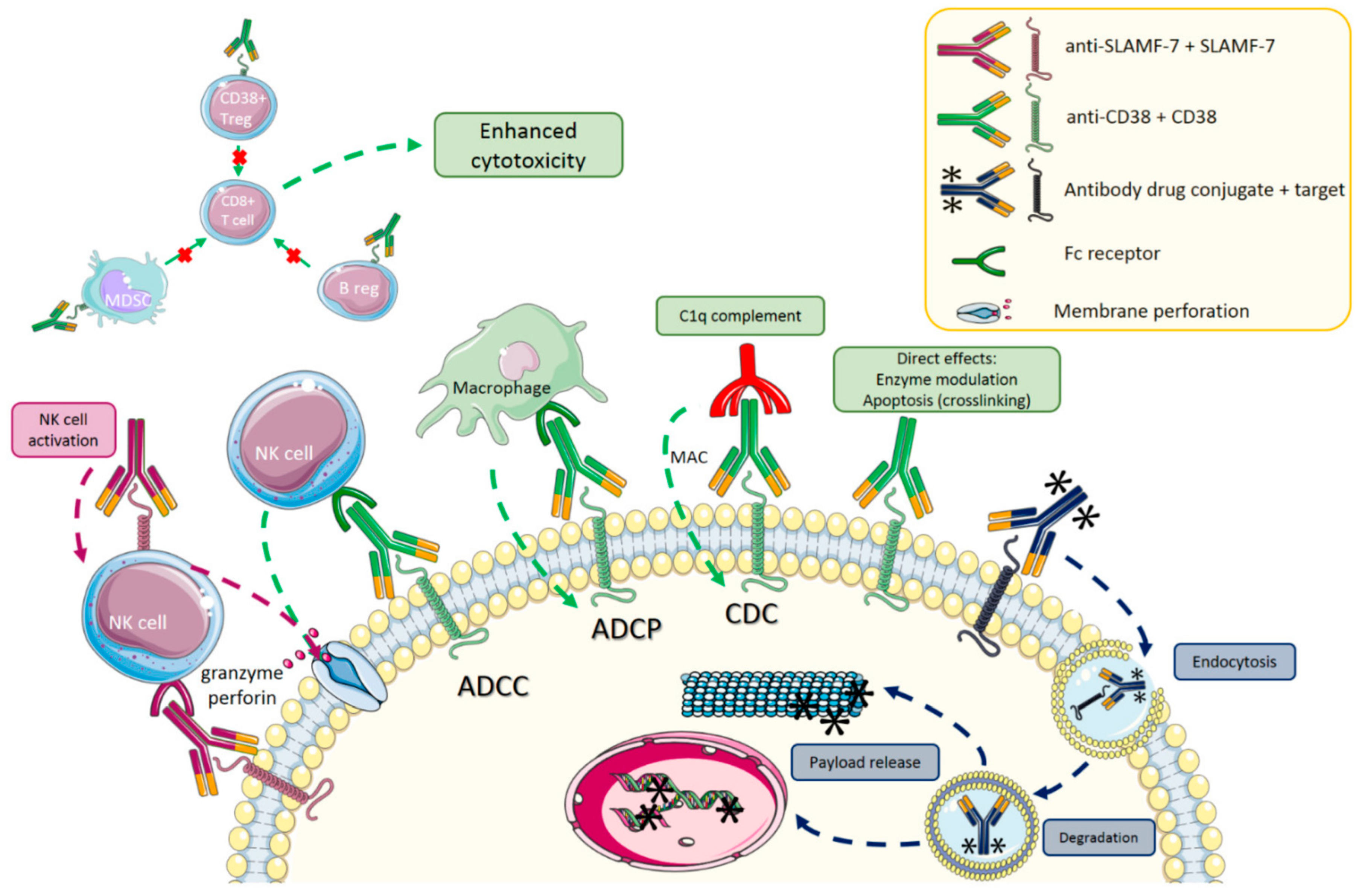
3.2.2. Monoclonal Antibodies
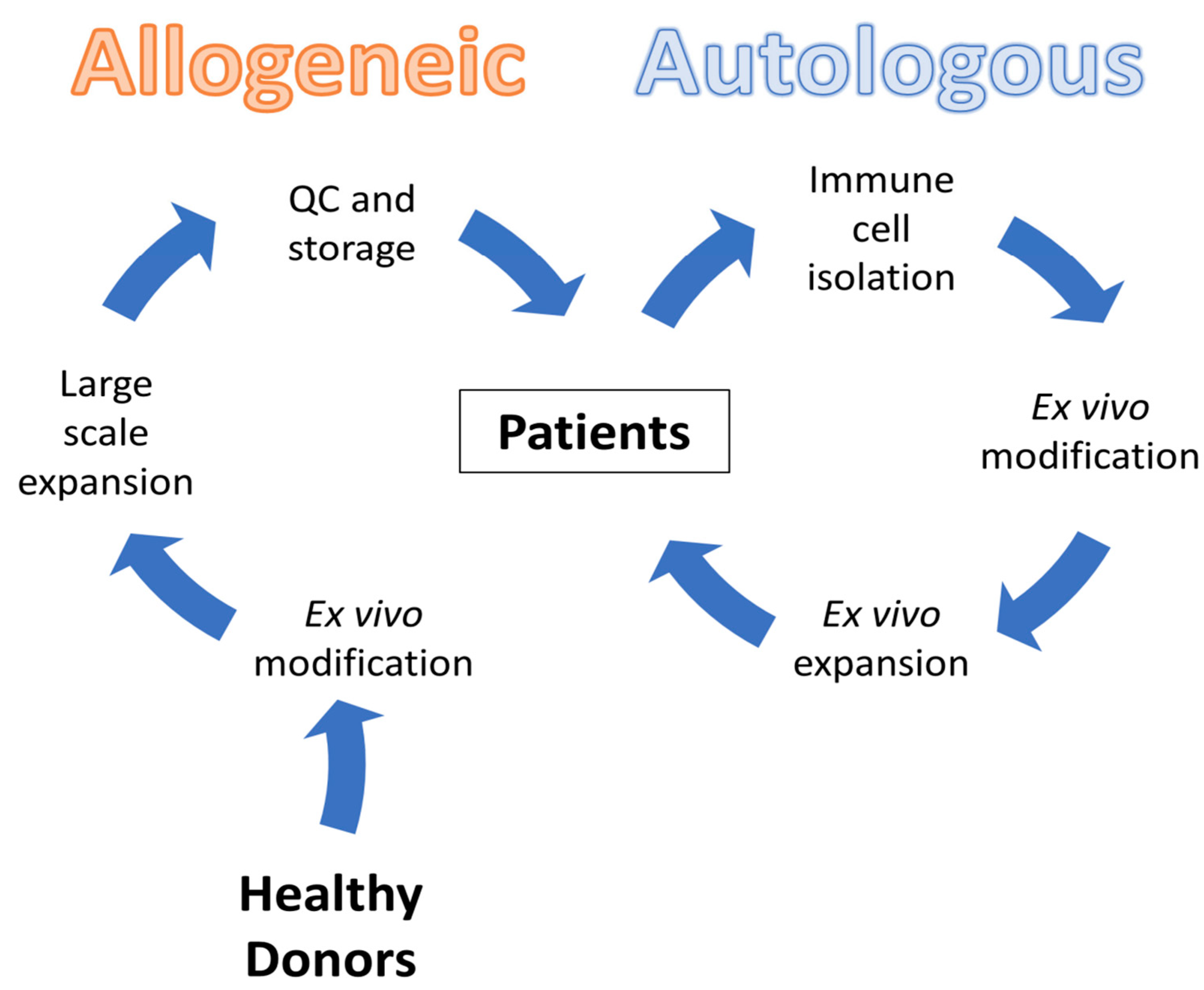
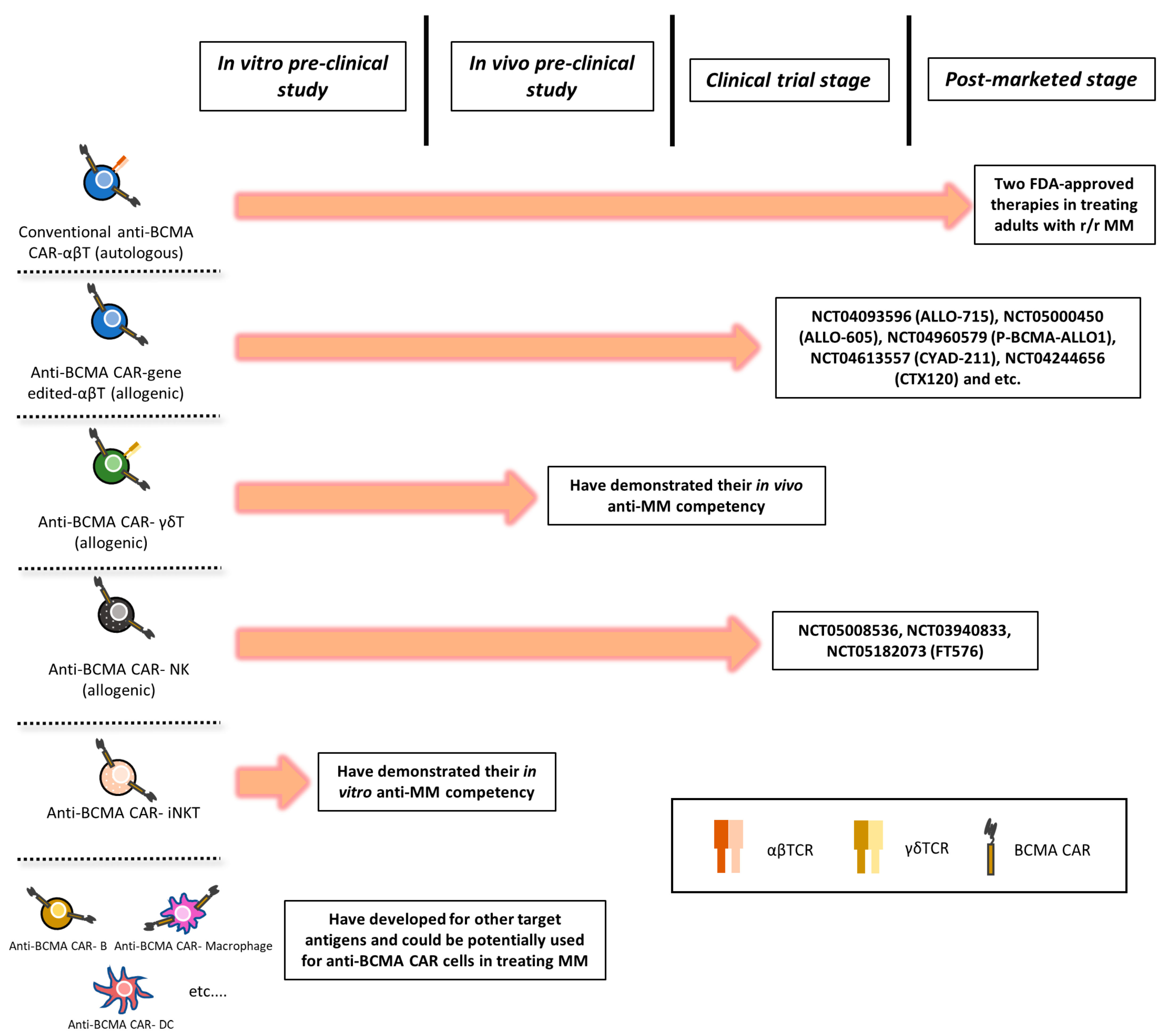
Monoclonal antibodies have emerged as a promising immunotherapeutic approach in the treatment of multiple myeloma (MM), targeting the complex interplay between the immune system and tumor cells. Among the various antibodies evaluated, anti-CD38 and anti-SLAMF7 antibodies have proven to be transformative, with approvals for clinical use in MM. CD38 is a type II transmembrane glycoprotein highly and uniformly expressed on myeloma cells, making it an attractive target for antibody-based therapy. Daratumumab, a human IgG1 antibody targeting CD38, exerts its anti-myeloma activity through multiple mechanisms, including antibody-dependent cellular cytotoxicity (ADCC), antibody-dependent cellular phagocytosis (ADCP), complement-dependent cytotoxicity (CDC), direct apoptosis induction, and immunomodulation, by depleting CD38-positive immune suppressor cells (Figure 3) [39]. Clinical trials have demonstrated notable activity with daratumumab, with rapid, deep, and durable responses. In a monotherapy study, the median overall survival was 20.1 months, and patients achieving stable disease or better experienced a significant survival benefit compared to those with progressive disease [47]. Another trial presented an impressive overall response rate of 90.9% in patients receiving daratumumab [48].
Elotuzumab, a humanized IgG1 antibody targeting SLAMF7 (Figure 3), is another promising agent in the MM immunotherapy landscape. SLAMF7 is highly expressed in myeloma cells, normal plasma cells, NK cells, and a subset of CD8+ T cells. Elotuzumab primarily exerts its anti-myeloma activity through NK-mediated ADCC [50]. While monotherapy studies did not show significant efficacy, combination therapy with elotuzumab in untreated, newly diagnosed MM patients has yielded promising results [51].
While monoclonal antibody therapy shows promising results, there still exist several limitations and challenges. Producing these therapeutic antibodies involves cultivating large quantities of mammalian cells and undergoing extensive purification processes in compliance with good manufacturing practices. This leads to a drastic increase in production costs and limits their widespread use. Moreover, studies indicate that high doses are necessary for clinical effectiveness [52]. For daratumumab, the recommended dosage is 16 mg/kg of body weight, administered as an intravenous infusion weekly for the first eight weeks, then biweekly for 16 weeks, and finally once every four weeks thereafter [53]. This poses a huge financial burden for patients. Patient eligibility is another challenge in monoclonal antibody treatment. It is essential to consider disease-specific factors, such as the stage of the disease and the gene expression profile of the clonal plasma cells, along with patient-related factors, including existing comorbidities and functional status [54]. A patient’s overall health and ability to tolerate treatment are also important considerations. Factors such as age, comorbidities, and performance status can impact eligibility for monoclonal antibody therapies. These criteria ensure that the most appropriate candidates receive these advanced therapies while maximizing the potential for positive outcomes [55].

3.3. CAR-T Therapy

Immunomodulatory drugs, proteasome inhibitors, and anti-CD38 monoclonal antibodies, which are the leading treatments for MM, prolong the survival of patients but often lead to relapse and the cancer becomes more refractory. CAR-T (Chimeric Antigen Receptor T cell) is a differing approach that utilizes a patient’s T cells to specifically target the myeloma cancer cells within their body. Autologous forms of CAR-T have several logistical challenges, such as (1) lymphopenic patients having scarce cells; (2) manufacturing constraints; and (3) demanding vein-to-vein process. This has led to several clinical studies being conducted to test the effectiveness of allogeneic CAR-T through healthy donors to bridge these gaps. Currently, there are multiple ongoing clinical trials for anti-BCMA CAR gene-edited αβ T cells and CAR-Natural Killer (NK) cells, while CAR-γδ T cells and CAR-invariant natural killer T (iNKT) cells are still in preclinical studies (Figure 4) [56].

3.3.1. ALLO-715 Therapy

A promising development within CAR-T cell therapy is the introduction of allogeneic therapies, which seek to overcome challenges associated with autologous CAR-T therapies by utilizing donor-derived T cells (Figure 5). ALLO-715 is an allogeneic therapy that incorporates a second-generation anti-BCMA CAR into donor T cells, as part of the first-in-human Phase 1 UNIVERSAL trial. The integrated CAR has a single-chain variable fragment (scFv) from a human anti-BCMA antibody and intracellular domains of 4-1BB and CD3ζ for the recognition of BCMA and activation of T cells. ALLO-715, the first in its field, was administered to 43 different patients who have been previously treated for MM. The primary objective of this clinical trial was to determine the safety and feasibility of ALLO-715 in human patients. The side effects observed post administration of ALLO-715 such as cytokine release syndrome (CRS) in 56% of patients and neurotoxicity in 14% are somewhat lower rates than what was observed in autologous anti-BCMA CAR T cell therapy. Additionally, there were similar rates of prolonged cytopenia (19%) compared to other autologous therapies. This suggests that allogeneic therapy is a promising candidate as an alternative to autologous therapy. This is a novel advancement in terms of MM treatment as this introduces an off-the-shelf form of therapy that has faster accessibility and manufacturing. A limitation of allogeneic therapy is graft-versus-host disease (GvHD) and the potential of patients rejecting the donor cells. The UNIVERSAL trial has addressed this risk by knocking out TRAC (T cell receptor alpha constant) to prevent TCR-αβ-mediated recognition of histocompatibility antigens. These precautions were deemed successful as no cases of GvHD were reported [57]. Numerous clinical studies are ongoing to fully understand the efficacy of ALLO-715 and other allogeneic CAR-T therapies.

3.3.2. Limitations and Challenges

While the advancements in CAR-T and Allo-715 therapies are novel, there are ongoing challenges in accessibility, cost, and patient eligibility. Delays in autologous CAR-T therapies occur due to the extended process required to collect a patient’s T cells, genetically modify them, and expand them for therapeutic use. This slow process is conflicting for patients who have aggressive disease progression. Allo-715 can counteract this delay by offering off-the-shelf potential but there are still barriers in regulation and collection of clinical data. Additionally, these targeting therapies require specialized facilities and expertise, which limits availability to patients in underdeveloped or rural areas. The cost of CAR-T therapy is extremely high per infusion due to the personalized manufacturing and hospitalization periods. These costs are addressed using Allo-715 by leveraging the pre-manufactured approach. However, these products require a high initial infrastructure and quality control assessment of the donor cells. Lastly, the eligibility criteria for patients to receive these targeted therapies such as CAR-T and Allo-715 are strict. The patients typically eligible are those with refractory or relapsed multiple myeloma who have already undergone prior treatments. Consequently, a past with extensive prior treatments compromises the efficacy of CAR-T therapy as the quality of their T cells may be reduced. These challenges must be overcome for targeted therapies to help more patients recover from multiple myeloma.

4. Relapse

Relapse of multiple myeloma can be categorized into early relapse and refractory. Firstly, early relapse occurs if the multiple myeloma is resistant to first, second, or third therapies (International Myeloma Foundation, 2021) [58]. Refractory relapse of multiple myeloma refers to a disease that is non-responsive to therapy or progresses within 60 days of the last line of therapy [59].
On the other hand, primary refractory refers to the rejection of myeloma’s response to frontline therapy or initially responded to therapy then progressed during or within 60 days of completing frontline therapy. This rejection of frontline therapy early on is indicative of the inferiority of survival in multiple myeloma. The first relapse of multiple myeloma can be diagnosed by the symptoms, which include bone pain, fatigue, breathlessness, peripheral neuropathy, or blood and urine tests such as blood count, chemistry panel, and myeloma protein measurements. In addition to this, early relapse of MM can be diagnosed by imaging tests of bone marrow or the monitoring of the M proteins or serum-free light chain levels (International Myeloma Foundation, 2021) [58]. First relapse of multiple myeloma is further subcategorized to whether patients had a lasting response of at least 18 months, treatment-free interval of 6 to 9 months or both then retreatment with the original schedule can be considered [60]. For more than 20 years now, bortezomib, thalidomide, and lenalidomide, as single agents or combined with dexamethasone were employed for relapsed MM. However, these agents gave rise to what is known now as next-generation IMiDs, PIs, monoclonal antibodies, and histone deacetylase inhibitors [60]. Bortezomib is a first-in-class reversible and selective proteasome inhibitor. To illustrate, the proteasome that functions as a quality control agent for proteins and destroys any misfolded proteins was thought that it could also malfunction and destroy normal beneficial ones in the mid-1990s [61]. Thus, bortezomib slows the action of proteasome leading to a slower destruction of the proteins needed to inhibit cancer progression and aggressiveness. Immunomodulatory drugs (IMiDs) are viewed to be a cornerstone [62]. IMiDs such as thalidomide and lenalidomide work by binding to the cereblon component of the CRL4 CRBN, IMiDs alter the substrate receptor’s specificity, which results in the ubiquitination and degradation of proteins that play an essential role in the survival of MM. For example, the Ikaros zinc-finger family transcription factors (IKF TFs), which are important regulators of lymphocyte development and B cell differentiation as in Ikzf1-deficient mice, lack CLP and fail to generate mature B/T lymphocytes, natural killer, and dendritic cells [63]. However, in multiple myeloma, Ikaros zinc-finger family transcription factors (IKF TFs) are important for its survival, and their degradation leads to anti-proliferation of myeloma cells [64].
The International Myeloma Working Group published a guideline for relapsed patients. This scheme categorizes relapsed myeloma patients depending on patient-specific, disease-specific, or treatment-specific parameters. Despite this, there is a consensus on following another rationale. According to the updated European Society for Medical Oncology guidelines in 2017, physicians subcategorize patients who suffer from the first relapse of multiple myeloma into two categories: First relapse after IMiDs and first relapse after bortezomib-based induction [60]. For the second and third relapse incidents drugs such as ixazomib have the same function as bortezomib, which inhibits the proteasome from protein degradation. However, it is described as having a higher efficiency and lower side effects than bortezomib. Pomalidomide works by enhancing the activity of natural killer and T cells to attack myeloma cells, preventing angiogenesis, and causing their destruction [65].
Overall, treatment of multiple myeloma should be carefully monitored due to the high and almost inevitable possibility of relapse. In addition to this, many parameters should be taken into consideration when dealing with a relapsed case of multiple myeloma such as the previously mentioned examples.

4.1. Novel Therapies in Relapsed MM

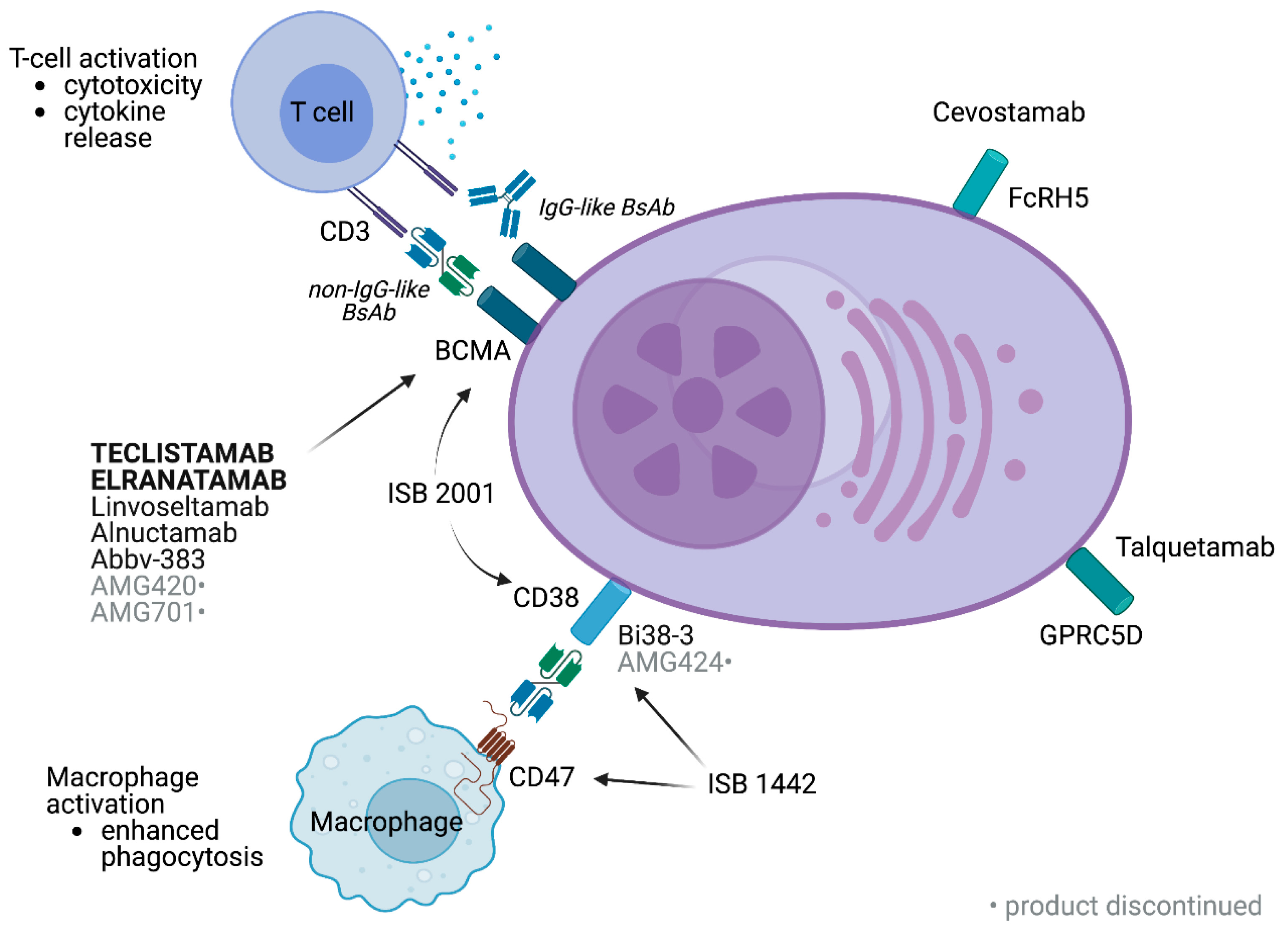
4.1.1. Bispecific Antibodies (BsAbs)

Bispecific antibodies (BsAbs) are novel immunotherapy designed to target two different epitopes or antigens simultaneously either on the same cell or two different ones. They are readily used in cancer where bispecific antibodies are designed to link cancer cells with cytotoxic T cells. Thus, they effectively bring the immune cells near the tumor cells and enhance the immune response. BsAbs were previously based on the heterologous recombination of heavy chains and matching of light chains. Bispecific antibodies, a rapidly advancing treatment for hematologic malignancies, form an immune synapse between T cells, via surface marker CD3 and surface markers on tumor cells [66]. In addition to this, they target B cell maturation antigen (BCMA), cell surface marker FcRH5, or G protein-coupled receptor GPRC5D in multiple myeloma. BCMA is a transmembrane glycoprotein member of the tumor necrosis factor receptor superfamily 17 (TNFRSF17), highly expressed in the plasma cells of multiple myeloma (MM) patients, as well as the normal population. Findings show that BCMA is considered to be an ideal target for chimeric antigen receptor (CAR) T cells in the treatment of relapsed or refractory (R/R) multiple myeloma (MM). Studies have shown that 81–97% of patients with R/R MM can achieve responses after infusion of anti-BCMA CAR T cells [67]. Similarly, FcRH5 has been identified as an attractive B cell lineage-specific surface marker in myeloma and is exclusively expressed in the B cell lineage [68]. GPRC5D is a promising marker for monitoring the tumor load and targeting multiple myeloma cells. Overexpression of G protein-coupled receptor 5D in the bone marrow is associated with poor prognosis in patients with multiple myeloma. Additionally, the overexpression of GPRC5D was associated with plasma cell burden and genetic aberrations in patients with MM and now has emerged as a novel therapeutic target for the treatment of multiple myeloma [69]. The mechanism of BsAbs is designed to simultaneously bind to a target moiety on tumor cells and to CD3 on T cells (Figure 6). Consequently, the cytotoxic T cells are activated leading to the destruction of tumor cells.

4.1.2. Immunocytokine Therapy

Immunocytokine therapy is an innovative approach that combines the specificity of antibodies with the immune-stimulating properties of cytokines. It aims to deliver cytokines directly to the tumor microenvironment, thereby minimizing systemic toxicity and maximizing therapeutic efficacy. When a specific type of immunocytokine, namely the antibody–cytokine fusion protein, is infused into the body, immune cytokines bind to targets on myeloma cells and activate the immune system to fight the myeloma. Immunocytokines use monoclonal antibodies (mAbs) to target specific antigens found on cancer cell surfaces. Proteins or other substances that are overexpressed on tumor cells relative to normal cells are known as antigens. A key component in controlling immune responses are proteins called cytokines. Interleukins (e.g., IL-2, IL-12), interferons, and granulocyte-macrophage colony-stimulating factor (GM-CSF) are frequently utilized cytokines in immunocytokine therapy [71]. The immunocytokine’s antibody component binds to a specific antigen on the surface of cancer cells, delivering the cytokine to the tumor site. Furthermore, the cytokine can alter the tumor microenvironment, increasing its aversion to cancer cells and increasing its support for immune cell activity (Figure 7).

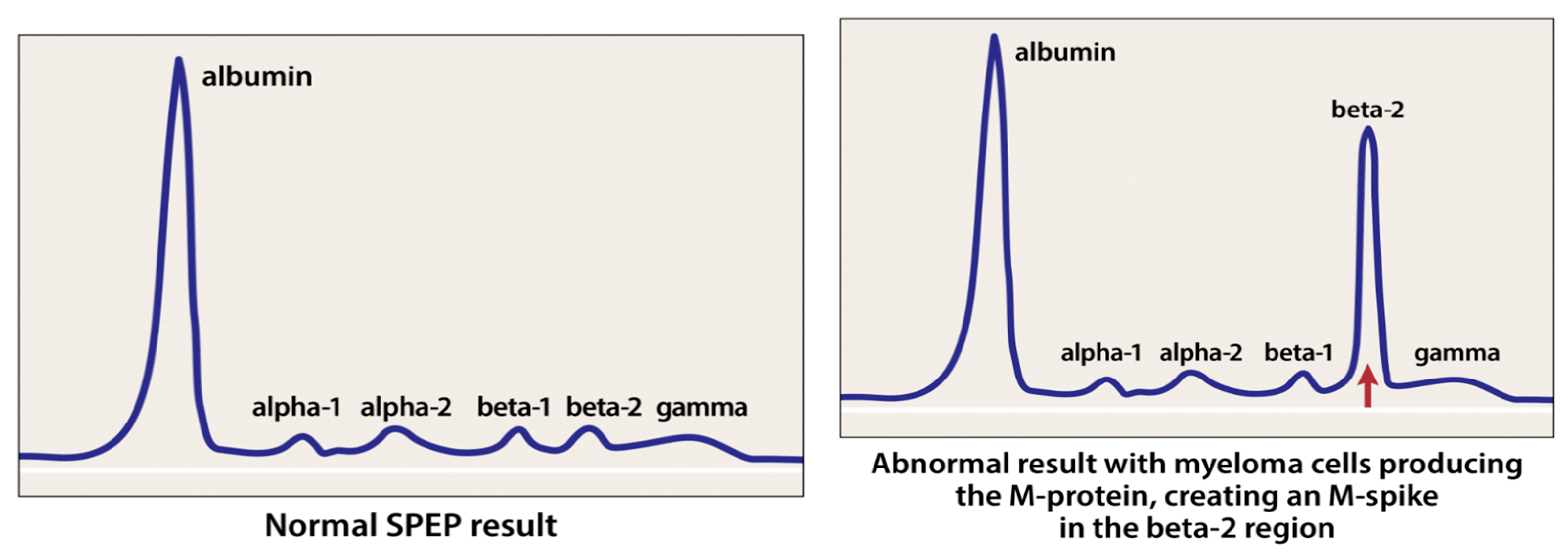
4.2. Mass Spectrometry in Detecting M-Proteins

Plasma cell disorders (PCDs) are identified in the clinical lab by detecting the monoclonal immunoglobulin (M-proteins) that they produce. Novel techniques based on mass spectrometry (MS) are emerging, which have improved clinical and analytical performance to detect the levels of M-proteins. One of the techniques to assess the levels of M-proteins is through serum protein electrophoresis. Serum protein electrophoresis (SPEP) works by separating and quantifying proteins depending on their electrical charge, size, and shape (Figure 8). Monoclonal immunoglobulin proteins appear on the results graph as a narrow spike, and the area under the curve of the spike (AUC) is calculated and subtracted from the amount representing normal immunoglobulins. Consequently, the pathologist can quantify the number of M-proteins (International Myeloma Foundation, 2021) [72].
Despite SPEP being an easy, inexpensive method of separating proteins, studies suggest that quantifying exceedingly low concentrations of M-proteins, although possible, may not yield adequate accuracy and precision between laboratories [73]. Thus, novel studies have found that mass spectrometry (MS) has resulted in increased resolution and sensitivity, which have outpaced improvements in electrophoresis. Each plasma cell is known to produce a unique immunoglobulin characterized by having a unique complementarity-determining region (CDR). Each CDR has a specific amino acid sequence, resulting in a unique overall mass, resulting in M-protein detection by MS. Thus, MM can be easily monitored through MS by detecting M-protein levels. According to the literature, two methods of MS have emerged that differ in the analytical target used to detect the M-protein [74]. It has been proved that MS has higher sensitivity and specificity that can detect very low levels of M-proteins and precisely quantify them. Additionally, it is faster than electrophoresis-based methods, with minimal manual interpretation required. Therefore, it has been concluded that the advantage of increased accuracy and documented clinical and analytic sensitivity of MS is easier for the detection and monitoring of M-proteins.
The implementation of novel therapies for relapsed multiple myeloma (MM) faces several barriers and global disparities in access. The disease course in patients with multiple myeloma can be highly heterogeneous, leading to significant variation in the standard practice and approach to therapy initiation for relapsed myeloma particularly regarding immunocytokine therapy and bispecific antibodies (BsAbs). The absence of a formal risk stratification system proposed or widely accepted for the relapsed setting makes it challenging to tailor treatments effectively. Additionally, there are limited data to suggest that basing therapy on the depth of response can alter long-term outcomes in relapsed disease. Patients with relapsed/refractory MM often have comorbidities such as renal impairment and cardiac issues, which increase their vulnerability to treatment-related toxicity, further complicating the management of their disease [75]. The discussion around immunomodulatory drugs (IMiDs) in relapsed/refractory multiple myeloma (RRMM) highlights several logistical considerations that can lead to dosing delays and what is referred to as “time toxicity”. Time toxicity is characterized by frequent healthcare-related encounters and phone calls that interfere with patient well-being, adding to the overall burden of the disease. In the US, IMiDs can only be prescribed under specialized REMS (Risk Evaluation and Mitigation Strategies) programs due to their teratogenic risks and are largely dispensed through specialty pharmacies rather than patients’ pharmacies. Additionally, the high cost of IMiDs in RRMM can lead to substantial financial toxicity (FT). For example, out-of-pocket costs for pomalidomide can exceed USD 21,000 per year, even for patients with Medicare insurance, highlighting the economic burden associated with these therapies [75].

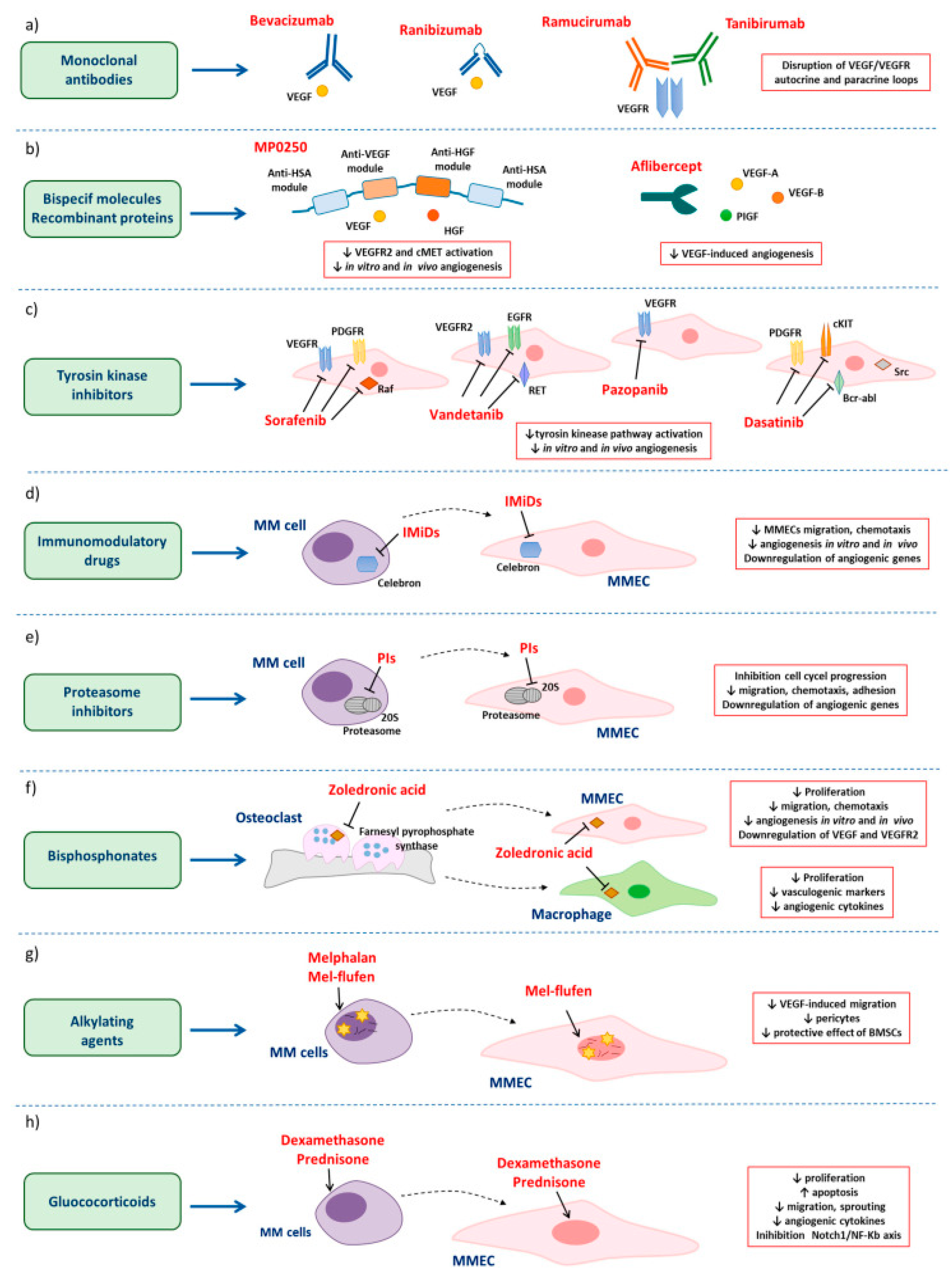
4.3. Angiogenesis and Its Effect on MM Progression

While monitoring M-protein levels is crucial for detecting disease progression, understanding the biological processes driving multiple myeloma, such as angiogenesis, is essential for developing effective treatment strategies. Angiogenesis is a constant hallmark of multiple myeloma (MM) progression. Under physiological conditions, angiogenesis depends on the balance of positive and negative angiogenic modulators within the vascular microenvironment. However, in the tumor environment angiogenesis is linked to switching this balance by depending on the release of growth factors of the neoplastic cells. According to research, it was concluded that the degree of angiogenesis is correlated with disease stage, prognosis, or response to therapy [76]. Additionally, angiogenesis promotes the progression from monoclonal gammopathies of undetermined significance (MGUS) to MM through the angiogenic factors that are released by the neoplastic cells. However, research has shown that patients with relapsed multiple myeloma showed a higher incidence of high-grade angiogenesis compared to those newly diagnosed with the disease [4]. In normal physiological conditions, healthy plasma cells express an excess amount of pro-angiogenic over anti-angiogenic genes to promote angiogenesis. However, in MM, this over-expression of pro-angiogenic genes is highly upregulated, while the expression of anti-angiogenic genes is markedly suppressed. Pro-angiogenic factors, such as VEGF and FGF-2, are induced by the active MM cell plasma, which stimulates tumor microenvironment cells to secrete additional VEGF, FGF-2, and HGF.
The induction of the cells of the tumor microenvironment by VEGF, FGF-2, and HGF results in the recruitment and activation of the MM-associated macrophages. These MM-associated macrophages protect MM cells from spontaneous and melphalan-induced apoptosis. Additionally, as a result of increased expression of pro-angiogenic factors, tumor macrophages become differentiated into cells that are similar to MM endothelial cells, which can generate in vitro capillary-like networks [11]. Thus, it can be concluded that the progression of MM readily depends on the over-expression of pro-angiogenic genes promoting angiogenesis. Fortunately, there are anti-angiogenic therapies that are emerging as a complementary approach to MM treatment. These therapies are segmented into two approaches: direct and indirect. Direct anti-angiogenic therapies that target angiogenic cytokines and/or pathways include monoclonal antibodies (mAbs), bispecific molecules, and tyrosine kinase inhibitors (TKIs). Indirect anti-angiogenic therapies include immunomodulatory drugs (IMiDs), proteasome inhibitors, alkylating agents, bisphosphonates, and glucocorticoids, indicated in Figure 9 [77].
Anti-angiogenic agents, such as thalidomide, lenalidomide, and VEGF inhibitors, have shown to be successful in suppressing angiogenesis in MM. Table 3 discusses the three anti-angiogenic agents and their effects [78,79,80,81,82,83]. Despite these agents’ effectiveness, relapsed MM had occurred due to the development of resistance as a consequence of mutations in key signaling pathways in plasma cell clones that emerge after initial therapy [84]. As a result, many trials have repeatedly shown that a three-drug combination improves both progression-free and overall survival, suggesting a multidrug combination that likely impacts the clonal diversity of the tumor will provide the best clinical results [85].

5. Comparative Discussion on the Safety Profiles of the Novel Therapies

The novel therapies available for treating multiple myeloma have distinct safety profiles that must be carefully considered when choosing a treatment strategy. Recognizing and managing these risks is critical in ensuring safe and effective MM care. Monoclonal antibody therapy plays a crucial role in the management of bone health and the treatment of multiple myeloma (MM). However, this treatment is associated with distinct safety concerns as shown in Table 4 [36,86]. In addition, CAR-T and Allo-715 therapies use cellular engineering to target cancer more effectively [56]. However, they also come with safety concerns that require careful management, mentioned as well in Table 4. While these challenges do not diminish their potential, they highlight the importance of monitoring and individualized care to ensure the best possible outcomes for patients.
Despite the significant clinical activity of bispecific antibodies (BsAbs), their safety profile and potential toxicities are key considerations as well. In a study of AMG420, a BCMAxCD3 bispecific T cell engagement, where forty-two patients with at least two prior lines of therapy were enrolled, a 36% double refractory to an IMiD and PI was obtained, and some dose-limiting toxicities arose [87]. For example, cytokine release syndrome (CRS) occurred in 38% of patients, serious infections in 33%, and grade 3 polyneuropathy was shown in two patients. CRS is caused by a large, rapid release of cytokines into the blood from immune cells affected by the immunotherapy. As a result of this study, the AMG420 compound was discontinued for a longer half-life of compounds of BsAbs, namely, AMG701, CC-93269, PF-06863135, REGN5458, JNJ-64007957 (teclistamab), TNB-383B, JNJ-64407564 (talquetamab), and BFCR4350A (cevostamab). Additionally, other complications were identified with other BsAbs compounds, such as neurotoxicity that ranged from 5% to 28% at any grade in the trials. Symptoms of neurotoxicity included confusion, aphasia, cognitive disorder, and encephalopathy. Hematological side effects were identified in the study such as anemia, neutropenia, lymphopenia, leukopenia, and thrombocytopenia. However, the dose–response relationship was unidentified from the early data. Finally, it was found that infection was reported in all of the trials including different compounds of BsAbs. Consequently, it can be concluded that infection is the most common treatment-related adverse event seen with these agents.
Moreover, resistance to BsAbs has been reported; the key resistance mechanisms to BsAbs include high tumor burden, T cell fitness and repertoire, and mutations and the loss of target on MM cells [88]. Resistance to BsAbs was a result of high tumor burden, (International Staging System) ISS stage 3, and extramedullary disease, which has been shown to adversely impact the clinical efficacy of BsAbs in MM [89]. Acquired resistance mechanisms include T cell exhaustion, antigen escape, and loss of MHC class I, BCMA mutation, or deletion as a result of selective pressure of continuous administration of BsAbs. This mutation or deletion of BCMA could lead to target antigen loss [88]. However, some strategies arose to account for the possibility of the resistance occurrence. For example, combining BsAbs with therapeutic agents that augment T cell function has been shown to increase efficiency. The addition of cyclophosphamide to BCMA-BsAb was also found to be effective. BsAbs represent a transformative modality in MM treatment, offering potent anti-myeloma activity. However, close monitoring and proactive management of toxicities such as CRS, ICANS, infections, and cytopenias are critical in optimizing their safety and therapeutic benefits.
Moreover, based on the preclinical evidence, many immunocytokines have progressed to phase I/II clinical trials for different indications. One pilot study in melanoma patients showed dose-limiting toxicities of hypoxia, thrombocytopenia, hypotension, and hyperglycemia in 7/33 patients, all of which were reversible. Additionally, a recent pilot trial in advanced resectable melanoma patients with a lower tumor burden resulted in a median recurrence-free survival (RFS) of 5.73 months but 6/18 RFS cases at approximately 57 months. The results of this study were as follows: five out of 45 patients had objective responses, and three of those had no events for up to five years. Four patients had dose-limiting toxicities [90]. Another study carried out for the optimization of this immunocytokine in neuroblastoma led to a phase II trial in combination with GM-CSF and the vitamin A-derivative isotretinoin. Immunocytokine therapy for MM showed severe side effects that include cytokine release syndrome similar to the BsAbs effect, immune effector cell-associated neurotoxicity syndrome, cytopenia, infections, hemophagocytic lymphohistiocytosis, and organ toxicity, which could sometimes be life threatening.
While immunocytokines are designed to target tumor antigens, cytokines can activate immune cells with nonspecifically autoimmune or inflammatory toxicities. Symptoms include rash, colitis, hepatitis, or pneumonitis due to immune dysregulation. However, such symptoms can be managed by immunosuppressive agents like corticosteroids, depending on severity. Despite toxicity posing a concern, particularly related to immune activation and CRS, these therapies generally have a manageable safety profile with appropriate precautions.

Funding

This research received no external funding.

Conflicts of Interest

The authors declare no conflict of interest.

References

  1. Rajkumar, S.V.; Kumar, S. Multiple myeloma current treatment algorithms. Blood Cancer J. 2020, 10, 1–10. [Google Scholar] [CrossRef]
  2. Gerecke, C.; Fuhrmann, S.; Strifler, S.; Schmidt-Hieber, M.; Einsele, H.; Knop, S. The Diagnosis and Treatment of Multiple Myeloma. Dtsch. Aerzteblatt Online 2016, 113, 470–476. [Google Scholar] [CrossRef]
  3. Hatjiharissi, P. The progress in multiple myeloma. Hell. J. Nucl. Med. 2023, 26, 30–35. [Google Scholar] [PubMed]
  4. Giuliani, N.; Storti, P.; Bolzoni, M.; Palma, B.D.; Bonomini, S. Angiogenesis and Multiple Myeloma. Cancer Microenviron. 2011, 4, 325. [Google Scholar] [CrossRef] [PubMed]
  5. San Miguel, J. Multiple myeloma: A model for scientific and clinical progress. Hematology 2014, 2014, 1–7. [Google Scholar] [CrossRef] [PubMed]
  6. Ahmed, A.; Killeen, R.B. Relapsed and Refractory Multiple Myeloma. In StatPearls [Internet]; StatPearls Publishing: Treasure Island, FL, USA, 2024. Available online: https://www.ncbi.nlm.nih.gov/books/NBK592405/ (accessed on 8 June 2023).
  7. Dadzie, T.G.; Green, A.C. The role of the bone microenvironment in regulating myeloma residual disease and treatment. Front. Oncol. 2022, 12, 999939. [Google Scholar] [CrossRef]
  8. Kaseb, H.; Annamaraju, P.; Babiker, H.M. Monoclonal Gammopathy of Undetermined Significance (MGUS); PubMed; StatPearls Publishing: Treasure Island, FL, USA, 2020. Available online: https://www.ncbi.nlm.nih.gov/books/NBK507880/ (accessed on 10 July 2022).
  9. Fonseca, R.; Gonzalez-Velez, M. Treatment of Smoldering Multiple Myeloma: Expectant Observation Should Still Be the Standard. Am. Soc. Clin. Oncol. Educ. Book/Educ. Book 2020, 40, 364–370. [Google Scholar] [CrossRef] [PubMed]
  10. Mateos, M.-V.; Hernandez, M.T.; Giraldo, P.; de la Rubia, J.; de Arriba, F.; Corral, L.L.; Rosiñol, L.; Paiva, B.; Palomera, L.; Bargay, J.; et al. Lenalidomide plus dexamethasone versus observation in patients with high-risk smouldering multiple myeloma (QuiRedex): Long-term follow-up of a randomised, controlled, phase 3 trial. Lancet Oncol. 2016, 17, 1127–1136. [Google Scholar] [CrossRef]
  11. Rajkumar, S.V.; Mesa, R.A.; Fonseca, R.; Schroeder, G.; Plevak, M.F.; Dispenzieri, A.; Lacy, M.Q.; Lust, J.A.; Witzig, T.E.; Gertz, M.A.; et al. Bone Marrow Angiogenesis in 400 Patients with Monoclonal Gammopathy of Undetermined Significance, Multiple Myeloma, and Primary Amyloidosis1. Clin. Cancer Res. 2002, 8, 2210–2216. [Google Scholar]
  12. Rajkumar, S.V.; Dimopoulos, M.A.; Palumbo, A.; Blade, J.; Merlini, G.; Mateos, M.-V.; Kumar, S.; Hillengass, J.; Kastritis, E.; Richardson, P.; et al. International Myeloma Working Group updated criteria for the diagnosis of multiple myeloma. Lancet Oncol. 2014, 15, e538–e548. [Google Scholar] [CrossRef]
  13. Kapoor, P.; Rajkumar, S.V. Smoldering Multiple Myeloma. Cancer J. 2019, 25, 65–71. [Google Scholar] [CrossRef] [PubMed]
  14. Hillengass, J.; Fechtner, K.; Weber, M.-A.; Bäuerle, T.; Ayyaz, S.; Heiss, C.; Hielscher, T.; Moehler, T.M.; Egerer, G.; Neben, K.; et al. Prognostic Significance of Focal Lesions in Whole-Body Magnetic Resonance Imaging in Patients with Asymptomatic Multiple Myeloma. J. Clin. Oncol 2010, 28, 1606–1610. [Google Scholar] [CrossRef]
  15. Merz, M.; Hielscher, T.; Wagner, B.; Sauer, S.; Shah, S.; Raab, M.S.; Jauch, A.; Neben, K.; Hose, D.; Egerer, G.; et al. Predictive value of longitudinal whole-body magnetic resonance imaging in patients with smoldering multiple myeloma. Leukemia 2014, 28, 1902–1908. [Google Scholar] [CrossRef] [PubMed]
  16. Rajkumar, S.V.; Kumar, S.; Lonial, S.; Mateos, M.V. Smoldering multiple myeloma current treatment algorithms. Blood Cancer J. 2022, 8, 2210–2216. [Google Scholar] [CrossRef]
  17. Mateos, M.-V.; Hernández, M.-T.; Giraldo, P.; de la Rubia, J.; de Arriba, F.; López Corral, L.; Rosiñol, L.; Paiva, B.; Palomera, L.; Bargay, J.; et al. Lenalidomide plus dexamethasone for high-risk smoldering multiple myeloma. N. Engl. J. Med. 2013, 369, 438–447. [Google Scholar] [CrossRef] [PubMed]
  18. D’Arena, G.; Gobbi, P.G.; Broglia, C.; Sacchi, S.; Quarta, G.; Baldini, L.; Iannitto, E.; Falcone, A.; Guariglia, R.; Pietrantuono, G.; et al. Pamidronate versus observation in asymptomatic myeloma: Final results with long-term follow-up of a randomized study. Leuk. Lymphoma 2011, 52, 771–775. [Google Scholar] [CrossRef] [PubMed]
  19. Rajkumar, S.V. Multiple myeloma: 2020 update on diagnosis, risk-stratification and management. Am. J. Hematol. 2020, 95, 548–567. [Google Scholar] [CrossRef] [PubMed]
  20. Korde, N.; Roschewski, M.; Zingone, A.; Kwok, M.; Manasanch, E.E.; Bhutani, M.; Tageja, N.; Kazandjian, D.; Mailankody, S.; Wu, P.; et al. Treatment with Carfilzomib-Lenalidomide-Dexamethasone With Lenalidomide Extension in Patients With Smoldering or Newly Diagnosed Multiple Myeloma. JAMA Oncol. 2015, 1, 746. [Google Scholar] [CrossRef] [PubMed]
  21. Charliński, G.; Jurczyszyn, A. Non-secretory multiple myeloma: Diagnosis and management. Adv. Clin. Exp. Med. 2021, 31, 95–100. [Google Scholar] [CrossRef]
  22. Dupuis, M.M.; Tuchman, S.A. Non-secretory multiple myeloma: From biology to clinical management. OncoTargets Ther. 2016, 9, 7583–7590. [Google Scholar] [CrossRef]
  23. Hill, E.; Dew, A.; Kazandjian, D. State of the science in smoldering myeloma: Should we be treating in the clinic? Semin. Oncol. 2019, 46, 112–120. [Google Scholar] [CrossRef] [PubMed]
  24. Boyce, B.; Yao, Z.; Xing, L. Osteoclasts have multiple roles in bone in addition to Bone Resorption. Crit. Rev. Eukaryot. Gene Expr. 2009, 19, 171–180. [Google Scholar] [CrossRef] [PubMed]
  25. Mundy, G.R.; Yoneda, T.; Hiraga, T. Preclinical studies with zoledronic acid and other bisphosphonates: Impact on the bone microenvironment. Semin. Oncol. 2001, 28, 35–44. [Google Scholar] [CrossRef]
  26. Hussain, M.; Khan, F.; Al Hadidi, S. The use of bone-modifying agents in multiple myeloma. Blood Rev. 2023, 57, 100999. [Google Scholar] [CrossRef] [PubMed]
  27. Terpos, E.; Ntanasis-Stathopoulos, I.; Dimopoulos, M.A. Myeloma bone disease: From biology findings to treatment approaches. Blood 2019, 133, 1534–1539. [Google Scholar] [CrossRef]
  28. Dunford, J.E.; Thompson, K.; Coxon, F.P.; Luckman, S.P.; Hahn, F.M.; Poulter, C.D.; Ebetino, F.H.; Rogers, M.J. Structure-activity relationships for inhibition of farnesyl diphosphate synthase in vitro and inhibition of bone resorption in vivo by nitrogen-containing bisphosphonates. J. Pharmacol. Exp. Ther. 2001, 296, 235–242. [Google Scholar] [CrossRef] [PubMed]
  29. Gao, J.; Chu, X.; Qiu, Y.; Wu, L.; Qiao, Y.; Wu, J.; Li, D. Discovery of potent inhibitor for farnesyl pyrophosphate synthase in the mevalonate pathway. Chem. Commun. 2010, 46, 5340. [Google Scholar] [CrossRef] [PubMed]
  30. Kanakis, I.; Kousidou, O.C.; Karamanos, N.K. In vitro and in vivo antiresorptive effects of bisphosphonates in metastatic bone disease. In Vivo 2005, 19, 311–318. [Google Scholar]
  31. Mhaskar, R.; Redzepovic, J.; Wheatley, K.; Clark, O.A.; Miladinovic, B.; Glasmacher, A.; Kumar, A.; Djulbegovic, B. Bisphosphonates in multiple myeloma: A network meta-analysis. Cochrane Database Syst. Rev. 2012. [Google Scholar] [CrossRef]
  32. Shipman, C.M.; Rogers, M.J.; Apperley, J.F.; Russell, R.G.; Croucher, P.I. Bisphosphonates induce apoptosis in human myeloma cell lines: A novel anti-tumour activity. Br. J. Haematol. 1997, 98, 665–672. [Google Scholar] [CrossRef] [PubMed]
  33. Noriega Aldave, A.P.; Jaiswal, S. Severe resistant hypocalcemia in multiple myeloma after zoledronic acid administration: A case report. J. Med Case Rep. 2014, 8, 353. [Google Scholar] [CrossRef] [PubMed]
  34. Duh, M.; Weide, R.; Koppler, H.; Antras, L.; Smith, M.; Eva Chang, M.; Green, J.; Wintfeld, N.; Neary, M. Renal toxicity in patients with multiple myeloma receiving zoledronic acid vs, ibandronate: A Retrospective Medical Records Review. J. Cancer Res. Ther. 2010, 6, 31. [Google Scholar] [CrossRef] [PubMed]
  35. Reiriz, A.B.; de Moraes De Zorzi, P.; Lovat, C.P. Bisphosphonates and Osteonecrosis of the jaw: A case report. Clinics 2008, 63, 281–284. [Google Scholar] [CrossRef] [PubMed]
  36. Raje, N.; Terpos, E.; Willenbacher, W.; Shimizu, K.; García-Sanz, R.; Durie, B.; Legieć, W.; Krejčí, M.; Laribi, K.; Zhu, L.; et al. Denosumab versus zoledronic acid in bone disease treatment of newly diagnosed multiple myeloma: An international, double-blind, double-dummy, randomised, controlled, phase 3 study. Lancet Oncol. 2018, 19, 370–381. [Google Scholar] [CrossRef]
  37. West, H. Denosumab for prevention of skeletal-related events in patients with bone metastases from solid tumors: Incremental benefit, debatable value. J. Clin. Oncol. 2011, 29, 1095–1098. [Google Scholar] [CrossRef]
  38. Body, J.-J.; Facon, T.; Coleman, R.E.; Lipton, A.; Geurs, F.; Fan, M.; Holloway, D.; Peterson, M.C.; Bekker, P.J. A study of the biological receptor activator of nuclear factor-kappab ligand inhibitor, denosumab, in patients with multiple myeloma or bone metastases from breast cancer. Clin. Cancer Res. 2006, 12, 1221–1228. [Google Scholar] [CrossRef]
  39. Wudhikarn, K.; Wills, B.; Lesokhin, A.M. Monoclonal antibodies in multiple myeloma: Current and emerging targets and mechanisms of action. Best Pract. Amp; Res. Clin. Haematol. 2020, 33, 101143. [Google Scholar] [CrossRef]
  40. Adams, J. The Proteasome: A suitable antineoplastic target. Nat. Rev. Cancer 2004, 4, 349–360. [Google Scholar] [CrossRef] [PubMed]
  41. Crawford, L.J.; Irvine, A.E. Targeting the ubiquitin proteasome system in haematological malignancies. Blood Rev. 2013, 27, 297–304. [Google Scholar] [CrossRef]
  42. Jayaweera, S.P.; Wanigasinghe Kanakanamge, S.P.; Rajalingam, D.; Silva, G.N. Carfilzomib: A promising proteasome inhibitor for the treatment of relapsed and refractory multiple myeloma. Front. Oncol. 2021, 11. [Google Scholar] [CrossRef]
  43. Ito, S. Proteasome inhibitors for the treatment of multiple myeloma. Cancers 2020, 12, 265. [Google Scholar] [CrossRef]
  44. Niewerth, D.; Jansen, G.; Assaraf, Y.G.; Zweegman, S.; Kaspers, G.J.L.; Cloos, J. Molecular basis of resistance to proteasome inhibitors in hematological malignancies. Drug Resist. Updates 2015, 18, 18–35. [Google Scholar] [CrossRef] [PubMed]
  45. Parlati, F.; Lee, S.J.; Aujay, M.; Suzuki, E.; Levitsky, K.; Lorens, J.B.; Micklem, D.R.; Ruurs, P.; Sylvain, C.; Lu, Y.; et al. Carfilzomib can induce tumor cell death through selective inhibition of the chymotrypsin-like activity of the proteasome. Blood 2009, 114, 3439–3447. [Google Scholar] [CrossRef] [PubMed]
  46. Demo, S.D.; Kirk, C.J.; Aujay, M.A.; Buchholz, T.J.; Dajee, M.; Ho, M.N.; Jiang, J.; Laidig, G.J.; Lewis, E.R.; Parlati, F.; et al. Antitumor activity of PR-171, a novel irreversible inhibitor of the Proteasome. Cancer Res. 2007, 67, 6383–6391. [Google Scholar] [CrossRef] [PubMed]
  47. Usmani, S.Z.; Weiss, B.M.; Plesner, T.; Bahlis, N.J.; Belch, A.; Lonial, S.; Lokhorst, H.M.; Voorhees, P.M.; Richardson, P.G.; Chari, A.; et al. Clinical efficacy of daratumumab monotherapy in patients with heavily pretreated relapsed or refractory multiple myeloma. Blood 2016, 128, 37–44. [Google Scholar] [CrossRef]
  48. Mateos, M.V.; Dimopoulos, M.A.; Cavo, M.; Suzuki, K.; Jakubowiak, A.; Knop, S.; Doyen, C.; Lúcio, P.; Nagy, Z.; Kaplan, P.; et al. Daratumumab plus bortezomib, melphalan, and prednisone for untreated myeloma. N. Engl. J. Med. 2018, 378, 518–528. [Google Scholar] [CrossRef] [PubMed]
  49. Radocha, J.; van de Donk, N.W.; Weisel, K. Monoclonal antibodies and antibody drug conjugates in multiple myeloma. Cancers 2021, 13, 1571. [Google Scholar] [CrossRef] [PubMed]
  50. Collins, S.M.; Bakan, C.E.; Swartzel, G.D.; Hofmeister, C.C.; Efebera, Y.A.; Kwon, H.; Starling, G.C.; Ciarlariello, D.; Bhaskar, S.; Briercheck, E.L.; et al. Elotuzumab directly enhances NK cell cytotoxicity against myeloma via CS1 ligation: Evidence for augmented NK cell function complementing ADCC. Cancer Immunol. Immunother. 2013, 62, 1841–1849. [Google Scholar] [CrossRef] [PubMed]
  51. Leow, C.C.-Y.; Low, M.S. Targeted therapies for multiple myeloma. J. Pers. Med. 2021, 11, 334. [Google Scholar] [CrossRef] [PubMed]
  52. Chames, P.; Van Regenmortel, M.; Weiss, E.; Baty, D. Therapeutic antibodies: Successes, limitations and hopes for the future. Br. J. Pharmacol. 2009, 157, 220–233. [Google Scholar] [CrossRef] [PubMed]
  53. Xu, X.; Yan, X.; Puchalski, T.; Lonial, S.; Lokhorst, H.M.; Voorhees, P.; Plesner, T.; Liu, K.; Khan, I.; Jansson, R.; et al. Clinical Implications of Complex Pharmacokinetics for Daratumumab Dose Regimen in Patients With Relapsed/Refractory Multiple Myeloma. Clin. Pharmacol. Ther./Clin. Pharmacol. Ther. 2017, 101, 721–724. [Google Scholar] [CrossRef] [PubMed]
  54. Michels, T.C.; Petersen, K.E. Multiple Myeloma: Diagnosis and Treatment. Am. Fam. Physician 2017, 95, 373–383. [Google Scholar]
  55. Palumbo, A.; Anderson, K. Multiple Myeloma. N. Engl. J. Med. 2011, 364, 1046–1060. [Google Scholar] [CrossRef] [PubMed]
  56. Du, Z.; Zhu, S.; Zhang, X.; Gong, Z.; Wang, S. Non-Conventional Allogeneic Anti-BCMA Chimeric Antigen Receptor-Based Immune Cell Therapies for Multiple Myeloma Treatment. Cancers 2023, 15, 567. [Google Scholar] [CrossRef] [PubMed]
  57. Mailankody, S.; Matous, J.V.; Chhabra, S.; Liedtke, M.; Sidana, S.; Oluwole, O.O.; Malik, S.; Nath, R.; Anwer, F.; Cruz, J.C.; et al. Allogeneic BCMA-targeting CAR T cells in relapsed/refractory multiple myeloma: Phase 1 UNIVERSAL trial interim results. Nat. Med. 2023, 29, 422–429. [Google Scholar] [CrossRef] [PubMed]
  58. International Myeloma Foundation. What Is Relapse? Available online: https://www.myeloma.org/treatment/relapse-definition (accessed on 19 July 2021).
  59. Rajkumar, S.V.; Harousseau, J.; Durie, B.; Anderson, K.C.; Dimopoulos, M.; Kyle, R.; Blade, J.; Richardson, P.; Orlowski, R.; Siegel, D.; et al. Consensus recommendations for the uniform reporting of clinical trials: Report of the International Myeloma Workshop Consensus Panel 1. Blood 2011, 117, 4691–4695. [Google Scholar] [CrossRef]
  60. Sonneveld, P. Management of multiple myeloma in the relapsed/refractory patient. Hematology 2017, 508–517. [Google Scholar] [CrossRef]
  61. Alwahsh, M.; Farhat, J.; Talhouni, S.; Hamadneh, L.; Hergenröder, R. Bortezomib advanced mechanisms of action in multiple myeloma, solid and liquid tumors along with its novel therapeutic applications. PubMed 2023, 22, 146–168. [Google Scholar] [CrossRef]
  62. Bird, S.A.; Pawlyn, C. IMID resistance in Multiple myeloma: Current understanding of the underpinning biology and clinical impact. Blood 2023, 142, 131–140. [Google Scholar] [CrossRef]
  63. Rivellese, F.; Manou-Stathopoulou, S.; Mauro, D.; Goldmann, K.; Pyne, D.; Rajakariar, R.; Gordon, P.; Schafer, P.; Bombardieri, M.; Pitzalis, C.; et al. Effects of targeting the transcription factors Ikaros and Aiolos on B cell activation and differentiation in systemic lupus erythematosus. Lupus Sci. Med. 2021, 8, e000445. [Google Scholar] [CrossRef]
  64. Frere, G.A.; De Araujo, E.D.; Gunning, P.T. Emerging mechanisms of targeted protein degradation by molecular glues. In Methods in Cell Biology; Academic Press: Cambridge, MA, USA, 2022; pp. 1–26. [Google Scholar] [CrossRef]
  65. Chanan-Khan, A.A.; Swaika, A.; Paulus, A.; Kumar, S.K.; Mikhael, J.R.; Rajkumar, S.V.; Dispenzieri, A.; Lacy, M.Q. Pomalidomide: The new immunomodulatory agent for the treatment of multiple myeloma. Blood Cancer J. 2013, 3, e143. [Google Scholar] [CrossRef] [PubMed]
  66. Reynolds, G.; Cliff, E.R.S.; Mohyuddin, G.R.; Popat, R.; Midha, S.; Hing, M.N.L.; Harrison, S.J.; Kesselheim, A.S.; Teh, B.W. Infections following bispecific antibodies in myeloma: A systematic review and meta-analysis. Blood Adv. 2023, 7, 5898–5903. [Google Scholar] [CrossRef]
  67. Xia, J.; Li, H.; Yan, Z.; Zhou, D.; Wang, Y.; Qi, Y.; Cao, J.; Li, D.; Cheng, H.; Sang, W.; et al. Anti–G Protein–Coupled Receptor, Class C Group 5 member D chimeric antigen receptor T cells in patients with relapsed or refractory multiple myeloma: A Single-Arm, Phase II trial. J. Clin. Oncol. 2023, 41, 2583–2593. [Google Scholar] [CrossRef]
  68. Elkins, K.; Zheng, B.; Go, M.; Slaga, D.; Du, C.; Scales, S.J.; Yu, S.; McBride, J.; De Tute, R.; Rawstron, A.; et al. FCRL5 as a target of Antibody–Drug conjugates for the treatment of multiple myeloma. Mol. Cancer Ther. 2012, 11, 2222–2232. [Google Scholar] [CrossRef] [PubMed]
  69. Rodriguez-Otero, P.; van de Donk, N.W.C.J.; Pillarisetti, K.; Cornax, I.; Vishwamitra, D.; Gray, K.; Hilder, B.; Tolbert, J.; Renaud, T.; Masterson, T.; et al. GPRC5D as a novel target for the treatment of multiple myeloma: A narrative review. Blood Cancer J. 2024, 14, 1–13. [Google Scholar] [CrossRef]
  70. Swan, D.; Murphy, P.; Glavey, S.; Quinn, J. Bispecific Antibodies in multiple myeloma: Opportunities to enhance Efficacy and improve safety. Cancers 2023, 15, 1819. [Google Scholar] [CrossRef]
  71. Runbeck, E.; Crescioli, S.; Karagiannis, S.N.; Papa, S. Utilizing immunocytokines for cancer therapy. Antibodies 2021, 10, 10. [Google Scholar] [CrossRef] [PubMed]
  72. International Myeloma Foundation. Tests to Assess Monoclonal Protein. Available online: https://www.myeloma.org/monoclonal-protein-tests (accessed on 19 July 2021).
  73. Turner, K.A.; Frinack, J.L.; Ettore, M.W.; Tate, J.R.; Graziani, M.S.; Jacobs, J.F.; Booth, R.A.; McCudden, C.R.; Keren, D.F.; Delgado, J.C.; et al. An international multi-center serum protein electrophoresis accuracy and M-protein isotyping study, Part I: Factors impacting limit of quantitation of serum protein electrophoresis. Clin. Chem. Lab. Med. (CCLM) 2020, 58, 533–546. [Google Scholar] [CrossRef]
  74. Murray, D.L.; Puig, N.; Kristinsson, S.; Usmani, S.Z.; Dispenzieri, A.; Bianchi, G.; Kumar, S.; Chng, W.J.; Hajek, R.; Paiva, B.; et al. Mass spectrometry for the evaluation of monoclonal proteins in multiple myeloma and related disorders: An International Myeloma Working Group Mass Spectrometry Committee Report. Blood Cancer J. 2021, 11, 1–6. [Google Scholar] [CrossRef]
  75. Banerjee, R.; Biru, Y.; Cole, C.E.; Faiman, B.; Midha, S.; Ailawadhi, S. Disparities in relapsed or refractory multiple myeloma: Recommendations from an interprofessional consensus panel. Blood Cancer J. 2024, 14, 1–10. [Google Scholar] [CrossRef] [PubMed]
  76. Ribatti, D.; Vacca, A. New Insights in Anti-Angiogenesis in Multiple Myeloma. Int. J. Mol. Sci. 2018, 19, 2031. [Google Scholar] [CrossRef] [PubMed]
  77. Saltarella, I.; Altamura, C.; Campanale, C.; Laghetti, P.; Vacca, A.; Frassanito, M.A.; Desaphy, F. Anti-Angiogenic Activity of Drugs in Multiple Myeloma. Cancers 2023, 15, 1990. [Google Scholar] [CrossRef] [PubMed]
  78. Chang, D.H.; Liu, N.; Klimek, V.; Hassoun, H.; Mazumder, A.; Nimer, S.D.; Jagannath, S.; Dhodapkar, M.V. Enhancement of ligand-dependent activation of human natural killer T cells by lenalidomide: Therapeutic implications. Blood 2006, 108, 618–621. [Google Scholar] [CrossRef]
  79. Dredge, K.; Horsfall, R.; Robinson, S.P.; Zhang, L.; Lu, L.; Tang, Y.; Shirley, M.A.; Muller, G.; Schafer, P.; Stirling, D.; et al. Orally administered lenalidomide (CC-5013) is anti-angiogenic in vivo and inhibits endothelial cell migration and Akt phosphorylation in vitro. Microvasc. Res. 2005, 69, 56–63. [Google Scholar] [CrossRef]
  80. Lu, L.; Payvandi, F.; Wu, L.; Zhang, L.; Hariri, R.J.; Man, H.; Chen, R.S.; Muller, G.W.; Hughes, C.C.; Stirling, D.I.; et al. The anti-cancer drug lenalidomide inhibits angiogenesis metastasis via multiple inhibitory effects on endothelial cell, f.u.n.ction in normoxic and hypoxic conditions. Microvasc. Res. 2009, 77, 78–86. [Google Scholar] [CrossRef] [PubMed]
  81. Gorgun, G.; Calabrese, E.; Soydan, E.; Hideshima, T.; Perrone, G.; Bandi, M.; Cirstea, D.; Santo, L.; Hu, Y.; Tai, Y.T.; et al. Immunomodulatory effects of lenalidomide and pomalidomide on interaction of tumor and bone marrow accessory cells in multiple myeloma. Blood 2010, 116, 3227–3237. [Google Scholar] [CrossRef]
  82. Hideshima, T.; Mitsiades, C.; Tonon, G.; Richardson, P.G.; Anderson, K.C. Understanding multiple myeloma pathogenesis in the bone marrow to identify new therapeutic targets. Nat. Rev. Cancer 2007, 7, 585–598. [Google Scholar] [CrossRef]
  83. Ria, R.; Melaccio, A.; Racanelli, V.; Vacca, A. Anti-VEGF Drugs in the Treatment of Multiple Myeloma Patients. J. Clin. Med. 2020, 9, 1765. [Google Scholar] [CrossRef] [PubMed]
  84. Bhatt, P.; Kloock, C.; Comenzo, R. Relapsed/Refractory Multiple Myeloma: A review of available therapies and clinical scenarios encountered in myeloma relapse. Curr. Oncol. 2023, 30, 2322–2347. [Google Scholar] [CrossRef] [PubMed]
  85. Kumar, S.; Baizer, L.; Callander, N.S.; Giralt, S.A.; Hillengass, J.; Freidlin, B.; Hoering, A.; Richardson, P.G.; Schwartz, E.I.; Reiman, A.; et al. Gaps and opportunities in the treatment of relapsed-refractory multiple myeloma: Consensus recommendations of the NCI Multiple Myeloma Steering Committee. Blood Cancer J. 2022, 12, 98. [Google Scholar] [CrossRef] [PubMed]
  86. Bringhen, S.; De Wit, E.; Dimopoulos, M.-A. New Agents in Multiple Myeloma: An Examination of Safety Profiles. Clin. Lymphoma Myeloma Leuk. 2017, 17, 391–407.e5. [Google Scholar] [CrossRef] [PubMed]
  87. Lancman, G.; Sastow, D.L.; Cho, H.J.; Jagannath, S.; Madduri, D.; Parekh, S.S.; Richard, S.; Richter, J.; Sanchez, L.; Chari, A. Bispecific antibodies in Multiple myeloma: Present and future. Blood Cancer Discov. 2021, 2, 423–433. [Google Scholar] [CrossRef] [PubMed]
  88. Devasia, A.J.; Chari, A.; Lancman, G. Bispecific antibodies in the treatment of multiple myeloma. Blood Cancer J. 2024, 14, 158. [Google Scholar] [CrossRef]
  89. Lesokhin, A.M.; Tomasson, M.H.; Arnulf, B.; Bahlis, N.J.; Miles Prince, H.; Niesvizky, R.; Koehne, G.; Touzeau, C.; Jethava, Y.; Quach, H.; et al. Elranatamab in relapsed or refractory multiple myeloma: Phase 2 MagnetisMM-3 trial results. Nat. Med. 2023, 29, 2259–2267. [Google Scholar] [CrossRef] [PubMed]
  90. Albertini, M.R.; Yang, R.K.; Ranheim, E.A.; Hank, J.A.; Zuleger, C.L.; Weber, S.; Neuman, H.; Hartig, G.; Weigel, T.; Mahvi, D.; et al. Pilot trial of the hu14.18-IL2 immunocytokine in patients with completely resectable recurrent stage III or stage IV melanoma. Cancer Immunol. Immunother. 2018, 67, 1647–1658. [Google Scholar] [CrossRef]
Figure 1. Action of bisphosphonates and denosumab against MM-induced hypercalcemia. Note: Osteoporosis in multiple myeloma (MM) occurs when the Notch receptor on MM cells binds to Jagged, triggering RANKL production. RANKL promotes osteoclast formation, leading to bone degradation and the release of factors that fuel MM growth. Bisphosphonates inhibit bone resorption by targeting osteoclasts, while denosumab binds to RANKL, preventing osteoclast activation.
Lymphatics 03 00002 g001
Figure 2. Mechanism of Action of Proteasome Inhibitors in Myeloma Cells. Note: Proteasome inhibition leads to ER stress, disrupting cell regulation and triggering apoptosis through JNK and p53 activation, which upregulates NOXA and Bax, causing mitochondrial dysfunction [42,43]. Bortezomib reversibly inhibits β5 and β1 activities, affecting NF-κB and cell adhesion [44], while carfilzomib irreversibly blocks chymotrypsin-like activity [45]. (Adapted from [43].)
Lymphatics 03 00002 g002
Figure 3. Mechanism of Action of Monoclonal Antibodies Daratumumab and Elotuzumab on Myeloma Cells. Note: Daratumumab, a CD38-targeting antibody, acts against myeloma cells via immune and apoptotic pathways, including ADCC, ADCP, CDC, and suppression of CD38-positive immune suppressor cells. Elotuzumab targets SLAMF7 on myeloma cells, inducing NK cell-mediated ADCC. (Adapted from [49].)
Lymphatics 03 00002 g003
Figure 4. Allogeneic and Autologous Adoptive Transfer of CAR-based Immune Cells. (Adapted from [56].)
Lymphatics 03 00002 g004
Figure 5. The Current Development Stages of Major Types of Anti-BCMA CAR-based Immune Cells. Note: Overview of the development stages of BCMA-targeting CAR therapies in multiple myeloma (MM). The figure illustrates the progress of various BCMA CAR therapies, including both autologous and allogeneic approaches, across different immune cell types (αβT, γδT, NK, iNKT, etc.) in the treatment pipeline. Conventional autologous BCMA CAR-T therapies have reached post-marketing stages, while several allogeneic BCMA-targeting CAR-T therapies (including ALLO-715) are in clinical trials (such as NCT04093596). Other CAR therapies using natural killer (NK) and invariant natural killer T cells (iNKT) are still in pre-clinical stages but demonstrate promising in vitro and in vivo results against MM. (Adapted from [56].)
Lymphatics 03 00002 g005
Figure 6. Mechanism of Action of BsAbs. Note: Illustration of the MM cell expressing BCMA, GPRC5D, and FcRH5 with BsAbs converging between BCMA and T cell, leading to the cytotoxic response of CD3. (Adapted from [70].)
Lymphatics 03 00002 g006
Figure 7. Mechanism of Action of Immunocytokine Therapy. Note: This diagram illustrates how immunocytokines function to combine the immune-stimulating qualities of cytokines with the targeting efficacy of antibodies. Immunocytokines boost the immune response against cancer by attaching to tumor cells and stimulating immune cells. This results in decreased tumor growth, higher tumor cell death, and a stronger immunological attack of the cancer cells. Immunocytokines are an effective therapeutic method in cancer treatment due to their dual mechanism. (Adapted from [71]).
Lymphatics 03 00002 g007
Figure 8. SPEP test results example. Note: Illustration of the difference between the M-protein spikes in the normal SPEP result and the abnormal result (International Myeloma Foundation, 2021) [72].
Lymphatics 03 00002 g008
Figure 9. Graphical Representation of the Main Drugs with Direct and Indirect Anti-angiogenic Effects in MM. (a) monoclonal antibodies; (b) bispecific molecules and recombinant proteins; (c) tyrosin kinase inhibitors; (d) immunomodulatory drugs; (e) proteasome inhibitors; (f) bisphosphonates; (g) alkylating agents; and (h) glucocorticoids. (Adapted from [77]).
Lymphatics 03 00002 g009
Table 1. Smoldering Multiple Myeloma Staging Criteria.
Monoclonal ProteinClonal Plasma CellsMyeloma-Defining Event
Serum Monoclonal Protein
≥3 g/dL
not necessaryNone
Urinary Monoclonal Protein ≥ 500 mg/24 h10–60% bone marrow plasma cell infiltrationNone
not necessary10–60% bone marrow plasma cell infiltrationNone
Table 2. Diagnosis and Progression of Multiple Myeloma Stages.
CriteriaMGUSSMMHigh-Risk SMMMultiple Myeloma
Serum M Protein<3 g/dL≥3 g/dL≥3 g/dLAny
Bone Marrow Monoclonal Plasma Cells<10%≥10%≥10%
Aberrant Cells ≥ 95%
≥10%
SymptomsAbsentAbsentAbsentCRAB Criteria
Other sFLCR ≥ 8
Immunoparesis
BM Plasmacytosis ≥ 60%
sFLCR ≥ 100
Focal BM lesions on MRI
Table 3. Key Mechanisms and Effects of Anti-Angiogenic Agents in MM Treatment.
Anti-Angiogenic Factors Type Effects
Thalidomide First-generation
immunomodulatory drug (IMIDs)
-
Inhibits tubulin cytoskeletal rearrangement and filopodial formation [78]
-
Reduces the proliferation, migration, and tube formation of stimulated ECs [79]
-
Inhibits the reassembly of microtubules [80]
-
Alters the dynamics of individual microtubules, decreases the growth rates, and shortens the excursions [81,82,83]
Lenalidomide Second-generation IMiD
-
Inhibits the VEGF-induced PI3K-Akt signaling pathway and HIF-1 expression [81]
-
Induces apoptosis of tumor cells [82]
-
Blocks the activity of TNF and modulates T cells and NK cells activities [83]
-
Inhibits MM PCs/stromal cell interaction by blocking cell adhesion [81]
-
Inhibits the proliferation and migration of ECs in patients with active MM by downregulating angiogenesis-related key genes and proteins [83]
-
Inhibits in vitro neovessel formation in the Matrigel assay and in vivo PCs-induced angiogenesis in the chorioallantoic membrane (CAM) assay [82]
Table 4. Safety profile and Toxicity of Monoclonal Antibodies, CAR-T, and ALLO-715 Therapies.
TherapyCommon Side EffectsKey Toxicities Recommendations for Monitoring
Monoclonal Antibodies
(Daratumumab and Elotuzumab) [86]
Infusion-related reactions (IRRs)
-
Severe infections (caused by immunosuppression).
-
Hemolytic anemia (rare).
Premedication to prevent IRRs; Prophylaxis and Monitor infection signs; Regular CBC monitoring.
CAR-T and ALLO-715 [56]Cytokine Release Syndrome (CRS)CRS (fever, hypotension, and organ dysfunction)Regular monitoring for early signs of CRS (fever and hypotension); assess for neurotoxicity symptoms like confusion or seizures; frequent blood counts for cytopenias
CAR-T [56]Neurotoxicity (ICANS)
-
Encephalopathy
-
Seizures
-
Aphasia
Close neurological monitoring, especially for signs of confusion, seizures, or altered mental status. Corticosteroids if symptoms worsen
InfectionsImmune suppression Prophylactic antimicrobials, regular screening for infections, and continuous monitoring for fever or other infection signs
ALLO-715 [56]Graft-versus-Host Disease (GvHD)Genetic engineering of T cells reduce alloreactivityMonitor for GvHD symptoms, such as rash, diarrhea, and liver dysfunction
Prolonged CytopeniasDecreased white blood cell count, hemoglobin, platelet count due to immune suppressionRegular blood counts to monitor for cytopenias; support with transfusions if needed
Disclaimer/Publisher’s Note: The statements, opinions and data contained in all publications are solely those of the individual author(s) and contributor(s) and not of MDPI and/or the editor(s). MDPI and/or the editor(s) disclaim responsibility for any injury to people or property resulting from any ideas, methods, instructions or products referred to in the content.

