Lymphatics 2025, 3(4), 36; https://doi.org/10.3390/lymphatics3040036 - 27 Oct 2025
Abstract
►
Show Figures
Introduction: Axillary reverse mapping (ARM) aims to reduce the risk of breast cancer-related lymphedema (BCRL) by preserving and limiting dissection of arm-draining lymphatics. The ideal type of dye and the location of injection, which maximize the sparing of lymphatics and improve outcomes of
[...] Read more.
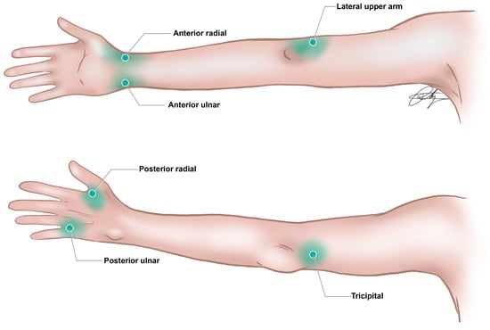
Introduction: Axillary reverse mapping (ARM) aims to reduce the risk of breast cancer-related lymphedema (BCRL) by preserving and limiting dissection of arm-draining lymphatics. The ideal type of dye and the location of injection, which maximize the sparing of lymphatics and improve outcomes of immediate lymphatic reconstruction (ILR), remain under-studied. The current literature reports inconsistent visualization of lymphatics using blue dye alone, whereas indocyanine green (ICG) near-infrared (NIR) lymphography has shown improved rates. However, optimized dual-dye workflows integrating breast–plastics co-surgery are lacking. Methods: A retrospective review of patients who underwent ILR following ALND for breast cancer between June 2021 and June 2023 was conducted. Patients who underwent ARM using our dual-dye technique were included, utilizing intradermal injections of indocyanine green (ICG) into the wrist and isosulfan blue (ISB) into the upper arm. Axillary reverse mapping channels were categorized by the type of dye used to visualize. Dye injection site, number of lymphatic channels visualized, channel diameter (mm), time-to-first channel, coordinates relative to fixed landmarks, ILR configuration, and pathologic findings were reviewed. Mann–Whitney U tests were used to compare channel visualization rates between types of dye. Results: Of 26 patients, 21 underwent dual-dye mapping and were included. A total of 115 ARM channels were identified: 99 (86%) via ICG and 29 (25%) via ISB. A total of 64 lymphaticovenous anastomoses were performed (mean: 2.46 per patient). Both dyes were identified in the axilla in only 11.7% of patients. At the end of the study, the lymphedema rate was 12%. Conclusions: We developed a reproducible dual-dye ARM technique for ALND with planned ILR, reducing lymphedema risk while maintaining oncologic safety. Dual-dye mapping reveals that proximal and distal lymphatics exhibit both overlapping and divergent drainage to axillary nodes. ICG’s higher axillary detection rate may reflect true anatomical differences or dye properties. These findings support the need for individualized lymphatic mapping during breast cancer surgery to guide preservation techniques and reduce the risk of BCRL.
Full article

Figure 1


