Communication

Assembly and Annotation of the Complete Genome Sequence of the Paenibacillus Bacteriophage phJNUCC32

Department of Beauty and Cosmetology, Jeju Inside Agency and Cosmetic Science Center, Jeju National University, Jeju 63243, Republic of Korea
*
Author to whom correspondence should be addressed.
Acta Microbiol. Hell. 2024, 69(3), 144-152; https://doi.org/10.3390/amh69030014
Submission received: 19 May 2024 / Revised: 17 June 2024 / Accepted: 25 June 2024 / Published: 5 July 2024

Abstract

:
A potential biocontrol agent for American foulbrood (AFB), the Paenibacillus bacteriophage phJNUCC32, was isolated from Baengnokdam in Halla Mountain. This study aimed to investigate its genomic characteristics through whole-genome sequencing. The genome of phJNUCC32 was found to be 62,871 base pairs in length, with a G + C content of 51.98%. Phylogenetic analysis classified phJNUCC32 within the unclassified Caudoviricetes bacteriophage category. The genome prediction confirmed the absence of virulence factors and antibiotic-resistance genes, ensuring its genetic safety. A total of 63 coding DNA sequences were identified, revealing a modular arrangement. Notably, the annotation of gene function indicates that phJNUCC32 harbors the holin/lysin system, suggesting significant potential for controlling bacterial infections in AFB and agriculture.

1. Introduction

Bacteriophages, viruses infecting various microorganisms, were discovered by Twort and d’Herelle in 1915 and 1917 [1]. With unique host specificity, they combat bacterial infections and are extensively used in medicine, dentistry, and agriculture. Molecular biology advancements have deepened our understanding of bacteriophages, aiding in their application as antibiotic alternatives. Leveraging their specificity, self-replication, and low cost, they are widely employed in treating infections in humans, animals, plants, and environmental decontamination.
Some species, such as Paenibacillus larvae, Paenibacillus apiarius, and Paenibacillus glabratella, are pathogens to honeybees and other invertebrates, with P. larvae causing lethal intestinal infections [2,3]. P. larvae is a Gram-positive bacterium responsible for inducing American foulbrood, a significant affliction in apiculture [4]. AFB detrimentally affects honeybee larvae, exacerbates colony collapse disorder, and diminishes agricultural productivity. In particular, strains of P. larvae are increasingly exhibiting resistance to antibiotics [5]; bacteriophages targeting and lysing P. larvae present a potentially promising avenue for therapeutic intervention. In summary, reported Paenibacillus bacteriophages include the P. larvae phage phiIBB_Pl23 [6]; five P. larvae bacteriophages from soil [7]; nine P. larvae bacteriophages from soil, propolis, and infected bees [8]; and eighteen P. larvae phages from the western United States [9], etc. The results of the genomic analysis of 48 P. larvae bacteriophages reveal that all phage genomes display a conserved N-acetylmuramoyl-l-alanine amidase, serving as an endolysin [10].
The holin/lysin system in bacteriophages inhibits the impact of P. larvae on honeybee larvae through a collaborative mechanism [11]. Endolysin, primarily sourced from phages targeting Gram-positive bacteria [12], serves as a key component of the holin/lysin system; it participates in the bacteriophage’s infection and cell lysis process by regulating the localization of endolysins to the specific cleavage sites of “cross-links” within the peptidoglycan (PG) layer of bacterial cell walls through the accumulation of holins and creation of lesions in the cytoplasmic membrane, ultimately triggering host cell lysis at a specific time point [13,14,15].
In previous studies, the majority of bacteriophages utilize endolysin to enzymatically degrade the PG layer of the host bacterium, exhibiting anti-biofilm properties. This includes the natural lysis of Salmonella enteritidis by the endolysin of the bacteriophage vB_Sal-S-S10 [16], the endolysin Ply113 acting as a potent antibacterial agent against polymicrobial biofilms formed by enterococci and Staphylococcus aureus [17], and the endolysin from Staphylococcus aureus bacteriophage 52 showing anti-biofilm and broad antibacterial activity against Gram-positive bacteria [18]. Additionally, endolysins such as LysAB1245 targeting different capsular types associated with Acinetobacter exhibit extended lytic activity [19], while endolysins LysCPD2 and LysCPQ7 act as biocontrol agents against Clostridium perfringens [20,21], etc.
The holin/lysin system offers a sustainable and environmentally friendly approach to selectively eradicate target pathogens without harming beneficial microorganisms [22]. Operating through physical mechanisms that disrupt bacterial cell membranes and walls rather than chemical agents, it minimizes the likelihood of inducing resistance, making it promising for controlling drug-resistant pathogens [23]. Its mechanism of action leads to rapid lysis and dissolution of bacterial cells, releasing numerous bacteriophage particles, thus swiftly reducing pathogen populations and aiding in disease control [24]. Compared to chemical pesticides, it reduces reliance on chemical substances and minimizes environmental pollution, aligning with principles of sustainable agriculture and environmental protection.
This study isolated a potential strain, Paenibacillus bacteriophage phJNUCC32, from Baengnokdam in Halla Mountain and conducted whole-genome sequencing. Through sequence comparisons, gene function annotation of the bacteriophage, as well as predictions of virulence factors and antibiotic-resistance genes, were conducted, resulting in the identification of genes with potential therapeutic effects against American foulbrood.

2. Materials and Methods

2.1. Bacterial Isolation

The Paenibacillus bacteriophage phJNUCC32 was isolated from Baengnokdam, Mt. Halla, South Korea, in September 2019. Soil samples (0.5 g) were suspended in 0.45 mL of 0.1% tris-buffer and shaken (180 rpm, 30 °C, 1 h). After serial dilution (10−5 to 10−9), 100 µL of the suspension was spread onto MRS medium (pH 6.5). Routine culture involved aerobic growth on LB solid/liquid medium at 30 °C for 1 day, with storage in 20% glycerol at −80 °C [25].

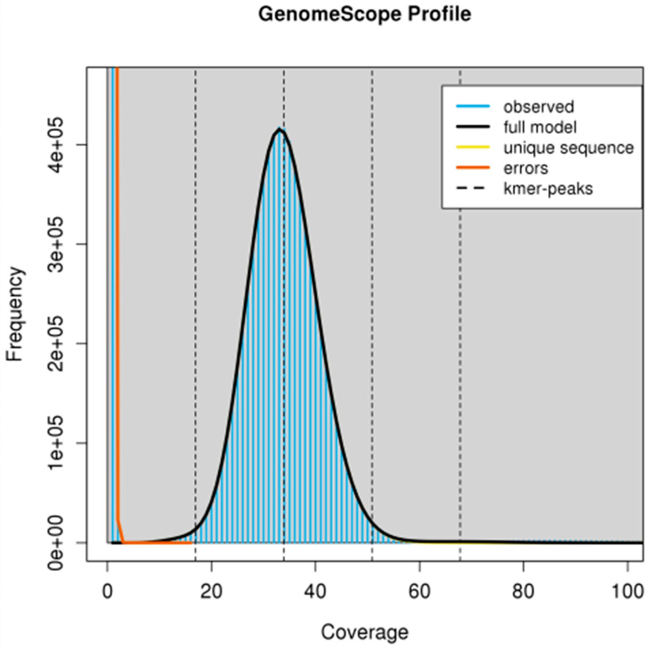
2.2. Sequencing and De Novo Assembly

The strain phJNUCC32’s DNA was extracted using a QIAGEN genomic-tip and sequenced with PacBio RSII and Illumina at Macrogen, Inc. (Seoul, Republic of Korea). K-mer analysis was performed to estimate the genome size of the sample. The k-mer distribution of the genome was analyzed using Jellyfish (v2.2.10), and GenomeScope 2.0. HGAP (v3.0) first assembled PacBio long reads. Then, Illumina reads were used to refine genome sequence accuracy with Pilon (v1.21). Finally, subreads were mapped against contigs to generate consensus sequences with coverage depth data.

2.3. Genome Annotation and Phylogenetic Tree

We used the online tool PHASTEST (PHAge Search Tool with Enhanced Sequence Translation) (https://phastest.ca/, accessed on 5 March 2024) to predict functional annotations of the genome of the bacteriophage phJNUCC32. Antibiotic-resistance gene prediction was performed by submitting the genome of the bacteriophage phJNUCC32 to the online database (http://arpcard.mcmaster.ca, accessed on 21 March 2024). Virulence gene prediction was conducted by submitting the genome of the bacteriophage phJNUCC32 to the online tool (http://www.mgc.ac.cn/VFs/search_VFs.htm, accessed on 21 March 2024). For the construction of phylogenetic trees, we employed full-length amino acid sequences of the terminase large subunit (TerL). Sequence alignment was performed using MAFFT. Sequence trimming was performed using trimAl, with commands to retain conserved regions. Phylogenetic tree construction was performed using IQ-TREE 2.0, which automatically determined the optimal model, utilizing the maximum likelihood method and 1000 bootstrap replicates. [26]. The resulting phylogenetic trees were visualized using TVBOT (https://www.chiplot.online/tvbot.html, accessed on 7 March 2024).

3. Results and Discussion

3.1. Genome Characteristics of the Paenibacillus Bacteriophage phJNUCC32

Jellyfish generated a 21-mer count histogram from a subset of short DNA reads. GenomeScope then used this histogram to estimate genome size, k-mer coverage, and heterozygosity. The graph was plotted with the coverage and frequency of k-mers. The genome size can be estimated using total k-mer number and volume peak (Figure 1). The genome of the phage JNUCC32 has a size of 62,871 base pairs, with a G + C content of 51.98%, a k-mer coverage of 33.9, and low heterozygosity of 0.022% (below 0.05%). Genome scanning identified 63 coding DNA sequences (CDSs) in the genome, with no RNA sequences detected. The genomic features of the phage JNUCC32 are summarized in Figure 2. The circular genome map was generated using Prokka (v1.12b). The draft genome sequence of the phage JNUCC32 has been submitted and deposited in the NCBI GenBank database under the accession ID CP062261.

3.2. Phylogenetic Analysis

To further elucidate the evolutionary relationships among bacteriophages, a phylogenetic tree was constructed by comparing conserved and evolutionarily significant sequences within these viruses. The sequence of the terminase large subunit (ORF2), a conserved region within bacteriophages, was utilized for this purpose, allowing for comparison of genetic relationships among different bacteriophages. As shown in Figure 3, phylogenetic analysis of the terminase large subunit of the Paenibacillus phage phJNUCC32 reveals a close genetic relationship with the Bacillus phage phBC6A51. This proximity within the evolutionary tree suggests their classification within the unclassified Caudoviricetes bacteriophage category. In summary, phylogenetic linkages provide a framework for understanding the evolutionary history, taxonomy, and functional diversity of bacteriophages. These insights are crucial for advancing phage research, including phage therapy, biotechnology, and microbial ecology.

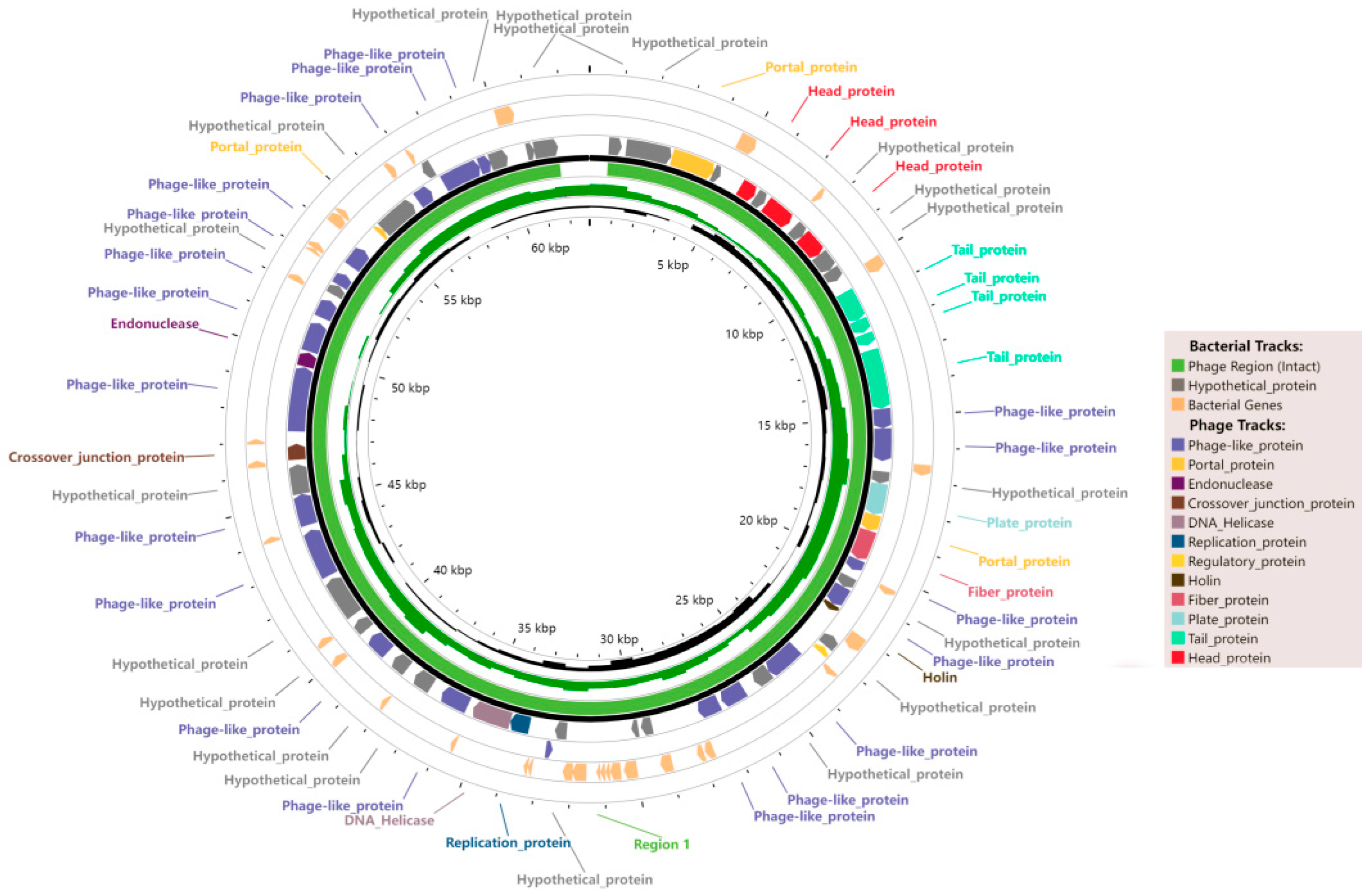
3.3. Functional Annotation

In the Paenibacillus bacteriophage phJNUCC32 genome, a total of 97 open reading frames (ORFs) have been identified. Of these, 63 are CDSs. Among the CDSs, 45 are predicted to encode proteins with known functions, constituting 71.4% of the total CDSs. (Figure 4, Table 1). The gene distribution of phJNUCC32 demonstrates a characteristic modular pattern, comprising various functional modules such as hypothetical protein, portal protein, head protein, tail protein, phage-like protein, plate protein, fiber protein, holin, regulatory protein, replication protein, DNA helicase, crossover junction protein, and endonuclease.
ORF2 in the DNA packaging module encodes the large subunit of the terminase enzyme. Terminase enzymes are essential for packaging phage DNA into the capsid during virion assembly, ensuring proper encapsulation of genetic material. These proteins play a critical role in phage replication and propagation [27].
Portal proteins are encoded by ORFs 3, 20, and 55. Portal proteins create a channel in the capsid for phage DNA injection into host cells during infection [28]. They facilitate early infection stages by aiding phage DNA entry into the host cell cytoplasm. Portal proteins are vital for phage infectivity, often conserved among different phage species. They are promising targets for phage therapy and genetic engineering.
Tail proteins are represented by ORFs 12 to 15. Tail proteins attach phage to host cell surfaces, recognize host receptors, and inject phage DNA into host cells [29]. They facilitate phage adsorption and penetration, initiating infection. Tail proteins exhibit diversity across phage types, reflecting host recognition specificity and range determination.
ORF22 encodes thiosulfate sulfurtransferase (TST), also known as rhodanese, pivotal in microbial metabolism, detoxifying thiosulfate by binding it with organic toxins, aiding their elimination [30]. TST also balances sulfur cycling by transferring sulfur atoms and may combat oxidative stress by neutralizing oxygen radicals [31]. In microorganisms, TST ensures detoxification, metabolic balance, antioxidation, and energy metabolism, vital for survival and adaptation.
ORF24 is predicted to encode the endolysin gene based on bioinformatic analysis, indicating that nucleotides 21,003–21,692 (690 bp) of ORF24 encoded amino acid endolysin and that the protein belongs to the N-acetylmuramoyl-L-alanine amidase CwlA family. Endolysins are phage-encoded peptidoglycan hydrolases (PGHs), also known as internal lysins or bacterial lysozymes. They exhibit high specificity and are involved in bacterial lysis at the end of the infection cycle. The bacteriophage perforin forms pores on the cell membrane, allowing endolysin to reach PG targets on the bacterial cell wall and cleave it hydrolytically, ultimately leading to bacterial lysis and death [32].
ORF25 of phJNUCC32 is annotated as the holin gene. Holin is a small transmembrane protein encoded by bacteriophages. Upon bacteriophage infection, holin forms non-specific pores on the host cell membrane within a specific time frame, thereby controlling the duration of the bacteriophage infection cycle and lysing the host cell at the optimal time point. Due to its role in regulating the bacteriophage infection cycle, holin is also referred to as the “clock” controlling bacteriophage infection [33].
ORF49 is predicted to encode the HNH endonuclease (H: Histidine, N: Asparagine, and H: Histidine), which participates in mediating the insertion of the bacteriophage genome into the host genome. The endonuclease can cleave specific sites in the host bacterium’s DNA, followed by homologous recombination between the bacteriophage’s single-stranded DNA and the host genome DNA, facilitated by host nuclease inhibitory proteins [34,35].
No known antibiotic-resistance genes or virulence factors were predicted in phJNUCC32, indicating the phage’s safety at the genetic level.

4. Conclusions

In this study, we isolated and characterized the Paenibacillus bacteriophage phJNUCC32, which shows promising biocontrol activity. Whole-genome sequencing and functional analysis revealed the presence of the holin/lysin system, enabling selective infection and eradication of P. larvae, effectively controlling the spread of American foulbrood while preserving beneficial microorganisms, thereby contributing to ecosystem balance and biodiversity conservation. Moreover, phJNUCC32 exhibited genetic safety by lacking virulence factors and antibiotic-resistance genes, thus mitigating potential harm to the environment and ecosystems. Overall, the bacteriophage phJNUCC32 demonstrates significant potential for biocontrol and ecological safety, offering a novel approach for sustainable bee health management and beekeeping industry development.

Author Contributions

Conceptualization, C.-G.H. and X.L.; methodology, Y.X. and X.L.; software, Y.X. and X.L.; writing—original draft preparation, Y.X. and X.L.; writing—review and editing, C.-G.H.; supervision, C.-G.H.; project administration, C.-G.H.; funding acquisition, C.-G.H. All authors have read and agreed to the published version of the manuscript.

Funding

This research was supported by the “Regional Innovation Strategy (RIS)” through the National Research Foundation of Korea funded by the Ministry of Education (2023RIS-009).

Institutional Review Board Statement

Not applicable.

Informed Consent Statement

Not applicable.

Data Availability Statement

Data are contained within the article.

Conflicts of Interest

The authors declare no conflicts of interest.

References

  1. Duckworth, D.H. Who discovered bacteriophage? Bacteriol. Rev. 1976, 40, 793–802. [Google Scholar] [CrossRef] [PubMed]
  2. Gkitsaki, I.; Papachristoforou, A.; Michailidou, S. The transmittable through stinging microbiota differs between honeybees and wasps: A potentially greater microbial risk of the wasp sting for humans. Int. Microbiol. 2023, 26, 663–674. [Google Scholar] [CrossRef] [PubMed]
  3. Rampacci, E.; Sforna, M.; Dentini, A. Paenibacillus amylolyticus osteomyelitis in a Poodle dog: Case report and literature review. J. Vet. Diagn. Investig. 2022, 34, 703–708. [Google Scholar] [CrossRef] [PubMed]
  4. Genersch, E.; Forsgren, E.; Pentikäinen, J. Reclassification of Paenibacillus larvae subsp. Pulvifaciens and Paenibacillus larvae subsp. larvae as Paenibacillus larvae without subspecies differentiation. Int. J. Syst. Evol. Microbiol. 2006, 56, 501–511. [Google Scholar] [PubMed]
  5. Evans, J.D. Diverse origins of tetracycline resistance in the honey bee bacterial pathogen Paenibacillus larvae. J. Invertebr. Pathol. 2003, 83, 46–50. [Google Scholar] [CrossRef] [PubMed]
  6. Oliveira, A.; Melo, L.D.R.; Kropinski, A.M. Complete genome sequence of the broad-host-range Paenibacillus larvae phage phiIBB_Pl23. Genome Announc. 2013, 1, 00438-13. [Google Scholar] [CrossRef] [PubMed]
  7. Sheflo, M.A.; Gardner, A.V.; Merrill, B.D. Complete genome sequences of five Paenibacillus larvae bacteriophages. Genome Announc. 2013, 1, 00668-13. [Google Scholar] [CrossRef] [PubMed]
  8. Philippos, K.T.; Diane, G.Y.; Andrew, K. Complete Genome Sequences of Nine Phages Capable of Infecting Paenibacillus larvae, the Causative Agent of American Foulbrood Disease in Honeybees. ASM Sci. J. 2015, 3, e01120-15. [Google Scholar]
  9. Merrill, B.D.; Fajardo, C.P.; Hilton, J.A. Complete genome sequences of 18 Paenibacillus larvae phages from the Western United States. Microbiol. Resour. Announc. 2018, 7, 00966-18. [Google Scholar] [CrossRef] [PubMed]
  10. Stamereilers, C.; Fajardo, C.P.; Walker, J.K. Genomic analysis of 48 Paenibacillus larvae bacteriophages. Viruses 2018, 10, 377. [Google Scholar] [CrossRef]
  11. Tsourkas, P.K. Paenibacillus larvae bacteriophages: Obscure past, promising future. Microb. Genom. 2020, 6, e000329. [Google Scholar] [CrossRef] [PubMed]
  12. Oliveira, H.; São José, C.; Azeredo, J. Phage-derived peptidoglycan degrading enzymes: Challenges and future prospects for in vivo therapy. Viruses 2018, 10, 292. [Google Scholar] [CrossRef] [PubMed]
  13. Shi, Y.; Yan, Y.; Ji, W. Characterization and determination of holin protein of Streptococcus suis bacteriophage SMP in heterologous host. Virol. J. 2012, 9, 1–11. [Google Scholar] [CrossRef]
  14. Hua, Y.; An, X.; Pei, G. Characterization of the morphology and genome of an Escherichia coli podovirus. Arch. Virol. 2014, 159, 3249–3256. [Google Scholar] [CrossRef]
  15. Schmelcher, M.; Donovan, D.M.; Loessner, M.J. Bacteriophage endolysins as novel antimicrobials. Future Microbiol. 2012, 7, 1147–1171. [Google Scholar] [CrossRef] [PubMed]
  16. Wang, X.; Han, L.; Rong, J. Endolysins of bacteriophage vB_Sal-S-S10 can naturally lyse Salmonella enteritidis. BMC Vet. Res. 2022, 18, 410. [Google Scholar] [CrossRef]
  17. Wang, J.; Liang, S.; Lu, X. Bacteriophage endolysin Ply113 as a potent antibacterial agent against polymicrobial biofilms formed by enterococci and Staphylococcus aureus. Front. Microbiol. 2023, 14, 1304932. [Google Scholar] [CrossRef]
  18. Abdurahman, M.A.; Durukan, İ.; Dinçer, T. Staphylococcus aureus Bacteriophage 52 endolysin exhibits anti-biofilm and broad antibacterial activity against gram-positive bacteria. Protein J. 2023, 42, 596–606. [Google Scholar] [CrossRef]
  19. Soontarach, R.; Srimanote, P.; Arechanajan, B. Characterization of a novel bacteriophage endolysin (LysAB1245) with extended lytic activity against distinct capsular types associated with Acinetobacter baumannii resistance. PLoS ONE 2024, 19, e0296453. [Google Scholar] [CrossRef]
  20. Mohammadi, T.N.; Lin, Y.; Maung, A.T. Characterization and antibacterial activity of highly thermo-and pH-stable endolysin LysCPQ7 and its application as a biocontrol agent against Clostridium perfringens in milk and cheese. Food Control. 2024, 156, 110157. [Google Scholar] [CrossRef]
  21. Pennone, V.; Sanz-Gaitero, M.; O’Connor, P. Inhibition of L. monocytogenes biofilm formation by the amidase domain of the phage vB_LmoS_293 endolysin. Viruses. 2019, 11, 722. [Google Scholar] [CrossRef] [PubMed]
  22. Cisek, A.A.; Dąbrowska, I.; Gregorczyk, K.P. Phage therapy in bacterial infections treatment: One hundred years after the discovery of bacteriophages. Curr. Microbiol. 2017, 74, 277–283. [Google Scholar] [CrossRef] [PubMed]
  23. Drulis-Kawa, Z.; Majkowska-Skrobek, G.; Maciejewska, B. Bacteriophages and phage-derived proteins–application approaches. Curr. Med. Chem. 2015, 22, 1757–1773. [Google Scholar] [CrossRef] [PubMed]
  24. Loessner, M.J. Bacteriophage endolysins-current state of research and applications. Curr. Opin. Microbiol. 2005, 8, 480–487. [Google Scholar] [CrossRef] [PubMed]
  25. Xu, Y.; Liang, X.; Hyun, C.G. Isolation, Characterization, Genome Annotation, and Evaluation of Tyrosinase Inhibitory Activity in Secondary Metabolites of Paenibacillus sp. JNUCC32: A Comprehensive Analysis through Molecular Docking and Molecular Dynamics Simulation. Int. J. Mol. Sci. 2024, 25, 2213. [Google Scholar] [CrossRef] [PubMed]
  26. Nguyen, L.T.; Schmidt, H.A.; Von, H.A. IQ-TREE: A fast and effective stochastic algorithm for estimating maximum-likelihood phylogenie. Mol. Biol. Evol. 2015, 32, 268–274. [Google Scholar] [CrossRef]
  27. Maluf, N.K.; Gaussier, H.; Bogner, E. Assembly of bacteriophage lambda terminase into a viral DNA maturation and packaging machine. Biochemistry 2006, 45, 15259–15268. [Google Scholar] [CrossRef]
  28. Prevelige, J.P.E.; Cortines, J.R. Phage assembly and the special role of the portal protein. Curr. Opin. Virol. 2018, 31, 66–73. [Google Scholar] [CrossRef]
  29. Taslem, M.J.; Awe, A.; Guo, W. Understanding bacteriophage tail fiber interaction with host surface receptor: The key “blueprint” for reprogramming phage host range. Int. J. Mol. Sci. 2022, 23, 12146. [Google Scholar] [CrossRef]
  30. Kruithof, P.D.; Lunev, S.; Lozano, S.P.A. Unraveling the role of thiosulfate sulfurtransferase in metabolic diseases. BBA-Mol. Basis Dis. 2020, 1866, 165716. [Google Scholar] [CrossRef]
  31. Nakajima, T. Roles of sulfur metabolism and rhodanese in detoxification and anti-oxidative stress functions in the liver: Responses to radiation exposure. Med. Sci. Monit. 2015, 21, 1721. [Google Scholar] [CrossRef] [PubMed]
  32. Rodríguez-Rubio, L.; Martínez, B.; Donovan, D.M. Bacteriophage virion-associated peptidoglycan hydrolases: Potential new enzybiotics. Crit. Rev. Microbiol. 2013, 39, 427–434. [Google Scholar] [CrossRef] [PubMed]
  33. Wang, I.N.; Smith, D.L.; Young, R. Holins: The protein clocks of bacteriophage infections. Annu. Rev. Microbiol. 2000, 54, 799–825. [Google Scholar] [CrossRef] [PubMed]
  34. Kim, Y.G.; Cha, J.; Chandrasegaran, S. Hybrid restriction enzymes: Zinc finger fusions to Fok I cleavage domain. Proc. Nat. Acad. Sci. USA 1996, 93, 1156–1160. [Google Scholar] [CrossRef]
  35. Williams, R.J. Restriction endonuclease: Classification, properties, and applications. Mol. Biotechnol. 2003, 23, 225–243. [Google Scholar] [CrossRef]
Figure 1. K-mer analysis of the genome.
Amh 69 00014 g001
Figure 2. Circular genome map of the phJNUCC32 chromosome. The genomic characteristics are illustrated from the outer to the central region, including forward-strand CDS, reverse-strand CDS, GC content, and GC skew. Forward CDS: Regions containing forward CDS are represented, with non-CDS regions indicated as blank. Reverse CDS: Areas with reverse CDS are presented, and non-CDS regions are denoted as blank. GC content: Regions with a higher GC percentage than the average are depicted in an exterior light green peak. GC skew: Calculated by (G − C)/(G + C), a positive value indicates G dominance, while a negative value indicates C dominance. The exterior light green peak signifies regions with higher G content, while the interior lavender peak represents regions with higher C content.
Amh 69 00014 g002
Figure 3. Maximum likelihood phylogeny of the phage terminase large subunit (TerL) proteins of the Paenibacillus bacteriophage phJNUCC32.
Amh 69 00014 g003
Figure 4. Gene functional annotation circle plot of the Paenibacillus phage phJNUCC32.
Amh 69 00014 g004
Table 1. General features of the CDSs predicted from the genome of the Paenibacillus phage phJNUCC32.
CDSPositionPrediction FunctionBLAST HitE-Value
1658..1101Helix–turn–helix proteinBacill_phBC6A51_NC_0048201.48 × 10−33
21252..2823Terminase large subunitBacill_phBC6A51_NC_0048200.0
32845..4356Portal Gp-6 family-like proteinBacill_Mgbh1_NC_0418798.21 × 10−154
44349..4618Hypothetical proteinPHAGE_Verruc_P8625_NC_0290471.29 × 10−12
55395..5970Capsid protein-like proteinBacill_Mgbh1_NC_0418791.15 × 10−25
66011..6385Hypothetical proteinBacill_Mgbh1_NC_0418792.85 × 10−46
76457..7482Major capsid proteinBacill_Mgbh1_NC_0418791.42 × 10−131
87689..8117Hypothetical proteinBacill_phBC6A51_NC_0048204.01 × 10−13
98114..8962MuF-like minor capsid proteinGordon_Sadboi_NC_0488158.92 × 10−7
108962..9603Hypothetical proteinPaenib_Tripp_NC_0289301.40 × 10−5
119603..10037HK97 gp10 family phage proteinPaenib_Tripp_NC_0289301.97 × 10−5
1210513..11559xkdK-like tail sheath proteinClostr_phiCTC2B_NC_0309518.04 × 10−23
1311575..12021Putative tail core proteinClostr_phiCDHM19_NC_0289961.06 × 10−15
1412089..12472Putative core tail proteinLactob_jlb1_NC_0242061.87 × 10−12
1512674..14686Tail tape measure proteinLister_LP_101_NC_0243877.65 × 10−71
1614699..15373XkdPBalMu_1_NC_0309454.36 × 10−42
1715373..16485Late control D proteinBrevib_Jimmer1_NC_0291044.17 × 10−23
1816836..17252DUF2634 domain-containing proteinBrevib_Abouo_NC_0290291.36 × 10−20
1917252..18334Baseplate JThermu_OH2_NC_0217842.68 × 10−70
2018331..18894Portal proteinClostr_phiCT9441A_NC_0290222.04 × 10−28
2118898..19938Putative tail fiber proteinBacill_BCD7_NC_0195156.31 × 10−7
2219953..20351Rhodanese-related sulfurtransferaseThermu_OH2_NC_0217841.04 × 10−24
2320607..20999Hpothetical proteinBacill_vB_BboS_125_NC_0487353.69 × 10−12
2421003..21692EndolysinBacill_Waukesha92_NC_0254243.66 × 10−30
2521694..21912HolinEntero_AUEF3_NC_0421341.48 × 10−6
2622530..22928Hypothetical proteinPaenib_Tripp_NC_0289305.86 × 10−21
2722981..23205Helix–turn–helix transcriptional regulatorBacill_BceA1_NC_0486281.85 × 10−15
2823654..24877DNA translocase FtsKBacill_BceA1_NC_0486284.29 × 10−81
2924877..25479Replication–relaxation family proteinBacill_PfEFR_4_NC_0486414.68 × 10−64
3025921..26772Conserved phage proteinBacill_WBeta_NC_0077343.53 × 10−21
3126888..27655Putative cobyrinic acid ac-diamide synthaseBrevib_Sudance_NC_0287493.91 × 10−102
3229301..29675Hypothetical proteinTripp_NC_0289301.29 × 10−12
3329777..30010Hypothetical proteinBacill_Staley_NC_0227671.71 × 10−24
3432215..32625Hypothetical proteinPaenib_Tripp_NC_0289301.10 × 10−35
3532620..32838Xre-like proteinBacter_Lily_NC_0288414.75 × 10−11
3633500..34174DNA replication proteinPaenib_Tripp_NC_0289308.27 × 10−79
3734178..35518DNA helicase-like proteinBacill_Mgbh1_NC_0418791.12 × 10−137
3835720..36700DNA primasePaenib_Tripp_NC_0289309.76 × 10−80
3937121..37783Sigma-70 family RNA polymerase sigma factorBacill_Mgbh1_NC_0418794.35 × 10−10
4038141..38707Hypothetical proteinPaenib_Tripp_NC_0289301.20 × 10−24
4139014..39790Single-stranded DNA-binding proteinPaenib_Tripp_NC_0289303.87 × 10−95
4240070..40516Hypothetical protein KLEB271_gp57 Bacillus phage Paenib_Tripp_NC_0289309.99 × 10−49
4340737..42182Hypothetical proteinBacill_Mgbh1_NC_0418796.27 × 10−19
4442311..43990DNA polymerasePaenib_Tripp_NC_0289300.0
4544173..45258DNA polymerasePaenib_Tripp_NC_0289300.0
4645264..46277hypothetical proteinPaenib_Tripp_NC_0289302.99 × 10−110
4746437..46979Crossover junction endodeoxyribonucleaseBacill_Mgbh1_NC_0418795.54 × 10−48
4847398..49614Ribonucleotide diphosphate reductase alpha subunitBacill_Eldridge_NC_0309200.0
4949634..50128Putative HNH homing endonucleaseBacill_BCP8_2_NC_0273553.09 × 10−30
5050166..51197Ribonucleotide diphosphate reductase beta subunitBacill_SP_15_NC_0312451.95 × 10−109
5151394..52047Phosphate starvation-inducible protein PhoH-like proteinBacill_SP_10_NC_0194872.71 × 10−56
5252274..52627Hypothetical proteinBacill_Blue_NC_0310569.58 × 10−16
5352645..53076dCTPaseAcinet_ZZ1_NC_0180877.08 × 10−9
5453407..54123Thymidylate synthaseBacill_Riggi_NC_0227651.53 × 10−105
5554881..55084Portal proteinBacill_T_NC_0242052.15 × 10−8
5655081..56412Hypothetical proteinPaenib_Tripp_NC_0289301.33 × 10−32
5756620..57171ATPase-like proteinPaenib_Tripp_NC_0289301.76 × 10−58
5857276..57605Hypothetical proteinPaenib_Tripp_NC_0289301.14 × 10−7
5957676..59025Modification methylaseBacill_SPbeta_NC_0018841.24 × 10−92
6059025..59426SigK-like proteinPaenib_Tripp_NC_0289302.46 × 10−43
6159413..60039Hypothetical proteinPaenib_Tripp_NC_0289304.38 × 10−33
6260715..60981Hypothetical proteinPaenib_Tripp_NC_0289306.08 × 10−6
6360968..61792Hypothetical proteinPaenib_Tripp_NC_0289301.42 × 10−106
Disclaimer/Publisher’s Note: The statements, opinions and data contained in all publications are solely those of the individual author(s) and contributor(s) and not of MDPI and/or the editor(s). MDPI and/or the editor(s) disclaim responsibility for any injury to people or property resulting from any ideas, methods, instructions or products referred to in the content.

