Article

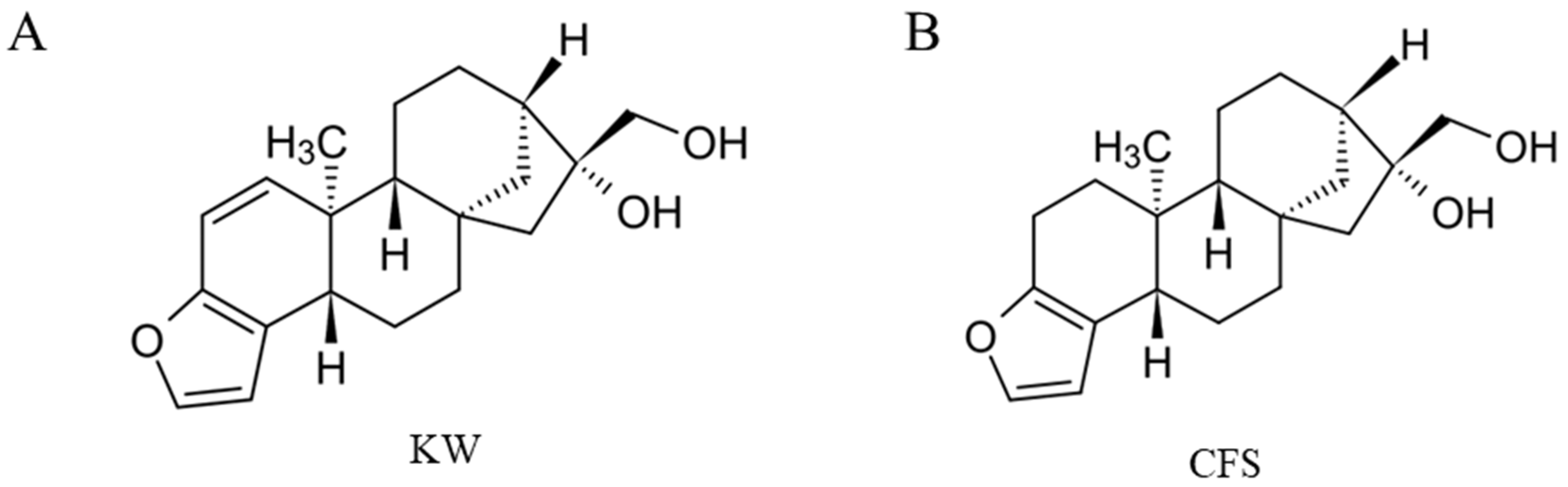
Two Coffee Diterpenes, Kahweol and Cafestol, Inhibit Extracellular Melanogenesis: An In Vitro Pilot Study

1
Department of Biochemistry and Cell Biology, Stony Brook University, Stony Brook, NY 11794-5215, USA
2
Department of Biomedical Engineering, Stony Brook University, Stony Brook, NY 11794-5281, USA
Biologics 2024, 4(2), 202-217; https://doi.org/10.3390/biologics4020014
Submission received: 12 April 2024 / Revised: 26 May 2024 / Accepted: 31 May 2024 / Published: 5 June 2024
(This article belongs to the Section Natural Products)

Abstract

:
Hyperpigmentation skin disorders are marked by an abnormal accumulation or export of melanin pigment synthesized within melanocytes and pose a significant aesthetic concern. The search for novel natural compounds that exhibit pharmacological potential for treating pigmentation disorders is growing. In this study, kahweol (KW) and cafestol (CFS), two structural analogs of coffee diterpenes, were evaluated and compared for their effects on melanogenesis using B16F10 mouse melanoma cells and primary human melanocytes derived from Asian and African American skin. To the best of our knowledge, there are no reports of the effects of KW and CFS on melanogenesis yet. We first screened nontoxic concentrations of both compounds using an MTS assay after 72 h incubations and subsequently tested their effects on melanin synthesis and export. Cellular tyrosinase activity and cell-free mushroom tyrosinase activity were assayed to study the mechanisms of melanogenesis suppression. Human melanocytes from a moderately pigmented donor (HEMn-MP cells) and from a darkly pigmented donor (HEMn-DP cells) were next examined, and effects on cellular viability, melanin content, cellular tyrosinase activity, and melanin export (quantitated via dendricity) were similarly examined for both compounds. Our results show that KW and CFS did not significantly affect intracellular melanin content but suppressed extracellular melanin in B16F10 cells and dendritic parameters in human melanocytes, indicating their unique capacity to target extracellular melanogenesis and melanin export. Although KW showed a greater extracellular melanogenesis inhibitory capacity in B16F10 cells, in both primary melanocyte cells, CFS emerged as a potent inhibitor of melanin export compared to KW. Together, these results reveal novel modes of action of both compounds and indicate a promise to use CFS as a novel candidate for treating hyperpigmentation disorders of the human skin for clinical and cosmetic use. Additional research is necessary to shed light on the molecular pathways and the efficacy of melanogenesis inhibition by CFS in 3D human skin equivalents and in vivo studies.

Graphical Abstract

1. Introduction

Coffee, a beverage widely consumed globally, contains various bioactive compounds such as caffeine, trigonelline, caffeic acid, chlorogenic acid, and diterpenes [1]. The diterpene chemicals kahweol (KW) and cafestol (CFS) are known for their significant presence in the lipid composition of coffee [2,3]. The concentration of diterpene in Arabica coffee (Coffea arabica) is in the range of 1.3–1.9% (w/w), while in Robusta coffee (Coffea canephora), it is in the range of 0.2–1.5%. The levels of free forms of KW and CFS range from 0.1% to 0.4% [4]. CFS is primarily found in unfiltered coffee brews, including Turkish/Greek coffee, French press, Scandinavian boiled coffees, and espresso. Filtered or instant coffee, conversely, contains only trace amounts of CFS [5]. KW and CFS are structurally similar, ent-kaurane pentacyclic diterpenes, with KW differing from CFS by a double bond on the furan ring (Figure 1A,B). When ingested orally, the coffee diterpenes KW and CFS have been demonstrated to raise serum cholesterol levels, with CFS causing significant effects [6,7,8,9]. Particularly, CFS was shown to increase plasma low-density lipoprotein levels in humans, thus being a risk factor for cardiovascular abnormalities [10,11]. KW and CFS are susceptible to degradation when exposed to acidic conditions, elevated temperatures, and light. However, KW, in its purified form, exhibits significant instability. Nevertheless, KW and CFS have been shown to demonstrate a multitude of biological benefits. For example, KW has been shown to exhibit anti-carcinogenic [12], anti-angiogenic [13], anti-adipogenic [14], antioxidant [15,16], anti-inflammatory [13,17], and skin-moisturizing effects [18]. CFS has been shown to demonstrate cardioprotective effects [19], anti-diabetic effects [20], and anti-cancer effects [21].
The excessive production of melanin pigment by specialized cells called melanocytes can lead to various dermatological concerns, including melasma, freckles, solar lentigo (age spots), and even skin cancer [22]. These issues go beyond mere aesthetics and have significant implications for dermatology. Tyrosinase is a crucial player in melanogenesis and is widely studied in skin pigmentation [23]. Melanin export is elevated in skin hyperpigmentation conditions, including post-inflammatory melanoderma, melasma, and solar lentigo [24,25]. Commonly used skin-depigmenting compounds are hydroquinone (HQ), arbutin, and kojic acid (KA), which lead to adverse effects. For instance, HQ has been linked to exogenous ochronosis [26,27], genotoxicity, and carcinogenic effects [28,29], while arbutin, a glycoside of HQ, causes contact dermatitis [30]. KA has been demonstrated to cause contact allergies [31]. In addition, a clinical study showed that the subjects who used the combination of KA and HQ in a topical gel developed adverse reactions, including erythema, burning sensation, and desquamation [32]. Given these limitations, there is ongoing research to discover novel natural products that can address pigmentation disorders and effectively reduce skin hyperpigmentation without causing harmful side effects and with a relatively safe profile. Numerous studies have been conducted to explore alternative methods of inhibiting melanogenesis, considering that some skin-whitening compounds function as tyrosinase inhibitors. There are a few that are focused on later stages of the melanogenesis cascade, like the transportation of melanin pigment through dendrites, which are the cytoplasmic structures of melanocytes that contain actin and microtubule [33]. Once synthesized within the cells, melanin-containing melanosomes are transported from melanocytes through a network of dendrites that may contact as many as forty keratinocytes and are ingested by keratinocytes through different pathways [34,35]. By utilizing compounds that can either suppress or enhance dendricity, it is possible to regulate skin pigmentation disorders.
Several prior studies have documented the suppression of melanin production and tyrosinase activity in B16F10 cells by an ethanolic extract of Arabica coffee [36] and a supercritical fluid extract of spent coffee [37]. A specific extract from Robusta coffee beans inhibited tyrosinase activity [38]. A clinical investigation revealed that the consumption of coffee was linked to a reduction in hyperpigmented facial spots caused by UV exposure [39]. In addition, the study [39] found that coffee mainly had a suppressive impact on areas with excessive pigmentation rather than basal pigmentation. It is important to mention that the study did not document the coffee consumption history of each participant, making it challenging to establish the impact of coffee usage duration on the results. Moreover, the study excluded individuals with melasma or other types of hyperpigmentation, focusing solely on those with UV-induced age spots (solar lentigines). Although the study did not pinpoint the exact compounds responsible for the effects, the authors suggested that coffee’s polyphenols might have contributed to antioxidant benefits and decreased pigmentation. Caffeic acid, a compound present in coffee, was shown to decrease melanin production in B16F10 cells [40] and also diminish UVB-induced hyperpigmentation and dermatitis in a mouse model after oral administration [41]. The potential of various coffee extracts and their bioactive compounds continues to increase, especially in the cosmetic industry [42]. For instance, a previous clinical study [43] reported that a coffee silverskin extract, enriched in KW and CFS fatty acid esters, contributed to skin protective and hydrating effects. However, to our knowledge, the effects of the purified coffee diterpenes KW and CFS on melanogenesis have not yet been explored. The objective of this study is to investigate the impact of two diterpenes, KW and CFS, which are commonly found in coffee, on the process of melanogenesis using the B16F10 mouse cell model and primary human melanocytes obtained from two distinct ethnic donors (African American and Asian donors). This is the first report to investigate the effects of natural compounds, KW and CFS, present in coffee, on melanogenesis utilizing the B16F10 cell model and further examining the impacts on normal human melanocytes from two distinct ethnicities.

2. Materials and Methods

2.1. Materials

The compounds KW and CFS (both > 98% purity) were procured from Cayman Chemicals (Ann Arbor, MI, USA). The stock solution of the two compounds was made using dimethyl sulfoxide (DMSO) and kept at −20 °C until usage. The CellTiter 96® Aqueous one MTS dye was obtained from Promega Corporation, located in Madison, WI, USA. The human melanocyte growth supplement (HMGS), basal medium 254, phosphate-buffered saline (PBS), Pierce™ bicinchoninic acid (BCA) protein assay kit, and the AKT/ERK Activation multispecies InstantOne™ ELISA kit were acquired from Thermo Fisher Scientific (Waltham, MA, USA). Kojic acid (KA), synthetic melanin, mushroom tyrosinase, L-DOPA, α-glucosidase from baker’s yeast, and p-nitrophenyl-α-D-glucopyranoside (pNG) substrate were purchased from Sigma-Aldrich (St. Louis, MO, USA).

2.2. Cell Culture

B16F10 mouse melanoma cells (CRL-6475™, ATCC, Manassas, VA, USA) were cultured in Dulbecco’s modified eagles’ medium (DMEM) with 10% heat-inactivated fetal bovine serum and 1% penicillin-streptomycin antibiotics. Human keratinocytes (HaCaT) were obtained from AddexBio (San Diego, CA, USA) and cultured in DMEM containing 10% heat-inactivated fetal bovine serum and 1% antibiotics. Primary human epidermal neonatal melanocytes (HEMn) from two different donors were obtained from Thermo Fisher Scientific, Inc. (Waltham, MA, USA). One donor had dark pigmentation (HEMn-DP cells; C2025C [44]), while the other had moderate pigmentation (HEMn-MP; C1025C [45]), representative of African American and Asian skin, respectively. These cells were cultivated using medium 254, which was supplemented with 1% HMGS and 1% antibiotics. All cells were grown in standard cell culture conditions (humidified atmosphere, 5% CO2, 37 °C).

2.3. MTS Cell Viability Assay

The B16F10 cells were cultured at 0.8 × 10⁴ cells/well in a 96-well plate for 24 h, followed by the addition of the compounds KW and CFS at different concentrations in a solution containing 0.4% DMSO. The cells were then incubated for 72 h. The HEMn-DP and HEMn-MP cells were seeded at a density of 2 × 10⁴ cells per well in a 96-well plate for 24 h and treated with the compounds for 72 h. In the case of HaCaT cells, 1 × 10⁴ cells were distributed into each well of a 96-well plate, compounds were added after 48 h, and cultures were incubated for 72 h. Control group treatment included solvent control (0.4% DMSO) in all experiments. After the 72 h incubation period of different cells, the culture medium was substituted with a new medium containing MTS dye, and the plates were incubated followed by measurement of absorbance at 490 nm.

2.4. Determination of Intracellular and Extracellular Melanin

The method used to quantify extracellular melanin and intracellular melanin is similar to that outlined in our previously published studies [46,47]. Briefly, B16F10 cells (0.6 × 10⁵ cells per well) were seeded into a 12-well tissue culture plate and cultured for 24 h. The HEMn-DP (1.5 × 10⁵ cells/well) and HEMn-MP cells (1.3 × 10⁵ cells/well) were grown in a 12-well plate for 24 h. The compounds were added, and the cultures were incubated for another 72 h. The culture medium was collected and centrifuged to remove cell debris. The supernatants were transferred in 96-well plates, and the absorbance was read at 475 nm. Cells were harvested, and cell pellets were then rinsed in PBS for the intracellular melanin assay. Melanin was dissolved in 1 N NaOH and heated to 70 °C after aspiration. A standard curve generated using synthetic melanin produced at various concentrations under similar conditions was used to convert the absorbance measured at 475 nm to μg melanin. Both the extracellular melanin content and intracellular melanin contents were reported as µg melanin/mg protein.

2.5. Intracellular Tyrosinase Activity

The B16F10 cells were cultivated in 12-well tissue culture plates at a 0.6 × 10⁵ cells/well density. After 24 h, the culture medium was renewed with the compounds and further incubated for 72 h. HEMn-DP cells (0.65 × 10⁵ cells/well) and HEMn-MP cells (1.3 × 10⁵ cells per well) were cultivated in 12-well culture plates for 24 h followed by KW and CFS treatments for 72 h. After the completion of the 72 h treatments, cells were subjected to trypsinization, rinsed with PBS, and then lysed. The lysates were clarified by centrifugation, and the resulting supernatants were transferred to a 96-well plate and combined with the L-DOPA substrate. Subsequently, the plate was read in kinetic mode in a microplate reader. The details of the method for determining tyrosinase activity for B16F10 cells have been described in our earlier study [48], while those for HEMn-DP/MP cells have been described in another study [49].

2.6. Quantitation of Melanocyte Dendricity Parameters

Similar to our previously published studies [46,47,50,51], dendricity was quantified as a metric for melanosome export by measuring three parameters: namely, (i) the number of dendrites per cell, (ii) total dendrite length, and (iii) the percentage of cells with more than two dendrites. Three days after treating HEMn-DP or HEMn-MP cells with KW/CFS, the cultures were photographed, and dendrite numbers per cell were manually counted. Next, using imaging software (NIS Elements 5.0), the lengths of all dendrites in each cell were measured and added to compute the overall dendrite length. Additionally, the percentage of cells with >2 dendrites was determined by counting and expressing it relative to the total number of cells.

2.7. Cell-Free Tyrosinase Activity

For the purpose of determining the effects of the compounds KW and CFS on tyrosinase activity in the absence of cells, mushroom tyrosinase enzyme was used in combination with L-DOPA as the substrate. The compounds KW and CFS were prepared in 0.05 M sodium phosphate buffer at various concentrations, and 80 microliters of each concentration were transferred to a 96-well plate. After this, 100 µL of 2 mM L-DOPA substrate solution was added, followed by the addition of the mushroom tyrosinase enzyme (with the final enzyme concentration in each well as 3.5 µg/mL). The progress of the reaction was monitored at 475 nm (for 10 min every 30 s). The linear portion of the slopes of the kinetic measurements were utilized to report the relative tyrosinase activity.

2.8. Intracellular and Cell-Free α-Glucosidase Enzymatic Assay

The enzyme α-glucosidase is involved in the glycosylation process, which controls tyrosinase’s initial maturation and transportation inside cells [52,53]. The method used to determine intracellular α-glucosidase enzymatic activity in B16F10 cells is similar to that described in our earlier report [46]. The direct (cell-free) α-glucosidase activity was estimated similar to the method outlined in our prior study [54] with minor modification. Briefly, in a 96-well plate, aliquots of different concentrations of KW and CFS were mixed with 100 microliters of 2 mM pNG solution. Subsequently, the enzyme was added, and the reaction rate was monitored kinetically for 20 min at 405 nm in a microplate reader.

2.9. ERK/AKT Phosphorylation ELISA Assay

B16F10 cells were treated with compounds KW and CFS over a concentration range of 10–30 μM for 72 h. After this, the cells were lysed, and the lysates were processed to determine the relative levels of phosphorylated ERK and phosphorylated AKT according to the instructions provided by the manufacturer of the AKT/ERK Activation multispecies InstantOne™ ELISA kit.

2.10. Statistical Analysis

The data for dendrite analysis were analyzed by Student’s t-test with Welch’s correction. The data for MTS cell viability for all experiments were analyzed by one-way analysis of variance (ANOVA) with Dunnett’s test. The data for all other experiments were analyzed by one-way ANOVA with Tukey’s test. All the analyses were run using GraphPad Prism software (version 10.2.2) and differences were regarded as statistically significant when p < 0.05. All data are reported as mean ± standard deviation (SD), and the symbols *, **, $, and # were used to denote significant levels of p < 0.05, p < 0.01, p < 0.001, and p < 0.0001, respectively, in comparison to the control.

3. Results

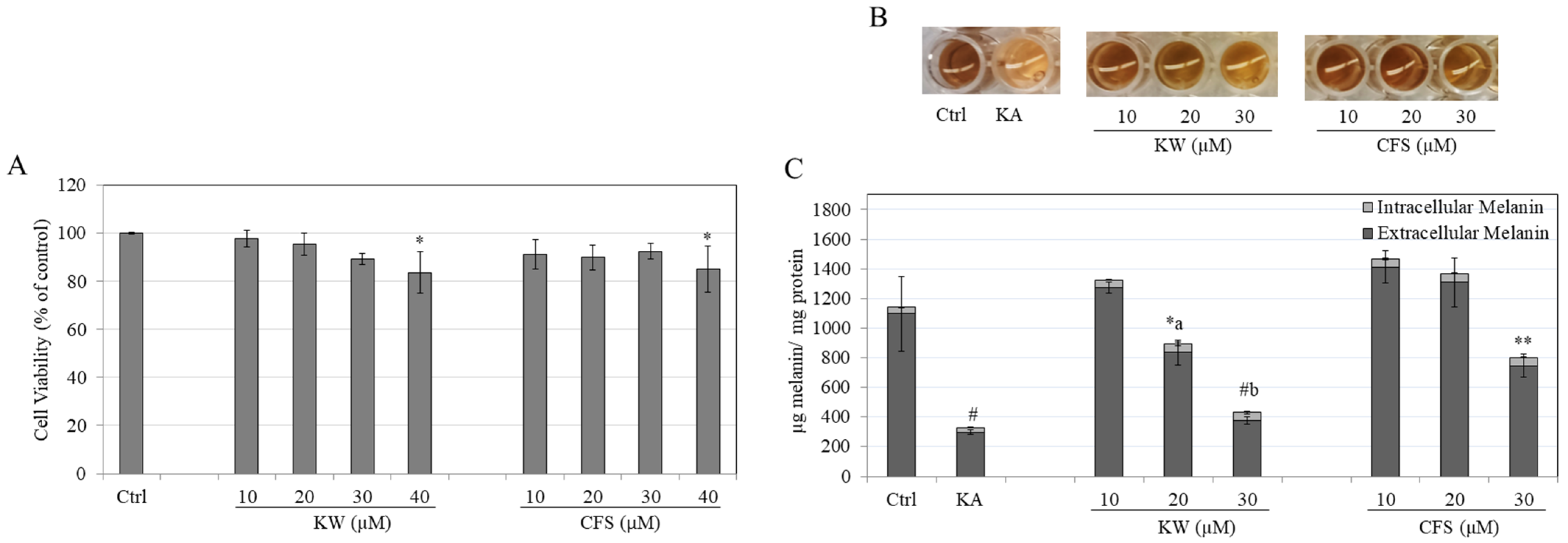
3.1. Effects of Compounds on B16F10 Cell Viability

Using an MTS assay, we conducted initial tests on KW and CFS for cytotoxicity. This allowed us to identify concentrations that were not toxic, which we then used for subsequent testing. The viability of B16F10 cells remained unaffected by both KW and CFS at concentrations ≤30 µM. Nonetheless, when the concentration reached 40 µM, both KW and CFS significantly reduced the cell viability to 83.80% and 85.18%, respectively (Figure 2A). Therefore, we only used the noncytotoxic concentration of both compounds in subsequent experiments.

3.2. Effects of Compounds on Melanin Content in B16F10 Cells

After KW and CFS treatment, the B16F10 cells’ culture media were noticeably lighter than the untreated control. Specifically, KA at 20 and 30 µM while CFS at 30 µM appeared to diminish the extracellular melanin (Figure 2B). Treatment with KA, a known melanogenesis inhibitor, also showed a lighter color medium. The intracellular melanin concentration of B16F10 cells did not show any significant changes when treated with either KW or CFS (p > 0.05), compared to the untreated control group (Figure 2C). Nevertheless, the extracellular melanin was significantly lowered by 13% and 60.71% when treated with KW at concentrations of 20 and 30 µM, respectively (Figure 2C). In addition, at a concentration of 30 µM, CFS reduced extracellular melanin by 22.23%. In contrast, lower concentrations did not show any effect (Figure 2C). Treatment with the positive control KA at a concentration of 500 µM significantly lowered extracellular melanin by 68.87% (Figure 2C). The data points for intracellular melanin and extracellular melanin amounts as shown in Figure 2C have also been summarized in a table for easier comparison between groups (Table S1).
Remarkably, the inhibitory impact of KW at a concentration of 30 µM on extracellular melanin was comparable to the effect of KA and substantially potent than CFS at the same concentration. Additionally, the quantitative data on extracellular melanin corroborated the earlier visual findings. These results indicate that KW and CFS exhibit the capacity to selectively suppress extracellular melanin without significantly impacting intracellular melanin, with KW having a superior effect than CFS.

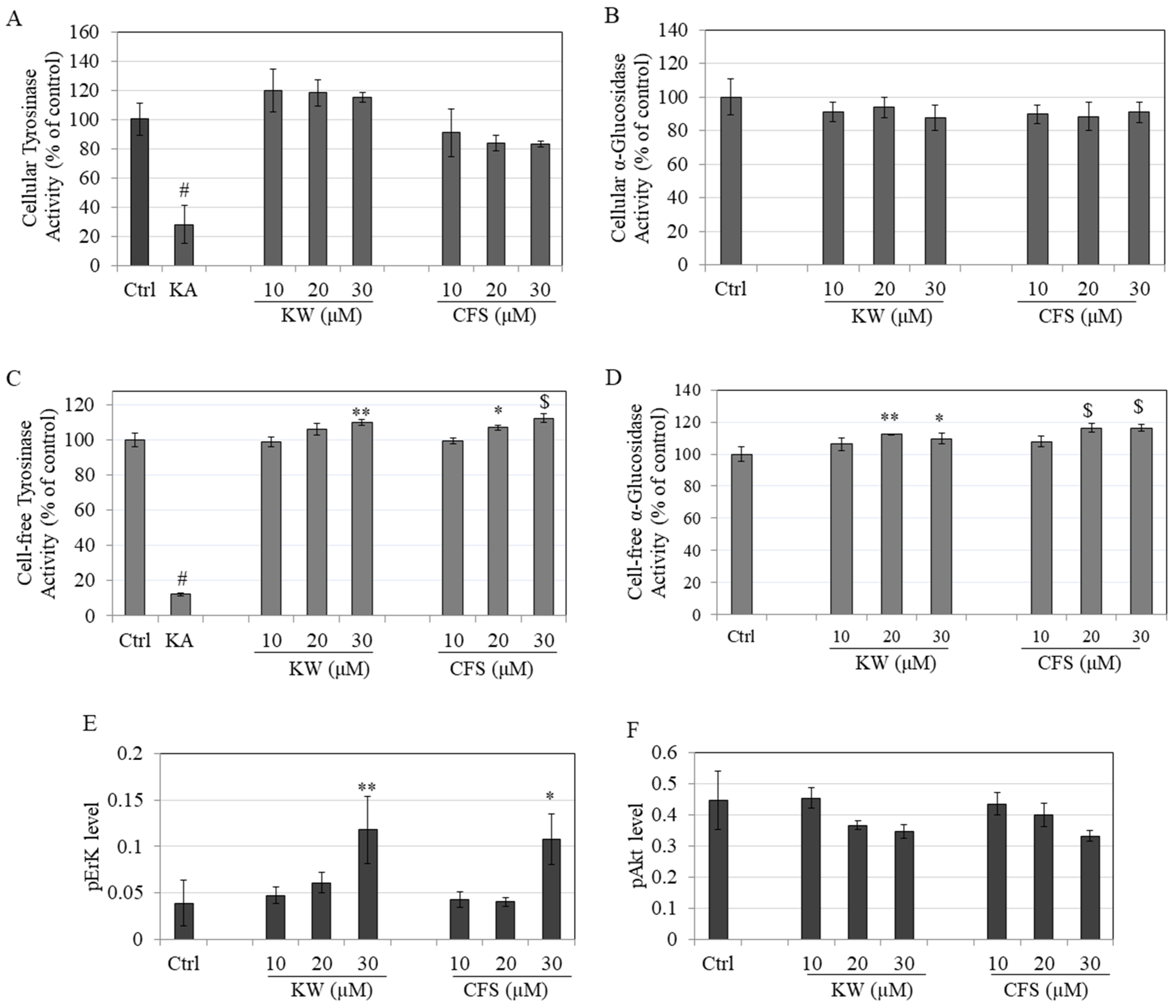
3.3. Effects on Tyrosinase and α-Glucosidase Enzymatic Activity

The effects of KW and CFS were next evaluated on tyrosinase enzyme activity in B16F10 cultures. The results of the comparison of the tyrosinase activities of KW and CFS showed a trend for increase while CFS showed a trend for decrease in the activity, although the levels were not significantly different from the untreated control group (Figure 3A). Next, the cellular α-glucosidase activities of B16F10 cells were also not affected after treatment with either KW or CFS at any concentration (Figure 3B).
Further, the effects of these compounds on enzymatic activities were also assessed in a cell-free system. Intriguingly, the results showed that KW and CFS enhanced the enzymatic activity of mushroom tyrosinase and α-glucosidase enzymes. Specifically, KW at 30 µM increased the tyrosinase activity by 10.07%, while CFS increased the tyrosinase activity by 6.99% and 12.44% at 20 and 30 µM concentrations, respectively (Figure 3C). KW significantly stimulated the activity of α-glucosidase enzyme by 12.26% and 9.64% at 20 and 30 µM concentrations, respectively (Figure 3D). Concurrently, at concentrations of 20 and 30 µM, CFS increased the α-glucosidase activity by 16.29% and 16.30%, respectively (Figure 3D). These results indicate that the inhibitory effects of KW and CFS on extracellular melanogenesis are not related to tyrosinase or α-glucosidase enzyme activities.

3.4. Effects on Phosphorylation of ERK/AKT

Treatment of B16F10 cells with both KW and CFS at 30 µM significantly increased the ERK ½ phosphorylation. Specifically, the phospho-ERK levels were elevated by 3.03-fold and 2.77-fold by KW (30 µM) and CFS (30 µM), respectively (Figure 3E). However, the levels of phosphorylated AKT seemed to show a trend for decrease although significance was not reached (Figure 3F). Overall, these results show that KW and CFS at 30 µM activate the ERK pathway that suppressed melanogenesis.

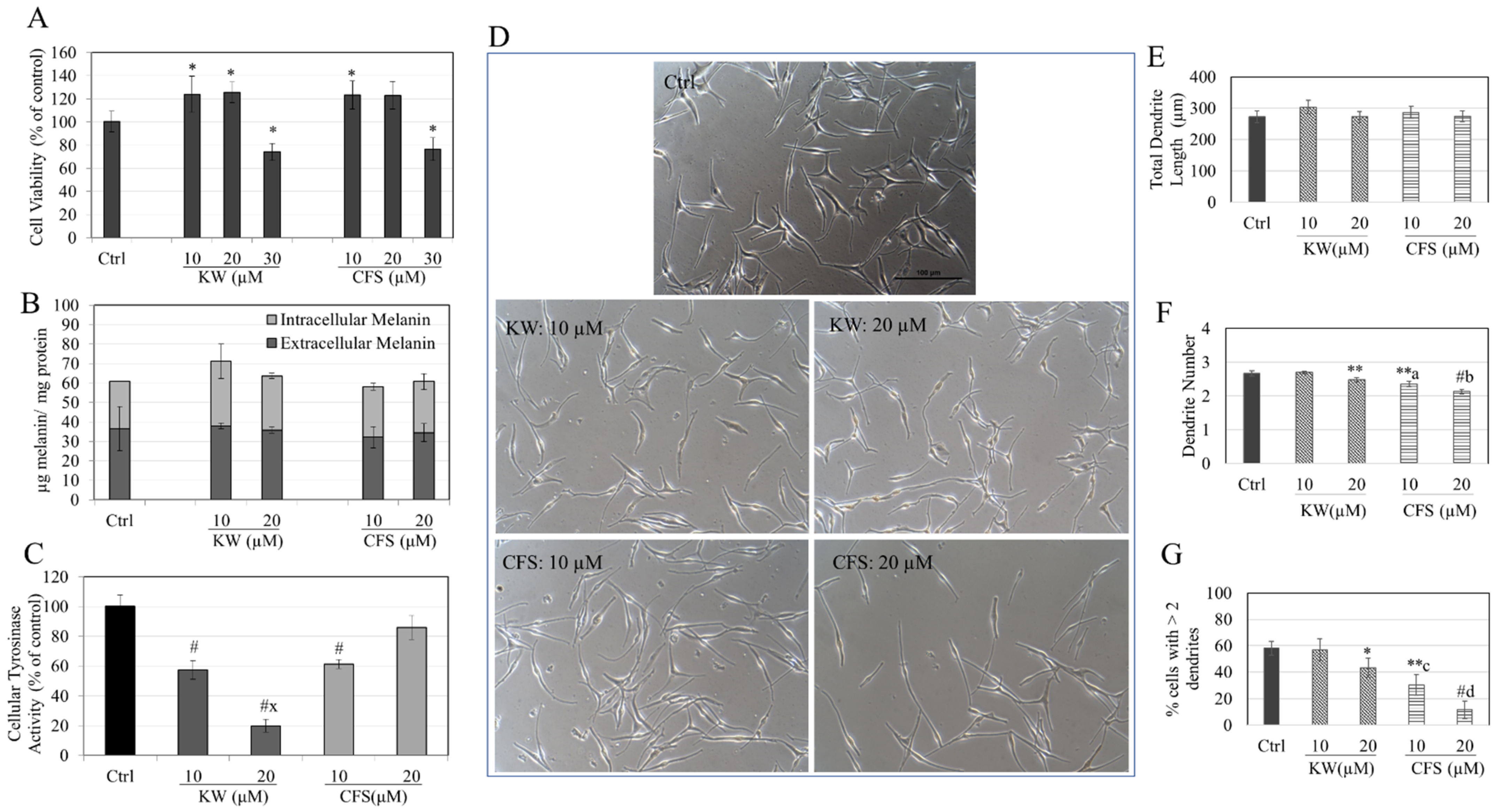
3.5. Effects of Compounds in HEMn-DP Cells

KW exerted cytotoxicity on HEMn-DP cells and significantly lowered the viability by 21.97% and 26.38% at concentrations of 20 and 30 µM, respectively (Figure 4A). On the other hand, CFS was nontoxic over the tested concentration range (Figure 4A). Accordingly, in further experiments with HEMn-DP cells, KW (10 µM) and CFS (10–30 µM) concentrations were utilized. The results on cellular melanin production (intracellular) and secretion (extracellular) showed no changes (p > 0.05) by KW or CFS at any concentration relative to the untreated control group (Figure 4B). The evaluation of the tyrosinase activity of HEMn-DP cells further showed that KW potently inhibited the cellular tyrosinase activity by 48.58% at 10 µM. In comparison, CFS significantly inhibited the tyrosinase activity by 31.82% and 64.99% at 20 and 30 µM, respectively (Figure 4C). KW had a stronger inhibitory effect than the equivalent concentration of CFS.
The morphological examination of cells after treatment with CFS at higher concentrations revealed striking changes, with the majority of cells having a lower number of dendrites, which was in contrast to the multidendritic morphology of the control group (Figure 4D). The quantitation of the dendritic parameters showed that the total dendrite lengths of cells were significantly decreased by treatment with both compounds. Specifically, KW (10 µM) significantly decreased the total dendrite length by 21.98%, while CFS significantly decreased this parameter by 17.05%, 16.89%, and 50.40% at 10, 20, and 30 µM, respectively (Figure 4E). The number of dendrites per cell was significantly diminished only by CFS in a concentration-dependent manner, with decreases of 22.62%, 32.89%, and 45.23% obtained at 10, 20, and 30 µM, respectively (Figure 4F). Lastly, the percentage of cells with >2 dendrites was significantly diminished from 75.45% of the control group to 53.47% and 20.36% by CFS at 20 and 30 µM, respectively (Figure 4G).
Taken together, these results show that at noncytotoxic concentrations, both KW and CFS do not impact the total melanin levels or melanin production within cells. However, they significantly inhibit cellular tyrosinase activity. However, CFS has a dramatic impact on the dendritic morphology by significantly suppressing all the dendritic parameters at higher concentrations, while KW diminished the total dendrite length, although it did not impact the dendrite number or the cell distribution. This suggests that KW has a much less significant impact on dendricity than CFS.

3.6. Effects of Compounds in HEMn-MP Cells

At a concentration of 30 µM, both KW and CFS significantly lowered the viability of HEMn-MP cells by 26.39% and 23.86%, respectively (Figure 5A). Based on this, further experiments were conducted using HEMn-MP cells with KW and CFS at concentrations of 10–20 µM. The findings regarding intracellular and extracellular melanin indicated that no significant changes were observed when comparing KW or CFS at any concentration to the control group (Figure 5B).
Next, the tyrosinase activity of HEMn-MP cells revealed that KW robustly inhibited the tyrosinase activity by 42.80% and 80.13% at 10 and 20 µM, respectively (Figure 5C). At the same time, CFS significantly inhibited the tyrosinase activity by 38.78% at 10 µM with no significant change at the higher concentration of 20 µM (Figure 5C). Similar to the DP cells, KW exerted a greater inhibitory effect on tyrosinase activity than that of CFS in MP cells.
The examination of the images of HEMn-MP cells showed that cells treated with CFS, especially at the higher concentration of 20 µM, displayed visibly shorter and a smaller number of dendrites (Figure 5D). The treatment with either KW or CFS did not have a significant impact on the total dendrite lengths of cells, as shown in Figure 5E. However, after treatment with KW (10 µM), CFS (10 µM), and CFS (20 µM), the dendrite number exhibited a significant reduction of 7.12%, 11.89%, and 20.41%, respectively (Figure 5F). The percentage of cells with >2 dendrites was significantly diminished from 58.17% in the control group to 30.27% and 11.62% in the presence of CFS at concentrations of 10 and 20 µM, respectively (Figure 5G). At the same time, KW significantly diminished this parameter to 43.10% at a concentration of 20 µM (Figure 5G).
Interestingly, it was observed that in MP cells, CFS and KW did not impact the total length of dendrites, which is different from their effects in DP cells. However, it was found that CFS concentration-dependently decreased the number of dendrites by 20.41% at 20 µM and robustly suppressed the percentage of polydendritic (>2 dendrites) by 46.55%. This suggests that CFS treatment drastically reduces dendrite outgrowth, resulting in a higher proportion (88.38%) of cells with two dendrites than the control group (41.83%). It is worth noting that when comparing CFS at 20 µM to KW, CFS showed a greater ability to suppress dendricity in HEMn-MP cells. This suggests that CFS is a more effective inhibitor of melanosome export.

3.7. Effects of Compounds HaCaT Cell Viability

KW and CFS were noncytotoxic to keratinocytes over the 10–30 µM concentration range for three days. Notably, KW substantially impacted the survival of HaCaT cells, increasing their viability by 30.29% at a concentration of 30 µM. Similarly, CFS significantly increased the cell viability by 20.77%, 22.97%, and 23.49% at concentrations of 10, 20, and 30 µM, respectively (Figure 6).

4. Discussion

The effects of two diterpenes found in coffee, CFS and KW, which are structurally related, were assessed on melanogenesis in this study. The evaluation used the B16F10 mouse cell model and primary human melanocytes from two distinct ethnic donors: Asians and African Americans. A novel anti-melanogenic activity of the compound CFS has been identified, which inhibits melanosome export rather than synthesis. Furthermore, the compounds’ strikingly distinct properties in mouse and human cell models have been demonstrated. We further demonstrated that CFS and KW did not induce cytotoxicity in human keratinocytes at the concentrations that suppress melanosome export in cells. In the absence of cytotoxic effects, we demonstrated that the compounds’ efficacy was reversed in primary human melanocytes, with CFS emerging as the most promising candidate. Because melanocytes and keratinocytes are in a symbiotic relationship where a single melanocyte may contact up to 30–40 keratinocytes, keratinocyte safety is crucial for skin pigmentary candidates [55,56]. The results show that at concentrations at which the compounds KW and CFS suppress melanin export, they are noncytotoxic to keratinocytes.
The findings of the current study indicate that the amount of extracellular melanin in B16F10 cells was 25-fold higher than intracellular melanin. This observation aligns with a prior study [57] that reported a 23-fold higher concentration of extracellular melanin than intracellular melanin in B16F10 cells. The results of this study further demonstrate that the coffee compounds KW and CFS have a selective inhibitory effect on extracellular melanogenesis. These results align with previous research that has shown the capacity of natural compounds such as arbutin, manassantin B, ugonin J, ugonin K, 6-gingerol, and 8-gingerol to inhibit extracellular melanogenesis without influencing intracellular melanin levels [57,58,59,60]. Extracellular melanin release in primary melanocytes was depicted in the plots alongside intracellular melanin, although it should be emphasized that typically, these cells do not secrete melanin into the culture medium, as observed in the current study. The B16F10 cells, in contrast, discharge a significant quantity of extracellular melanin into the culture supernatants. The results of this study bear similarity to our previously published studies [46,47,51,61], where different compounds such as thermorubin and chemically modified tetracycline (CMT) analogs were shown to selectively suppress extracellular melanogenesis with no significant impact on intracellular melanin amounts. In the current study, KW emerged as a potent extracellular melanin-suppressing compound in B16F10 cells. Still, in primary human melanocytes, KW was ineffective or moderately effective at a single concentration, while CFS showed a potent efficacy at multiple concentrations. The opposite result of the compounds can be attributed to the different sources of the cells; B16F10 cells and HEMn-DP/MP cells differ in several aspects, including species (mouse vs. human), cell type (tumor vs. normal), status (immortalized vs. primary), and basal melanin content (high melanin vs. low melanin).
Our results showed a greater potency of CFS at suppressing dendricity in HEMn-DP cells than in HEMn-MP cells. Intriguingly, this observation is somewhat similar to the findings of a prior clinical trial [39] that showed that the intake of coffee beverage diminished pigmentation on the face at spots that had greater pigmentation (hyperpigmented) than the spots that had less pigmentation (lower or basal pigmentation). Given that CFS at a concentration of 30 µM had the most significant inhibitory impact on dendricity in HEMn-DP cells, the cells were subsequently cultured for an extended duration in a culture medium devoid of CFS in order to investigate the potential restoration of dendritic morphological alterations. The findings demonstrated that the CFS-induced reduction in dendritic density was fully restored in the CFS group, as shown by the comparable multidendritic morphology seen in both the Ctrl and CFS groups at the conclusion of the recovery period (Figure S1).
Phosphatidylinositol 3-kinase (PI3K)/Akt and Extracellular signal-related kinase 1/2 (ERK) are key signaling pathways involved in melanogenesis [62,63,64,65,66,67]. ERK activation results in the inhibition of melanogenesis via the regulation of microphthalmia transcription factor (MITF) [66]. Akt activation also results in a reduction in melanin by attenuating MITF [65]. MITF is a well-established transcription factor for the tyrosinase gene that regulates melanocyte differentiation and dendricity [68,69]. Furthermore, MITF plays a crucial role in regulating the transportation of melanosomes via its interaction with the melanosomal proteins, including Rab27A, myosin VA, and melanophilin [70]. Although KW and CFS activated the ERK pathway in B16F10 cells, further delineation of this pathway was not undertaken, as this preliminary study did not focus on investigating the mechanisms underlying the suppression of melanosome export by KW or CFS. This may be accomplished by assessing the expression of the proteins responsible for melanosome trafficking. Further research is necessary to clarify the mechanism of action of CFS and KW on the protein expression of MITF and the melanosome export proteins, such as Rab27A, myosin VA, and melanophilin [71,72].
It should be noted that the composition of the diterpenes KW and CFS in coffee varies depending on the coffee species [73], post-harvesting technique [74], roasting method [75], and coffee brewing method [2,76]. One cup of coffee made using a French press, measuring 150 mL, can contain 19.7 mg of CFS and 17.2 mg of KW [77]. On average, an adult’s blood volume is approximately 5 L. When coffee is consumed, approximately 70% of the ingested CFS and KW are absorbed in the small intestines [78]. In order to achieve a physiological concentration of 30 µM of CFS/KW in the body, it is estimated that approximately 3–4 cups of coffee would be required [79]. Therefore, it appears feasible to attain a concentration of 30 µM in vivo for both CFS and KW, which exhibited an anti-melanogenic effect in our experiment.
The current study is not without limitations. First, the effects of KW and CFS on extracellular melanogenesis and dendricity were only studied in melanocyte monocultures, which do not fully replicate the in vivo scenario where keratinocytes play a role in regulating melanogenesis through cellular communication [80,81,82]. Therefore, additional research is needed to utilize melanocyte cocultures with keratinocytes that closely mimic in vivo conditions. This will facilitate the evaluation of melanosome transfer from melanocytes to keratinocytes to validate the extracellular melanogenesis inhibitory capacity of KW/CFS. Another limitation of this study is the lack of in-depth mechanistic studies that can explain the results of the suppression of dendricity in human melanocytes when treated with KW/CFS. Further research is necessary to delve into the molecular mechanisms of melanosome export proteins that regulate dendricity. While this research focused on analyzing the separate impacts of KW and CFS, it is essential to note that both chemicals may exist together as a combination in coffee drinks. Previous research has investigated the biological effects of a combination of KW and CFS [9,83]. Hence, it will be interesting to conduct future research to assess the effects of a combination of KW and CFS on melanogenesis.

5. Conclusions

Our research presents a novel study that demonstrates the varying abilities of coffee diterpenes, KW and CFS, to inhibit extracellular melanogenesis while leaving intracellular melanin production unaffected. With its ability to effectively suppress melanocyte dendricity and tyrosinase activity without affecting melanin content, CFS shows promise for potential use in treating hyperpigmentation in clinical dermatology and cosmetic applications. Our findings suggest that CFS could be a promising option for treating human skin hyperpigmentation disorders and encourage further study on formulations that include the diterpene CFS. Further research will be necessary to clarify the specific molecular mechanisms as well as the validation of CFS’s anti-melanogenic efficacy using in vivo studies.

Supplementary Materials

The following supporting information can be downloaded at https://www.mdpi.com/article/10.3390/biologics4020014/s1: Table S1: Summary of the extracellular and intracellular melanin amounts of B16F10 cells after a 3-day treatment with KW and CFS; Figure S1: Microscopic images of HEMn-DP cells taken after a (A) 3-day exposure with CFS (30 µM) and after a (B) 4-day recovery period with culture in CFS-free medium.

Funding

The research was supported, in part, by funds from the Research Foundation for The State University of New York (85184–1155067).

Institutional Review Board Statement

Not applicable.

Informed Consent Statement

Not applicable.

Data Availability Statement

The data used in this study are available from the corresponding author upon reasonable request.

Acknowledgments

The use of resources under Sanford R. Simon (Department of Biochemistry and Cell Biology, Stony Brook University) is acknowledged.

Conflicts of Interest

The author declares no conflicts of interest.

References

  1. Stoikidou, T.; Koidis, A. Coffee and tea bioactive compounds. In Functional Foods and Their Implications for Health Promotion; Elsevier: Amsterdam, The Netherlands, 2023; pp. 29–53. [Google Scholar]
  2. Moeenfard, M.; Silva, J.A.; Borges, N.; Santos, A.; Alves, A. Diterpenes in espresso coffee: Impact of preparation parameters. Eur. Food Res. Technol. 2015, 240, 763–773. [Google Scholar] [CrossRef]
  3. Lee, K.-A.; Chae, J.-I.; Shim, J.-H. Natural diterpenes from coffee, cafestol and kahweol induce apoptosis through regulation of specificity protein 1 expression in human malignant pleural mesothelioma. J. Biomed. Sci. 2012, 19, 60. [Google Scholar] [CrossRef] [PubMed]
  4. De Roos, B.; Van Der Weg, G.; Urgert, R.; Van De Bovenkamp, P.; Charrier, A.; Katan, M.B. Levels of cafestol, kahweol, and related diterpenoids in wild species of the coffee plant Coffea. J. Agric. Food Chem. 1997, 45, 3065–3069. [Google Scholar] [CrossRef]
  5. Moeenfard, M.; Erny, G.L.; Alves, A. Variability of some diterpene esters in coffee beverages as influenced by brewing procedures. J. Food Sci. Technol. 2016, 53, 3916–3927. [Google Scholar] [CrossRef] [PubMed]
  6. Urgert, R.; Katan, M. The cholesterol-raising factor from coffee beans. Annu. Rev. Nutr. 1997, 17, 305–324. [Google Scholar] [CrossRef] [PubMed]
  7. Weusten-Van Der Wouw, M.; Katan, M.; Viani, R.; Huggett, A.; Liardon, R.; Lund-Larsen, P.; Thelle, D.; Ahola, I.; Aro, A. Identity of the cholesterol-raising factor from boiled coffee and its effects on liver function enzymes. J. Lipid Res. 1994, 35, 721–733. [Google Scholar] [CrossRef] [PubMed]
  8. Urgert, R.; Van Der Weg, G.; Kosmeijer-Schuil, T.G.; Van De Bovenkamp, P.; Hovenier, R.; Katan, M.B. Levels of the cholesterol-elevating diterpenes cafestol and kahweol in various coffee brews. J. Agric. Food Chem. 1995, 43, 2167–2172. [Google Scholar] [CrossRef]
  9. Rustan, A.C.; Halvorsen, B.; Huggett, A.C.; Ranheim, T.; Drevon, C.A. Effect of coffee lipids (cafestol and kahweol) on regulation of cholesterol metabolism in HepG2 cells. Arterioscler. Thromb. Vasc. Biol. 1997, 17, 2140–2149. [Google Scholar] [CrossRef] [PubMed]
  10. Godos, J.; Pluchinotta, F.R.; Marventano, S.; Buscemi, S.; Li Volti, G.; Galvano, F.; Grosso, G. Coffee components and cardiovascular risk: Beneficial and detrimental effects. Int. J. Food Sci. Nutr. 2014, 65, 925–936. [Google Scholar] [CrossRef]
  11. De Roos, B.; Van Tol, A.; Urgert, R.; Scheek, L.; Van Gent, T.; Buytenhek, R.; Princen, H.; Katan, M. Consumption of french-press coffee raises cholesteryl ester transfer protein activity levels before LDL cholesterol in normolipidaemic subjects. J. Intern. Med. 2000, 248, 211–216. [Google Scholar] [CrossRef]
  12. Lee, J.H.; Kim, J.; Kim, H.S.; Kang, Y.J. Unraveling Connective Tissue Growth Factor as a Therapeutic Target and Assessing Kahweol as a Potential Drug Candidate in Triple-Negative Breast Cancer Treatment. Int. J. Mol. Sci. 2023, 24, 16307. [Google Scholar] [CrossRef] [PubMed]
  13. Cárdenas, C.; Quesada, A.R.; Medina, M.A. Anti-angiogenic and anti-inflammatory properties of kahweol, a coffee diterpene. PLoS ONE 2011, 6, e23407. [Google Scholar] [CrossRef]
  14. Kim, J.S.; Lee, S.G.; Kang, Y.J.; Kwon, T.K.; Nam, J.-O. Kahweol inhibits adipogenesis of 3T3-L1 adipocytes through downregulation of PPARγ. Nat. Prod. Res. 2018, 32, 1216–1219. [Google Scholar] [CrossRef] [PubMed]
  15. Seo, H.-Y.; Lee, S.-H.; Lee, J.-H.; Hwang, J.S.; Kim, M.K.; Jang, B.K. Kahweol activates the Nrf2/HO-1 pathway by decreasing Keap1 expression independently of p62 and autophagy pathways. PLoS ONE 2020, 15, e0240478. [Google Scholar] [CrossRef] [PubMed]
  16. Lee, K.J.; Jeong, H.G. Protective effects of kahweol and cafestol against hydrogen peroxide-induced oxidative stress and DNA damage. Toxicol. Lett. 2007, 173, 80–87. [Google Scholar] [CrossRef] [PubMed]
  17. Shen, T.; Park, Y.C.; Kim, S.H.; Lee, J.; Cho, J.Y. Nuclear factor-κB/signal Transducers and activators of transcription-1-mediated inflammatory responses in Lipopolysaccharide-activated macrophages are a major inhibitory target of kahweol, a coffee diterpene. Biol. Pharm. Bull. 2010, 33, 1159–1164. [Google Scholar] [CrossRef] [PubMed]
  18. Chen, H.; Hossain, M.A.; Kim, J.-H.; Cho, J.Y. Kahweol Exerts Skin Moisturizing Activities by Upregulating STAT1 Activity. Int. J. Mol. Sci. 2021, 22, 8864. [Google Scholar] [CrossRef] [PubMed]
  19. Al-Kenany, S.A.; Al-Shawi, N.N. Protective effect of cafestol against doxorubicin-induced cardiotoxicity in rats by activating the Nrf2 pathway. Front. Pharmacol. 2023, 14, 1206782. [Google Scholar] [CrossRef] [PubMed]
  20. Mellbye, F.B.; Jeppesen, P.B.; Shokouh, P.; Laustsen, C.; Hermansen, K.; Gregersen, S. Cafestol, a bioactive substance in coffee, has antidiabetic properties in KKAy mice. J. Nat. Prod. 2017, 80, 2353–2359. [Google Scholar] [CrossRef]
  21. Feng, Y.; Yang, J.; Wang, Y.; Wang, X.; Ma, Q.; Li, Y.; Zhang, X.; Wang, S.; Zhang, Q.; Mi, F. Cafestol inhibits colon cancer cell proliferation and tumor growth in xenograft mice by activating LKB1/AMPK/ULK1-dependent autophagy. J. Nutr. Biochem. 2024, 129, 109623. [Google Scholar] [CrossRef]
  22. Pandya, A.G.; Guevara, I.L. Disorders of hyperpigmentation. Dermatol. Clin. 2000, 18, 91–98. [Google Scholar] [CrossRef] [PubMed]
  23. Masum, M.N.; Yamauchi, K.; Mitsunaga, T. Tyrosinase inhibitors from natural and synthetic sources as skin-lightening agents. Rev. Agric. Sci. 2019, 7, 41–58. [Google Scholar] [CrossRef]
  24. Oh, T.S.; Bae, E.J.; Ro, K.W.; Seo, S.H.; Son, S.W.; Kim, I.-H. Acral lentiginous melanoma developing during long-standing atypical melanosis: Usefulness of dermoscopy for detection of early acral melanoma. Ann. Dermatol. 2011, 23, 400. [Google Scholar] [CrossRef] [PubMed]
  25. Iriyama, S.; Ono, T.; Aoki, H.; Amano, S. Hyperpigmentation in human solar lentigo is promoted by heparanase-induced loss of heparan sulfate chains at the dermal–epidermal junction. J. Dermatol. Sci. 2011, 64, 223–228. [Google Scholar] [CrossRef]
  26. Ishack, S.; Lipner, S.R. Exogenous ochronosis associated with hydroquinone: A systematic review. Int. J. Dermatol. 2022, 61, 675–684. [Google Scholar] [CrossRef]
  27. Charoo, N.A. Hyperpigmentation: Looking beyond hydroquinone. J. Cosmet. Dermatol. 2022, 21, 4133–4145. [Google Scholar] [CrossRef] [PubMed]
  28. Luo, L.; Jiang, L.; Geng, C.; Cao, J.; Zhong, L. Hydroquinone-induced genotoxicity and oxidative DNA damage in HepG2 cells. Chem.-Biol. Interact. 2008, 173, 1–8. [Google Scholar] [CrossRef] [PubMed]
  29. Kooyers, T.; Westerhof, W. Toxicology and health risks of hydroquinone in skin lightening formulations. J. Eur. Acad. Dermatol. Venereol. 2006, 20, 777–780. [Google Scholar] [CrossRef]
  30. Numata, T.; Tobita, R.; Tsuboi, R.; Okubo, Y. Contact dermatitis caused by arbutin contained in skin-whitening cosmetics. Contact Dermat. 2016, 75, 187–188. [Google Scholar] [CrossRef]
  31. Nakagawa, M.; Kawai, K.; Kawai, K. Contact allergy to kojic acid in skin care products. Contact Dermat. 1995, 32, 9–13. [Google Scholar] [CrossRef]
  32. Lim, J.T.E. Treatment of melasma using kojic acid in a gel containing hydroquinone and glycolic acid. Dermatol. Surg. 1999, 25, 282–284. [Google Scholar] [CrossRef] [PubMed]
  33. Scott, G. Rac and rho: The story behind melanocyte dendrite formation. Pigment. Cell Res. 2002, 15, 322–330. [Google Scholar] [CrossRef] [PubMed]
  34. Ando, H.; Niki, Y.; Ito, M.; Akiyama, K.; Matsui, M.S.; Yarosh, D.B.; Ichihashi, M. Melanosomes are transferred from melanocytes to keratinocytes through the processes of packaging, release, uptake, and dispersion. J. Investig. Dermatol. 2012, 132, 1222–1229. [Google Scholar] [CrossRef] [PubMed]
  35. Van Den Bossche, K.; Naeyaert, J.M.; Lambert, J. The quest for the mechanism of melanin transfer. Traffic 2006, 7, 769–778. [Google Scholar] [CrossRef] [PubMed]
  36. Kim, I.H.; Lee, J.H. Antibacterial and whitening activities of Coffea arabica ethanol extract. Korean Chem. Eng. Res. 2018, 56, 245–251. [Google Scholar]
  37. Sung, H.M.; Jung, H.J.; Sin, J.S.; Kim, K.M.; Wee, J.-H. Skin whitening activity of supercritical fluid extract from spent coffee in B16F10 melanoma cell. Food Sci. Biotechnol. 2015, 24, 1087–1096. [Google Scholar] [CrossRef]
  38. Elfiah, U.; Perdanakusuma, D.S.; Saputro, I.D. Robusta BP-42 coffee bean extract is a new anti-tyrosinase candidate to reduce melanogenesis activity. Bali Med. J. 2022, 11, 2022–2026. [Google Scholar]
  39. Fukushima, Y.; Takahashi, Y.; Hori, Y.; Kishimoto, Y.; Shiga, K.; Tanaka, Y.; Masunaga, E.; Tani, M.; Yokoyama, M.; Kondo, K. Skin photoprotection and consumption of coffee and polyphenols in healthy middle-aged Japanese females. Int. J. Dermatol. 2015, 54, 410–418. [Google Scholar] [CrossRef] [PubMed]
  40. Maruyama, H.; Kawakami, F.; Lwin, T.-T.; Imai, M.; Shamsa, F. Biochemical characterization of ferulic acid and caffeic acid which effectively inhibit melanin synthesis via different mechanisms in B16 melanoma cells. Biol. Pharm. Bull. 2018, 41, 806–810. [Google Scholar] [CrossRef]
  41. Yamate, Y.; Hiramoto, K.; Sato, E.F. The preventive effect of coffee compounds on dermatitis and epidermal pigmentation after ultraviolet irradiation in mice. Ski. Pharmacol. Physiol. 2017, 30, 24–35. [Google Scholar] [CrossRef]
  42. De Mello, V.; De Mesquita Júnior, G.A.; Alvim, J.G.E.; Costa, J.D.C.D.; Vilela, F.M.P. Recent patent applications for coffee and coffee by-products as active ingredients in cosmetics. Int. J. Cosmet. Sci. 2023, 45, 267–287. [Google Scholar] [CrossRef]
  43. Grigolon, G.; Nowak, K.; Poigny, S.; Hubert, J.; Kotland, A.; Waldschütz, L.; Wandrey, F. From Coffee Waste to Active Ingredient for Cosmetic Applications. Int. J. Mol. Sci. 2023, 24, 8516. [Google Scholar] [CrossRef]
  44. Human Epidermal Melanocytes, Neonatal, Darkly Pigmented Donor, (Hemn-DP). Available online: https://www.thermofisher.com/order/catalog/product/C2025C?SID=srch-srp-C2025C (accessed on 3 April 2024).
  45. Human Epidermal Melanocytes, Neonatal, Moderately Pigmented Donor, (Hemn-MP). Available online: https://www.thermofisher.com/order/catalog/product/C2025C (accessed on 3 April 2024).
  46. Goenka, S.; Simon, S.R. Cmt-308, a nonantimicrobial chemically-modified tetracycline, exhibits anti-melanogenic activity by suppression of melanosome export. Biomedicines 2020, 8, 411. [Google Scholar] [CrossRef] [PubMed]
  47. Goenka, S.; Simon, S.R. Inhibitory effects of the bioactive thermorubin isolated from the fungus thermoactinomyces antibioticus on melanogenesis. Cosmetics 2020, 7, 61. [Google Scholar] [CrossRef]
  48. Goenka, S.; Simon, S.R. Comparative study of curcumin and its hydrogenated metabolites, tetrahydrocurcumin, hexahydrocurcumin, and octahydrocurcumin, on melanogenesis in B16F10 and MNT-1 cells. Cosmetics 2021, 8, 4. [Google Scholar] [CrossRef]
  49. Goenka, S. Comparative study of Δ9-tetrahydrocannabinol and cannabidiol on melanogenesis in human epidermal melanocytes from different pigmentation phototypes: A pilot study. J. Xenobiot. 2022, 12, 131–144. [Google Scholar] [CrossRef]
  50. Goenka, S. Exploring the effect of butyric acid, a metabolite from periodontopathic bacteria, on primary human melanocytes: An in vitro study. J. Oral Biosci. 2024, 66, 253–259. [Google Scholar] [CrossRef] [PubMed]
  51. Goenka, S.; Golub, L.M. In Vitro Study of the Effects of Five Chemically Modified Tetracycline (CMT) Analogs on Human Epidermal Melanogenesis: Potential as Novel Anti-Melanogenic Agents. Drugs Drug Candidates 2023, 2, 810–826. [Google Scholar] [CrossRef]
  52. Mikami, M.; Sonoki, T.; Ito, M.; Funasaka, Y.; Suzuki, T.; Katagata, Y. Glycosylation of tyrosinase is a determinant of melanin production in cultured melanoma cells. Mol. Med. Rep. 2013, 8, 818–822. [Google Scholar] [CrossRef] [PubMed]
  53. Wang, N.; Hebert, D.N. Tyrosinase maturation through the mammalian secretory pathway: Bringing color to life. Pigment Cell Res. 2006, 19, 3–18. [Google Scholar] [CrossRef]
  54. Goenka, S.; Nagabhushanam, K.; Majeed, M.; Simon, S.R. Calebin-A, a Curcuminoid analog inhibits α-MSH-induced melanogenesis in B16F10 mouse melanoma cells. Cosmetics 2019, 6, 51. [Google Scholar] [CrossRef]
  55. Fitzpatrick, T.B.; Breathnach, A. The epidermal melanin unit system. Dermatol. Wochenschr. 1963, 147, 481–489. [Google Scholar] [PubMed]
  56. De Luca, M. Keratinocyte-melanocyte interactions in in vitro reconstituted normal human epidermis. Chron. Dermatol. 1994, 4, 935–940. [Google Scholar]
  57. Yamauchi, K.; Mitsunaga, T.; Itakura, Y.; Batubara, I. Extracellular melanogenesis inhibitory activity and the structure-activity relationships of ugonins from Helminthostachys zeylanica roots. Fitoterapia 2015, 104, 69–74. [Google Scholar] [CrossRef] [PubMed]
  58. Yamauchi, K.; Natsume, M.; Yamaguchi, K.; Batubara, I.; Mitsunaga, T. Structure-activity relationship for vanilloid compounds from extract of Zingiber officinale var rubrum rhizomes: Effect on extracellular melanogenesis inhibitory activity. Med. Chem. Res. 2019, 28, 1402–1412. [Google Scholar] [CrossRef]
  59. Yamauchi, K.; Mitsunaga, T.; Muddathir, A.M. Screening for melanogenesis-controlled agents using Sudanese medicinal plants and identification of active compounds in the methanol extract of Terminalia brownii bark. J. Wood Sci. 2016, 62, 285–293. [Google Scholar] [CrossRef]
  60. Chang, H.; Choi, H.; Joo, K.M.; Kim, D.; Lee, T.R. Manassantin B inhibits melanosome transport in melanocytes by disrupting the melanophilin–myosin Va interaction. Pigment. Cell Melanoma Res. 2012, 25, 765–772. [Google Scholar] [CrossRef] [PubMed]
  61. Goenka, S.; Simon, S.R. Comparative study of doxycycline, sancycline, and 4-dedimethylamino sancycline (CMT-3) on epidermal melanogenesis. Arch. Dermatol. Res. 2021, 315, 249–257. [Google Scholar] [CrossRef] [PubMed]
  62. Chae, J.K.; Subedi, L.; Jeong, M.; Park, Y.U.; Kim, C.Y.; Kim, H.; Kim, S.Y. Gomisin N Inhibits Melanogenesis through Regulating the PI3K/Akt and MAPK/ERK Signaling Pathways in Melanocytes. Int. J. Mol. Sci. 2017, 18, 471. [Google Scholar] [CrossRef]
  63. D’mello, S.A.; Finlay, G.J.; Baguley, B.C.; Askarian-Amiri, M.E. Signaling Pathways in Melanogenesis. Int. J. Mol. Sci. 2016, 17, 1144. [Google Scholar] [CrossRef]
  64. Jang, J.Y.; Lee, J.H.; Kang, B.W.; Chung, K.T.; Choi, Y.H.; Choi, B.T. Dichloromethane fraction of Cimicifuga heracleifolia decreases the level of melanin synthesis by activating the ERK or AKT signaling pathway in B16F10 cells. Exp. Dermatol. 2009, 18, 232–237. [Google Scholar] [CrossRef] [PubMed]
  65. Li, X.; Guo, L.; Sun, Y.; Zhou, J.; Gu, Y.; Li, Y. Baicalein inhibits melanogenesis through activation of the ERK signaling pathway. Int. J. Mol. Med. 2010, 25, 923–927. [Google Scholar] [CrossRef] [PubMed]
  66. Song, Y.S.; Balcos, M.C.; Yun, H.Y.; Baek, K.J.; Kwon, N.S.; Kim, M.K.; Kim, D.S. ERK Activation by Fucoidan Leads to Inhibition of Melanogenesis in Mel-Ab Cells. Korean J. Physiol. Pharmacol. 2015, 19, 29–34. [Google Scholar] [CrossRef] [PubMed]
  67. Su, T.R.; Lin, J.J.; Tsai, C.C.; Huang, T.K.; Yang, Z.Y.; Wu, M.O.; Zheng, Y.Q.; Su, C.C.; Wu, Y.J. Inhibition of melanogenesis by gallic acid: Possible involvement of the PI3K/Akt, MEK/ERK and Wnt/beta-catenin signaling pathways in B16F10 cells. Int. J. Mol. Sci. 2013, 14, 20443–20458. [Google Scholar] [CrossRef] [PubMed]
  68. Yamaguchi, Y.; Brenner, M.; Hearing, V.J. The regulation of skin pigmentation. J. Biol. Chem. 2007, 282, 27557–27561. [Google Scholar] [CrossRef]
  69. Widlund, H.R.; Fisher, D.E. Microphthalamia-associated transcription factor: A critical regulator of pigment cell development and survival. Oncogene 2003, 22, 3035–3041. [Google Scholar] [CrossRef] [PubMed]
  70. Chiaverini, C.; Beuret, L.; Flori, E.; Abbe, P.; Bille, K.; Bahadoran, P.; Ortonne, J.-P.; Bertolotto, C.; Ballotti, R. Microphthalmia-associated transcription factor regulates RAB27A gene expression and controls melanosome transport. J. Biol. Chem. 2008, 283, 12635–12642. [Google Scholar] [CrossRef] [PubMed]
  71. Robinson, C.L.; Evans, R.D.; Sivarasa, K.; Ramalho, J.S.; Briggs, D.A.; Hume, A.N. The adaptor protein melanophilin regulates dynamic myosin-Va: Cargo interaction and dendrite development in melanocytes. Mol. Biol. Cell 2019, 30, 742–752. [Google Scholar] [CrossRef] [PubMed]
  72. Strom, M.; Hume, A.N.; Tarafder, A.K.; Barkagianni, E.; Seabra, M.C. A family of Rab27-binding proteins: Melanophilin links Rab27a and myosin Va function in melanosome transport. J. Biol. Chem. 2002, 277, 25423–25430. [Google Scholar] [CrossRef]
  73. Bicho, N.C.; Leitao, A.E.; Ramalho, J.C.; Lidon, F.C. Identification of chemical clusters discriminators of the roast degree in Arabica and Robusta coffee beans. Eur. Food Res. Technol. 2011, 233, 303–311. [Google Scholar] [CrossRef]
  74. Kitzberger, C.S.G.; Dos Santos Scholz, M.B.; Pereira, L.F.P.; Vieira, L.G.E.; Sera, T.; Silva, J.B.G.D.; De Toledo Benassi, M. Diterpenes in green and roasted coffee of Coffea arabica cultivars growing in the same edapho-climatic conditions. J. Food Compos. Anal. 2013, 30, 52–57. [Google Scholar] [CrossRef]
  75. Dias, R.C.E.; De Faria-Machado, A.F.; Mercadante, A.Z.; Bragagnolo, N.; Benassi, M.D.T. Roasting process affects the profile of diterpenes in coffee. Eur. Food Res. Technol. 2014, 239, 961–970. [Google Scholar] [CrossRef]
  76. Gross, G.; Jaccaud, E.; Huggett, A. Analysis of the content of the diterpenes cafestol and kahweol in coffee brews. Food Chem. Toxicol. 1997, 35, 547–554. [Google Scholar] [CrossRef] [PubMed]
  77. Moeenfard, M.; Alves, A. New trends in coffee diterpenes research from technological to health aspects. Food Res. Int. 2020, 134, 109207. [Google Scholar] [CrossRef] [PubMed]
  78. Ren, Y.; Wang, C.; Xu, J.; Wang, S. Cafestol and kahweol: A review on their bioactivities and pharmacological properties. Int. J. Mol. Sci. 2019, 20, 4238. [Google Scholar] [CrossRef] [PubMed]
  79. Makino, T.; Izumi, K.; Hiratsuka, K.; Kano, H.; Shimada, T.; Nakano, T.; Kadomoto, S.; Naito, R.; Iwamoto, H.; Yaegashi, H. Anti-proliferative and anti-migratory properties of coffee diterpenes kahweol acetate and cafestol in human renal cancer cells. Sci. Rep. 2021, 11, 675. [Google Scholar] [CrossRef] [PubMed]
  80. Shin, Y.-H.; Seo, Y.-K.; Yoon, H.-H.; Song, K.-Y.; Park, J.-K. Effect of keratinocytes on regulation of melanogenesis in culture of melanocytes. Biotechnol. Bioprocess Eng. 2012, 17, 203–210. [Google Scholar] [CrossRef]
  81. Chen, N.; Hu, Y.; Li, W.H.; Eisinger, M.; Seiberg, M.; Lin, C.B. The The role of keratinocyte growth factor in melanogenesis: A possible mechanism for the initiation of solar lentigines. Exp. Dermatol. 2010, 19, 865–872. [Google Scholar] [CrossRef]
  82. Prospéri, M.-T.; Giordano, C.; Gomez-Duro, M.; Hurbain, I.; Macé, A.-S.; Raposo, G.; D’angelo, G. Extracellular vesicles released by keratinocytes regulate melanosome maturation, melanocyte dendricity, and pigment transfer. Proc. Natl. Acad. Sci. USA 2024, 121, e2321323121. [Google Scholar] [CrossRef] [PubMed]
  83. Iwamoto, H.; Izumi, K.; Natsagdorj, A.; Naito, R.; Makino, T.; Kadomoto, S.; Hiratsuka, K.; Shigehara, K.; Kadono, Y.; Narimoto, K. Coffee diterpenes kahweol acetate and cafestol synergistically inhibit the proliferation and migration of prostate cancer cells. Prostate 2019, 79, 468–479. [Google Scholar] [CrossRef]
Figure 1. Chemical structures of (A) kahweol (KW) and (B) cafestol (CFS).
Biologics 04 00014 g001
Figure 2. (A) Viability of B16F10 cells determined by MTS assay after treatment with KW and CFS over concentration range (10–40 µM) for three days; (B) photos of culture medium showing extracellular melanin and quantitation of (C) extracellular and intracellular melanin contents of B16F10 cells after treatment with KW and CFS; KA at 500 µM was used as a positive control [extracellular melanin contents showed significance denoted with symbols *, **, and #; letter a: p < 0.05 vs. CFS (20 µM); letter b: p < 0.01 vs. CFS (30 µM)]. Data for (A) are from three independent experiments, while data for (C) are from one representative of two independent experiments.
Biologics 04 00014 g002
Figure 3. (A) B16F10 cell tyrosinase activity and (B) α-glucosidase activity after treatment with KW and CFS for three days over a concentration range (10–30 µM); KA at 500 µM is used as a positive control. (C) Cell-free tyrosinase activity and (D) cell-free α-glucosidase activity after treatment with compounds KW and CFS; Phosphorylation levels of (E) ERK and (F) AKT in B16F10 cells after treatment with various concentrations of KW and CFS. Data for (A,B) are the mean ± SD of values combined from two separate experiments. Data for (C,D) are mean ± SD of triplicates (n = 3 per group).
Biologics 04 00014 g003
Figure 4. (A) Viability of HEMn-DP cells cultured in the presence of different concentrations of KW and CFS for three days; data are from three separate experiments. (B) Melanin content (intracellular and extracellular) in cultures of HEMn-DP cells after 3-day treatment with noncytotoxic concentrations of KW and CFS compounds; data are from triplicates. (C) Tyrosinase activity of HEMn-DP cells after treatment with KW and CFS; (letter a: p < 0.01 vs. CFS at 10 µM; data are from triplicates). (D) Representative phase-contrast images of HEMn-DP cells at objective magnification 20× after treatment with KW and CFS; blue arrows denote the bidendritic cells. Dendritic parameters quantified by (E) total dendrite length; (F) dendrite number; and (G) % of cells with >2 dendrites. From each treatment group an average of ~80 cells were counted (n = 4 per group).
Biologics 04 00014 g004
Figure 5. (A) HEMn-MP cell viability after treatment with various concentrations of KW and CFS for three days. (B) Melanin content (intracellular and extracellular) of HEMn-MP cells after 3-day treatment with noncytotoxic concentrations of KW and CFS. (C) Tyrosinase activity of HEMn-MP cells after treatment with KW and CFS; (letter x: p < 0.001 vs. CFS at 20 µM. Data for (AC) are from triplicates. (D) Representative images of HEMn-MP cells (20× magnification) after treatment with KW and CFS. Dendritic parameters of (E) total dendrite length; (F) dendrite number; and (G) % of cells with >2 dendrites (* p < 0.05; ** p < 0.01; # p < 0.0001 vs. Ctrl; letter a: p < 0.001 vs. KW at 10 µM; letter b: p < 0.001 vs. KW at 20 µM; letter c: p < 0.01 vs. KW at 10 µM; letter d: p < 0.001 vs. KW at 20 µM. From each group, 70–80 cells on average were counted (n = 4 per group)).
Biologics 04 00014 g005
Figure 6. Viability of HaCaT cells after treatment with KW and CFS for three days; data represent the mean ± SD of triplicates from one representative of two independent experiments.
Biologics 04 00014 g006
Disclaimer/Publisher’s Note: The statements, opinions and data contained in all publications are solely those of the individual author(s) and contributor(s) and not of MDPI and/or the editor(s). MDPI and/or the editor(s) disclaim responsibility for any injury to people or property resulting from any ideas, methods, instructions or products referred to in the content.

