


Abstract
Obesity-related chronic kidney disease (ob-CKD) is an increasingly prevalent condition whose pathogenesis extends beyond excess body mass to the metabolic dysfunction of visceral adipose tissue (VAT). This narrative review synthesizes the mechanistic, diagnostic, and therapeutic dimensions of the adiporenal axis: the bidirectional crosstalk between dysfunctional visceral fat and the kidney. From a pathophysiological perspective, VAT promotes renal injury through four converging pathways: hemodynamic overload via the renin–angiotensin–aldosterone system and sympathetic activation; adipokine imbalance (leptin excess/adiponectin deficiency); chronic inflammation mediated by tumor necrosis factor-α (TNF-α) and interleukin-6 (IL-6); and direct lipotoxicity from ectopic renal fat deposition. Because body mass index (BMI)fails to capture these pathogenic mechanisms, visceral adiposity-centered assessment (using waist circumference, the visceral adiposity index, and cross-sectional imaging) is essential for accurate risk stratification. The recently proposed ob-CKD classification (types 1–5) links disease stage to therapeutic strategy across the full CKD continuum. The therapeutic landscape now includes agents with combined weight-loss and nephroprotective properties: glucagon-like peptide-1 (GLP-1) receptor agonists (FLOW trial), dual glucose-dependent insulinotropic polypeptide (GIP)/GLP-1 agonists (tirzepatide), sodium-glucose co-transporter 2 inhibitors (SGLT2i), and non-steroidal mineralocorticoid receptor antagonists (finerenone), alongside metabolic surgery for refractory cases. This review provides an integrative framework for shifting clinical practice from BMI-centric to visceral adiposity-driven approaches in the prevention and management of ob-CKD.
1. Introduction
Obesity has become a major global driver of CKD, but its renal impact is determined less by total body mass than by the biological dysfunction of VAT. Traditional metrics illustrate a progressive refinement in risk capture: BMI, the simplest tool, conflates lean mass with fat and ignores distribution; waist circumference better reflects central fat accumulation and predicts albuminuria even in individuals with BMI < 25 kg/m2 [1]. However, only VAT assessment identifies the pathogenic compartment: the inflamed, metabolically dysregulated depot whose secretory products directly engage the kidney through what has been termed the adiporenal axis [2,3]. This progression from BMI to waist circumference to VAT parallels a deeper conceptual shift: from adiposity as an anthropometric trait to adiposopathy, the recognition that tissue dysfunction, not mass alone, underlies cardiometabolic and renal injury [4].
Ob-CKD encompasses the structural and functional renal alterations driven by hemodynamic overload, chronic low-grade inflammation, oxidative stress, and metabolic derangements associated with excess visceral adiposity [5]. Epidemiological data indicate that obesity has become one of the fastest-growing contributors to CKD worldwide [6], and Mendelian randomization supports a causal, not merely associative, link between genetically predicted VAT and CKD risk [2]. Despite this evidence, VAT assessment remains underutilized in clinical practice, leading to persistent risk misclassification. This review synthesizes the pathways through which VAT promotes renal injury, establishes the biological framework of the adiporenal axis, and evaluates how this framework informs clinical assessment and emerging therapeutic strategies.
For clarity, several related but distinct terms are used throughout this review. ‘Obesity-related CKD’ (ob-CKD) is an umbrella term encompassing the full spectrum of renal structural and functional alterations caused or exacerbated by excess adiposity, as defined by the recent SEN-SLANH-SEEDO consensus [5]. ‘Obesity-related glomerulopathy’ (ORG) refers specifically to the histopathological entity characterized by glomerulomegaly with or without secondary focal segmental glomerulosclerosis (ob-CKD type 2). The ‘adiporenal axis’ describes the bidirectional pathophysiological crosstalk between dysfunctional visceral adipose tissue and the kidney, representing the mechanistic framework underlying ob-CKD. ‘Fatty kidney’ refers to ectopic lipid deposition within renal parenchymal cells, a contributor to lipotoxic injury but not synonymous with ORG or ob-CKD as a whole.
For this narrative review, we searched PubMed, Scopus, and Web of Science from inception through June 2026, using the terms ‘visceral adiposity,’ ‘obesity-related kidney disease,’ ‘obesity-related glomerulopathy,’ ‘adiporenal,’ ‘renal lipotoxicity,’ and individual drug class names combined with ‘kidney.’ Reference lists of selected articles and recent systematic reviews were also screened. No formal systematic search protocol or risk-of-bias assessment was applied. Throughout the manuscript, we distinguish findings derived from preclinical or mechanistic models from those supported by human observational data or randomized controlled trials.
2. Pathophysiological Mechanisms of VAT-Induced Renal Damage
The pathophysiological mechanisms of VAT-induced renal damage are multipronged. They include hemodynamic alterations, adipokine dysregulation, systemic inflammation and oxidative stress, and lipotoxicity and ectopic fat deposition (Figure 1).
Figure 1.
Stepwise cascade from visceral adiposity to chronic kidney disease. Schematic representation of the seven-step pathophysiological cascade linking visceral adipose tissue accumulation to CKD. The sequence encompasses: (1) visceral fat expansion, (2) adipokine dysregulation and inflammatory signaling, (3) hemodynamic stress (RAAS activation, sympathetic overactivity), (4) glomerular hyperfiltration and hypertension, (5) podocyte injury and proteinuria, (6) tubulointerstitial inflammation and fibrosis, and (7) progressive nephron loss leading to established CKD. Abbreviations: CKD: chronic kidney disease; GFR: glomerular filtration rate; RAAS: renin–angiotensin–aldosterone system; SNS: sympathetic nervous system. Arrows indicate the direction of change: ↑ increased; ↓ decreased.
2.1. Neurohormonal and Hemodynamic Alterations
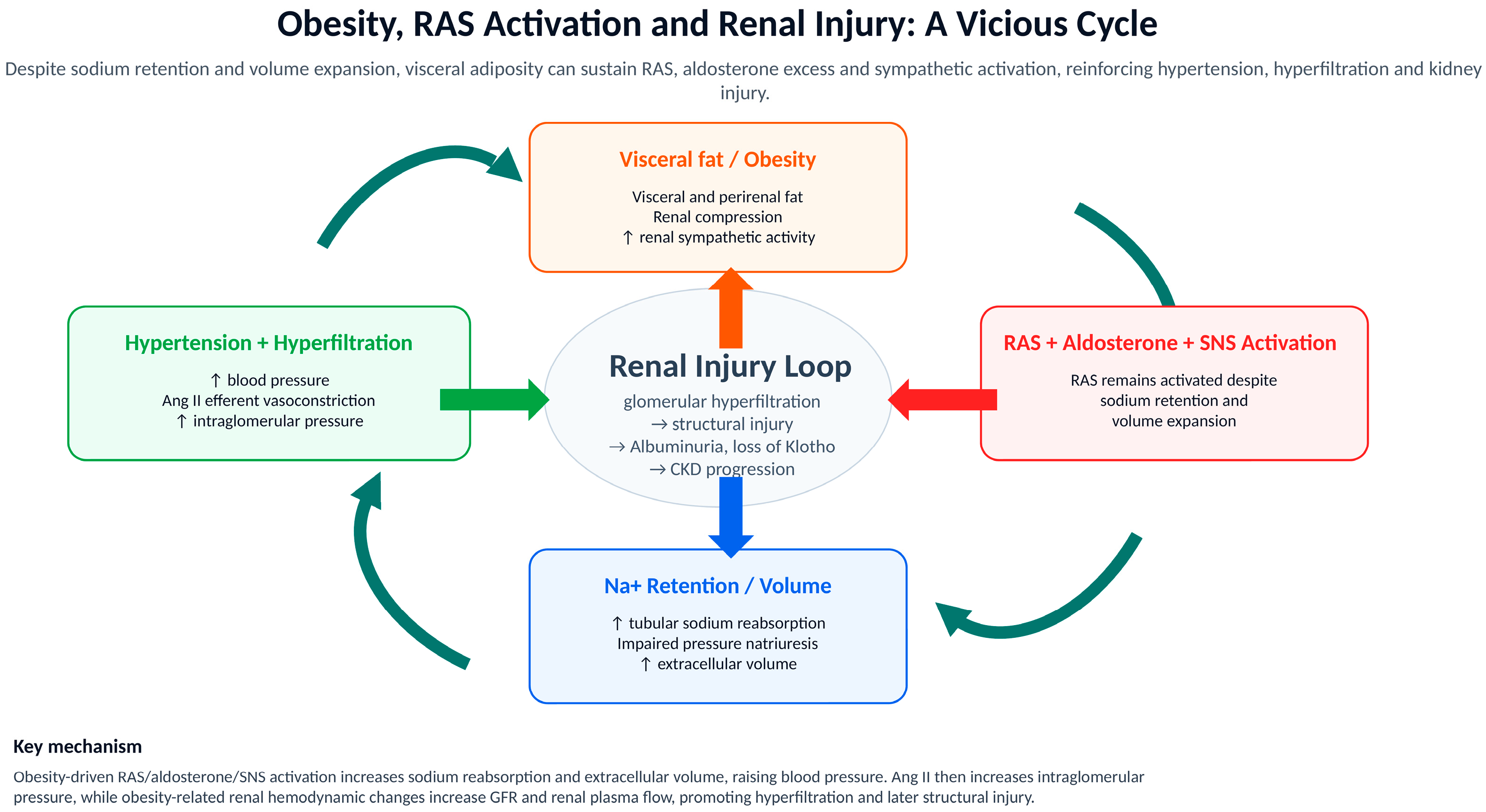
Obesity triggers a neurohormonal cascade from adipose and intrarenal renin–angiotensin system (RAS) activation to aldosterone excess and sympathetic overactivity, causing glomerular hypertension and single-nephron hyperfiltration [7,8,9,10] (Figure 2). Glomerular hyperfiltration is now recognized as the main driver of CKD progression, as it is decreased by all current nephron-sparing drugs [11]. The resulting glomerular hypertension directly injures podocytes, causes podocyte loss, glomerulosclerosis, and increases albumin filtration [12,13]. Excess filtered albumin overwhelms proximal tubular reabsorption capacity, producing proteotoxicity, loss of gerosuppressor functions (Klotho), and an inflammatory response that magnifies kidney injury [14,15]. Adipose tissue produces angiotensinogen, aldosterone, and inflammatory mediators [16,17], and excess adiposity chronically activates both the RAS and the sympathetic nervous system through an adipose–brain–kidney axis [18]. Sympathetic overactivity promotes vasoconstriction, sodium retention, and hypertension [19,20], while insulin resistance further stimulates sodium reabsorption and RAS activation, creating a vicious cycle [12].
Importantly, aldosterone is regulated not only by angiotensin II but also by Klotho deficiency and hyperkalemia, among others [21]; thus, RAS blockade alone may not sufficiently suppress aldosterone, and the addition of a mineralocorticoid receptor antagonist (MRA) may help to further optimize cardiorenal outcomes [22,23]. Aldosterone promotes inflammation, oxidative stress, endothelial dysfunction, and fibrosis, contributing to CKD progression even in the absence of other cardiovascular conditions [9,24]. Although mechanistic and animal studies strongly support adipose-derived RAS and aldosterone as contributors to renal injury [25,26], interventional human data remain limited. Nonetheless, Mendelian randomization supports a causal role of VAT in CKD, with hypertension mediating approximately 46% of this effect [2], and perirenal fat correlates with decreased estimated glomerular filtration rate (eGFR) and CKD burden in patients with resistant hypertension [27].
Figure 2.
Vicious cycle of renin–angiotensin–aldosterone system (RAAS) activation in obesity-related kidney disease. This figure illustrates how excess visceral adipose tissue activates the intrarenal and systemic RAAS, leading to angiotensin II-mediated efferent arteriolar vasoconstriction, aldosterone excess, sympathetic nervous system overactivation, and sodium retention. These hemodynamic disturbances converge on glomerular hypertension and hyperfiltration, which drive podocyte injury, proteinuria, and progressive glomerulosclerosis, establishing a self-perpetuating cycle of renal damage. Abbreviations: Ang II: angiotensin II; CKD: chronic kidney disease; GFR: glomerular filtration rate; RAS: renin–angiotensin system; SNS: sympathetic nervous system. ↑ indicates an increase; → indicates a causal or sequential step in the cycle. Based on [10,12,14,27,28].
2.2. Adipokine Dysregulation
Adipose tissue acts as an endocrine organ secreting adipokines that regulate metabolism, inflammation, and vascular function. In obesity, adipocyte hypertrophy shifts this balance toward a pro-inflammatory and pro-fibrotic profile characterized by increased leptin and resistin and reduced adiponectin, alongside elevated cytokines such as TNF-α, IL-6, and monocyte chemotactic protein-1 (MCP-1) [12,17]. Leptin, physiologically involved in appetite regulation via the hypothalamic POMC-MC4R pathway [29,30], increases proportionally with fat mass and is elevated in CKD. In experimental models, leptin stimulates transforming growth factor-beta 1 (TGF-β1) production, extracellular matrix deposition, mesangial proliferation, sympathetic activation, and adrenal aldosterone secretion [12,31], and directly impairs podocyte homeostasis [31,32]. Resistin, predominantly secreted by macrophages in humans, stimulates proinflammatory cytokine production and reduces endothelial nitric oxide (NO) availability [33,34]. In contrast, adiponectin, which decreases as visceral adiposity increases, preserves podocyte structure by maintaining nephrin and podocin expression through AdipoR1/R2 activation [35,36].
Beyond classical adipokines, additional adipose-derived mediators (autotaxin, platelet-derived growth factor D, lipocalin-2, fatty acid binding proteins, lipoxygenases) have been implicated in experimental models of obesity-related renal and cardiovascular injury within the cardiovascular–kidney–metabolic syndrome [37]. Targeted suppression of these factors within adipose tissue mitigates inflammation, fibrosis, insulin resistance, and organ damage in murine studies [26]. Despite robust mechanistic and preclinical evidence, human interventional data confirming causal relationships between adipokine dysregulation and kidney injury remain scarce.
2.3. Inflammation and Oxidative Stress
Expansion and dysfunction of visceral and perirenal adipose tissue promote macrophage activation and release of TNF-α and IL-6 (Figure 3), both of which cause kidney and cardiovascular injury [38,39]. Although TNF-α has been implicated in kidney injury since the 1990s [40], no anti-TNF-α therapy is currently approved for kidney protection. Clinical trials assessing anti-IL-6 therapies for CKD are ongoing [41,42]. In phase 2 trials, targeting IL-6 reduced lipoprotein(a), improved anemia control, and decreased systemic inflammation. Inflammatory cytokines amplify oxidative stress, promote endothelial activation and dysfunction, and reduce nitric oxide bioavailability, contributing to microvascular dysfunction, podocyte injury, and fibrosis [13,43]. A characteristic histological feature is the crown-like structure, in which macrophages aggregate around dying hypertrophic adipocytes and release inflammatory mediators [44]; visceral fat exhibits stronger pro-inflammatory macrophage phenotypes than subcutaneous fat [45].
Figure 3.
Inflammatory and oxidative stress pathways in visceral adiposity-driven kidney injury. Overview of the molecular mechanisms by which inflamed visceral and perirenal adipose tissue promotes renal damage. As demonstrated in human adipose tissue biopsies and animal models, hypertrophic adipocytes and crown-like structures release pro-inflammatory cytokines (TNF-α, IL-6) that activate NF-κB and JAK/STAT signaling, stimulate NADPH oxidase-dependent reactive oxygen species generation, and impair endothelial nitric oxide bioavailability. These processes lead to glomerular endothelial dysfunction, podocyte injury, tubular stress, and progressive interstitial fibrosis. The figure also highlights the paracrine effects of perirenal fat and the downregulation of kidney-protective factors (Klotho, PGC1α, NLRP6). Abbreviations: IL-6: interleukin-6; NLRP6: NLR family pyrin domain containing 6; NO: nitric oxide; PGC1α: peroxisome proliferator-activated receptor gamma coactivator 1-alpha; ROS: reactive oxygen species; TNF-α: tumor necrosis factor alpha. ↑ increased; ↓ decreased.
TNF-α and IL-6 activate NF-κB and JAK/STAT signaling, stimulate reactive oxygen species generation through NADPH oxidases and mitochondrial dysfunction, and impair endothelial nitric oxide synthase activation, leading to endothelial dysfunction [46,47]. Human obesity is associated with microvascular endothelial dysfunction driven by enhanced oxidative stress [48,49], while systemic cytokines increase endothelin-mediated vasoconstriction and leukocyte adhesion, damaging glomerular endothelium and favoring albuminuria [50]. The peritubular microcirculation is also affected, promoting local hypoxia, tubular stress, and interstitial fibrosis [28,49]. Notably, GLP-1 receptor agonists (GLP-1 RAs) exert kidney-protective effects partly through modulation of glomerular endothelial cell biology [51,52]. Systemic inflammation additionally decreases kidney resilience through downregulation of protective factors such as Klotho, PGC1α, and Nlrp6 [15,53]. Perirenal adipose tissue can also release cytokines and vasoactive factors in close proximity to the renal vasculature, exerting local paracrine effects [9,13,24]. Notably, several nephroprotective agents discussed in Section 5, including SGLT2i, GLP-1 RAs, and non-steroidal MRAs, also exert anti-inflammatory effects that may contribute to their renal benefits beyond their primary mechanisms of action.
2.4. Ectopic Fat Deposition and Lipotoxicity
Renal sinus and perirenal fat accumulation damage the kidney through mechanical compression and lipotoxicity. Mechanical compression disrupts renal hemodynamics by increasing intrarenal/interstitial pressure, reducing vasa recta blood flow, impairing natriuresis, and activating the RAS [43]. In dogs and humans with obesity, this compression increases extracellular matrix deposition, displaces renal parenchyma, and contributes to an enlarged, rounded kidney [12,54]. Reduced renal blood flow activates the RAS, which further increases intrarenal venous pressure and promotes sodium reabsorption, creating a self-perpetuating cycle.
In preclinical studies, ectopic fatty acid uptake by podocytes and proximal tubular cells promotes lipid droplet accumulation, mitochondrial dysfunction, ROS generation, NLRP3 activation, and fibrosis [43]. Imbalanced fatty acid uptake and oxidation leads to accumulation of toxic lipid metabolites such as ceramides and diacylglycerols [55]. In proximal tubular cells, CD36/KIM-1-mediated fatty acid uptake impairs mitochondrial function and promotes inflammation and fibrosis [56,57]. KIM-1 is itself an injury marker, creating a vicious circle in which injury facilitates fatty acid uptake and fatty acid uptake causes further injury. Circulating fatty acids also accumulate as ectopic lipid droplets in podocytes, inducing cytoskeletal damage and apoptosis [43,55].
3. Clinical Assessment: Moving Beyond BMI
BMI is the most widely used metric to classify obesity in nephrology practice, yet it conflates lean mass, bone, and fat, and is agnostic to fat distribution. These limitations are amplified in CKD, where sarcopenia, fluid overload, and muscle wasting distort the weight-adiposity relationship and produce the “obesity paradox” [40]. Accurately staging ob-CKD therefore requires measures that identify the pathogenic visceral compartment.
3.1. Imaging Modalities
Clinical assessment of adiposity has evolved along a trajectory of increasing specificity: BMI captures total mass but ignores distribution; waist circumference reflects central fat but cannot distinguish visceral from subcutaneous depots; and cross-sectional imaging directly quantifies VAT, the depot most strongly linked to renal injury. Computed tomography (CT) segments VAT using fat-specific attenuation thresholds (approximately −190 to −30 HU) on single axial slices at L3-L4 or across volumetric stacks. Magnetic resonance imaging (MRI) achieves comparable quantification without ionising radiation using Dixon or proton-density fat-fraction sequences, and shows close agreement with CT. [58]. Importantly, imaging also enables quantification of renal sinus fat, an ectopic depot invisible to anthropometric surrogates; higher renal sinus fat volumes are associated with hypertension and future >50% eGFR decline in biopsied CKD patients, making it a promising kidney-specific biomarker [3] (Figure 4).
Figure 4.
Imaging modalities for visceral and renal fat quantification. Comparative overview of the imaging techniques available for quantifying visceral adipose tissue and renal sinus fat, including computed tomography (CT), magnetic resonance imaging (MRI), ultrasound (US), dual-energy X-ray absorptiometry (DXA), and bioelectrical impedance analysis (BIA). For each modality, the figure presents the measurement principle, anatomical targets, key advantages and limitations, and clinical applicability for risk stratification in obesity-related kidney disease. Abbreviations: CT: computed tomography; PDFF: proton-density fat fraction; VAT: visceral adipose tissue. HU: Hounsfield units; LoA: limits of agreement. ✓ indicates an advantage or strength of the modality; ✗ indicates a limitation.
The principal drawback of CT and MRI is limited feasibility for routine screening: CT entails radiation, MRI is constrained by cost and access, and dedicated scans solely for adiposity are rarely justified. Opportunistic analysis of abdominal CT obtained for other indications, increasingly aided by automated and deep-learning tools, provides a pragmatic solution. Ultrasound offers a genuine point-of-care alternative (inexpensive, radiation-free, and bedside) measuring visceral fat thickness as a surrogate; meta-analyses show minimal systematic bias versus CT/MRI, although operator dependence widens limits of agreement [58]. Separately, bioelectrical impedance analysis consistently underestimates visceral fat compared with CT/MRI [58] and is not recommended as a primary assessment tool. Across all modalities, heterogeneous acquisition protocols and the absence of standardized, kidney-specific cut-offs still limit clinical translation.
3.2. Clinical Surrogate Markers
Where imaging is unavailable, anthropometric and composite indices provide accessible estimates of central adiposity. Waist circumference predicts albuminuria even in individuals with BMI < 25 kg/m2 and is endorsed within the cardiovascular–kidney–metabolic framework [1], though it cannot separate visceral from subcutaneous fat. The waist-to-hip ratio (WHR) partially addresses this; in the MESA cohort, WHR was associated with rapid kidney-function decline [59], and in meta-analyses of CKD patients, high WHR predicted increased mortality even as higher BMI appeared paradoxically protective [60,61]. Composite indices such as the Visceral Adiposity Index (VAI) and lipid accumulation product (LAP) combine anthropometry with serum triglycerides and HDL cholesterol. In a population-based cohort, ascending VAI quartiles independently predicted incident albuminuria and CKD [62], although in the REGARDS cohort VAI and LAP did not add predictive information beyond waist circumference and BMI [63]. These surrogates are attractive for scalability but lack ethnicity-specific cut-offs (Section 6) and may miss sarcopenic obesity; pairing a distribution-sensitive marker with an estimate of lean mass is preferable [43,58].
3.3. Epidemiological Evidence
The clinical case for visceral-fat-centered assessment rests on consistent cohort and genetic evidence (Table 1). Mendelian randomization using UK Biobank and CKDGen/FinnGen data supports a causal effect of genetically predicted VAT on CKD risk (odds ratios 1.18–1.47), with hypertension mediating roughly half the effect [2]. At the tissue level, renal sinus fat predicts >50% eGFR loss [3], while VAI and LAP predict incident albuminuria and CKD across diverse populations [62,63,64]. Across study designs, distribution-sensitive markers are more consistently associated with renal outcomes than BMI, and the obesity paradox in advanced CKD largely reflects BMI’s inability to separate protective lean mass from harmful visceral fat [60,61]. Clinically, these data support a tiered approach: standardized waist circumference and VAI/LAP at minimum, opportunistic VAT and renal sinus fat quantification when imaging is available, and dedicated MRI for research or selected patients requiring serial monitoring. This BMI-to-waist-circumference (WC)-to-imaging hierarchy underscores a central message: progressing from global to distribution-sensitive to depot-specific assessment progressively improves risk capture and should guide the tiered evaluation of every patient with obesity and CKD.
Table 1.
Epidemiological cohort studies and meta-analyses linking visceral adiposity markers to renal outcomes.
4. Clinical Implications: Obesity-Related Chronic Kidney Disease (ob-CKD)
Ob-CKD may be recognized by clinical and histopathological features and prognostic indicators that support an integrated clinical perspective.
4.1. Clinical Presentation
Ob-CKD encompasses a spectrum of functional and structural renal alterations driven by the interplay between obesity, hemodynamic stress, metabolic dysfunction, and chronic inflammation [13]. According to the SEN-SLANH-SEEDO consensus classification (Table 2), it should be noted that this classification represents an expert-consensus proposal and has not been formally endorsed by KDIGO or other international guideline bodies; prospective validation studies are warranted. ob-CKD type 1 corresponds to reversible functional abnormalities, while type 2 (corresponding to obesity-related glomerulopathy (ORG)) is characterized by persistent subnephrotic proteinuria with bland sediment, preserved renal function, and absence of alternative etiologies [5]. Full nephrotic syndrome is uncommon (<5%); structural injury including glomerulomegaly and early FSGS may be present even in normoalbuminuric individuals, thus preceding the albuminuric stage [6,66]. The clinical trajectory is one of slowly progressive proteinuria with variable rates of eGFR decline, commonly associated with cardiometabolic comorbidities that integrate ob-CKD within the cardiovascular–kidney–metabolic syndrome (type 3) [67]. Metabolically unhealthy phenotypes, characterized by visceral adiposity, insulin resistance, and a pro-inflammatory milieu, confer substantially higher risk [16].
Table 2.
Classification of obesity-related kidney disease (ob-CKD)
Once patients reach kidney failure requiring kidney replacement therapy (type 4), the obesity paradox becomes apparent, likely reflecting the confounding effect of malnutrition [61,68]. In the post-transplant setting (type 5), obesity is associated with higher surgical complications, infections, delayed graft function, and reduced access to transplantation [5,69].
4.2. Renal Biopsy Findings and Histopathological Features
When performed, renal biopsy provides the most definitive diagnostic information, revealing chronic exposure to glomerular hyperfiltration and metabolic stress (Figure 5). The hallmark lesion is glomerulomegaly, reflecting adaptive hypertrophy that ultimately becomes maladaptive. Secondary FSGS, typically perihilar, follows a more indolent course than primary FSGS (Table 3). Podocyte injury is central to progression and is characterized by hypertrophy, cytoskeletal stress, and progressive depletion, with foot process effacement that is typically mild and segmental (<50%) [24,70]. Ectopic lipid accumulation within podocytes, mesangial cells, and proximal tubular cells (the “fatty kidney”) contributes to progression through lipotoxicity [43,55]. Additional features include mesangial expansion with mild “diabetoid” changes, tubulointerstitial fibrosis, and arteriolar hyalinosis. Immunofluorescence is typically nonspecific [71,72]. Importantly, ORG often coexists with other renal pathologies within ob-CKD type 3, underscoring the role of biopsy in identifying overlapping lesions [5,73]. It should be noted that no single histological feature is pathognomonic for ob-CKD; the diagnosis relies on a characteristic pattern (glomerulomegaly, secondary FSGS, podocyte depletion) in the appropriate clinical context, after exclusion of alternative etiologies. Although ectopic lipid accumulation (“fatty kidney”) is pathophysiologically relevant, its systematic histological evaluation is limited by the dissolution of lipid during conventional tissue processing, and standardized criteria or cut-offs for renal lipid content have not been established.
Figure 5.
Histopathological features of obesity-related glomerulopathy (ORG). Representative renal biopsy findings. (A) Glomerulomegaly: the double-headed arrow indicates a maximal glomerular diameter exceeding 250 µm (periodic acid–Schiff). (B) Secondary focal segmental glomerulosclerosis with a perihilar distribution; the arrow indicates the segmental lesion (Masson trichrome). (C) Secondary focal segmental glomerulosclerosis with a perihilar distribution; the arrow indicates the segmental lesion (periodic acid–Schiff). (D) Foam cells with optically empty, finely vacuolated cytoplasm (thin arrow), consistent with lipid dissolved during routine processing, adjacent to an area of segmental sclerosis (block arrow) (Masson trichrome). Abbreviations: FSGS, focal segmental glomerulosclerosis; ORG, obesity-related glomerulopathy; PAS, periodic acid–Schiff.
Table 3.
Clinical, histopathological, prognostic and therapeutic differences between ORG (obesity-related glomerulopathy) and primary FSGS [5].
4.3. Silent Progression and Prognostic Indicators
Despite its apparently benign initial presentation, ob-CKD is progressive, with 10–33% of patients advancing to advanced CKD or kidney failure [66,74]. As for other kidney diseases, proteinuria magnitude and persistence are the strongest predictors of outcome; additional determinants include baseline renal function, obesity duration, histological chronicity, and metabolic factors (insulin resistance, hypertension, dyslipidemia) [75]. Emerging evidence underscores adipose tissue dysfunction and ectopic lipid deposition as key drivers, while organ-specific fat depots (renal sinus, perirenal) exacerbate injury through mechanical compression and local inflammation. Reduced nephron endowment further increases susceptibility to hyperfiltration [76]. A particularly important clinical concept is the silent nature of progression: conventional biomarkers lack sensitivity for early structural damage, and despite therapeutic choices including RAS blockade, SGLT2i, and incretins, a significant proportion of patients continue to progress [77].
4.4. Integrated Clinical Perspective
Ob-CKD should not be considered an isolated glomerular entity but rather a dynamic, potentially modifiable continuum of metabolic-inflammatory nephropathy in which adaptive responses to increased functional demand ultimately become maladaptive [6]. The recognition of the “fatty kidney” phenotype and the role of dysfunctional adipose tissue as a source of pro-inflammatory cytokines provides a mechanistic link between systemic metabolic disease and local kidney injury [5,16]. Therapeutic strategies should adopt a comprehensive, multifactorial approach targeting both hemodynamic and metabolic pathways, including optimized RAS blockade (ACEi/ARB), non-steroidal MRAs, SGLT2i, incretin-based therapies, and sustained weight reduction aimed at reducing ectopic fat burden [77].
5. Therapeutic Strategies Targeting the AdipoRenal Axis
The recognition of the adipo–renal axis as a pivotal pathophysiological pathway linking obesity and CKD has substantially reshaped therapeutic strategies. Figure 6 shows a therapeutic algorithm categorized by CKD stage, and Table 4 summarizes the key clinical trials evaluating nephroprotection and weight-loss efficacy.
Figure 6.
Proposed therapeutic algorithm for the management of obesity-related chronic kidney disease (ob-CKD). Stepwise therapeutic algorithm for the integrated management of obesity-related CKD, stratified by disease stage. The algorithm proposes a tiered approach: (1) universal lifestyle interventions (caloric restriction, structured aerobic exercise targeting visceral adipose tissue reduction) for all patients; (2) first-line pharmacotherapy with SGLT2 inhibitors (hemodynamic nephroprotection) and GLP-1 receptor agonists or dual GIP/GLP-1 agonists (weight loss and metabolic benefits); (3) addition of non-steroidal mineralocorticoid receptor antagonists (finerenone) for residual cardiorenal risk; (4) consideration of bariatric/metabolic surgery for patients with severe obesity (BMI ≥ 35 kg/m2) or inadequate response to medical therapy; and (5) stage-specific adaptations for dialysis (ob-CKD type 4) and kidney transplant recipients (ob-CKD type 5). Decision nodes incorporate visceral adiposity assessment (VAI, waist circumference, imaging when available) rather than BMI alone. Note: This algorithm represents a pragmatic expert-opinion framework based on currently available evidence and should not be interpreted as formal guideline recommendations or mandatory clinical guidance. Abbreviations: BMI: body mass index; CKD: chronic kidney disease; eGFR: estimated glomerular filtration rate; GIP: glucose-dependent insulinotropic polypeptide; GLP-1 RA: glucagon-like peptide-1 receptor agonist; MRA: mineralocorticoid receptor antagonist; ns-MRA: non-steroidal mineralocorticoid receptor antagonist; ob-CKD: obesity-related chronic kidney disease; RAASi: renin–angiotensin–aldosterone system inhibitor; SGLT2i: sodium-glucose cotransporter 2 inhibitor; UACR: urinary albumin-to-creatinine ratio; VAI: Visceral Adiposity Index; VAT: visceral adipose tissue. ↓ indicates a reduction in the corresponding parameter.
Table 4.
Major clinical trials evaluating nephroprotection, kidney outcomes and weight-loss efficacy in patients with obesity and metabolic disease.
† STEP 1 and SURMOUNT-1 are included to contextualize the magnitude of weight reduction achievable with incretin-based therapies, which is directly relevant to visceral adipose tissue reduction in ob-CKD. The FLOW trial subsequently demonstrated kidney-specific benefits for semaglutide, and the TREASURE-CKD trial (NCT05536804) is evaluating renal outcomes for tirzepatide.
5.1. Lifestyle Interventions
Caloric restriction and structured exercise reduce visceral adipose tissue and improve renal outcomes, including albuminuria regression [78]. A meta-analysis of 40 RCTs (2190 participants) demonstrated that both interventions reduce visceral adiposity, but only exercise shows a dose–response relationship with VAT reduction [79]. The Look AHEAD trial demonstrated that intensive lifestyle intervention in overweight/obese individuals with T2D reduced the incidence and progression of diabetic nephropathy [80]. The renal benefits are attributed to reduced glomerular hyperfiltration, improved insulin sensitivity, decreased RAS activation, attenuated inflammation, and improved adiponectin signaling [81].
5.2. Pharmacological Advances
Recent pharmacological advances include incretin analogues (GLP-1 RAs and dual GIP/GLP-1 Agonists), SGLT2i and non-steroidal MRAs.
5.2.1. GLP-1 RAs and Dual GIP/GLP-1 Agonists
Semaglutide promotes weight loss through GLP-1 receptor activation, with the STEP programme demonstrating 14.9–15% sustained weight reduction [82]. Beyond weight loss, semaglutide exerts direct renoprotective effects by attenuating renal inflammation, oxidative stress, endothelial dysfunction, and profibrotic signaling, partly through NHE3 inhibition in the proximal tubule, restoring tubuloglomerular feedback [52,77]. The FLOW trial [83] provided the first kidney-specific evidence: among 3533 participants with T2D and CKD, semaglutide reduced major kidney disease events by 24% (HR 0.76; 95% CI 0.66–0.88) and also reduced cardiovascular events and all-cause mortality [84], with consistent benefits across CKD stages and regardless of concomitant SGLT2i or MRA use [85]. The REMODEL trial (NCT04865770) is investigating the structural renal mechanisms underlying this protection.
Tirzepatide, a dual GIP/GLP-1 agonist, achieves even greater weight reductions (20.9% at the highest dose, SURMOUNT-1 [86]) and has demonstrated superiority over semaglutide (20.2% vs. 13.7%, SURMOUNT-5 [87]). The TREASURE-CKD trial (NCT05536804) is investigating its renoprotective mechanisms.
5.2.2. SGLT2 Inhibitors
SGLT2i slow CKD progression through mechanisms extending well beyond glycemic control. By blocking proximal tubular sodium-glucose reabsorption, they restore tubuloglomerular feedback, promote afferent arteriolar vasoconstriction, and reduce intraglomerular pressure [88]. The DAPA-CKD trial demonstrated a 39% reduction in the composite kidney endpoint (HR 0.61; 95% CI 0.51–0.72), regardless of diabetes status [89]. EMPA-KIDNEY confirmed these findings in a broader CKD population, with a 28% reduction in kidney disease progression or cardiovascular death (HR 0.72; 95% CI 0.64–0.82) [90]. Beyond hemodynamics, SGLT2i decrease weight and promote metabolic reprogramming: the negative energy balance reduces visceral adiposity, improves insulin sensitivity, and shifts energy metabolism toward ketone utilization [91]. Emerging evidence suggests that empagliflozin reduces renal lipid accumulation and inflammation [92], while dapagliflozin decreases epicardial adipose tissue [93].
5.2.3. Non-Steroidal MRAs
MR overactivation in obesity is driven by adipocyte aldosterone production and leptin-mediated pathways, independently of the classical RAS, creating a state of chronic MR overactivation that accelerates kidney injury [94]. Finerenone provides significant anti-inflammatory and antifibrotic effects while maintaining a favorable safety profile [95]. The FIDELIO-DKD trial demonstrated an 18% reduction in the primary renal composite outcome (HR 0.82; p = 0.001) and a 14% reduction in cardiovascular events [96]. FIGARO-DKD showed a 13% reduction in cardiovascular composite outcomes [97], and FIND-CKD extended benefits to non-diabetic CKD with a 23% reduction in composite kidney/cardiovascular outcomes [23]. In the FIDELITY pooled analysis, finerenone was effective regardless of obesity status, with greater benefits in patients with increased waist circumference [98]. While no clinically significant impact on body weight or visceral fat has been reported [95], finerenone reduced the risk of new-onset diabetes by 24% in the FINEARTS-HF trial [99], with consistent benefits regardless of baseline insulin resistance [100], suggesting potential metabolic benefits beyond mineralocorticoid blockade.
5.3. Bariatric and Metabolic Surgery
Bariatric and metabolic surgery (BMS) represents the most effective intervention for sustained weight loss in severe obesity, with renoprotective effects attributed not only to weight reduction but also to the marked decrease in VAT and associated metabolic, inflammatory, and hemodynamic improvements [101]. The SOS study demonstrated sustained reductions in albuminuria, remission from microalbuminuria to normoalbuminuria, and slower eGFR decline over 15 years of follow-up [102]. Multiple cohort studies [103] and meta-analyses confirm significant reductions in albuminuria, proteinuria, and glomerular hyperfiltration [104]. Importantly, a meta-analysis showed that BMS improves renal outcomes independently of total weight reduction, supporting the concept of metabolic rather than purely weight-based intervention [105]. Mechanisms include improved insulin sensitivity, modified incretin secretion, reduced RAS activation, and decreased pro-inflammatory adipokines [106]. The marked VAT reduction correlates more closely with renal improvement than overall weight loss [66,107].
BMS is particularly beneficial in obese individuals with T2D, where diabetes remission attenuates hyperglycemia-driven renal injury. BMS significantly lowered MACE risk over 8 years versus medical care [108]. Direct comparisons show greater weight loss with surgery versus GLP-1 RAs (28.3% vs. 10.3%) [109], and randomized trials confirm superiority in inducing albuminuria remission [110]. BMS is relatively safe in CKD patients, though renal adverse events (AKI, nephrolithiasis, rare oxalate nephropathy) can occur [111,112].
5.4. Combination Therapy: GLP-1 RAs and SGLT2i
The combination of GLP-1 RAs and SGLT2i is emerging as a promising strategy for ob-CKD, though robust randomized evidence is still lacking, particularly in non-diabetic CKD. Current data from trials such as DECLARE-TIMI 58 and EMPA-REG OUTCOME show consistent renal protection across BMI categories [113,114]. A recent meta-analysis demonstrated that long-term SGLT2i use is associated with significant reductions in fat mass and body weight while preserving fat-free mass [115], suggesting a more favorable body composition effect than initially appreciated . Post hoc analyses from FLOW and real-world cohorts suggest additive benefits, including greater reductions in weight, HbA1c, albuminuria, and improved eGFR trajectories, with no safety concerns [116,117]. However, no dedicated sequencing or head-to-head trials exist, and optimal use of combination therapy remains to be defined.
Non-steroidal MRAs represent a complementary pillar in multimodal therapy. A prespecified pooled participant-level analysis of FIDELIO-DKD, FIGARO-DKD, and FINEARTS-HF (FINE-HEART) demonstrated that finerenone consistently reduced cardiovascular death or heart failure hospitalization regardless of baseline BMI (interaction p = 0.27) or waist-to-height ratio (p = 0.26), with greater absolute benefits in patients with higher adiposity [118]. These data, together with the FIDELITY analysis showing enhanced efficacy in patients with increased waist circumference [98,118] and the FIND-CKD extension to non-diabetic CKD [23], with a pooled individual participant-level analysis (INFINITY) confirming consistent efficacy and safety across diabetic and non-diabetic CKD [119], support the integration of finerenone within combination strategies targeting the adipo–renal axis across the CKD spectrum.
6. Future Directions and Unmet Needs
Several important questions remain unanswered. First, the VAI lacks universally validated thresholds; the original equation was derived from white European adults [120], and ethnicity- and age-specific cut-offs are needed for global applicability. Table 5 summarizes available ethnicity-specific thresholds for identifying metabolic syndrome and visceral adipose dysfunction, but prospective studies validating their capacity to predict renal outcomes, cardiovascular events, and mortality are still required [121].
Table 5.
Visceral Adiposity Index (VAI) cut-off thresholds by ethnic group.
Second, standardized diagnostic criteria for ob-CKD remain an important unmet need. Currently, no consensus biomarker panel or histological scoring system exists to reliably differentiate ob-CKD from other nephropathies or to diagnose it as a concomitant condition when a second cause of CKD is present. The histological hallmarks (glomerulomegaly, secondary FSGS) are not pathognomonic, ectopic lipid evaluation is limited by conventional tissue processing, and clinical diagnosis relies largely on exclusion. Development of validated diagnostic criteria, including potential integration of imaging biomarkers (renal sinus fat), urinary lipidomics, and histological scoring, would strengthen clinical recognition and enable future interventional trials.
Third, long-term data on the synergistic effects of GLP-1 RAs and SGLT2i in obese patients with early-stage CKD are needed. Although the SELECT trial demonstrated that semaglutide 2.4 mg reduced major cardiovascular events by 20% and slowed kidney-function decline in overweight/obese individuals with established cardiovascular disease without requiring diabetes, including participants receiving concomitant SGLT2i [123], no trial has been specifically designed to evaluate this combination in non-diabetic CKD. Fourth, the development of novel biomarkers for early detection of renal lipotoxicity and adipose-mediated kidney injury represents an important unmet need. Emerging approaches, including lipidomics, urinary metabolomics, and machine-learning-based imaging analysis, hold promise for improved risk stratification but are not yet validated for clinical use [124]. Finally, aldosterone synthase inhibitors (baxdrostat, lorundrostat) represent an emerging pharmacological class that directly suppresses aldosterone production upstream of the mineralocorticoid receptor; preliminary data in hypertensive CKD patients are promising [125], but dedicated renal outcome trials are needed to define their role in ob-CKD.
7. Conclusions
The evidence reviewed converges on a central guiding concept: the adiporenal axis, the bidirectional crosstalk between dysfunctional visceral adipose tissue and the kidney, represents both the mechanistic core of ob-CKD and its most actionable therapeutic target. The progression from BMI to waist circumference to VAT as the preferred assessment metric reflects a parallel maturation in understanding: from total mass through central distribution to depot-specific dysfunction. Just as BMI misclassifies risk by ignoring fat distribution and muscle mass, and waist circumference improves but still conflates subcutaneous with visceral fat, only direct VAT quantification or validated surrogates such as the VAI capture the pathogenic compartment driving hemodynamic, inflammatory, adipokine-mediated, and lipotoxic injury.
From a therapeutic standpoint, the landscape has evolved from supportive measures to mechanism-targeted interventions that simultaneously address weight, metabolic dysfunction, and renal protection. GLP-1 RAs and dual GIP/GLP-1 agonists achieve unprecedented weight loss with direct kidney-protective mechanisms beyond metabolic improvement. SGLT2i restore tubuloglomerular feedback and reduce hyperfiltration independent of glycemic status. Non-steroidal MRAs (finerenone) address the residual inflammatory and fibrotic burden amplified by visceral fat and may improve the metabolic component. BMS remains the most effective intervention for sustained visceral fat reduction and albuminuria remission in severe obesity, with emerging evidence of benefits independent of total weight loss. Anchoring both assessment and treatment in visceral adiposity rather than BMI corrects the systematic misclassification that has historically undermined prevention, identifies high-risk individuals earlier, and aligns therapeutic intensity with the pathogenic compartment that drives kidney disease.
Author Contributions
Conceptualization, M.M.V., B.Q., J.P., A.O. and M.A.B.; methodology, M.M.V. and B.Q.; investigation, M.M.V.; writing—original draft preparation, M.M.V.; writing—review and editing, M.M.V., B.Q., J.P., A.O., B.F.-F., A.S.H., M.J.S., C.G.-C., E.M. and M.A.B.; supervision, J.P., A.O. and M.A.B. All authors have read and agreed to the published version of the manuscript.
Funding
M.M.V. and J.P.’s research is supported by the Instituto de Salud Carlos III (ISCIII) through public research grants PI25/01000 and by the RICORS programs RICORS2040-Renal (RD24/0004/0028) supported by Instituto de Salud Carlos III, co-funded by the European Union. Additional support was provided through unrestricted grants from the Sociedad Madrileña de Nefrología SOMANE 2024. MAB and BQ research are funded by Instituto de Salud Carlos III (ISCIII): RICORS program to RICORS2040-Renal (RD24/0004/0028), co-funded by the European Union (FEDER funds) and public research grants (PI25/00413). AO research is supported by Instituto de Salud Carlos III (ISCIII), (PI22/00469, PI22/00050, PI21/00251, PI25/00145, ERA-PerMed-JTC2022 (SPAREKID AC22/00027), Plataformas ISCIII de apoyo a la I + D + I en Biomedicina y Ciencias de la Salud. IP: PT23/00114, RICORS program to RICORS2040-renal (RD24/0004/0001) co-funded by European Union and SPACKDc PMP21/00109, FEDER funds; COST Action PERMEDIK CA21165 and CA25128 Implementation of Advanced Management Strategies in Chronic Kidney Disease: Towards Kidney Health (KidneyHealth) supported by COST (European Cooperation in Science and Technology); PREVENTCKD Consortium Project ID 101101220 Programme EU4H DG/Agency HADEA; KitNewCare Project ID 101137054, Call HORIZON-HLTH-2023-CARE-04, Programme HORIZON, DG/Agency HADEA; and PICKED Project ID 101168626 HORIZON-MSCA-2023-DN-01-01 MSCA Doctoral Networks 2023. BFF research is supported by ISCIII ERAPERMED2022-248—SIGNAL (AC22/00028) PI23/00627, co-funded by the European Union.
Institutional Review Board Statement
Not applicable.
Informed Consent Statement
Not applicable.
Data Availability Statement
No new data were created or analyzed in this study. Data sharing is not applicable to this article.
Conflicts of Interest
M.M.V. has received grants or contracts from Amgen, AstraZeneca, Bayer, Boehringer Ingelheim, Menarini, Novartis, Novo Nordisk, and Vifor CSL, and consulting fees from AstraZeneca, Bayer, Bioinnova Consulting, Boehringer Ingelheim, and Novo Nordisk. C.G.C. is a member of the Clinical Kidney Journal’s editorial board and serves as a board member of the ONCO-ERA Working Group of the European Renal Association and the Onconephrology Working Group of the Spanish Society of Nephrology. She is PI of the Oberce Project, funded by Novo Nordisk. She has received travel grants and congress registration support from AstraZeneca, Esteve, Novo Nordisk, Boehringer Ingelheim–Lilly, Astellas, CSL Vifor, Otsuka, Novartis, Bayer, Alexion, GSK, and Baxter. She has received honoraria for scientific lectures and participation in advisory boards organized by AstraZeneca, Boehringer Ingelheim–Lilly, Bayer, GSK, Otsuka, Esteve, Bristol Myers Squibb, Serb Pharmaceuticals, Sobi, CSL Vifor and Novo Nordisk. J.P. has received travel grants and speaker fees from CSL Vifor, Astellas and Vantive. AO has received consultancy or speaker fees from AstraZeneca, Boehringer Ingelheim, Fresenius Medical Care, Bayer, Sanofi-Genzyme, Sobi, Otsuka, Novo Nordisk, Sysmex and CSL-Vifor. M.A.B. has received consultancy, speaker fees or travel grants from AstraZeneca, Boehringer Ingelheim, Fresenius Medical Care, Astellas Pharma, Spa Farma Iberica, Laboratorios Rubió and CSL-Vifor. B.Q., B.F.-F., A.S.H., M.J.S. and E.M. declare no conflicts of interest with respect to this work.
References
- Ndumele, C.E.; Rangaswami, J.; Chow, S.L.; Neeland, I.J.; Tuttle, K.R.; Khan, S.S.; Coresh, J.; Mathew, R.O.; Baker-Smith, C.M.; Carnethon, M.R.; et al. Cardiovascular-Kidney-Metabolic Health: A Presidential Advisory from the American Heart Association. Circulation 2023, 148, 1606–1635. [Google Scholar] [CrossRef] [Scilit] [PubMed]
- Gao, T.Y.; Wang, J.C.; Zhao, Q.J.; Zhou, X.; Jiang, Y.; Ren, L.; Zhang, C. Causality of visceral adipose tissue on chronic kidney disease and renal function measure indicators, and mediation role of hypertension: Mendelian randomization study. Obes. Rev. 2024, 26, e13845. [Google Scholar] [CrossRef] [Scilit] [PubMed]
- Fujioka, H.; Koike, T.; Imamura, T.; Kakeshita, K.; Yamazaki, H.; Kinugawa, K. Prognostic impact of renal sinus fat accumulation in patients with chronic kidney disease. Clin. Exp. Nephrol. 2023, 27, 613–621. [Google Scholar] [CrossRef] [Scilit] [PubMed]
- Bays, H. Adiposopathy, “sick fat,” Ockham’s razor, and resolution of the obesity paradox. Curr. Atheroscler. Rep. 2014, 16, 409. [Google Scholar] [CrossRef] [Scilit] [PubMed]
- Rico-Fontalvo, J.; Ciudin, A.; Correa-Rotter, R.; Díaz-Crespo, F.J.; Bonanno, C.; Lecube, A.; Daza-Arnedo, R.; Morales, E.; Quiroga, B.; Lorca, E.; et al. S.E.N., SLANH, and SEEDO consensus report on obesity-related kidney disease: Proposal for a new classification. Kidney Int. 2025, 108, 572–583. [Google Scholar] [CrossRef] [Scilit] [PubMed]
- Kramer, H.J.; Lavenburg, L.M.; Navaneethan, S.D. Obesity in CKD: Core Curriculum 2026. Am. J. Kidney Dis. 2026, 88, 142–153. [Google Scholar] [CrossRef] [Scilit] [PubMed]
- Li, L.; Liu, C.; Gu, J. Emerging insights into the role of visceral adipose tissue in diabetic kidney disease. Front. Nutr. 2025, 12, 1715701. [Google Scholar] [CrossRef] [Scilit] [PubMed]
- Hall, J.E.; Mouton, A.J.; da Silva, A.A.; Omoto, A.C.M.; Wang, Z.; Li, X.; Carmo, J.M.D. Obesity, kidney dysfunction, and inflammation: Interactions in hypertension. Cardiovasc. Res. 2021, 117, 1859–1876. [Google Scholar] [CrossRef] [Scilit] [PubMed]
- Kanbay, M.; Copur, S.; Demiray, A.; Sag, A.A.; Covic, A.; Ortiz, A.; Tuttle, K.R. Fatty kidney: A possible future for chronic kidney disease research. Eur. J. Clin. Investig. 2022, 52, e13748. [Google Scholar] [CrossRef] [Scilit] [PubMed]
- Thethi, T.; Kamiyama, M.; Kobori, H. The link between the renin-angiotensin-aldosterone system and renal injury in obesity and the metabolic syndrome. Curr. Hypertens. Rep. 2012, 14, 160–169. [Google Scholar] [CrossRef] [Scilit] [PubMed]
- Kanbay, M.; Copur, S.; Bakir, C.N.; Covic, A.; Ortiz, A.; Tuttle, K.R. Glomerular hyperfiltration as a therapeutic target for CKD. Nephrol. Dial. Transplant. 2024, 39, 1228–1238. [Google Scholar] [CrossRef] [Scilit] [PubMed]
- Hall, J.E.; do Carmo, J.M.; da Silva, A.A.; Wang, Z.; Hall, M.E. Obesity, kidney dysfunction and hypertension: Mechanistic links. Nat. Rev. Nephrol. 2019, 15, 367–385. [Google Scholar] [CrossRef] [Scilit] [PubMed]
- Martínez-Montoro, J.I.; Morales, E.; Cornejo-Pareja, I.; Tinahones, F.J.; Fernández-García, J.C. Obesity-related glomerulopathy: Current approaches and future perspectives. Obes. Rev. 2022, 23, e13450. [Google Scholar] [CrossRef] [Scilit] [PubMed]
- Fernandez-Fernandez, B.; Izquierdo, M.C.; Valiño-Rivas, L.; Nastou, D.; Sanz, A.B.; Ortiz, A.; Sanchez-Niño, M.D. Albumin downregulates Klotho in tubular cells. Nephrol. Dial. Transplant. 2018, 33, 1712–1722. [Google Scholar] [CrossRef] [Scilit] [PubMed]
- Moreno, J.A.; Izquierdo, M.C.; Sanchez-Niño, M.D.; Suárez-Alvarez, B.; Lopez-Larrea, C.; Jakubowski, A.; Blanco, J.; Ramirez, R.; Selgas, R.; Ruiz-Ortega, M.; et al. The inflammatory cytokines TWEAK and TNFα reduce renal klotho expression through NFκB. J. Am. Soc. Nephrol. 2011, 22, 1315–1325. [Google Scholar] [CrossRef] [Scilit] [PubMed]
- Furth, S.L.; Colhoun, H.M.; Kanbay, M.; Kukla, A.; Lim, L.L.; MacLaughlin, H.L.; Navaneethan, S.D.; Shroff, R.; Stenvinkel, P.; Cheung, M.; et al. The relationship between obesity and chronic kidney disease: Conclusions from a Kidney Disease: Improving Global Outcomes (KDIGO) Controversies Conference. Kidney Int. 2025, 109, 442–464. [Google Scholar] [CrossRef] [Scilit] [PubMed]
- Engeli, S.; Negrel, R.; Sharma, A.M. Physiology and pathophysiology of the adipose tissue renin-angiotensin system. Hypertension 2000, 35, 1270–1277. [Google Scholar] [CrossRef] [Scilit] [PubMed]
- Ahmed, S.K.; Mohammed, R.A. Obesity: Prevalence, causes, consequences, management, preventive strategies and future research directions. Metab. Open 2025, 27, 100375. [Google Scholar] [CrossRef] [Scilit] [PubMed]
- Bell, B.B.; Rahmouni, K. Leptin as a Mediator of Obesity-Induced Hypertension. Curr. Obes. Rep. 2016, 5, 397–404. [Google Scholar] [CrossRef] [Scilit] [PubMed]
- Da Silva, A.A.; do Carmo, J.M.; Wang, Z.; Hall, J.E. Melanocortin-4 Receptors and Sympathetic Nervous System Activation in Hypertension. Curr. Hypertens. Rep. 2019, 21, 46. [Google Scholar] [CrossRef] [Scilit] [PubMed]
- Zhou, X.; Chen, K.; Wang, Y.; Schuman, M.; Lei, H.; Sun, Z. Antiaging Gene Klotho Regulates Adrenal CYP11B2 Expression and Aldosterone Synthesis. J. Am. Soc. Nephrol. 2015, 27, 1765–1776. [Google Scholar] [CrossRef] [Scilit] [PubMed]
- Ortiz, A.; Fernandez-Fernandez, B. Finerenone: Kidney protection beyond type 2 diabetes. Lancet 2026, 407, 2348–2350. [Google Scholar] [CrossRef] [Scilit] [PubMed]
- Heerspink, H.J.L.; Neuen, B.L.; Agarwal, R.; Cherney, D.Z.I.; Lam, C.S.P.; Tuttle, K.R.; Wanner, C.; Sarafidis, P.; Jongs, N.; Smeijer, J.D.; et al. Finerenone in Persons with Chronic Kidney Disease without Diabetes. N. Engl. J. Med. 2026. Epub ahead of print. [Google Scholar] [CrossRef] [Scilit] [PubMed]
- D’Agati, V.D.; Chagnac, A.; de Vries, A.P.; Levi, M.; Porrini, E.; Herman-Edelstein, M.; Praga, M. Obesity-related glomerulopathy: Clinical and pathologic characteristics and pathogenesis. Nat. Rev. Nephrol. 2016, 12, 453–471. [Google Scholar] [CrossRef] [Scilit] [PubMed]
- Hall, J.E.; do Carmo, J.M.; da Silva, A.A.; Wang, Z.; Hall, M.E. Obesity-induced hypertension: Interaction of neurohumoral and renal mechanisms. Circ. Res. 2015, 116, 991–1006. [Google Scholar] [CrossRef] [Scilit] [PubMed]
- Packer, M. Causal demonstration of adiposity-induced adipose-specific signaling derangements in the pathogenesis of the clinical features of the cardiovascular-kidney metabolic syndrome. Cardiovasc. Diabetol. 2026, 25, 86. [Google Scholar] [CrossRef] [Scilit] [PubMed]
- Zyubanova, I.; Ryumshina, N.; Mordovin, V.; Manukyan, M.; Lichikaki, V.; Solonskaya, E.; Gusakova, A.; Suslova, T.; Pekarskiy, S.; Khunkhinova, S.; et al. Sex-specific differences in the efficacy of renal denervation in patients with resistant hypertension depending on visceral obesity and kidney function. Front. Cardiovasc. Med. 2025, 12, 1501296. [Google Scholar] [CrossRef] [Scilit] [PubMed]
- Virdis, A.; Colucci, R.; Bernardini, N.; Blandizzi, C.; Taddei, S.; Masi, S. Microvascular Endothelial Dysfunction in Human Obesity: Role of TNF-α. J. Clin. Endocrinol. Metab. 2019, 104, 341–348. [Google Scholar] [CrossRef] [Scilit] [PubMed]
- Minocci, A.; Savia, G.; Lucantoni, R.; Berselli, M.E.; Tagliaferri, M.; Calò, G.; Petroni, M.; de Medici, C.; Viberti, G.; Liuzzi, A. Leptin plasma concentrations are dependent on body fat distribution in obese patients. Int. J. Obes. Relat. Metab. Disord. 2000, 24, 1139–1144. [Google Scholar] [CrossRef] [Scilit] [PubMed]
- Mao, S.; Fang, L.; Liu, F.; Jiang, S.; Wu, L.; Zhang, J. Leptin and chronic kidney diseases. J. Recept. Signal Transduct. Res. 2018, 38, 89–94. [Google Scholar] [CrossRef] [Scilit] [PubMed]
- Nasrallah, M.P.; Ziyadeh, F.N. Overview of the physiology and pathophysiology of leptin with special emphasis on its role in the kidney. Semin. Nephrol. 2013, 33, 54–65. [Google Scholar] [CrossRef] [Scilit] [PubMed]
- Zhao, J.; Rui, H.L.; Yang, M.; Sun, L.J.; Dong, H.R.; Cheng, H. CD36-Mediated Lipid Accumulation and Activation of NLRP3 Inflammasome Lead to Podocyte Injury in Obesity-Related Glomerulopathy. Mediat. Inflamm. 2019, 2019, 3172647. [Google Scholar] [CrossRef] [Scilit] [PubMed]
- Romejko, K.; Rymarz, A.; Szamotulska, K.; Bartoszewicz, Z.; Rozmyslowicz, T.; Niemczyk, S. Resistin Contribution to Cardiovascular Risk in Chronic Kidney Disease Male Patients. Cells 2023, 12, 999. [Google Scholar] [CrossRef] [Scilit] [PubMed]
- Axelsson, J.; Bergsten, A.; Qureshi, A.R.; Heimbürger, O.; Bárány, P.; Lönnqvist, F.; Lindholm, B.; Nordfors, L.; Alvestrand, A.; Stenvinkel, P. Elevated resistin levels in chronic kidney disease are associated with decreased glomerular filtration rate and inflammation, but not with insulin resistance. Kidney Int. 2006, 69, 596–604. [Google Scholar] [CrossRef] [Scilit] [PubMed]
- Coimbra, S.; Rocha, S.; Valente, M.J.; Catarino, C.; Bronze-da-Rocha, E.; Belo, L.; Santos-Silva, A. New Insights into Adiponectin and Leptin Roles in Chronic Kidney Disease. Biomedicines 2022, 10, 2642. [Google Scholar] [CrossRef] [Scilit] [PubMed]
- Rutkowski, J.M.; Scherer, P.E. Isolation and quantitation of adiponectin higher order complexes. Methods Enzymol. 2014, 537, 243–259. [Google Scholar] [CrossRef] [Scilit] [PubMed]
- Cases, A.; Broseta, J.J.; Marqués, M.; Cigarrán, S.; Julián, J.C.; Alcázar, R.; Ortiz, A. Cardiovascular-kidney-metabolic syndrome definition and its role in the prevention, risk staging, and treatment. An opportunity for the Nephrology. Nefrologia (Engl. Ed.) 2024, 44, 771–783. [Google Scholar] [CrossRef] [Scilit] [PubMed]
- Hammoud, S.H.; AlZaim, I.; Al-Dhaheri, Y.; Eid, A.H.; El-Yazbi, A.F. Perirenal Adipose Tissue Inflammation: Novel Insights Linking Metabolic Dysfunction to Renal Diseases. Front. Endocrinol. 2021, 12, 707126. [Google Scholar] [CrossRef] [Scilit] [PubMed]
- Sharma, K. Obesity, oxidative stress, and fibrosis in chronic kidney disease. Kidney Int. Suppl. 2014, 4, 113–117. [Google Scholar] [CrossRef] [Scilit] [PubMed]
- Gómez-Chiarri, M.; Ortíz, A.; Lerma, J.L.; López-Armada, M.J.; Mampaso, F.; González, E.; Egido, J. Involvement of tumor necrosis factor and platelet-activating factor in the pathogenesis of experimental nephrosis in rats. Lab. Investig. 1994, 70, 449–459. [Google Scholar] [PubMed]
- Ridker, P.M.; Baeres, F.M.M.; Hveplund, A.; Engelmann, M.M.D.; Hovingh, G.K.; Lincoff, A.M.; Marx, N.; Navar, A.M.; Sattar, N.; Tuttle, K.; et al. Rationale, Design, and Baseline Clinical Characteristics of the Ziltivekimab Cardiovascular Outcomes Trial: Interleukin-6 Inhibition and Atherosclerotic Event Rate Reduction. JAMA Cardiol. 2026, 11, 89–97. [Google Scholar] [CrossRef] [Scilit] [PubMed]
- Catanese, B.; Heise, M.; Chang, A.M.; Chandhok, G.; Velkoska, E.; Luo, C.; Chertow, G.M.; Wolf, M.; Szecsödy, P.; Edmonston, D. Effect of Clazakizumab on Neutrophil-Lymphocyte Ratio in Patients Receiving Hemodialysis: Secondary Analysis of the POSIBIL6ESKD Phase 2b Trial. Clin. J. Am. Soc. Nephrol. 2026, 21, 860–862. [Google Scholar] [CrossRef] [Scilit] [PubMed]
- Ahmed, N.; Dalmasso, C.; Turner, M.B.; Arthur, G.; Cincinelli, C.; Loria, A.S. From fat to filter: The effect of adipose tissue-derived signals on kidney function. Nat. Rev. Nephrol. 2025, 21, 417–434. [Google Scholar] [CrossRef] [Scilit] [PubMed]
- Murano, I.; Barbatelli, G.; Parisani, V.; Latini, C.; Muzzonigro, G.; Castellucci, M.; Cinti, S. Dead adipocytes, detected as crown-like structures, are prevalent in visceral fat depots of genetically obese mice. J. Lipid Res. 2008, 49, 1562–1568. [Google Scholar] [CrossRef] [Scilit] [PubMed]
- Zeyda, M.; Stulnig, T.M. Adipose tissue macrophages. Immunol. Lett. 2007, 112, 61–67. [Google Scholar] [CrossRef] [Scilit] [PubMed]
- Engin, A. Endothelial Dysfunction in Obesity and Therapeutic Targets. Adv. Exp. Med. Biol. 2024, 1460, 489–538. [Google Scholar] [CrossRef] [Scilit] [PubMed]
- Gkrinia, E.M.M.; Belančić, A. The Mechanisms of Chronic Inflammation in Obesity and Potential Therapeutic Strategies: A Narrative Review. Curr. Issues Mol. Biol. 2025, 47, 357. [Google Scholar] [CrossRef] [Scilit] [PubMed]
- Virdis, A.; Masi, S.; Colucci, R.; Chiriacò, M.; Uliana, M.; Puxeddu, I.; Bernardini, N.; Blandizzi, C.; Taddei, S. Microvascular Endothelial Dysfunction in Patients with Obesity. Curr. Hypertens. Rep. 2019, 21, 32. [Google Scholar] [CrossRef] [Scilit] [PubMed]
- Jang, K.W.; Hur, J.; Lee, D.W.; Kim, S.R. Metabolic Syndrome, Kidney-Related Adiposity, and Kidney Microcirculation: Unraveling the Damage. Biomedicines 2024, 12, 2706. [Google Scholar] [CrossRef] [Scilit] [PubMed]
- Mima, A.; Yasuzawa, T.; King, G.L.; Ueshima, S. Obesity-associated glomerular inflammation increases albuminuria without renal histological changes. FEBS Open Bio 2018, 8, 664–670. [Google Scholar] [CrossRef] [Scilit] [PubMed]
- Pruijm, M.; Belmar, N.; Bjornstad, P.; Cherney, D.Z.I.; Das, V.; Gunnarsson, T.; Hodgin, J.B.; Schytz, P.A.; Tuttle, K.R.; Kretzler, M.; et al. REMODELing mechanistic trials for kidney disease: A multimodal, tissue-centered approach to understand the renal mechanism of action of semaglutide. Kidney Int. 2025, 109, 6–16. [Google Scholar] [CrossRef] [Scilit] [PubMed]
- Abasheva, D.; Ortiz, A.; Fernandez-Fernandez, B. GLP-1 receptor agonists in patients with chronic kidney disease and either overweight or obesity. Clin. Kidney J. 2024, 17, 19–35. [Google Scholar] [CrossRef] [Scilit] [PubMed]
- Rayego-Mateos, S.; Rodrigues-Diez, R.R.; Fernandez-Fernandez, B.; Mora-Fernández, C.; Marchant, V.; Donate-Correa, J.; Navarro-González, J.F.; Ortiz, A.; Ruiz-Ortega, M. Targeting inflammation to treat diabetic kidney disease: The road to 2030. Kidney Int. 2022, 103, 282–296. [Google Scholar] [CrossRef] [Scilit] [PubMed]
- Mende, C.; Einhorn, D. Fatty kidney disease: The importance of ectopic fat deposition and the potential value of imaging. J. Diabetes 2021, 14, 73–78. [Google Scholar] [CrossRef] [Scilit] [PubMed]
- Mitrofanova, A.; Merscher, S.; Fornoni, A. Kidney lipid dysmetabolism and lipid droplet accumulation in chronic kidney disease. Nat. Rev. Nephrol. 2023, 19, 629–645. [Google Scholar] [CrossRef] [Scilit] [PubMed]
- Mori, Y.; Ajay, A.K.; Chang, J.H.; Mou, S.; Zhao, H.; Kishi, S.; Li, J.; Brooks, C.R.; Xiao, S.; Woo, H.-M.; et al. KIM-1 mediates fatty acid uptake by renal tubular cells to promote progressive diabetic kidney disease. Cell Metab. 2021, 33, 1042–1061.e7. [Google Scholar] [CrossRef] [Scilit] [PubMed]
- Zhao, X.; Chen, X.; Zhang, Y.; George, J.; Cobbs, A.; Wang, G.; Li, L.; Emmett, N. Kidney Injury Molecule-1 Is Upregulated in Renal Lipotoxicity and Mediates Palmitate-Induced Tubular Cell Injury and Inflammatory Response. Int. J. Mol. Sci. 2019, 20, 3406. [Google Scholar] [CrossRef] [Scilit] [PubMed]
- Murphy, J.; Bacon, S.L.; Morais, J.A.; Tsoukas, M.A.; Santosa, S. Intra-abdominal adipose tissue quantification by alternative versus reference methods: A systematic review and meta-analysis. Obesity 2019, 27, 1115–1122. [Google Scholar] [CrossRef] [Scilit] [PubMed]
- Malkina, A.; Katz, R.; Shlipak, M.G.; Ix, J.H.; de Boer, I.H.; Sarnak, M.J.; Allison, M.; Kramer, H.J.; Lin, J.; Siscovick, D.; et al. Association of obesity and kidney function decline among non-diabetic adults with eGFR > 60 mL/min/1.73 m2: Results from the Multi-Ethnic Study of Atherosclerosis (MESA). Open J. Endocr. Metab. Dis. 2013, 3, 103–112. [Google Scholar] [CrossRef] [PubMed]
- Zimmermann, S.; Mathew, A.; Schöppe, R.; Mangova, G.; Biemann, R.; Surov, A.; Meyer, H.-J.; Isermann, B. Fat tissue quantity, waist circumference or waist-to-hip ratio in patients with chronic kidney disease: A systematic review and meta-analysis. Obes. Res. Clin. Pract. 2024, 18, 81–87. [Google Scholar] [CrossRef] [Scilit] [PubMed]
- Kusuma, H.; Lee, H.F.; Yen, M.; Fetzer, S.J.; Lam, L.T. Obesity measurement methods estimated mortality risk in patients undergoing hemodialysis: A systematic review and meta-analysis. Int. Urol. Nephrol. 2024, 57, 1585–1600. [Google Scholar] [CrossRef] [Scilit] [PubMed]
- Yu, J.; Huang, C.; Lin, J.; Lin, D.; Li, F.; Qi, Y.; Liang, Y.; Wang, C.; Cai, L.; Feng, W.; et al. Visceral Adiposity Index Is Associated With the Development of Increased Urinary Albumin Excretion and Chronic Kidney Disease: A Population-Based Study. Int. J. Endocrinol. 2025, 2025, 1173231. [Google Scholar] [CrossRef] [Scilit] [PubMed]
- Bullen, A.L.; Katz, R.; Kumar, U.; Gutierrez, O.M.; Sarnak, M.J.; Kramer, H.J.; Shlipak, M.G.; Ix, J.H.; Judd, S.E.; Cushman, M.; et al. Lipid accumulation product, visceral adiposity index and risk of chronic kidney disease. BMC Nephrol. 2022, 23, 401. [Google Scholar] [CrossRef] [Scilit] [PubMed]
- Li, X.; Wang, L.; Zhou, H.; Xu, H. Association between triglyceride-glucose index and chronic kidney disease: Results from NHANES 1999-2020. Int. Urol. Nephrol. 2024, 56, 3605–3616. [Google Scholar] [CrossRef] [Scilit] [PubMed]
- Oh, S.W.; Ahn, S.Y.; Jianwei, X.; Kim, K.W.; Kim, S.; Na, K.Y.; Chae, D.W.; Kim, S.; Chin, H.J. Relationship between changes in body fat and a decline of renal function in the elderly. PLoS ONE 2014, 9, e84052. [Google Scholar] [CrossRef] [Scilit] [PubMed]
- Sandino, J.; Martín-Taboada, M.; Medina-Gómez, G.; Vila-Bedmar, R.; Morales, E. Novel insights in the physiopathology and management of obesity-related kidney disease. Nutrients 2022, 14, 3937. [Google Scholar] [CrossRef] [Scilit] [PubMed]
- Pinto, K.R.D.; Feckinghaus, C.M.; Hirakata, V.N. Obesity as a predictive factor for chronic kidney disease in adults: Systematic review and meta-analysis. Braz. J. Med. Biol. Res. 2021, 54, e10022. [Google Scholar] [CrossRef] [Scilit] [PubMed]
- Friedman, A.N. Obesity in patients undergoing dialysis and kidney transplantation. Adv. Chronic Kidney Dis. 2013, 20, 128–134. [Google Scholar] [CrossRef] [Scilit] [PubMed]
- Tzvetanov, I.G.; Spaggiari, M.; Tulla, K.A.; Di Bella, C.; Okoye, O.; Di Cocco, P.; Jeon, H.; Oberholzer, J.; Giulianotti, P.C.; Benedetti, E. Robotic kidney transplantation in the obese patient: 10-year experience from a single center. Am. J. Transplant. 2019, 20, 430–440. [Google Scholar] [CrossRef] [Scilit] [PubMed]
- Haruhara, K.; Okabayashi, Y.; Sasaki, T.; Kubo, E.; D’Agati, V.D.; Bertram, J.F.; Tsuboi, N.; Yokoo, T. Podocyte density as a predictor of long-term kidney outcome in obesity-related glomerulopathy. Kidney Int. 2024, 106, 496–507. [Google Scholar] [CrossRef] [Scilit] [PubMed]
- Choung, H.G.; Bomback, A.S.; Stokes, M.B.; Santoriello, D.; Campenot, E.S.; Batal, I.; Markowitz, G.S.; D’agati, V.D. The spectrum of kidney biopsy findings in patients with morbid obesity. Kidney Int. 2019, 95, 647–654. [Google Scholar] [CrossRef] [Scilit] [PubMed]
- Rodríguez-Rodríguez, R.; Hornum, M.; Rodríguez Rodríguez, A.E.; Bevc, S.; Trevisani, F.; Fernández, G.; Hojs, R.; Fernández-Fernández, B.; Corona, C.M.C.; Cruzado, J.M.; et al. Renal Disease in Metabolic Syndrome: The Hidden Role of Intrarenal Ischemia. Kidney Int. Rep. 2024, 9, 1419–1428, Correction in Kidney Int. Rep. 2024, 10, 627. https://doi.org/10.1016/j.ekir.2024.12.015. [Google Scholar] [CrossRef] [Scilit] [PubMed]
- Sharma, S.; Sharma, A.; Kapoor, N.; Sharma, N.; Saravanan, P.; Kalra, S. Obesity-related glomerulopathy: A lesser known complication of obesity. Curr. Opin. Endocrinol. Diabetes Obes. 2025, 32, 258–268. [Google Scholar] [CrossRef] [Scilit] [PubMed]
- Praga, M.; Morales, E. The Fatty Kidney: Obesity and Renal Disease. Nephron 2016, 136, 273–276. [Google Scholar] [CrossRef] [Scilit] [PubMed]
- Morales, E.; Martin, W.P.; Bevc, S.; Jenssen, T.G.; Miglinas, M.; Trillini, M. How to individualize renoprotective therapy in obese patients with chronic kidney disease: A commentary by the Diabesity Working Group of the ERA. Nephrol. Dial. Transplant. 2025, 40, 1977–1988. [Google Scholar] [CrossRef] [Scilit] [PubMed]
- Sandino, J.; Cordero Garcia-Galán, L.; Aubert Girbal, L.; Praga, M.; Pascual, J.; Morales, E. Anything New in the Treatment of Obesity in Obese Patients with CKD? Nephron 2022, 146, 616–623. [Google Scholar] [CrossRef] [Scilit] [PubMed]
- Morales, E.; Jenssen, T.G.; Bevc, S.; Miglinas, M.; Martin, W.P.; Theodorakopoulou, M.; Jørgensen, M.B.; Trillini, M. Obesity as a modifiable risk factor in patients with chronic kidney disease. Nephrol. Dial. Transplant. 2026, 41, 846–858. [Google Scholar] [CrossRef] [Scilit] [PubMed]
- Abe, T.; Song, J.S.; Bell, Z.W.; Wong, V.; Spitz, R.W.; Yamada, Y.; Loenneke, J.P. Comparisons of calorie restriction and structured exercise on reductions in visceral and abdominal subcutaneous adipose tissue: A systematic review. Eur. J. Clin. Nutr. 2022, 76, 184–195. [Google Scholar] [CrossRef] [Scilit] [PubMed]
- Recchia, F.; Leung, C.K.; Yu, A.P.; Leung, W.; Yu, D.J.; Fong, D.Y.; Montero, D.; Lee, C.-H.; Wong, S.H.; Siu, P.M. Dose-response effects of exercise and caloric restriction on visceral adiposity in overweight and obese adults: A systematic review and meta-analysis of randomised controlled trials. Br. J. Sports Med. 2023, 57, 1035–1041. [Google Scholar] [CrossRef] [Scilit] [PubMed]
- Look of AHEAD Research Group. Effect of a long-term behavioural weight loss intervention on nephropathy in overweight or obese adults with type 2 diabetes: A secondary analysis of the Look AHEAD randomised clinical trial. Lancet Diabetes Endocrinol. 2014, 2, 801–809. [Google Scholar] [CrossRef] [Scilit] [PubMed]
- Kramer, H.; Reboussin, D.; Bertoni, A.G.; Marcovina, S.; Lipkin, E.; Greenway, F.L.; Brancati, F.L. the Look Ahead Research Group. Obesity and albuminuria among adults with type 2 diabetes: The Look AHEAD (Action for Health in Diabetes) Study. Diabetes Care 2009, 32, 851–853. [Google Scholar] [CrossRef] [Scilit] [PubMed]
- Wilding, J.P.H.; Batterham, R.L.; Calanna, S.; Davies, M.; Van Gaal, L.F.; Lingvay, I.; McGowan, B.M.; Rosenstock, J.; Tran, M.T.; Wadden, T.A.; et al. Once-weekly semaglutide in adults with overweight or obesity. N. Engl. J. Med. 2021, 384, 989–1002. [Google Scholar] [CrossRef] [Scilit] [PubMed]
- Rossing, P.; Baeres, F.M.M.; Bakris, G.; Bosch-Traberg, H.; Gislum, M.; Gough, S.C.; Idorn, T.; Lawson, J.; Mahaffey, K.W.; Mann, J.F.; et al. The rationale, design and baseline data of FLOW, a kidney outcomes trial with once-weekly semaglutide in people with type 2 diabetes and chronic kidney disease. Nephrol. Dial. Transplant. 2023, 38, 2041–2051, Erratum in Nephrol. Dial. Transplant. 2024, 39, 724. https://doi.org/10.1093/ndt/gfad252. [Google Scholar] [CrossRef] [Scilit] [PubMed]
- Mahaffey, K.W.; Tuttle, K.R.; Arici, M.; Baeres, F.M.M.; Bakris, G.; Charytan, D.M.; Cherney, D.Z.; Chernin, G.; Correa-Rotter, R.; Gumprecht, J.; et al. Cardiovascular outcomes with semaglutide by severity of chronic kidney disease in type 2 diabetes: The FLOW trial. Eur. Heart J. 2025, 46, 1096–1108. [Google Scholar] [CrossRef] [Scilit] [PubMed]
- Perkovic, V.; Tuttle, K.R.; Rossing, P.; Mahaffey, K.W.; Mann, J.F.; Bakris, G.; Baeres, F.M.; Idorn, T.; Bosch-Traberg, H.; Lausvig, N.L.; et al. Effects of semaglutide on chronic kidney disease in patients with type 2 diabetes. N. Engl. J. Med. 2024, 391, 109–121. [Google Scholar] [CrossRef] [Scilit] [PubMed]
- Jastreboff, A.M.; Aronne, L.J.; Ahmad, N.N.; Wharton, S.; Connery, L.; Alves, B.; Kiyosue, A.; Zhang, S.; Liu, B.; Bunck, M.C.; et al. Tirzepatide once weekly for the treatment of obesity. N. Engl. J. Med. 2022, 387, 205–216. [Google Scholar] [CrossRef] [Scilit] [PubMed]
- Aronne, L.J.; Horn, D.B.; Le Roux, C.W.; Ho, W.; Falcon, B.L.; Valderas, E.G.; Das, S.; Lee, C.J.; Glass, L.C.; Senyucel, C.; et al. Tirzepatide as compared with semaglutide for the treatment of obesity. N. Engl. J. Med. 2025, 393, 26–36. [Google Scholar] [CrossRef] [Scilit] [PubMed]
- Upadhyay, A. SGLT2 inhibitors and kidney protection: Mechanisms beyond tubuloglomerular feedback. Kidney360 2024, 5, 771–782. [Google Scholar] [CrossRef] [Scilit] [PubMed]
- Heerspink, H.J.L.; Stefánsson, B.V.; Correa-Rotter, R.; Chertow, G.M.; Greene, T.; Hou, F.-F.; Mann, J.F.E.; McMurray, J.J.V.; Lindberg, M.; Rossing, P.; et al. Dapagliflozin in patients with chronic kidney disease. N. Engl. J. Med. 2020, 383, 1436–1446. [Google Scholar] [CrossRef] [Scilit] [PubMed]
- The EMPA-KIDNEY Collaborative Group; Herrington, W.G.; Staplin, N.; Wanner, C.; Green, J.B.; Hauske, S.J.; Emberson, J.R.; Preiss, D.; Judge, P.; Mayne, K.J.; et al. Empagliflozin in patients with chronic kidney disease. N. Engl. J. Med. 2023, 388, 117–127. [Google Scholar] [CrossRef] [Scilit] [PubMed]
- Szekeres, Z.; Sandor, B.; Bognar, Z.; Ramadan, F.H.J.; Palfi, A.; Bodis, B.; Toth, K.; Szabados, E. Clinical study of metabolic parameters, leptin and the SGLT2 inhibitor empagliflozin among patients with obesity and type 2 diabetes. Int. J. Mol. Sci. 2023, 24, 4405. [Google Scholar] [CrossRef] [Scilit] [PubMed]
- Shi, S.; Li, W.; Yang, L.; Zhang, J.; Wu, X. Empagliflozin alleviates lipid deposition and inflammation in diabetic kidney disease by downregulating C1QC. Mol. Cell Biochem. 2026, 481, 885–898. [Google Scholar] [CrossRef] [Scilit] [PubMed]
- Alghamdi, M.; Dihoum, A.; Hakami, K.; Bhattacharjee, A.; Brown, A.J.M.; Singh, J.; Altalabany, S.; Lang, C.; Mordi, I.; Khan, F. Dapagliflozin reduces epicardial adipose tissue in patients with heart failure and type 2 diabetes. Diabetes Obes. Metab. 2025, 27, 7561–7569. [Google Scholar] [CrossRef] [Scilit] [PubMed]
- Alicic, R.Z.; Neumiller, J.J.; Tuttle, K.R. Combination therapy: An upcoming paradigm to improve kidney and cardiovascular outcomes in chronic kidney disease. Nephrol. Dial. Transplant. 2025, 40, i3–i17. [Google Scholar] [CrossRef] [Scilit] [PubMed]
- Butt, J.H.; Henderson, A.D.; Jhund, P.S.; Claggett, B.L.; Desai, A.S.; Lay-Flurrie, J.; Viswanathan, P.; Lage, A.; Scheerer, M.F.; Lam, C.S.; et al. Finerenone, obesity, and heart failure with mildly reduced/preserved ejection fraction: Prespecified analysis of FINEARTS-HF. J. Am. Coll. Cardiol. 2025, 85, 140–155. [Google Scholar] [CrossRef] [Scilit] [PubMed]
- Bakris, G.L.; Agarwal, R.; Anker, S.D.; Pitt, B.; Ruilope, L.M.; Rossing, P.; Kolkhof, P.; Nowack, C.; Schloemer, P.; Joseph, A.; et al. Effect of finerenone on chronic kidney disease outcomes in type 2 diabetes. N. Engl. J. Med. 2020, 383, 2219–2229. [Google Scholar] [CrossRef] [Scilit] [PubMed]
- Pitt, B.; Filippatos, G.; Agarwal, R.; Anker, S.D.; Bakris, G.L.; Rossing, P.; Joseph, A.; Kolkhof, P.; Nowack, C.; Schloemer, P.; et al. Cardiovascular events with finerenone in kidney disease and type 2 diabetes. N. Engl. J. Med. 2021, 385, 2252–2263. [Google Scholar] [CrossRef] [Scilit] [PubMed]
- Rossing, P.; Anker, S.D.; Filippatos, G.; Pitt, B.; Ruilope, L.M.; Billings, L.K.; Green, J.B.; Koya, D.; Mosenzon, O.; Pantalone, K.M.; et al. The impact of obesity on cardiovascular and kidney outcomes in patients with chronic kidney disease and type 2 diabetes treated with finerenone: Post hoc analysis of the FIDELITY study. Diabetes Obes. Metab. 2023, 25, 2989–2998. [Google Scholar] [CrossRef] [Scilit] [PubMed]
- Butt, J.H.; Jhund, P.S.; Henderson, A.D.; Claggett, B.L.; Desai, A.S.; Packer, M.; Rouleau, J.L.; Zannad, F.; Lefkowitz, M.P.; Claggett, B.; et al. Finerenone and new-onset diabetes in heart failure: A prespecified analysis of the FINEARTS-HF trial. Lancet Diabetes Endocrinol. 2025, 13, 107–118. [Google Scholar] [CrossRef] [Scilit] [PubMed]
- Ostrominski, J.W.; Claggett, B.L.; Desai, A.S.; Jhund, P.S.; Lam, C.S.; Senni, M.; Shah, S.J.; Voors, A.A.; Zannad, F.; Pitt, B.; et al. Finerenone according to insulin resistance in heart failure: Insights from the FINEARTS-HF trial. Eur. J. Heart Fail. 2025, 27, 2788–2799. [Google Scholar] [CrossRef] [Scilit] [PubMed]
- Eisenberg, D.; Shikora, S.A.; Aarts, E.; Aminian, A.; Angrisani, L.; Cohen, R.V.; de Luca, M.; Faria, S.L.; Goodpaster, K.P.S.; Haddad, A.; et al. 2022 American society of metabolic and bariatric surgery (ASMBS) and international federation for the surgery of obesity and metabolic disorders (IFSO) indications for metabolic and bariatric surgery. Obes. Surg. 2023, 33, 3–14, Erratum in Obes. Surg. 2023, 33, 15–16. https://doi.org/10.1007/s11695-022-06332-1. [Google Scholar] [CrossRef] [Scilit] [PubMed]
- Carlsson, L.M.; Romeo, S.; Jacobson, P.; Burza, M.A.; Maglio, C.; Sjöholm, K.; Svensson, P.-A.; Haraldsson, B.; Peltonen, M.; Sjöström, L. The incidence of albuminuria after bariatric surgery and usual care in Swedish Obese Subjects (SOS): A prospective controlled intervention trial. Int. J. Obes. 2015, 39, 169–175. [Google Scholar] [CrossRef] [Scilit] [PubMed]
- McIsaac, M.; Kaban, G.; Clay, A.; Berry, W.; Prasad, B. Long-term impact of bariatric surgery on renal outcomes at a community-based publicly funded bariatric program: The Regina Bariatric Study. Can. J. Kidney Health Dis. 2019, 6, 2054358119884903. [Google Scholar] [CrossRef] [Scilit] [PubMed]
- Bilha, S.C.; Nistor, I.; Nedelcu, A.; Kanbay, M.; Scripcariu, V.; Timofte, D.; Siriopol, D.; Covic, A. The effects of bariatric surgery on renal outcomes: A systematic review and meta-analysis. Obes. Surg. 2018, 28, 3815–3833. [Google Scholar] [CrossRef] [Scilit] [PubMed]
- Scheurlen, K.M.; Probst, P.; Kopf, S.; Nawroth, P.P.; Billeter, A.T.; Müller-Stich, B.P. Metabolic surgery improves renal injury independent of weight loss: A meta-analysis. Surg. Obes. Relat. Dis. 2019, 15, 1006–1020. [Google Scholar] [CrossRef] [Scilit] [PubMed]
- Moriconi, D.; Nannipieri, M.; Dadson, P.; Rosada, J.; Tentolouris, N.; Rebelos, E. The beneficial effects of bariatric-surgery-induced weight loss on renal function. Metabolites 2022, 12, 967. [Google Scholar] [CrossRef] [Scilit] [PubMed]
- Yau, K.; Kuah, R.; Cherney, D.Z.I.; Lam, T.K.T. Obesity and the kidney: Mechanistic links and therapeutic advances. Nat. Rev. Endocrinol. 2024, 20, 321–335. [Google Scholar] [CrossRef] [Scilit] [PubMed]
- Aminian, A.; Zajichek, A.; Arterburn, D.E.; Wolski, K.E.; Brethauer, S.A.; Schauer, P.R.; Kattan, M.W.; Nissen, S.E. Association of metabolic surgery with major adverse cardiovascular outcomes in patients with type 2 diabetes and obesity. JAMA 2019, 322, 1271–1282. [Google Scholar] [CrossRef] [Scilit] [PubMed]
- Barrett, T.S.; Hafermann, J.O.; Richards, S.; LeJeune, K.; Eid, G.M. Obesity treatment with bariatric surgery vs GLP-1 receptor agonists. JAMA Surg. 2025, 160, 1232–1239. [Google Scholar] [CrossRef] [Scilit] [PubMed]
- Cohen, R.V.; Pereira, T.V.; Aboud, C.M.; Petry, T.B.Z.; Correa, J.L.L.; Schiavon, C.A.; Pompílio, C.E.; Pechy, F.N.Q.; Silva, A.C.C.d.C.; de Melo, F.L.G.; et al. Effect of gastric bypass vs best medical treatment on early-stage chronic kidney disease in patients with type 2 diabetes and obesity: A randomized clinical trial. JAMA Surg. 2020, 155, e200420. [Google Scholar] [CrossRef] [Scilit] [PubMed]
- Chang, A.R.; Grams, M.E.; Navaneethan, S.D. Bariatric Surgery and Kidney-Related Outcomes. Kidney Int. Rep. 2017, 2, 261–270. [Google Scholar] [CrossRef] [Scilit] [PubMed]
- Friedman, A.N.; Chang, A.R.; Chuah, L.L.; Favre, G.A.; Grangeon-Chapon, C.; Lane, K.A.; Li, Y.; le Roux, C.W.; Lieske, J.C.; Morales, E.; et al. Measurement, estimation, and correlates of the GFR before and after bariatric surgery. J. Am. Soc. Nephrol. 2026, 37, 110–119. [Google Scholar] [CrossRef] [Scilit] [PubMed]
- Oyama, K.; Raz, I.; Cahn, A.; Kuder, J.; Murphy, S.A.; Bhatt, D.L.; Leiter, L.A.; McGuire, D.K.; Wilding, J.P.H.; Park, K.S.; et al. Obesity and effects of dapagliflozin on cardiovascular and renal outcomes in patients with type 2 diabetes mellitus in the DECLARE-TIMI 58 trial. Eur. Heart J. 2022, 43, 2958–2967. [Google Scholar] [CrossRef] [Scilit] [PubMed]
- Ji, Q.; Ji, L.; Mu, Y.; Zhao, J.; Zinman, B.; Wanner, C.; George, J.T.; Zwiener, I.; Ueki, K.; Yokote, K.; et al. Effect of empagliflozin on cardiorenal outcomes and mortality according to body mass index: A subgroup analysis of the EMPA-REG OUTCOME trial with a focus on Asia. Diabetes Obes. Metab. 2021, 23, 1886–1891. [Google Scholar] [CrossRef] [Scilit] [PubMed]
- Yamamoto, S.; Yuge, H.; Okada, H.; Saijo, Y.; Hasegawa, Y.; Kitagawa, N.; Nakajima, H.; Okamura, T.; Nakatani, R.; Ushigome, E.; et al. A meta-analysis of body composition changes associated with long-term use of sodium-glucose cotransporter 2 inhibitors. Endocr. Pract. 2026, 32, 644–650. [Google Scholar] [CrossRef] [Scilit] [PubMed]
- Mann, J.F.E.; Rossing, P.; Bakris, G.; Belmar, N.; Bosch-Traberg, H.; Busch, R.; Charytan, D.M.; Hadjadj, S.; Gillard, P.; Górriz, J.L.; et al. Effects of semaglutide with and without concomitant SGLT2 inhibitor use in participants with type 2 diabetes and chronic kidney disease in the FLOW trial. Nat. Med. 2024, 30, 2849–2856. [Google Scholar] [CrossRef] [Scilit] [PubMed]
- Chaiyakunapruk, N.; Tan, X.; Liang, Y.; Guevarra, M.; Xie, L.; Cheng, A.Y.Y. Real-world effectiveness of adding newer generation GLP-1RA to SGLT2i in type 2 diabetes. Cardiovasc. Diabetol. 2025, 24, 177. [Google Scholar] [CrossRef] [Scilit] [PubMed]
- Ostrominski, J.W.; Harrington, J.; Claggett, B.L.; Filippatos, G.; Desai, A.S.; Jhund, P.S.; Henderson, A.D.; Lam, C.S.; Senni, M.; Shah, S.J.; et al. Anthropometric measures, cardiovascular outcomes, and treatment effects of finerenone in cardiovas-cular-kidney-metabolic disease: Pooled participant-level analysis of 3 global trials. J. Am. Coll. Cardiol. 2025, 86, 1781–1801. [Google Scholar] [CrossRef] [Scilit] [PubMed]
- Neuen, B.L.; Heerspink, H.J.L.; Perkovic, V.; Cherney, D.Z.I.; Lam, C.S.P.; Tuttle, K.R.; Wanner, C.; Sarafidis, P.; Anker, S.D.; Filippatos, G.; et al. Efficacy and safety of finerenone in patients with chronic kidney disease: An individual participant data pooled analysis (INFINITY). Lancet 2026, 407, 2339–2347. [Google Scholar] [CrossRef] [Scilit] [PubMed]
- Amato, M.C.; Giordano, C.; Pitrone, M.; Galluzzo, A. Cut-off points of the visceral adiposity index (VAI) identifying a visceral adipose dysfunction associated with cardiometabolic risk in a Caucasian Sicilian population. Lipids Health Dis. 2011, 10, 183. [Google Scholar] [CrossRef] [Scilit] [PubMed]
- Bermudez, V.J.; Salazar, J.; Añez, R.; Rivas-Ríos, J.R.; Chávez-Castillo, M.; Torres, W.; Núñez, V.; Mejías, J.; Wilches-Durán, S.; Cerda, M.; et al. Optimal cutoff for visceral adiposity index in the prediction of metabolic syndrome and dysfunctional adiposity in a Hispanic/Latino population. Rev. Argent. Endocrinol. Metab. 2017, 54, 176–183. [Google Scholar] [CrossRef] [Scilit]
- DiNatale, J.C.; Ellis, A.C.; Ard, J.D.; Locher, J.L.; Crowe-White, K.M. The Visceral Adiposity Index (VAI) as a predictor for metabolic syndrome in a sample of older adults with obesity. Nutrition 2026, 147, 113158. [Google Scholar] [CrossRef] [Scilit] [PubMed]
- MacIsaac, R.J.; Jerums, G. Kidney Outcomes Across the Semaglutide Clinical Trials. Expert Rev. Endocrinol. Metab. 2025, 20, 295–311. [Google Scholar] [CrossRef] [Scilit] [PubMed]
- Rico-Fontalvo, J.; Raad-Sarabia, M.; Montejo Hernández, J.; Rodríguez Yánez, T.; Caparroso Ramos, L.R.; Parra Sánchez, P.; Gomez, A.A.O.; Quintero, J.J.; Daza-Arnedo, R. Inflammatory and lipotoxicity mechanisms in obesity related CKD. Front. Nephrol. 2026, 5, 1684004. [Google Scholar] [CrossRef] [Scilit] [PubMed]
- Theodorakopoulou, M.; Iatridi, F.; Theodosiadi, A.; Hecking, M.; Strippoli, G.; Sarafidis, P. Efficacy and safety of selective aldosterone synthase inhibitors for blood pressure reduction in patients with CKD: A meta-analysis of randomized controlled trials. J. Hypertens. 2026, 44, 1684–1691. [Google Scholar] [CrossRef] [Scilit] [PubMed]
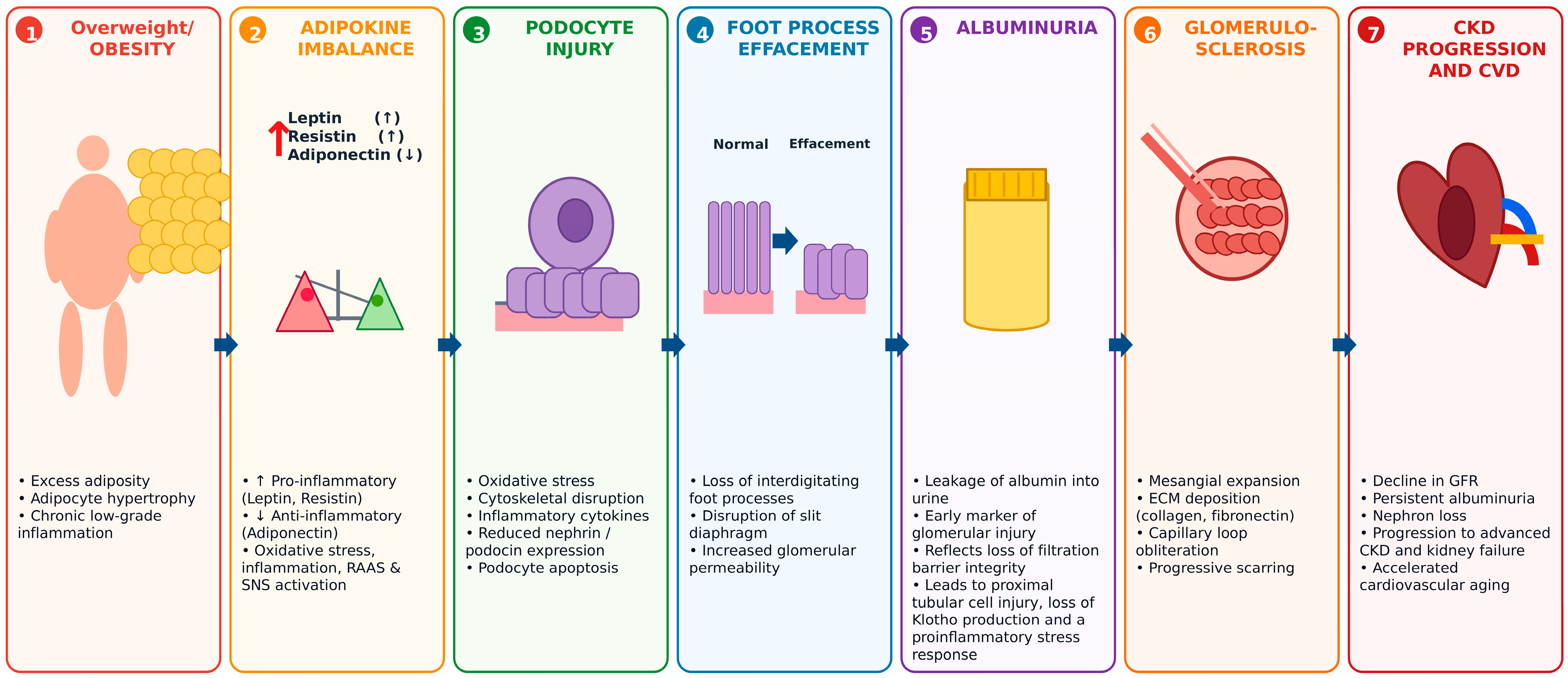
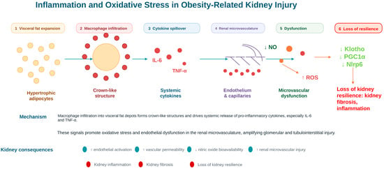
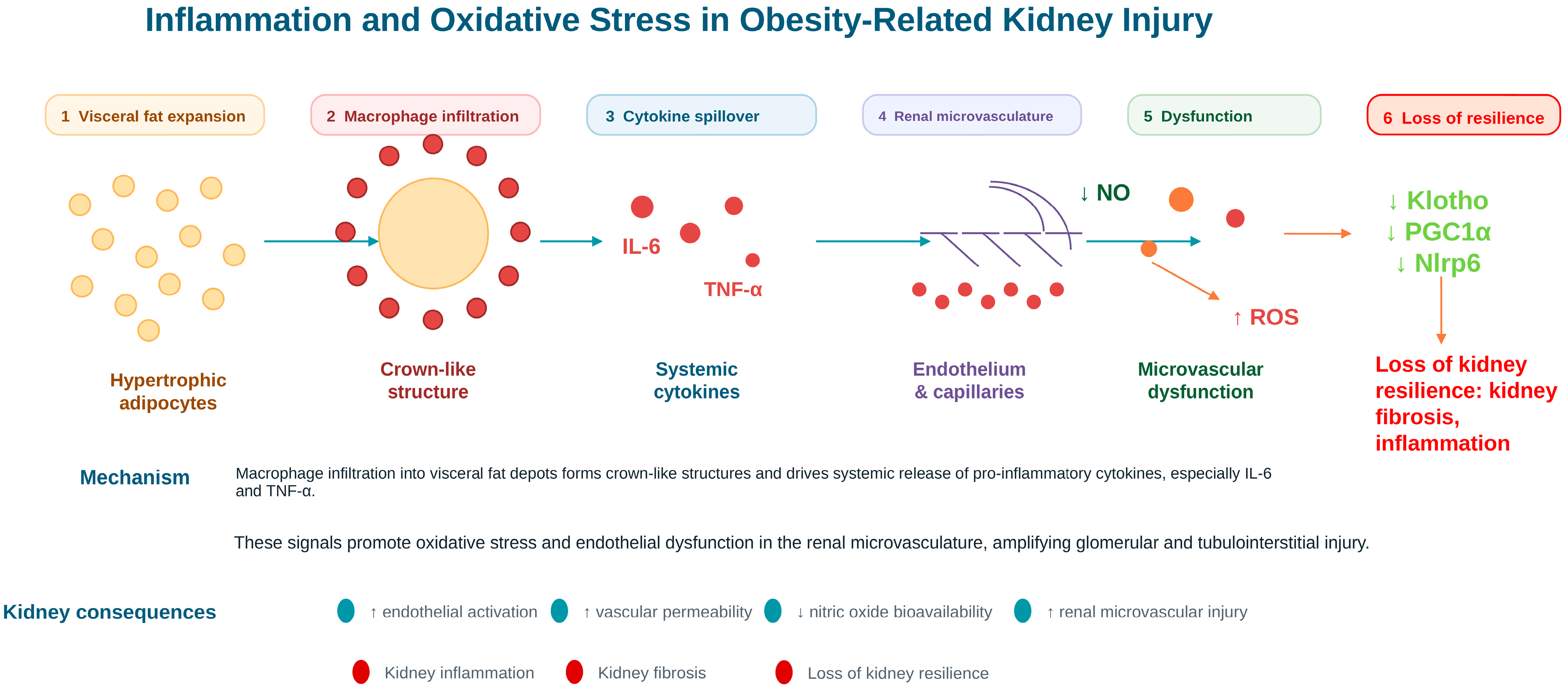
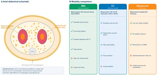
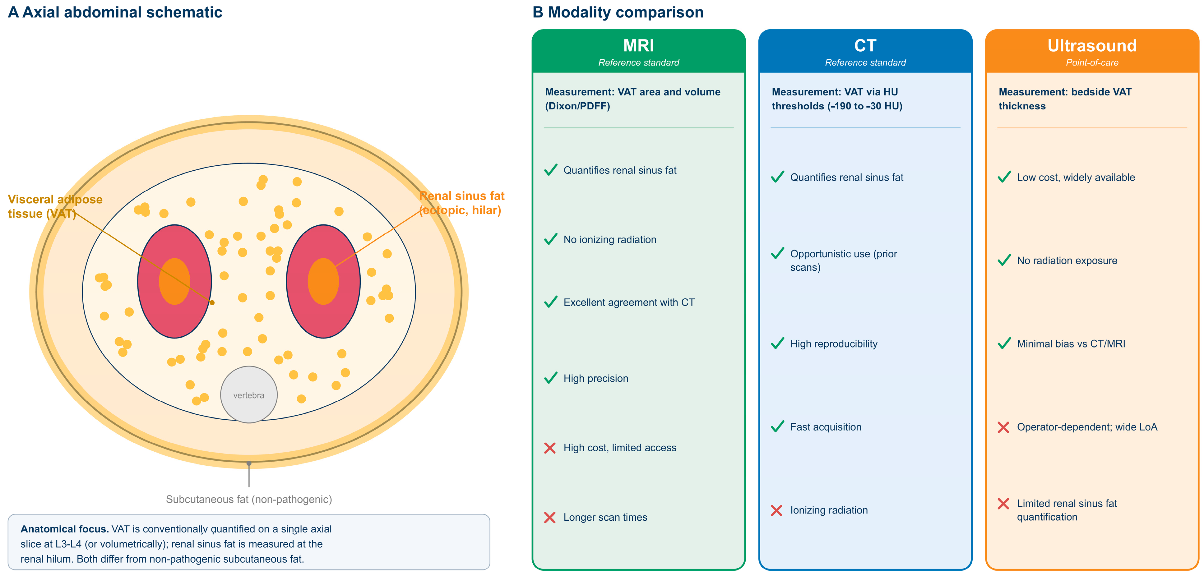
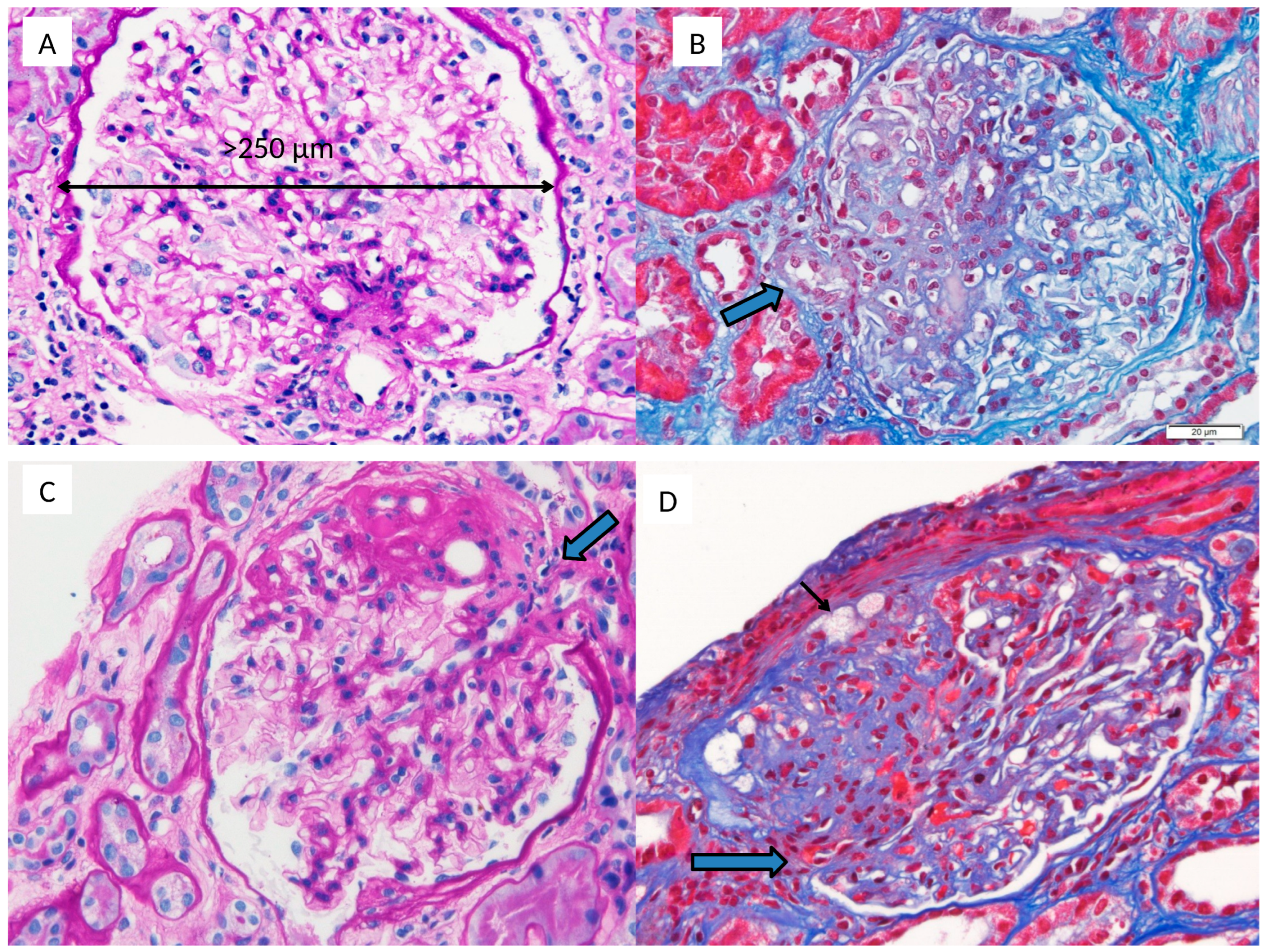
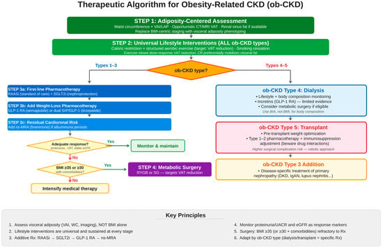
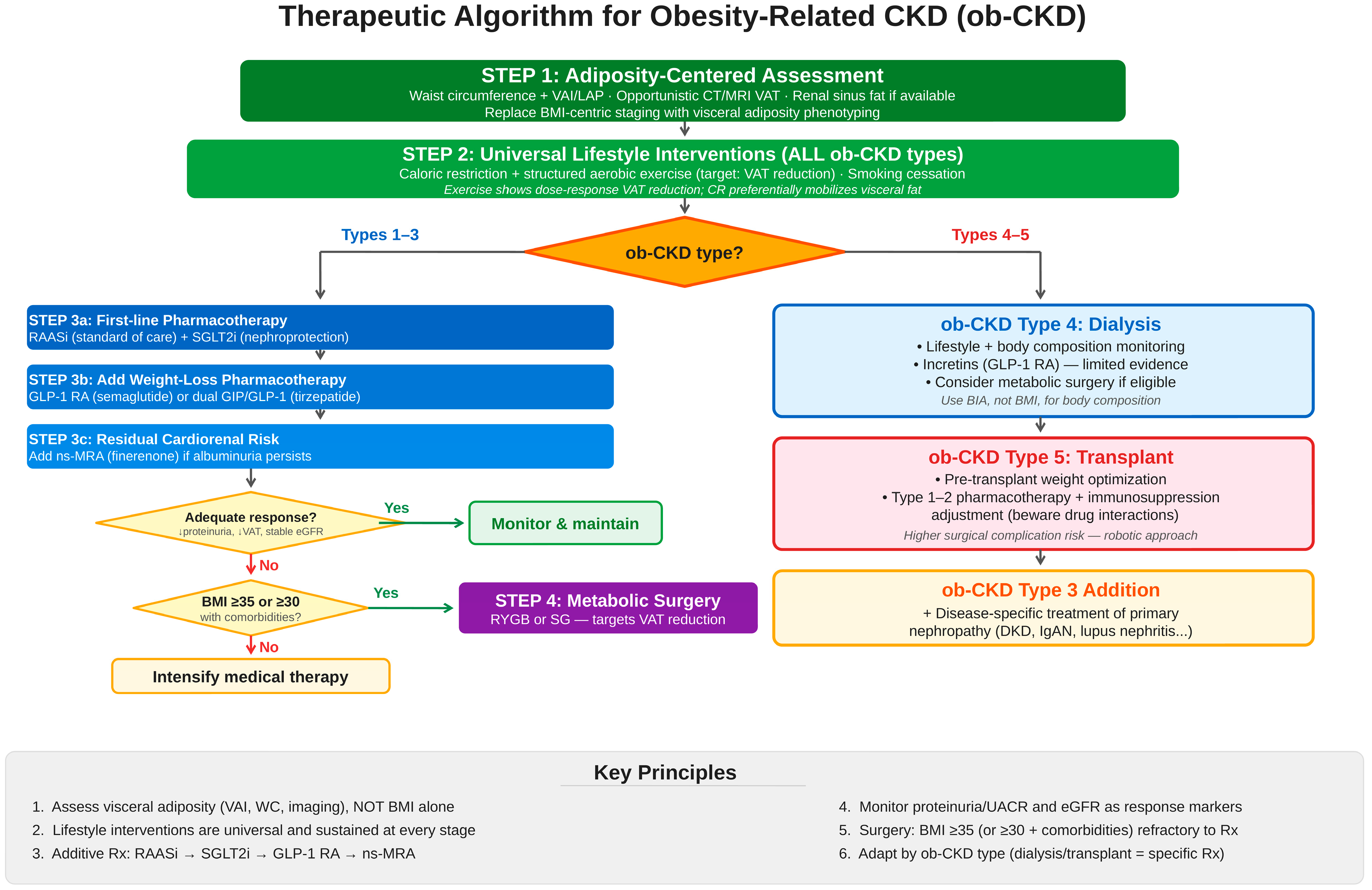

Disclaimer/Publisher’s Note: The statements, opinions and data contained in all publications are solely those of the individual author(s) and contributor(s) and not of MDPI and/or the editor(s). MDPI and/or the editor(s) disclaim responsibility for any injury to people or property resulting from any ideas, methods, instructions or products referred to in the content. |
© 2026 by the authors. Licensee MDPI, Basel, Switzerland. This article is an open access article distributed under the terms and conditions of the Creative Commons Attribution (CC BY) license.