Article

Cosmeceutical Potential of Grateloupia turuturu: Using Low-Cost Extraction Methodologies to Obtain Added-Value Extracts

1
MARE–Marine and Environmental Sciences Centre, ESTM, Politécnico de Leiria, 2520-641 Peniche, Portugal
2
REQUIMTE/LAQV, Laboratório de Farmacognosia, Faculdade de Farmácia, Universidade do Porto, 4050-313 Porto, Portugal
*
Authors to whom correspondence should be addressed.
Appl. Sci. 2021, 11(4), 1650; https://doi.org/10.3390/app11041650
Submission received: 15 January 2021 / Revised: 31 January 2021 / Accepted: 8 February 2021 / Published: 12 February 2021
(This article belongs to the Special Issue Marine Resources Application Potential for Biotechnological Purposes)

Abstract

:
The invasive macroalga Grateloupia turuturu is known to contain a diversity of bioactive compounds with different potentialities. Among them are compounds with relevant bioactivities for cosmetics. Considering this, this study aimed to screen bioactivities with cosmeceutical potential, namely, antioxidant, UV absorbance, anti-enzymatic, antimicrobial, and anti-inflammatory activities, as well as photoprotection potential. Extractions with higher concentrations of ethanol resulted in extracts with higher antioxidant activities, while for the anti-enzymatic activity, high inhibition percentages were obtained for elastase and hyaluronidase with almost all extracts. Regarding the antimicrobial activity, all extracts showed to be active against E. coli, S. aureus, and C. albicans. Extracts produced with higher percentages of ethanol were more effective against E. coli and with lower percentages against the other two microorganisms. Several concentrations of each extract were found to be safe for fibroblasts, but no photoprotection capacity was observed. However, one of the aqueous extracts was responsible for reducing around 40% of the nitric oxide production on macrophages, showing its anti-inflammatory potential. This work highlights G. turuturu’s potential in the cosmeceutical field, contributing to the further development of natural formulations for skin protection.

1. Introduction

Marine organisms’ environments are known to be deeply demanding due to competition and extreme conditions, forcing them to develop defense mechanisms and produce secondary metabolites to survive and protect themselves against external threats [1,2]. These produced compounds make marine organisms great sources of bioactive compounds with a myriad of applications. Among them, macroalgae are one of the most ecologically and economically relevant marine resources to obtain this type of compound, having in their constitution fibers, proteins, amino acids, minerals, polyunsaturated fatty acids, and vitamins [2].
Grateloupia turuturu (Yamada, 1941) is the largest edible red macroalga in the world. It is native to Korea and Japan and was classified as an invasive species in the Atlantic Ocean, being the first report in Portugal from 1997 [3]. It is typically characterized by a high content of carbohydrates (such as sulfated polysaccharides, known antioxidants, and antimicrobials), proteins (such as chromoproteins, with known antioxidant activity), and secondary metabolites (such as mycosporine-like amino acids (MAAs), known for their UV-shielding activity) and a low content of lipids [4,5,6,7]. The presence of such compounds is responsible for avoiding damages caused by the frequent exposure to UV radiation and high oxidative stress levels, typically found in mid-intertidal areas and intertidal pools, where this species is mostly located [8]. Developing extraction methodologies that are industrially feasible will create added-value extracts (such as cosmeceutical ingredients) that can turn to be an opportunity to promote the harvesting of this species, with positive consequences for the local invaded environments. For that, solid–liquid extraction (SLE) is one of the most suitable solutions due to the ease to up-scale the production. In fact, for the specific case of G. turuturu, solid–liquid extraction using ethanol and water as solvents is an already optimized method to extract the main bioactive compounds [9].
Personal care and image are receiving more attention every day, resulting in an unprecedented increase in cosmetic products use [1]. In 2016, Europeans spent a total of EUR 77 billion in this field, followed by the United Sates with EUR 64 billion and Brazil with EUR 24 billion [2]. The current concept of beauty includes healthy skin and a young appearance. Thus, the formulations to control the signs of aging are one of the industry’s biggest demands [10].
Skin aging is a natural and progressive process that is influenced by two main factors: intrinsic factors, such as genetics and physiological alterations, and extrinsic factors, such as environment, exposure to UV radiation or even smoking [1,2,11]. The signs of skin aging include thinning, fragility and continuous losses of elasticity of the skin, as well as the inability to maintain hydration, resulting in the formation of wrinkles [2,10]. In this process, the antioxidant defense system loses the capacity to block reactive oxygen species (ROS), leading to oxidative stress [10]. Together with reactive nitrogen species (RNS), they participate in regular cellular functions, being responsible for several regulatory mechanisms of cells to protect them against oxidative stress [2]. However, an overproduction of these molecules can play a different role, inducing damages in different cell structures, such as membranes, DNA, proteins and lipids, among others [2]. Thus, products able to reduce the symptoms of aging and consequently increase the quality of life and the self-esteem of consumers are among the most wanted, being used on a daily basis by millions of people [11]. Currently, an increased demand for natural solutions by customers [1,10] that replace the use of synthetic chemicals exists, due to the latter having high costs and being more pollutant and less sustainable, while also being perceived by the public as less safe.
Therefore, the main goal of this study is the evaluation of the bioactivities of several extracts from Grateloupia turuturu, taking into consideration the solvents used in the extraction procedure, with potential to be applied in natural skincare formulations, adding value to this species. For that, antioxidant, UV absorbance, anti-enzymatic, antimicrobial, and anti-inflammatory bioactivities are evaluated, as well as the cytotoxicity of extracts in fibroblasts and their photoprotection potential.

2. Materials and Methods

2.1. Seaweed Collection

The red seaweed Grateloupia turuturu was collected at Aguda Beach in Arcozelo, Portugal (41.054826, −8.656865), in July of 2017. The collected biomass was sorted for epibionts and then dried in a wind tunnel at 25 °C. The dried biomass was milled to flour-like powder (particle size 150 ± 50 µm) and stored under vacuum in the dark, at room temperature, until use.

2.2. Seaweed Extracts

Optimal conditions for 4 hydroethanolic solid–liquid extracts of G. turuturu and 2 aqueous extracts were selected (see Table 1) according to the optimization of the extraction process performed by Félix and co-workers [9], and their extraction methodology was followed. Briefly, two optimization assays were performed using a response surface methodology with a Box–Benhken design. Firstly, the solid-liquid ratio (SLR), the time of extraction (min) and the ethanol percentage were addressed. Then, using the results obtained for these 3 independent parameters, the influence of the extraction temperature (°C), pH and ethanol percentage was evaluated [9].
For the production of the selected extracts of Grateloupia turuturu, 5 g of biomass with the selected volume of solvent was mixed under constant magnetic stirring and thermostatized during the selected time of extraction. Each extract was then centrifuged for 5 min at 10,000× g and the obtained supernatant was filtered using filter paper (Whatmann no. 1). The evaporation of the extracts was performed under reduced pressure at 40 °C and then desiccated at room temperature using a vacuum concentrator (Vacufuge, Eppendorf, Germany). Yield of dry extracts was calculated (g extract·g−1 biomass) and then they were resuspended: aqueous extracts were resuspended in water at 25 mg·mL−1; 25% (v/v) ethanol extracts were resuspended in 25% (v/v) DMSO in water at 50 mg·mL−1; and 50% (v/v) ethanol extracts were resuspended in 50% (v/v) DMSO in water at 100 mg·mL−1.

2.3. Antioxidant Activity and UV Absorbance

The antioxidant activity was measured by ORAC assay, according to Félix and colleagues [9] and Dávalos and co-workers [12]. Briefly, a Trolox stock solution (VWR, Radnor, PA, USA) was used to prepare the dilutions from 8 to 0.5 µM. The obtained extracts were tested at 1 mg·mL−1 (diluted in 75 mM phosphate buffer). A fluorescein solution at 70 nM was used and the AAPH (2,2′-Azobis(2-methylpropionamidine) dihydrochloride) reagent (Sigma, Darmstadt, Germany) at 12 mM was prepared. A total of 20 μL of each sample was used and 120 μL of a fluorescein solution (70 nM) (Sigma, Darmstadt, Germany) was added to all samples in a 96-well black microplate (Greiner, Austria), including the standard curve. Phosphate buffer, at 75 mM, was used as control. Fluorescence was read for 15 min with a 1-min interval at an excitation wavelength of 485 nm and an emission wavelength of 525 nm in a microplate reader (Synergy H1, Biotek, Winooski, VT, USA) at 37 °C. After the incubation period, 60 μL of AAPH at 37 °C was added. The fluorescence was read for 80 min with 1-min intervals. Results were expressed as μmol of Trolox equivalents per gram of extract (μmol TE·−1 ext) and are reported as the mean of three replicates and standard deviation.
UV absorption was also performed according to [9]. Briefly, 200 μL of each extract (0.1 mg·mL−1) was added to a 96-well microplate for UV readings (Greiner UV-Star®, Kremsmünster, Austria) as well as the respective blanks. The absorbance was read between 280 and 400 nm (Synergy H1, Biotek, Winooski, VT, USA). The integral of the absorbance (Abs) was used to calculate the area under the curve (AUC), which was reported as the mean of three replicates and standard deviation.

2.4. Anti-Enzymatic Activity

2.4.1. Elastase Inhibition

The inhibition of elastase activity of the six extracts was performed using the EnzChek® Elastase Assay Kit (Invitrogen, Carlsbad, CA, USA) following the manufacturer’s instructions. A total of 50 µL of each extract at 2 mg·mL−1 was incubated with 50 µL of DQ-elastin from bovine neck ligament, BODIPY FL conjugate, in reaction buffer. Enzymatic release of fluorescent signal from DQ-elastin by elastase was quantitated using a fluorescent microplate reader (Synergy H1, Biotek, Winooski, VT, USA) at 486 nm excitation and 525 nm emission. To stop the enzymatic activity, N-Methoxysuccinyl-Ala-Ala-Pro-Val-chloromethyl ketone was added to the reaction buffer at a final concentration of 0.25 mg·mL−1. Elastase from pig pancreas was used at a final concentration of 0.025 mg·mL−1. Results are expressed as percentage of elastase inhibition.

2.4.2. Hyaluronidase Inhibition

The inhibition of hyaluronidase activity of the 6 extracts was performed according to Madan et al. and Adamczyk and colleagues [13,14], with some modifications. Hyaluronidase solution (4 U·mL−1) was prepared using a stock solution containing sodium phosphate buffer (200 mM, pH 7, 37 °C), 77 mM sodium chloride and 0.01% BSA. In a 1.5 mL tube, 200 µL of hyaluronidase solution and 25 µL of extract at 5 mg·mL−1 were incubated at 37 °C, for 10 min. Then, 100 µL hyaluronic acid solution (prepared in 300 mM of sodium phosphate monobasic solution at 0.06%) was added and the mixture was incubated at 37 °C, for 75 min. After the incubation period, 1 mL of acidic BSA (0.1% bovine serum albumin, 24 mM sodium acetate and 79 mM acetic acid, pH 3.75) was added and mixed by inversion, transferred to 96-well microplates and incubated for 15 min at room temperature. The absorbance was measured at 600 nm, in a microplate reader (Epoch2, Biotek, Winooski, VT, USA), and the data are presented as inhibition percentage.

2.5. Antimicrobial Activity

Antimicrobial activity of the six extracts of G. turuturu was evaluated through the microdilution technique [15,16] with slight modifications, using a fungal strain of Candida albicans (DSM-1386), the Gram-negative bacterium Escherichia coli (DSM-1103) and the Gram-positive bacterium Staphylococcus aureus (DSM-1104). The C. albicans two-day grown culture (Yeast and Mold Agar; VWR, cc) and the E. coli and S. aureus over-night grown cultures (Nutrient Agar; Sigma, Germany) were dissolved in saline solution (0.85% NaCl; Merck Millipore, Germany) and adjusted to a concentration of 1 × 10⁷ (for bacteria) and 2 × 10⁴ CFU·mL−1 (for fungus). The final inoculum concentrations on the microplates were 5 × 10⁵ (bacteria) and 1 × 10³ CFU.mL−1 (fungus), using Mueller-Hinton broth 2 (Sigma, Darmstadt, Germany) and RPMI-1640 (Sigma, Darmstadt, Germany). The positive control of inhibition used for E. coli was Ciprofloxacin (4 μg·mL−1; Sigma, Darmstadt, Germany), for S. aureus was Tetracycline (16 μg·mL−1, Sigma, Darmstadt, Germany) and Amphotericin B (4 μg·mL−1; Sigma, Darmstadt, Germany) was used for the C. albicans positive control; 4% (v/v) DMSO (Dimethyl sulfoxide; Carlo Erba, Spain) was used as negative control of microbial inhibition. Grateloupia turuturu extracts were tested at 0.0075, 0.75, 1.5 and 3 mg·mL−1 (diluted in phosphate saline buffer), using sterile round-bottom microplates (Thermo Scientific, Waltham, MA, EUA). For E. coli and S. aureus, the incubation period was 20 h at 35 °C, and for C. albicans, it was 48 h at 35 °C. After this time, the optical density (DO) was measured at 625 (bacteria) or 530 nm (fungus), in a microplate reader (Epoch2, BioTek, Winooski, VT, USA). The test was performed using 3 independent assays. Results are expressed in percentage of bacterial growth inhibition.

2.6. Photoprotection Activity

A 3T3 cell line (DSMZ–ACC 173, mouse fibroblasts) was grown and maintained according to supplier’s instructions. The cytotoxicity of the extracts was evaluated using the neutral red method described by Repetto et al. with slight modifications [17]. The 96-well microplates containing 5 × 10⁴ cells/well were incubated at 37 °C in 5% CO2 for 24 h in Dulbecco’s modified Eagle medium (DMEM) (Sigma, Darmstadt, Germany), 10% FBS (Biowest, Nuaillé, France). Cells were treated for 24 h with extracts (1:1 in DMEM, 10% FBS). A dose–response evaluation with eight different concentrations of each extract was performed (0.01, 0.062, 0.125, 0.25, 0.5, 1, 2 and 4 mg·mL−1 in phosphate-buffered saline [PBS]) in order to find the non-cytotoxic concentrations for the cells. After the incubation period, the medium was removed by aspiration and washed with 100 µL of PBS. After that, 100 µL of DMEM with 5% FBS, without phenol red and supplemented with neutral red (40 µg·mL−1 in PBS) (Sigma, Darmstadt, Germany), was added to each well to assess cell viability. The microplates were incubated at 37 °C in 5% CO2 for 4 h and then washed with PBS. After aspiration, 100 µL of desorption solution containing glacial acetic acid, ultrapure water and absolute ethanol (1:49:50) was added and the microplates were agitated until complete homogenization. The absorbance was read at 540 nm wavelength in a microplate spectrophotometer (Epoch2, BioTek, Winooski, VT, USA). PBS supplemented with the respective concentration of DMSO (vehicle) present in each sample and DMEM medium were used as controls. Data presented are the result of 3 independent replicas.
Knowing the non-cytotoxic concentrations of each extract, the concentration closest to 100% of cell viability was selected to perform a phototoxicity assay. The same 3T3 cell line was used and the assay was performed according to the OECD “Guidelines for Testing of Chemicals-In Vitro 3T3 NRU Phototoxicity Test” [18], with slight modifications. For the photoprotection evaluation of the extracts against UV radiation, the 96-well microplates containing 5 × 10⁴ cells/well were incubated at 37 °C in 5% CO2 for 24 h in DMEM, 10% FBS. After that period, the medium was removed and cells were treated for 1 h with 100 µL of each extract (E1—0.01 mg·mL−1, E2—0.062 mg·mL−1, E3—0.5 mg·mL−1, E4—0.5 mg·mL−1, E5—0.25 mg·mL−1, and E6—0.5 mg·mL−1, diluted in PBS) and then exposed for 40 min to UVA radiation (200 mJ/cm2) using a sun simulator chamber (UVA Cube 400, SOL500, Hönle UV Technology, Gräfelfing, Germany) equipped with a UVA filter (H1) (Hönle UV Technology, Gräfelfing, Germany) and a UVA sensor (FS UV-A D0, Hönle UV Technology, Gräfelfing, Germany) with a spectral range of 330–400 nm. After the exposure period, extracts were removed and the wells were washed with PBS, substituted by new medium and incubated at 37 °C in 5% CO2 for 24 h. After the incubation period, the medium was removed by aspiration and washed with 100 µL of PBS and the cytotoxicity was evaluated following the neutral red assay described above. PBS supplemented with the respective concentration of DMSO present in each sample and DMEM medium were used as positive controls and DMSO and empty wells were used as negative controls. Each condition was tested using 6 technical replicates and 3 independent assays.

2.7. NO Measurement

A RAW 264.7 cell line (ATCC-TIB 71, mouse macrophages) was grown and maintained according to the supplier’s instructions.
The effect of different concentrations of the macroalgal extracts on cell toxicity was determined using the 3-(4,5-dimethylthiazol-2-yl)-2,5-diphenyl tetrazolium bromide (MTT) assay according to Bahiense and colleagues, with slight modifications [19]. RAW 264.7 cells were treated for 24 h with extracts at increasing concentrations (0.01, 0.062, 0.125, 0.25, 0.5, 1, 2 and 4 mg·mL−1) in PBS. After the incubation period, the medium was removed by aspiration and washed with 100 µL of PBS. After that, 100 µL of DMEM with 5% FBS, without phenol red and supplemented with MTT solution (0.5 mg·mL−1 in PBS) (Sigma, Darmstadt, Germany), was added to each well to assess cell viability. The microplates were incubated at 37 °C in 5% CO2 for 4 h and then washed with PBS. After aspiration, 100 µL of DMSO was added and the microtiter plates were agitated for a few minutes and kept in the absence of light until complete solubilization of formazan. The absorbance was read at 570 nm wavelength in a microplate spectrophotometer (Epoch2, BioTek, Winooski, VT, USA). Each condition was tested in 3 independent assays.
Nitric oxide was then measured to determine the anti-inflammatory potential of the extracts. For that, all the concentrations whose cell viability was above 90% were selected for the assay. A Griess diazotization reaction was used to measure the production of NO in RAW 264.7 cells according to Bahiense et al. with slight modifications [19]. Briefly, the microplates were seeded with 1 × 10⁵ cells/well and incubated at 37 °C in 5% CO2 for 24 h. After that period, cells were treated with the extracts for 6 h, following the addition of LPS (lipopolysaccharide) solution from E. coli (Sigma, Darmstadt, Germany) at a final concentration of 1.5 µg·mL−1 for 22 h. Then, 150 µL of the supernatants of the cell culture was mixed with 50 µL of Griess reagent (Sigma, Germany) and incubated for 15 min at room temperature. The absorbance was measured at 540 nm using a microplate spectrophotometer (Epoch2, BioTek, Winooski, VT, EUA). Each condition was tested using 6 technical replicates and 3 independent assays.

2.8. Data Treatment

The values of antioxidant activity were studied as specific activity (activity per mass unit of extract) and total activity (activity per unit of seaweed extracted), the latter calculated by multiplying the values of the respective activities by the yield of extract. All the graphs and statistical analysis were performed with GraphPad Prism v.6 (GraphPad Software, La Jolla, San Diego, CA, USA).
For ORAC and UV AUC activities, Holm–Sidak’s multiple comparisons test was performed to understand the significant differences between extracts (different letters represent statistically significant differences, with p < 0.05).
For anti-enzymatic activity, cytotoxicity evaluation, photoprotection and anti-inflammatory potential, a one-way ANOVA was performed followed by Dunnett’s multiple comparisons test to evaluate the significant differences between the extracts and the respective controls (* p < 0.05, ** p < 0.01, *** p < 0.001, **** p < 0.0001).
For antimicrobial activity, a two-way ANOVA followed by Tukey’s multiple comparisons test was performed to evaluate the significant differences between extracts and between extracts and the inhibition control at each concentration (* p < 0.05, ** p < 0.01, *** p < 0.001, **** p < 0.0001).

3. Results

A total of four hydroethanolic and two aqueous extracts, obtained from the biomass of Grateloupia turuturu, were selected according the previous study of Félix and co-workers [9]. Several bioactivities related to cosmetic/cosmeceutical applications were analyzed to understand the potential of these extracts in this field, specifically their antioxidant and UV absorbance capacity, and anti-enzymatic and antimicrobial activities, as well as their photoprotection and anti-inflammatory potential.

3.1. Antioxidant Activity and UV Absorbance

Two different concentrations of ethanol were used in the extraction procedure, generating hydroethanolic extracts with different compositions (Table 2). Regarding the yield obtained, it was possible to verify that in the presence of ethanol, yields were lower when compared with the aqueous extracts, reaching almost twice the percentage of the yield (minimum obtained for E4 with 23.50% and maximum for E6 with 50.84%). However, higher values of antioxidant activity using the ORAC method were found for the extracts with higher concentrations of ethanol, reaching, for E1, the maximum with 153.09 µmol of Trolox equivalents· −1 extract and, for E6, the minimum with 45.00 µmol of Trolox equivalents·g−1 extract (p < 0.05). Similarly, E1 and E2 were the extracts presenting the highest values of UV absorbance.

3.2. Anti-Enzymatic Activity

Two different enzymes, known to be involved in skin degradation, were selected for this study. The inhibition of elastase (Figure 1A) and hyaluronidase (Figure 1B) activities was analyzed using the six seaweed extracts at 2 mg·mL−1. Results show that for elastase, all the extracts were able to inhibit nearly 100% of enzymatic activity when compared with the control (Figure 1A). For hyaluronidase, the inhibition percentages were also above 77% for all extracts, except for extract 1 (E1), which presented the lowest value of inhibition for this enzymatic activity (close to 40% inhibition).

3.3. Antimicrobial Activity

The antimicrobial potential of the extracts was evaluated against three representative microorganisms, namely, a Gram-negative bacterium, Escherichia coli, a Gram-positive bacterium, Staphylococcus aureus, and a fungal species, Candida albicans (Table 3). Regarding the bacterial inhibition of E. coli, extracts E1 and E2 (with higher percentages of ethanol used for the extraction procedure—50%) should be highlighted since the lower concentrations of extracts tested (0.0075 and 0.75 mg·mL−1) were significantly different (p < 0.05 or less) from the same concentrations for the other extracts (Table S1—complete statistical analysis). However, for the other concentrations (1.5 and 3 mg·mL−1), no significant differences were found between extracts, with the exception of E2 and E4 that significantly differ from each other (p < 0.05). The highest values of inhibition were found for E1 and E2, for 0.75 and 1.5 mg·mL−1, reaching values near to 40% of inhibition.
In the case of S. aureus inhibition, E3 showed to be the most promising extract against this bacterium, reaching values of inhibition close to 60% between 0.75 and 3 mg·mL−1 (significantly different from the other extracts, p < 0.05 or less), and only the lowest concentration, 0.0075 mg·mL−1, presented values of inhibition below 10%. It is also possible to verify that hydroethanolic extracts (E1–E4) were more efficient at inhibiting the growth of S. aureus (mostly at 0.75 and 3 mg·mL−1) when compared with aqueous extracts (E5–E6), with E6 at 0.0075 mg·mL−1 being responsible for the opposite effect—bacterial growth promotion.
The antimicrobial activity of the extracts against C. albicans showed a more variable profile between extracts and concentrations when compared to bacteria. In fact, the highest values of inhibition correspond to the E4 and E6 extracts at 0.0075 mg·mL−1 (p < 0.0001) and the lowest values (fungal growth promotion) were found for the same extracts but at 0.75 mg·mL−1 (p < 0.05 or less). Except for those cases, E1 and E2 were the extracts that reached lower values of inhibition.
Comparing the ability of the extracts in the study against the three microorganisms, globally, the higher inhibition (near to 60%) was found against S. aureus (E3) and C. albicans (E4 and E6) with different extracts, while against E. coli were the extracts E1 and E2 that were responsible for the higher antibacterial inhibition (near to 40%).

3.4. Photoprotection Activity

A dose–response evaluation of each extract was performed in a fibroblast cell line, 3T3, using a range of concentrations between 0.01 and 4 mg·mL−1, in order to evaluate the security of the extracts for skin applications (Figure S1). E1 and E2 (extraction with 50% ethanol/50% water) were the extracts with cell toxicity associated with more concentrations, especially E2, where cell viability was above 80% only in two of the eight concentrations tested. For E4, no concentration revealed a cytotoxic effect on 3T3 when compared with the control, and for E3 and E6, only the highest concentration (4 mg·mL−1) was responsible for a reduction in cell viability of close to 80% (p < 0.0001), with all the other concentrations being above 80% of cell viability. E5 showed significant differences when compared to the control (p < 0.0001) for the concentrations of 2 and 4 mg·mL−1, but also kept the values of cell viability near to 80%. Mostly in extracts with a lower or no concentration of ethanol in the extraction procedure (E3–E6), it was also possible to verify that the lowest concentrations tested were responsible for an increase in lysosomal activity, which might indicate growth promotion, being significantly different from the control.
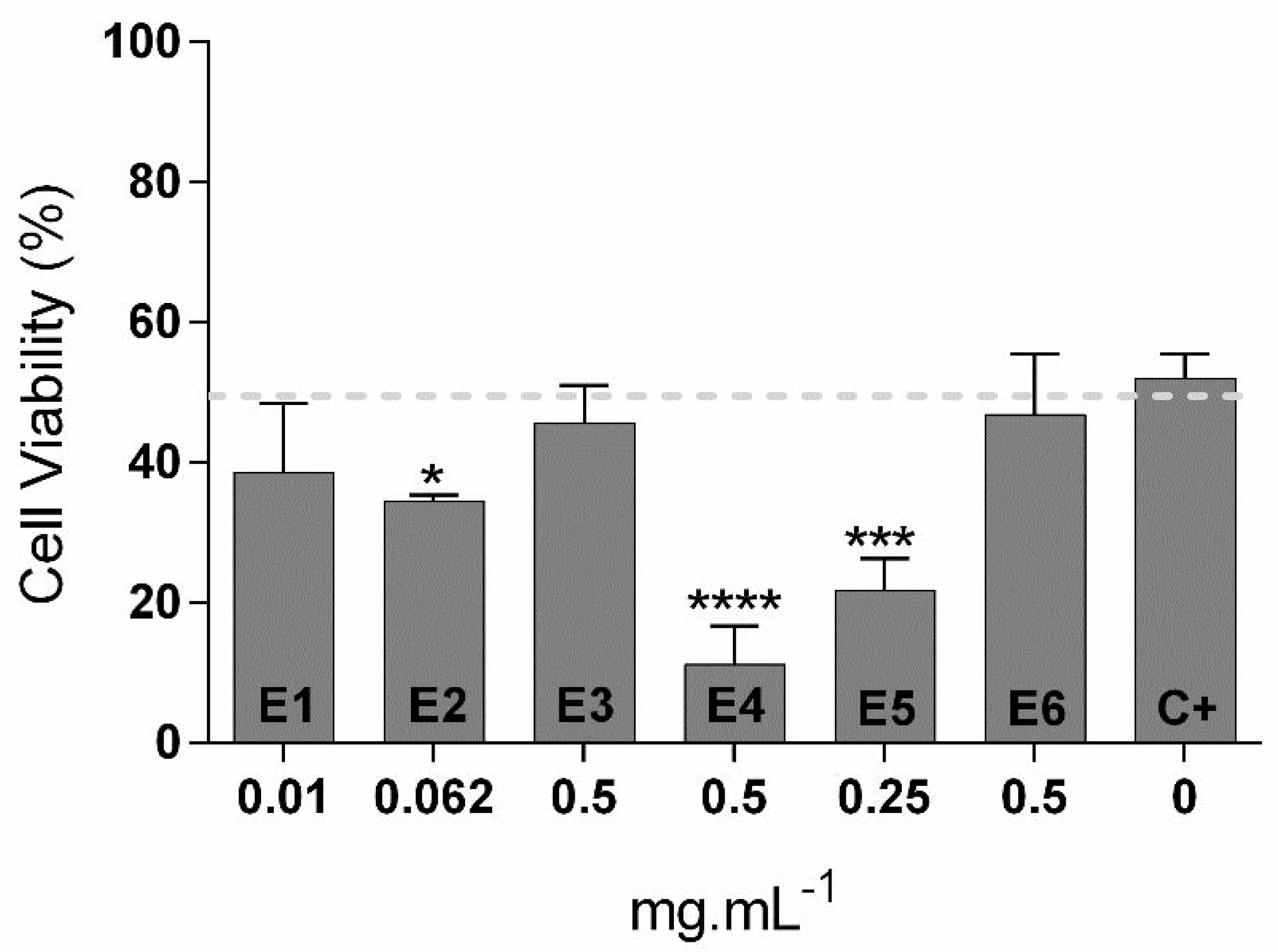
Based on the results obtained for cytotoxicity in 3T3 cells, a photoprotection assay using the concentrations closer to 100% of cell viability found for each extract was performed (Figure 2). Cells were exposed to a UV radiation dose capable of killing 50% of cells in the presence and absence of extracts. The results showed that none of the extracts tested presented a photoprotection capacity. From the six extracts, E1, E3 and E6 did not show any differences when compared with the control (cells without extracts and exposed to UV radiation), while E2 (p < 0.05), E4 (p < 0.0001) and E5 (p < 0.001) revealed a phototoxic behavior.

3.5. Nitric Oxide (NO) Measurement

A dose–response evaluation of each extract was performed in a macrophage cell line using the same range of concentrations used for 3T3 cells (Figure S2). Similar patterns were found in both cases: extracts with a higher concentration of ethanol in the extraction procedure (E1 and E2) showed higher cytotoxic effects when compared with the other four extracts. For the hydroethanolic extracts with 25% ethanol, the highest decrease in cell viability was reached for the concentration of 4 mg·mL−1 (p < 0.0001) with less than 20% of cell viability. The same trend was found for aqueous extracts, but although the concentration of 4 mg·mL−1 was significantly different from the control, cell viability percentages for that concentration were still high (above 70%).
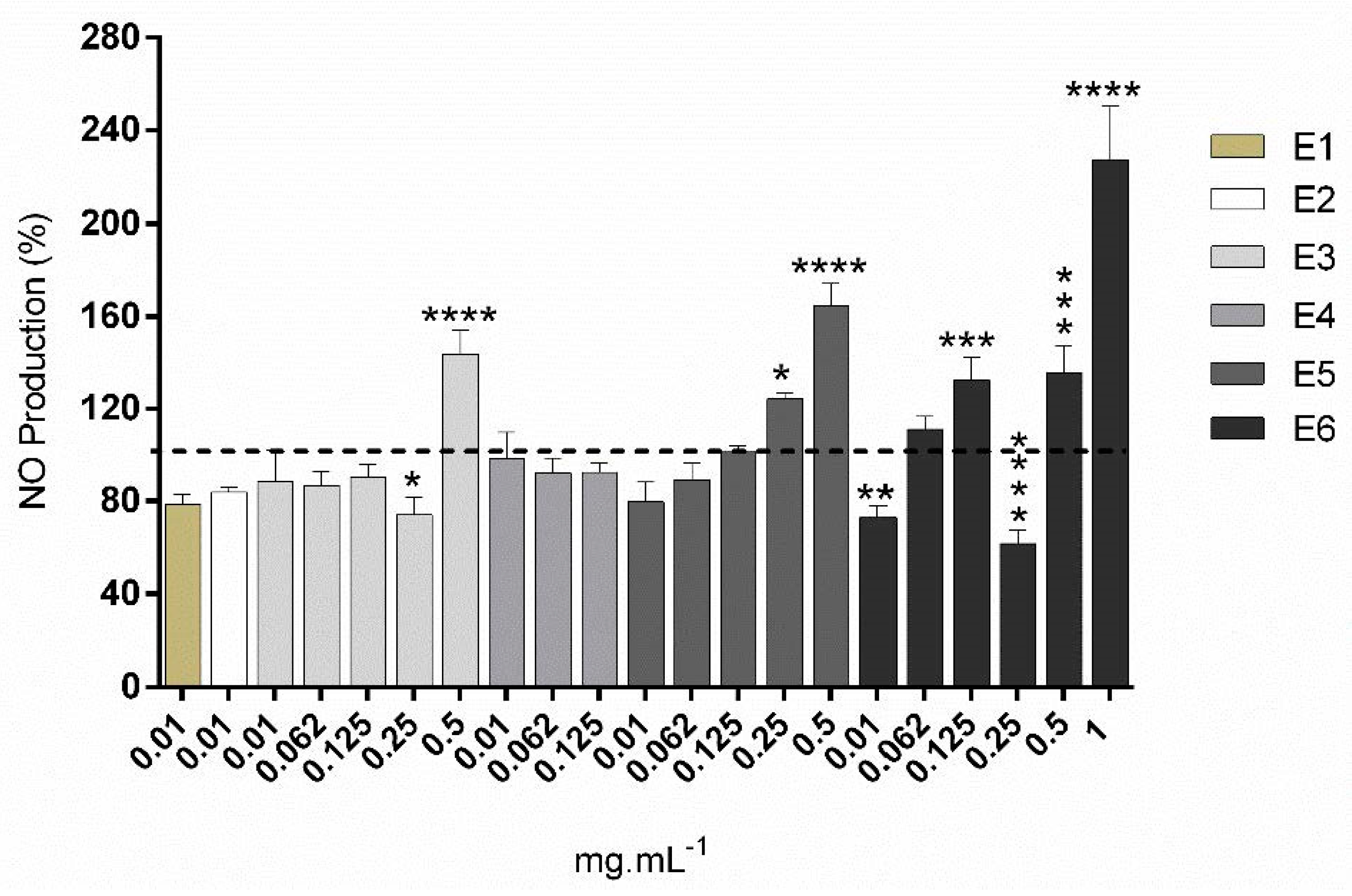
For each extract, all the concentrations above 90% cell viability were used to analyze the nitric oxide production and consequently the anti-inflammatory potential (Figure 3). Comparing with the control (cells subjected only to LPS solution), two extracts showed significant differences: E3 at 0·25 mg·mL−1 (p < 0.05), reducing the NO production 20%, and E6 at 0.01 (p < 0.01) and 0·25 mg.mL−1 (p < 0.0001), reducing 27% and 38.3%, respectively. However, higher concentrations increased the NO production on macrophages cells.

4. Discussion

The introduction of natural ingredients in the cosmetic industry is continuously increasing in an attempt to find effective, safer and sustainable solutions. Therefore, the screening of bioactives from marine resources to apply in the cosmetic/cosmeceutical field is a great contribution to achieve that. Building upon the work of Felix et al. [9], where the effects of the percentage of ethanol, temperature, time, pH, and solid-to-liquid ratio were all characterized in the solid–liquid extraction of Grateloupia turuturu’s antioxidant and UV-shielding compounds, six selected extracts were chosen and produced to further evaluate their properties of interest for the cosmeceutical industry.
The yield obtained for the six extracts (Table 1) showed that the increasing concentrations of ethanol are responsible for the decrease in the yield percentage (minimum obtained for E4 with 23.5% and maximum for E6 with 50.8%). This is in agreement with the fact that water is able to extract not only the galactans but also the proteins of this species, both presenting a significant massic contribution, while the presence of ethanol is responsible for their solubility decrease [9,20,21].
For the antioxidant activity by ORAC (Table 1), the opposite result was found: higher values of antioxidant activity in the presence of higher percentages of ethanol (reaching, for E1, the maximum with 153.09 and, for E6, the minimum with 45.00 µmol of Trolox equivalents·g−1 extract). This is also in accordance with the bibliography, since alcohols are known to be more efficient in the recovery process of antioxidants [20]. The presence of ROS, mostly originated from UV exposure, is responsible for triggering several processes in the skin (such as inflammation, oxidation of surface skin, hyperpigmentation and degradation of the dermal matrix, among others), promoting skin damages. Therefore, the use of molecules with antioxidant activity is a widely used approach to control and prevent symptoms related to skin damage [21].
Specifically, in G. turuturu, the presence of ethanol during the extraction procedure will contribute to recover molecules such as chlorophylls, polyphenols, and polar carotenoids and tocopherols, known for their antioxidant capacity [22,23,24], and thus, desired compounds to apply in cosmetic formulations. Although the main antioxidants are recovered using solvents such as ethanol, this does not prevent the study of aqueous extracts, since sulfated carrageenans, water-soluble compounds typically found in red algae, are also known for their antioxidant activity [25,26]. Apart from that, these polysaccharides are widely used in several industries due to their biocompatibility and high viscosity and gel forming properties [27].
Concerning the UV absorbance (Table 1), the highest ethanol concentration resulted in extracts with higher values of UV absorbance. This may be related to the extraction of compounds such as MAAs and polyphenols using this hydroethanolic mixture (50% each), while compounds with no activity, such as carbohydrates, are poorly extracted [9,28].
Between the different types of damages caused by the oxidative stress is the degradation of the extracellular matrix, which leads to a decrease in components responsible for the structure, elasticity and hydration of the skin (such as collagen, elastin and hyaluronic acid) and, consequently, to signs of skin aging, such as thinner skin, fine lines and wrinkles [2]. Thus, compounds able to enhance the inhibition of collagenase, elastase and hyaluronidase, among others, may be potential targets to use as bioactive ingredients in products with anti-aging properties [29]. In this context, the inhibition of elastase (Figure 1A) and hyaluronidase (Figure 1B) activities was analyzed using the six seaweed extracts at 2 mg·mL−1. For elastase, all the extracts were able to inhibit near to 100% of enzymatic activity when compared with the control (Figure 1A), and for hyaluronidase, the inhibition percentages were also above 77% for all extracts (with the exception of E1), showing a great potential as active ingredients for anti-wrinkle formulations. The high percentages of inhibition for all extracts suggest that more than one type of compound is responsible for these bioactivities, since the presence of different concentrations of ethanol during the extraction, or even the absence, would result in the extraction of different classes of compounds. While sulfated polysaccharides and proteins are almost exclusively soluble in water, compounds such as carotenoids, sterols and fatty acids, among others, are preferentially extracted using a compromise between ethanol and water due to their medium polarity. For MAAs, it is expected that from water to higher percentages of ethanol, the extraction of these compounds would occur, possibly presenting different relative contents for each extraction condition [9]. Peptides from seaweed, such as signal peptides, were described to stimulate the extracellular matrix, increasing neocollagenesis and elastin synthesis, resulting in wrinkle reduction and skin firming [2,30]. Moreover, secondary metabolites, such as MAAs, have been described by their anti-wrinkle ability [29], mostly by their ability to inhibit the collagenase and elastase activities and to stimulate the secretion of hyaluronic acid by human fibroblasts [31,32,33]. Another group of secondary metabolites produced by red macroalgae, known for their capacity to maintain the extracellular matrix as healthier, are phenolic compounds [10]. In fact, a study conducted using a red macroalgae resulted in a methanolic extract rich in phenolic compounds, which was able to inhibit the overexpression of metalloproteinases, preventing the formation of wrinkles [31].
The antimicrobial properties of seaweed are also well established for a wide range of macroalgae [34,35]. They are known to produce bioactive compounds to inhibit/reduce the growth of other competitive microorganisms [34]. For the six hydroethanolic extracts, three microorganisms were selected to evaluate the antimicrobial activity of G. turuturu: a Gram-negative and a Gram-positive bacterium, E. coli and S. aureus, respectively, and a fungal strain of C. albicans (Table 3). Results show that for E. coli, extracts with higher percentages of ethanol were responsible for higher percentages of growth inhibition (E1 and E2, reaching near to 40% of inhibition). In the case of the Gram-positive S. aureus, E3 was the most promising extract with almost 60% of growth inhibition for three of the four concentrations tested, while for C. albicans, the antimicrobial profile obtained was not so clear, with some concentrations of the extracts E3–E6 being among the most effective against this species. The recent study of Cardoso and co-workers [7] showed that ethanolic and polysaccharide extracts of G. turuturu presented antibacterial activity against E. coli and S. aureus, corroborating the results obtained and showing that the polysaccharides, such as carrageenan, present in this species, have antimicrobial activity. Further, the antifungal activity of this species was already confirmed for several species, as stated by Plouguerné and co-workers, who found extracts of G. turuturu highly active against five fungi species [36]. Regarding the hydroethanolic extracts of G. turuturu (E1–E4), bioactive compounds such as sulfated polysaccharides, phenolic compounds and carotenoids may be present and responsible for the antimicrobial activity, since they are known to alter the microbial cell permeability and to interfere with the membrane, leading to the loss of cellular integrity [34]. The wide antimicrobial activity of these extracts shows the potential for the cosmetic industry, as functional ingredients, but also as natural preservatives of cosmetic formulations, increasing the shelf-life of the product by reducing the microbial contamination [2,34].
In this study, a dose–response evaluation of each extract was performed in a mouse fibroblast cell line, 3T3, since these cells are one of the mains constituents of the skin, using a range of concentrations between 0.01 and 4 mg·mL−1, in order to evaluate the security of the extracts for skincare applications (Figure S1). E1 and E2 (extraction with 50% ethanol/50% water) were the extracts with cell toxicity associated with more concentrations, while for E3 to E6, only the highest concentrations were responsible for a reduction in cell viability, the lowest concentrations tested being responsible for an increase in the neutral red signal, suggesting a growth promotion. The presented results show that for all the extracts tested, several concentrations were not cytotoxic to fibroblast cells, being an excellent preliminary result about their security for potential applications in skincare products. Another important feature of red seaweeds for the cosmetic industry is the production of bioactive compounds with photoprotection activity, able to protect the skin from damages such as sunburn, photo-aging, photo-dermatoses and skin cancer, among others [2,11]. The production of such compounds by macroalgae consists of ecophysiological strategies developed to avoid the deleterious effects of the constant exposure to UV radiation, through the absorption of UV radiation [11,37]. In fact, bioactive compounds able to absorb UV radiation were found to protect human fibroblasts from cell death and to retard the signs of aging induced by UV radiation [2,38]. Red macroalgae are known to produce a variety of compounds with this ability, such as phenolic compounds, pigments and MAAs. Between them, MAAs are known to be the most relevant for this function [2,37]. These secondary metabolites present high antioxidant and UV absorbing capacities, acting as excellent UV filters and thus having a great potential for the cosmetic industry as antioxidants and photoprotectors [2,29].
Based on the results obtained for cytotoxicity in 3T3 cells, a photoprotection assay using the concentrations closer to 100% of cell viability found for each extract was performed (Figure 2). Cells were exposed to a UV radiation dose capable of reaching the IC50 of cells in the presence and absence of extracts. The results show that none of the extracts tested presented photoprotection capacity. From the six extracts, E1, E3, and E6 did not show any differences compared with the control (cells without extracts and exposed to UV radiation), while E2 (p < 0.05), E4 (p < 0.0001), and E5 (p < 0.001) revealed a phototoxic behavior, which was not an expected result for hydroethanolic extracts of the red seaweed G. turuturu. Comparing with the UV absorbance capacity of each extract, it is not possible to correlate the data, since E1 and E2, extracts with higher ethanol concentrations and consequently more phenolic compounds, were not the ones presenting less phototoxicity. However, several factors may contribute to explain these results. Specifically, a dose–response evaluation for the phototoxicity test could help to understand the effect of extract concentrations on this bioactivity. Since we are working with crude extracts that present a mixture of compounds with synergistic/antagonistic effects, it is possible that the best solution for the desired function may be related to specific concentrations where the dilution of certain compounds would be beneficial. Another interesting fact is that not all the MAAs have the same ability to act as photoprotectors [37]. At this point, further chemical characterization of the extracts could help to discriminate the presence of MAAs and which of them are present/at a relative quantity. Regarding also the production of MAAs, it is known that their production is affected by several abiotic factors, preferring the summer period and moderate depth [29,39]. A recent study also focused on the fact that G. turuturu tends to reduce the production of MAAs in the presence of intense UV radiation [40], suggesting that the production of such compounds may be highly influenced by different factors that are not totally controlled when the macroalga is grown in a natural environment.
The anti-inflammatory potential of seaweed has also been explored in an attempt to find potential sustainable and safer solutions with less side effects, especially for treatments of chronic inflammation [2]. During inflammation, oxidative stress increases and the cellular antioxidant capacity decreases, leading to large quantities of produced free radicals that will interact with fatty acids, cell membranes, proteins, and other components, promoting permanent alterations in cellular functions [41]. This process is mediated by a system of soluble factors that differ in their source and composition, one of them being the production of nitric oxide by macrophages. This compound is responsible for inducing vasodilatation, acting as a cytotoxic agent for pathogens [41]. Therefore, the discovery of novel compounds able to act as anti-inflammatories could be a new insight in this field.
A dose–response evaluation of each extract was performed in a macrophage cell line due to the direct implication of these cells in inflammatory processes, using the same range of concentrations used for 3T3 cells (Figure S2). Similar patterns were found in both cases: extracts with a higher concentration of ethanol in the extraction procedure (E1 and E2) showed higher cytotoxic effects when compared with the other four extracts. For each extract, all the concentrations above 90% cell viability were used to analyze the nitric oxide production (Figure 3). Comparing with the control (cells subjected only to LPS solution), two extracts showed significant differences: E3 at 0.25 mg·mL−1 (p < 0.05), reducing the NO production to 80%, and E6 at 0.01 (p < 0.01) and 0.25 mg·mL−1 (p < 0.0001), reducing 27% and 38.3%, respectively. However, higher concentrations stimulated NO production on macrophages cells.
In macroalgae, several types of bioactive compounds have already been described for their anti-inflammatory potential, namely, pigments (such as carotenoids), sulfated polysaccharides, proteins and their derivatives (such as phycobiliproteins), fatty acids (such as polyunsaturated fatty acids) and other compounds such as halogenated compounds or terpenes [10,29]. Some of them are known to be produced by red macroalgae and specifically by G. turuturu, such as carotenoids, phycobiliproteins, and sulfated polysaccharides, among others, which could help to explain the reduction in NO production in those extracts. However, different methods for anti-inflammatory evaluation, such as Western blot quantification of inflammatory markers’ expression (e.g., TNF-α, interleukins, among others), should be implemented, since different pathways may be activated and the specificity of the technique is higher.

5. Conclusions

The potential of the invasive macroalga G. turuturu for the cosmetic industry was investigated. Several bioactivities concerning skin protection of the hydroethanolic extracts were analyzed and the results obtained show that different concentrations of ethanol led to extracts with different bioactivities. Noticeably, among the tested extracts, good antioxidant and antimicrobial activities were found, which promotes the added value of these extracts both for skin benefits and for formula’s benefits. Additionally, significant inhibition of skin aging-related enzymes was attained, as well as some degree of inhibition of the inflammatory marker NO. A photoprotection assessment allowed the discovery of a phototoxicity of some extracts from G. turuturu, which is unexpected but very important information concerning this biomass use and valorization. This study is, therefore, an important contribution to understanding that seaweed extracts obtained from simple solvents (ethanol and water) and techniques (SLE), compatible with the industrial scale, have potential to be applied in the cosmetic field, bridging the demand for natural, greener and more sustainable products. However, further fractionation and/or characterization of these crude extracts is essential to understand the active ingredients of each extract responsible for the analyzed bioactivities.

Supplementary Materials

The following are available online at https://www.mdpi.com/2076-3417/11/4/1650/s1. Table S1: Statistical analysis of antimicrobial data using a two-Way ANOVA followed by Tukey’s multiple comparisons test. Significant differences between extracts and between extracts and inhibition control at each concentration tested were analyzed and discriminated (* p < 0.05, ** p < 0.01, *** p < 0.001, **** p < 0.0001). Figure S1: Dose–response cytotoxic evaluation of the E1 (A), E2 (B), E3 (C), E4 (D), E5 (E) and E6 (F) extracts of Grateloupia turuturu at 8 different concentrations (0.01, 0.062, 0.125, 0.25, 0.5, 1, 2 and 4 mg·mL-1) in 3T3 cells. Figure S2: Dose–response cytotoxic evaluation of the E1 (A), E2 (B), E3 (C), E4 (D), E5 (E) and E6 (F) extracts of Grateloupia turuturu at 8 different concentrations (0.01, 0.062, 0.125, 0.25, 0.5, 1, 2 and 4 mg·mL−1) in RAW 264.7 cells.

Author Contributions

Conceptualization, A.M.C., R.F., S.C.N. and M.F.L.L.; methodology, R.F.; formal analysis, R.F. and C.F.; investigation, C.F., A.M.C., A.J., P.D. and T.V.; resources, M.F.L.L. and R.P.; writing—original draft preparation, C.F.; writing—review and editing, C.F., R.F. and M.F.L.L.; supervision, J.S., C.A. and M.F.L.L.; project administration, M.F.L.L; funding acquisition, M.F.L.L. All authors have read and agreed to the published version of the manuscript.

Funding

This study was supported by UID/MAR/04292/2020 with funding from FCT/MCTES through national funds, and by the grant awarded to Rafael Félix (SFRH/BD/139763/2018). The authors also wish to acknowledge the support of the European Union through the EASME Blue Labs project AMALIA, Algae-to-MArket Lab IdeAs (EASME/EMFF/2016/1.2.1.4/03/SI2.750419), project VALORMAR (Mobilizing R&TD Programs, Portugal 2020), co-funded by COMPETE (POCI-01-0247-FEDER-024517), the Integrated Programme of SR&TD “Smart Valorization of Endogenous Marine Biological Resources Under a Changing Climate” (reference Centro-01-0145-FEDER-000018) and RD&T co-promotion project ORCHESTRA (nº 70155), co-funded by Centro 2020 program, Portugal 2020, European Union, European Regional Development Fund, and SAICTPAC/0019/2015—LISBOA-01-0145-FEDER-016405 Oncologia de Precisão: Terapias e Tecnologias Inovadoras (POINT4PAC), through the European Regional Development Fund.

Institutional Review Board Statement

Not applicable.

Informed Consent Statement

Not applicable.

Data Availability Statement

Data is contained within the article or supplementary material.

Conflicts of Interest

The authors declare no conflict of interest. The funders had no role in the design of the study; in the collection, analyses, or interpretation of data; in the writing of the manuscript, or in the decision to publish the results.

References

  1. Jesumani, V.; Du, H.; Aslam, M.; Pei, P.; Huang, N. Potential use of seaweed bioactive compounds in skincare—a review. Mar. Drugs 2019, 17, 688. [Google Scholar] [CrossRef] [Green Version]
  2. Pimentel, F.B.; Alves, R.C.; Rodrigues, F.; Oliveira, M.B.P.P. Macroalgae-derived ingredients for cosmetic industry-an update. Cosmetics 2018, 5, 4–9. [Google Scholar] [CrossRef] [Green Version]
  3. Freitas, C.; Araújo, R.; Bertocci, I. Patterns of benthic assemblages invaded and non-invaded by Grateloupia turuturu across rocky intertidal habitats. J. Sea Res. 2016, 115, 26–32. [Google Scholar] [CrossRef]
  4. Sekar, S.; Chandramohan, M. Phycobiliproteins as a commodity: Trends in applied research, patents and commercialization. J. Appl. Phycol. 2008, 20, 113–136. [Google Scholar] [CrossRef]
  5. Terasaki, M.; Narayan, B.; Kamogawa, H.; Nomura, M.; Stephen, N.M.; Kawagoe, C.; Hosokawa, M.; Miyashita, K. Carotenoid profile of edible japanese seaweeds: An improved hplc method for separation of major carotenoids. J. Aquat. Food Prod. Technol. 2012, 21, 468–479. [Google Scholar] [CrossRef]
  6. Li, W.; Su, H.N.; Pu, Y.; Chen, J.; Liu, L.N.; Liu, Q.; Qin, S. Phycobiliproteins: Molecular structure, production, applications, and prospects. Biotechnol. Adv. 2019, 37, 340–353. [Google Scholar] [CrossRef]
  7. Cardoso, I.; Cotas, J.; Rodrigues, A.; Ferreira, D.; Osório, N.; Pereira, L. Extraction and analysis of compounds with antibacterial potential from the red alga Grateloupia turuturu. J. Mar. Sci. Eng. 2019, 7, 220. [Google Scholar] [CrossRef] [Green Version]
  8. Araújo, R.; Violante, J.; Pereira, R.; Abreu, H.; Arenas, F.; Sousa-Pinto, I. Distribution and population dynamics of the introduced seaweed Grateloupia turuturu (halymeniaceae, rhodophyta) along the Portuguese coast. Phycologia 2011, 50, 392–402. [Google Scholar] [CrossRef]
  9. Félix, R.; Carmona, A.M.; Félix, C.; Novais, S.C.; Lemos, M.F.L. Industry-friendly hydroethanolic extraction protocols for grateloupia turuturu UV-shielding and antioxidant compounds. Appl. Sci. 2020, 10, 5304. [Google Scholar] [CrossRef]
  10. Lourenço-Lopes, C.; Fraga-Corral, M.; Jimenez-Lopez, C.; Pereira, A.G.; Garcia-Oliveira, P.; Carpena, M.; Prieto, M.A.; Simal-Gandara, J. Metabolites from macroalgae and its applications in the cosmetic industry: A circular economy approach. Resources 2020, 9, 101. [Google Scholar] [CrossRef]
  11. Ariede, M.B.; Candido, T.M.; Jacome, A.L.M.; Velasco, M.V.R.; de Carvalho, J.C.M.; Baby, A.R. Cosmetic attributes of algae - A review. Algal Res. 2017, 25, 483–487. [Google Scholar] [CrossRef]
  12. Dávalos, A.; Gómez-Cordovés, C.; Bartolomé, B. Extending Applicability of the Oxygen Radical Absorbance Capacity (ORAC-Fluorescein) Assay. J. Agric. Food Chem. 2004, 52, 48–54. [Google Scholar] [CrossRef]
  13. Madan, K.; Nanda, S. In-vitro evaluation of antioxidant, anti-elastase, anti-collagenase, anti-hyaluronidase activities of safranal and determination of its sun protection factor in skin photoaging. Bioorg. Chem. 2018, 77, 159–167. [Google Scholar] [CrossRef] [PubMed]
  14. Adamczyk, K.; Olech, M.; Abramek, J.; Pietrzak, W.; Kuźniewski, R.; Bogucka-Kocka, A.; Nowak, R.; Ptaszyńska, A.A.; Rapacka-Gackowska, A.; Skalski, T.; et al. Eleutherococcus species cultivated in Europe: A new source of compounds with antiacetylcholinesterase, antihyaluronidase, anti-DPPH, and cytotoxic activities. Oxid. Med. Cell. Longev. 2019, 2019. [Google Scholar] [CrossRef] [PubMed] [Green Version]
  15. CLSI. Methods for Dilution Antimicrobial Susceptibility Tests for Bacteria That Grow Aerobically. In Clinical and Laboratory Standards Institute; Approved Standard - M7-A7; Clinical and Laboratory Standards Institute: Wayne, PA, USA, 2006; Volume 26, ISBN 1562386255. [Google Scholar]
  16. CLSI. Reference Method for Broth Dilution Antifungal Susceptibility Testing of Yeasts; Approved Standard - M27-A3; Clinical and Laboratory Standards Institute: Wayne, PA, USA, 2006; Volume 28, pp. 0–13. [Google Scholar]
  17. Repetto, G.; del Peso, A.; Zurita, J.L. Neutral red uptake assay for the estimation of cell viability/ cytotoxicity. Nat. Protoc. 2008, 3, 1125–1131. [Google Scholar] [CrossRef] [PubMed]
  18. OECD. OECD Test Guideline 432: In Vitro 3T3 NRU Phototoxicity Test. 2004, pp. 1–15. Available online: https://www.oecd-ilibrary.org/environment/test-no-432-in-vitro-3t3-nru-phototoxicity-test_9789264071162-en (accessed on 12 February 2021).
  19. Bahiense, J.B.; Marques, F.M.; Figueira, M.M.; Vargas, T.S.; Kondratyuk, T.P.; Endringer, D.C.; Scherer, R.; Fronza, M. Potential anti-inflammatory, antioxidant and antimicrobial activities of Sambucus australis. Pharm. Biol. 2017, 55, 991–997. [Google Scholar] [CrossRef] [PubMed] [Green Version]
  20. Sultana, B.; Anwar, F.; Ashraf, M. Effect of extraction solvent/technique on the antioxidant activity of selected medicinal plant extracts. Molecules 2009, 14, 2167–2180. [Google Scholar] [CrossRef]
  21. Masaki, H. Role of antioxidants in the skin: Anti-aging effects. J. Dermatol. Sci. 2010, 58, 85–90. [Google Scholar] [CrossRef] [PubMed]
  22. Tiwari, B.K.; Troy, D.J. Seaweed Sustainability - Food and Non-Food Applications. Tiwari, B.K., Troy, D.J., Eds.; Elsevier Inc.: New York, NY, USA, 2015; ISBN 9780124186972. [Google Scholar]
  23. Shalaby, E.A. Algae as promising organisms for environment and health. Plant Signal. Behav. 2011, 6, 1338–1350. [Google Scholar] [CrossRef] [Green Version]
  24. Garcia-Vaquero, M.; Rajauria, G.; O’Doherty, J.V.; Sweeney, T. Polysaccharides from macroalgae: Recent advances, innovative technologies and challenges in extraction and purification. Food Res. Int. 2017, 99, 1011–1020. [Google Scholar] [CrossRef] [Green Version]
  25. Ye, D.; Jiang, Z.; Zheng, F.; Wang, H.; Zhang, Y.; Gao, F.; Chen, P.; Chen, Y.; Shi, G. Optimized extraction of polysaccharides from Grateloupia livida (Harv.) yamada and biological activities. Molecules 2015, 20, 16817–16832. [Google Scholar] [CrossRef]
  26. Tang, L.; Chen, Y.; Jiang, Z.; Zhong, S.; Chen, W.; Zheng, F.; Shi, G. Purification, partial characterization and bioactivity of sulfated polysaccharides from Grateloupia livida. Int. J. Biol. Macromol. 2017, 94, 642–652. [Google Scholar] [CrossRef] [PubMed]
  27. Pacheco-Quito, E.M.; Ruiz-Caro, R.; Veiga, M.D. Carrageenan: Drug Delivery Systems and Other Biomedical Applications. Mar. Drugs 2020, 18, 583. [Google Scholar] [CrossRef] [PubMed]
  28. Athukorala, Y.; Lee, K.; Song, C.; Ahn, C.; Shin, T.; Cha, Y.-J.; Shahid, F.; Jeon, Y.-J. Potential antioxidant activity of marine red alga grateloupia filicina extracts. J. Food Lipids 2003, 10, 251–265. [Google Scholar] [CrossRef]
  29. Pereira, L. Seaweeds as source of bioactive substances and skin care therapy-Cosmeceuticals, algotheraphy, and thalassotherapy. Cosmetics 2018, 5. [Google Scholar] [CrossRef] [Green Version]
  30. Malerich, S.; Berson, D. Next generation cosmeceuticals. The latest in peptides, growth factors, cytokines, and stem cells. Dermatol. Clin. 2014, 32, 13–21. [Google Scholar] [CrossRef] [PubMed]
  31. Ryu, B.M.; Qian, Z.J.; Kim, M.M.; Nam, K.W.; Kim, S.K. Anti-photoaging activity and inhibition of matrix metalloproteinase (MMP) by marine red alga, Corallina pilulifera methanol extract. Radiat. Phys. Chem. 2009, 78, 98–105. [Google Scholar] [CrossRef]
  32. Terazawa, S.; Nakano, M.; Yamamoto, A.; Imokawa, G. Mycosporine-like amino acids stimulate hyaluronan secretion by up-regulating hyaluronan synthase 2 via activation of the p38/MSK1/CREB/c-Fos/AP-1 axis. J. Biol. Chem. 2020, 295, 7274–7288. [Google Scholar] [CrossRef] [Green Version]
  33. Orfanoudaki, M.; Hartmann, A.; Alilou, M.; Gelbrich, T.; Planchenault, P.; Derbré, S.; Schinkovitz, A.; Richomme, P.; Hensel, A.; Ganzera, M. Absolute configuration of mycosporine-like amino acids, their wound healing properties and in vitro anti-aging effects. Mar. Drugs 2020, 18, 35. [Google Scholar] [CrossRef] [PubMed] [Green Version]
  34. Pérez, M.J.; Falqué, E.; Domínguez, H. Antimicrobial action of compounds from marine seaweed. Mar. Drugs 2016, 14, 52. [Google Scholar] [CrossRef] [Green Version]
  35. Silva, A.; Silva, S.A.; Carpena, M.; Garcia-Oliveira, P.; Gullón, P.; Barroso, M.F.; Prieto, M.A.; Simal-Gandara, J. Macroalgae as a source of valuable antimicrobial compounds: Extraction and applications. Antibiotics 2020, 9, 642. [Google Scholar] [CrossRef] [PubMed]
  36. Plouguerné, E.; Hellio, C.; Deslandes, E.; Véron, B.; Stiger-Pouvreau, V. Anti-microfouling activities in extracts of two invasive algae: Grateloupia turuturu and Sargassum muticum. Bot. Mar. 2008, 51, 202–208. [Google Scholar] [CrossRef]
  37. Álvarez-Gómez, F.; Korbee, N.; Casas-Arrojo, V.; Abdala-Díaz, R.T.; Figueroa, F.L. UV photoprotection, cytotoxicity and immunology capacity of red algae extracts. Molecules 2019, 24, 341. [Google Scholar] [CrossRef] [Green Version]
  38. Bedoux, G.; Hardouin, K.; Burlot, A.S.; Bourgougnon, N. Bioactive components from seaweeds: Cosmetic applications and future development; Elsevier: New York, NY, USA, 2014; Volume 71, ISBN 9780124080621. [Google Scholar]
  39. Pereira, L. Seaweed flora of the european north atlantic and mediterranean. In Springer Handbook of Marine Biotechnology; Springer: Cham, Switzerland, 2015; pp. 65–178. ISBN 9783642539718. [Google Scholar]
  40. de Ramos, B.; da Costa, G.B.; Ramlov, F.; Maraschin, M.; Horta, P.A.; Figueroa, F.L.; Korbee, N.; Bonomi-Barufi, J. Ecophysiological implications of UV radiation in the interspecific interaction of Pyropia acanthophora and Grateloupia turuturu (Rhodophyta). Mar. Environ. Res. 2019, 144, 36–45. [Google Scholar] [CrossRef] [PubMed]
  41. Fernando, I.P.S.; Nah, J.W.; Jeon, Y.J. Potential anti-inflammatory natural products from marine algae. Environ. Toxicol. Pharmacol. 2016, 48, 22–30. [Google Scholar] [CrossRef]
Figure 1. Evaluation of anti-enzymatic activity of the six extracts of Grateloupia turuturu at 2 mg·mL−1: inhibition of elastase (A) and hyaluronidase (B) activities. Control of inhibition is represented as a dashed line. A one-way ANOVA followed by Dunnett’s multiple comparisons test was performed to evaluate the significant differences between the extracts and the inhibition control (* p < 0.05, ** p < 0.01). Values presented are the mean of 3 independent assays.
Applsci 11 01650 g001
Figure 2. Photoprotection assay using the closest concentration to 100% of cell viability identified for each extract of Grateloupia turuturu. Cells were subjected to UV radiation in the presence and absence of extracts until IC50 of control without extract was reached to evaluate the photoprotection potential. Control of cell viability is represented as a dashed line. A one-way ANOVA followed by Dunnett’s multiple comparisons test was performed to evaluate the significant differences between the extracts and the control (* p < 0.05, *** p < 0.001, **** p < 0.0001). Values presented are the mean of 3 independent assays.
Applsci 11 01650 g002
Figure 3. Nitric oxide assay was performed using all the non-cytotoxic concentrations identified for each extract of Grateloupia turuturu to evaluate their anti-inflammatory potential, using a final concentration of 1.5 µg·mL−1 of LPS. Control of cell viability is represented as a dashed line. A one-way ANOVA followed by Dunnett’s multiple comparisons test was performed to evaluate the significant differences between the extracts and the control (* p < 0.05, ** p < 0.01, *** p < 0.001, **** p < 0.0001). Values presented are the mean of 3 independent assays.
Applsci 11 01650 g003
Table 1. Selected extracts and respective extraction conditions: temperature, pH, percentage of ethanol, time and solid–liquid ratio.
ExtractsTemperature (°C)pH% EtOHTime (min)SLR
E130950601:40
E2100950601:40
E31007251001:10
E4204251001:10
E52090201:40
E610090201:40
Table 2. Selected extracts of Grateloupia turuturu and respective ethanol percentage, yield, antioxidant capacity by ORAC assay (expressed as µmol of Trolox equivalents (TE) per gram of extract) and UV absorbance, using the area under the curve. Holm–Sidak’s multiple comparisons test was performed to understand the significant differences between extracts (different letters represent statistically significant differences, with p < 0.05).
ExtractsEtOH (%)Yield (%)ORAC (µmol TE· −1 ext)UV AUC
MeanSDSignificant DifferencesMeanSDSignificant Differences
E15024.39153.111.37a5.820.25a
E25028.56102.38.33b4.060.16b
E32524.2845.982.82c1.630.13c
E42523.5066.816.79d3.200.03d
E5043.3750.262.89c3.080.25d
E6050.8445.003.77c2.260.30e
Table 3. Antimicrobial activity of Grateloupia turuturu extracts against the bacteria Staphylococcus aureus and E. coli and the fungus Candida albicans at 4 different concentrations of extracts: 0.0075, 0.75, 1.5 and 3 mg·mL−1. A two-way ANOVA followed by Tukey’s multiple comparisons test was performed to evaluate the significant differences between extracts and between extracts and the inhibition control at each concentration (see Supplementary Table S1). Values presented are the mean of 3 independent assays.
Staphylococcus aureus
E1E2E3E4E5E6C + (Tetracycline)
mg·mL−1MeanSDMeanSDMeanSDMeanSDMeanSDMeanSDMeanSD
317.225.2537.775.8454.011.4828.374.5032.407.9535.804.68100.560.26
1.515.144.3626.885.8552.993.6725.604.4123.726.7124.907.99101.181.23
0.7529.588.9335.138.6052.643.6638.332.8810.7921.084.2823.15100.540.22
0.0075−8.932.33−3.493.31−9.235.29−5.865.99−10.586.05−10.635.72100.231.02
Escherichia coli
E1E2E3E4E5E6C + (Ciprofloxacin)
mg·mL−1MeanSDMeanSDMeanSDMeanSDMeanSDMeanSDMeanSD
321.141.7025.790.1716.673.2811.334.8817.585.8013.876.57106.102.57
1.530.690.7232.992.0125.687.0423.109.5427.0710.5226.1810.79106.102.57
0.7533.894.4336.275.3110.515.237.003.974.612.9215.734.40105.690.71
0.007520.040.1729.2811.344.851.003.950.443.451.924.511.70106.010.80
Candida albicans
E1E2E3E4E5E6C + (Amphotericin B)
mg·mL−1MeanSDMeanSDMeanSDMeanSDMeanSDMeanSDMeanSD
37.402.224.772.1641.326.9531.541.8737.642.4237.182.04101.420.54
1.53.870.475.451.1521.085.3131.110.9823.944.9732.563.03101.420.54
0.7520.986.6528.091.68−26.5512.72−14.588.44−8.714.34−3.0614.70105.272.30
0.007523.892.228.696.1441.534.4455.262.4411.454.4855.390.54103.903.62
Publisher’s Note: MDPI stays neutral with regard to jurisdictional claims in published maps and institutional affiliations.