

Craniomaxillofac. Trauma Reconstr. 2025, 18(4), 47; https://doi.org/10.3390/cmtr18040047 - 25 Oct 2025
Abstract
Background: The high perimandibular approach (HPA) is a feasible surgical technique for open reduction and internal fixation (OR-IF) of mandibular condylar fractures, offering reduced complication rates. In this study, we retrospectively evaluated the treatment outcomes and complications associated with HPA use. Patients and
[...] Read more.
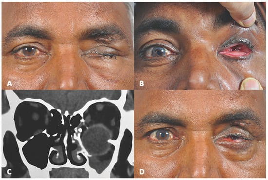
Background: The high perimandibular approach (HPA) is a feasible surgical technique for open reduction and internal fixation (OR-IF) of mandibular condylar fractures, offering reduced complication rates. In this study, we retrospectively evaluated the treatment outcomes and complications associated with HPA use. Patients and Methods: Patients who underwent OR-IF for mandibular condylar fractures using the HPA at three hospitals in Shimane between June 2019 and March 2024 were included. Data collected included the mechanism of injury, AO classification of the fracture site, fracture type and mode, surgical duration, mouth-opening range at 6 months post-operatively, and peri- and post-operative complications. Results: A total of 42 patients (46 condylar fractures; 18 males and 24 females; mean age, 63.0 years) were included. The fracture pattern included dislocations in 18 cases (42.8%). The mean surgical duration was 75.0 min. Post-operative trismus occurred in 16 patients (38.1%) at 6 months. Longer surgical duration and dislocated fractures were significantly associated with post-operative trismus (p < 0.05). Conclusions: The HPA is safe and effective for managing mandibular condylar fractures. However, post-operative trismus may be influenced by longer surgical duration and fracture types, warranting further investigation and potential post-surgical management.
Full article
(This article belongs to the Special Issue Advances in Facial Trauma Surgery)
►
Show Figures

Figure 1


