The Development and Application of Echocardiography in Heart Disease

A special issue of Journal of Clinical Medicine (ISSN 2077-0383). This special issue belongs to the section "Cardiology".

Deadline for manuscript submissions: 23 November 2024 | Viewed by 49049

Special Issue Editors


E-Mail Website
Guest Editor
Department of Cardiology, Umberto I Hospital, 84014 Nocera Inferiore, SA, Italy
Interests: advanced cardiovascular imaging; CMR; echocardiography; cardiovascular emergencies
Special Issues, Collections and Topics in MDPI journals

E-Mail Website
Guest Editor
1. Division of Cardiology, Department of Advanced Biomedical Sciences, University of Naples Federico II, 80138 Naples, Italy
2. Mediterranea Cardiocentro, 80122 Naples, Italy
Interests: echocardiography; speckle tracking echocardiography; valvular heart disease; ischemic heart disease; cardiomyopathies
Special Issues, Collections and Topics in MDPI journals

Special Issue Information

Dear Colleagues,

Cardiovascular diseases are the leading causes of mortality and morbidity in developed countries, with ischemic heart disease and non-ischemic cardiomyopathies playing the most relevant role. Many imaging techniques, either non-invasive or invasive, are being developed to allow a prompt detection of myocardial structural and functional abnormalities, as well as to guide therapeutic strategies.

Echocardiography is one of the most powerful diagnostic and monitoring tools available in clinical practice. Its wide availability, non-invasiveness and high diagnostic accuracy make echocardiography a fundamental step in the management of patients with cardiovascular disease.

Moreover, the ultimate role of echocardiography in managing heart disease has been highlighted, not only from a diagnostic perspective, but also for prognostic purposes.

The advent of more advanced techniques, such as three-dimensional echocardiography, speckle-tracking-derived longitudinal strain, and, more recently, non-invasive myocardial work assessment, has provided further insight into the detection of the structural and pathophysiological mechanisms underlying cardiovascular disease and has further extended the application of such technics in many clinical scenarios.

This Special Issue of Journal of Clinical Medicine is now open for submission, welcoming papers that aim to present the current role of echocardiography in the diagnosis and management of cardiovascular diseases, with particular attention on novel techniques that could contribute to relevant achievements in the near future.

Prof. Dr. Antonello D'Andrea
Dr. Federica Ilardi
Guest Editors

Manuscript Submission Information

Manuscripts should be submitted online at www.mdpi.com by registering and logging in to this website. Once you are registered, click here to go to the submission form. Manuscripts can be submitted until the deadline. All submissions that pass pre-check are peer-reviewed. Accepted papers will be published continuously in the journal (as soon as accepted) and will be listed together on the special issue website. Research articles, review articles as well as short communications are invited. For planned papers, a title and short abstract (about 100 words) can be sent to the Editorial Office for announcement on this website.

Submitted manuscripts should not have been published previously, nor be under consideration for publication elsewhere (except conference proceedings papers). All manuscripts are thoroughly refereed through a single-blind peer-review process. A guide for authors and other relevant information for submission of manuscripts is available on the Instructions for Authors page. Journal of Clinical Medicine is an international peer-reviewed open access semimonthly journal published by MDPI.

Please visit the Instructions for Authors page before submitting a manuscript. The Article Processing Charge (APC) for publication in this open access journal is 2600 CHF (Swiss Francs). Submitted papers should be well formatted and use good English. Authors may use MDPI's English editing service prior to publication or during author revisions.

Keywords

  • Speckle tracking echocardiography
  • Global longitudinal strain
  • Strain rate
  • Non-invasive myocardial work
  • 3D echocardiography
  • Stress echocardiography
  • Ischemic cardiomyopathy
  • Non-ischemic cardiomyopathy
  • Valvular heart disease

Benefits of Publishing in a Special Issue

  • Ease of navigation: Grouping papers by topic helps scholars navigate broad scope journals more efficiently.
  • Greater discoverability: Special Issues support the reach and impact of scientific research. Articles in Special Issues are more discoverable and cited more frequently.
  • Expansion of research network: Special Issues facilitate connections among authors, fostering scientific collaborations.
  • External promotion: Articles in Special Issues are often promoted through the journal's social media, increasing their visibility.
  • e-Book format: Special Issues with more than 10 articles can be published as dedicated e-books, ensuring wide and rapid dissemination.

Further information on MDPI's Special Issue polices can be found here.

Published Papers (15 papers)

Order results
Result details
Select all
Export citation of selected articles as:

Research

Jump to: Review

10 pages, 1274 KiB  
Article
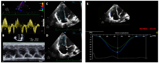
Effects on Right Ventricular Function One Year after COVID-19-Related Pulmonary Embolism
by Federica Ilardi, Mario Crisci, Cecilia Calabrese, Anna Scognamiglio, Fortunato Arenga, Rachele Manzo, Domenica F. Mariniello, Valentino Allocca, Anna Annunziata, Antonello D’Andrea, Raffaele Merenda, Vittorio Monda, Giovanni Esposito and Giuseppe Fiorentino
J. Clin. Med. 2023, 12(11), 3611; https://doi.org/10.3390/jcm12113611 - 23 May 2023
Cited by 2 | Viewed by 1599
Abstract
The aim of this study was to investigate the presence of subclinical cardiac dysfunction in recovered coronavirus disease 2019 (COVID-19) patients, who were stratified according to a previous diagnosis of pulmonary embolism (PE) as a complication of COVID-19 pneumonia. Out of 68 patients [...] Read more.
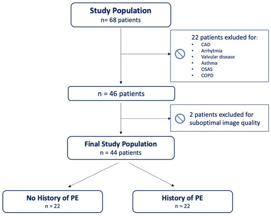
The aim of this study was to investigate the presence of subclinical cardiac dysfunction in recovered coronavirus disease 2019 (COVID-19) patients, who were stratified according to a previous diagnosis of pulmonary embolism (PE) as a complication of COVID-19 pneumonia. Out of 68 patients with SARS-CoV-2 pneumonia followed up for one year, 44 patients (mean age 58.4 ± 13.3, 70% males) without known cardiopulmonary disease were divided in two groups (PE+ and PE−, each comprising 22 patients) and underwent clinical and transthoracic echocardiographic examination, including right-ventricle global longitudinal strain (RV-GLS), and RV free wall longitudinal strain (RV-FWLS). While no significant differences were found in the left- or right-heart chambers’ dimensions between the two study groups, the PE+ patients showed a significant reduction in RV-GLS (−16.4 ± 2.9 vs. −21.6 ± 4.3%, p < 0.001) and RV-FWLS (−18.9 ± 4 vs. −24.6 ± 5.12%, p < 0.001) values compared to the PE- patients. According to the ROC-curve analysis, RV-FWLS < 21% was the best cut-off with which to predict PE diagnosis in patients after SARS-CoV-2 pneumonia (sensitivity 74%, specificity 89%, area under the curve = 0.819, p < 0.001). According to the multivariate logistic regression model, RV-FWLS < 21% was independently associated with PE (HR 34.96, 95% CI:3.24–377.09, p = 0.003) and obesity (HR 10.34, 95% CI:1.05–101.68, p = 0.045). In conclusion, in recovered COVID-19 patients with a history of PE+, there is a persistence of subclinical RV dysfunction one year after the acute phase of the disease, detectable by a significant impairment in RV-GLS and RV-FWLS. A reduction in RV-FWLS of lower than 21% is independently associated with COVID-related PE. Full article
(This article belongs to the Special Issue The Development and Application of Echocardiography in Heart Disease)
Show Figures

Figure 1

14 pages, 3408 KiB  
Article
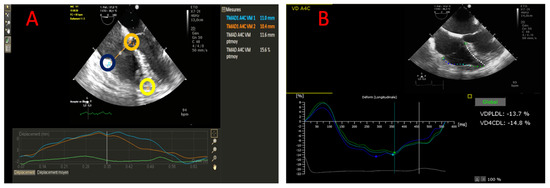
Echocardiographic Parameters to Predict Malignant Events in Arrhythmic Mitral Valve Prolapse Population
by Alessandro Vairo, Paolo Desalvo, Andrea Rinaudo, Francesco Piroli, Anna Tribuzio, Andrea Ballatore, Gianluca Marcelli, Lorenzo Pistelli, Veronica Dusi, Nicolò Montali, Gianluca Alunni, Cristina Barbero, Stefano Salizzoni, Marco Pocar, Mauro Rinaldi, Fiorenzo Gaita, Gaetano Maria De Ferrari and Carla Giustetto
J. Clin. Med. 2023, 12(3), 1232; https://doi.org/10.3390/jcm12031232 - 3 Feb 2023
Cited by 3 | Viewed by 4842
Abstract
Bileaflet Mitral Valve Prolapse (bMVP) has been linked to major arrhythmic events and sudden cardiac death (SCD). Consistent predictors in this field are still lacking. Echocardiography is the best tool for the analysis of the prolapse and its impact on the ventricular mechanics. [...] Read more.
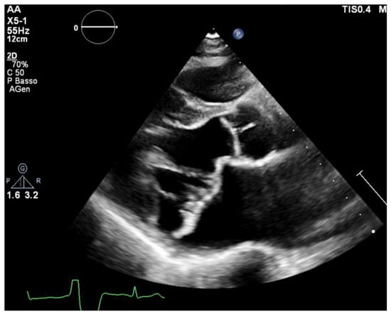
Bileaflet Mitral Valve Prolapse (bMVP) has been linked to major arrhythmic events and sudden cardiac death (SCD). Consistent predictors in this field are still lacking. Echocardiography is the best tool for the analysis of the prolapse and its impact on the ventricular mechanics. The aim of this study was to find new echocardiographic predictors of malignant events within an arrhythmic MVP population. We evaluated 22 patients with arrhythmic bMVP with a transthoracic echocardiogram focused on mitral valve anatomy and ventricular contraction. Six of them had major arrhythmic events that required ICD implantation (ICD-MVP group), while sixteen presented with a high arrhythmic burden without major events (A-MVP group). The best predictors of malignant events were the Anterior Mitral Leaflet (AML) greater length and greater Mechanical Dispersion (MD) of basal and mid-ventricular segments, while other significant predictors were the larger mitral valve annulus (MVA) indexed area, lower MVA anteroposterior diameter/AML length ratio, higher inferolateral basal segment S3 velocity. Full article
(This article belongs to the Special Issue The Development and Application of Echocardiography in Heart Disease)
Show Figures

Figure 1

11 pages, 1313 KiB  
Article
Feasibility, Prediction and Association of Right Ventricular Free Wall Longitudinal Strain with 30-Day Mortality in Severe COVID-19 Pneumonia: A Prospective Study
by Christophe Beyls, Tristan Ghesquières, Alexis Hermida, Thomas Booz, Maxime Crombet, Nicolas Martin, Pierre Huette, Vincent Jounieaux, Hervé Dupont, Osama Abou-Arab and Yazine Mahjoub
J. Clin. Med. 2022, 11(13), 3629; https://doi.org/10.3390/jcm11133629 - 23 Jun 2022
Cited by 4 | Viewed by 1653
Abstract
Introduction: Right ventricular (RV) systolic dysfunction (RVsD) is a common complication of coronavirus infection 2019 disease (COVID-19). The right ventricular free wall longitudinal strain parameter (RV-FWLS) is a powerful predictor of mortality. We explored the performance of RVsD parameters for predicting 30-day mortality [...] Read more.
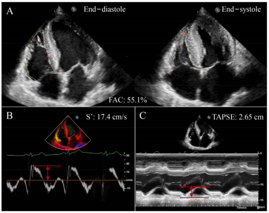
Introduction: Right ventricular (RV) systolic dysfunction (RVsD) is a common complication of coronavirus infection 2019 disease (COVID-19). The right ventricular free wall longitudinal strain parameter (RV-FWLS) is a powerful predictor of mortality. We explored the performance of RVsD parameters for predicting 30-day mortality and the association between RV-FWLS and 30-day mortality. Methods: COVID-19 patients hospitalized at Amiens University Hospital in the critical care unit with transthoracic echocardiography were included. We measured tricuspid annular plane systolic excursion (TAPSE), the RV S’ wave, RV fractional area change (RV-FAC), and RV-FWLS. The diagnostic performance of RVsD parameters as predictors for 30-day mortality was evaluated by the area under the receiver operating characteristic (ROC) curve (AUC). RVsD was defined by an RV-FWLS < 21% to explore the association between RVsD and 30-day mortality. Results: Of the 116 patients included, 20% (n = 23/116) died and 47 had a RVsD. ROC curve analysis showed that RV-FWLS failed to predict 30-day mortality, as did conventional RV parameters (all p > 0.05). TAPSE (21 (19–26) mm vs. 24 (21–27) mm; p = 0.024) and RV-FAC (40 (35–47)% vs. 47 (41–55)%; p = 0.006) were lowered in the RVsD group. In Cox analysis, RVsD was not associated with 30-day mortality (hazard ratio = 1.12, CI 95% (0.49–2.55), p = 0.78). Conclusion: In severe COVID-19 pneumonia, RV-FWLS was not associated with 30-day mortality. Full article
(This article belongs to the Special Issue The Development and Application of Echocardiography in Heart Disease)
Show Figures

Figure 1

12 pages, 1621 KiB  
Article
Association between the Right Ventricular Longitudinal Shortening Fraction and Mortality in Acute Respiratory Distress Syndrome Related to COVID-19 Infection: A Prospective Study
by Christophe Beyls, Camille Daumin, Alexis Hermida, Thomas Booz, Tristan Ghesquieres, Maxime Crombet, Nicolas Martin, Pierre Huette, Vincent Jounieaux, Hervé Dupont, Osama Abou-Arab and Yazine Mahjoub
J. Clin. Med. 2022, 11(9), 2625; https://doi.org/10.3390/jcm11092625 - 6 May 2022
Cited by 5 | Viewed by 1837
Abstract
Introduction: Right ventricular systolic dysfunction (RVsD) increases acute respiratory distress syndrome mortality in COVID-19 infection (CARDS). The RV longitudinal shortening fraction (RV-LSF) is an angle-independent and automatically calculated speckle-tracking parameter. We explored the association between RV-LSF and 30-day mortality in CARDS patients. Methods: [...] Read more.
Introduction: Right ventricular systolic dysfunction (RVsD) increases acute respiratory distress syndrome mortality in COVID-19 infection (CARDS). The RV longitudinal shortening fraction (RV-LSF) is an angle-independent and automatically calculated speckle-tracking parameter. We explored the association between RV-LSF and 30-day mortality in CARDS patients. Methods: Moderate-to-severe CARDS patients hospitalized at Amiens University Hospital with transesophageal echocardiography performed within 48 h of intensive care unit admission were included. RVsD was defined by an RV-LSF of <20%. The patients were divided into two groups according to the presence of RVsD. Using multivariate Cox regression, clinical and echocardiographic risk factors predicting 30-day mortality were evaluated. Results: Between 28 February 2020 and 1 December 2021, 86 patients were included. A total of 43% (n = 37/86) of the patients showed RVsD and 22% (n = 19/86) of the patients died. RV-LSF was observed in 26 (23.1–29.7)% of the no-RVsD function group and 16.5 (13.7–19.4)% (p < 0.001) of the RVsD group. Cardiogenic shock (n = 7/37 vs. 2/49, p = 0.03) and acute cor pulmonale (n = 18/37 vs. 10/49, p = 0.009) were more frequent in the RVsD group. The 30-day mortality was higher in the RVsD group (15/37 vs. 4/49, p = 0.001). In a multivariable Cox model, RV-LSF was an independent mortality factor (HR 4.45, 95%CI (1.43–13.8), p = 0.01). Conclusion: in a cohort of moderate-to-severe CARDS patients under mechanical ventilation, RVsD defined by the RV-LSF was associated with higher 30-day mortalities. Full article
(This article belongs to the Special Issue The Development and Application of Echocardiography in Heart Disease)
Show Figures

Figure 1

14 pages, 2265 KiB  
Article
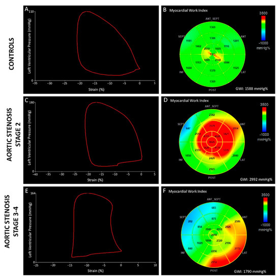
Prognostic Value of Non-Invasive Global Myocardial Work in Asymptomatic Aortic Stenosis
by Federica Ilardi, Adriana Postolache, Raluca Dulgheru, Mai-Linh Nguyen Trung, Nils de Marneffe, Tadafumi Sugimoto, Yun Yun Go, Cécile Oury, Giovanni Esposito and Patrizio Lancellotti
J. Clin. Med. 2022, 11(6), 1555; https://doi.org/10.3390/jcm11061555 - 11 Mar 2022
Cited by 12 | Viewed by 2498
Abstract
This study aimed to evaluate the modification of non-invasive myocardial work (MW) indices related to aortic stenosis (AS) stages of cardiac damage and their prognostic value. The echocardiographic and outcome data of 170 patients, with asymptomatic moderate-to-severe AS and left ventricular ejection fraction [...] Read more.
This study aimed to evaluate the modification of non-invasive myocardial work (MW) indices related to aortic stenosis (AS) stages of cardiac damage and their prognostic value. The echocardiographic and outcome data of 170 patients, with asymptomatic moderate-to-severe AS and left ventricular ejection fraction (LVEF) ≥ 50%, and 50 age- and sex-comparable healthy controls were analysed. Primary endpoints were the occurrence of all-cause and cardiovascular death. Increased values of the global work index (GWI), global constructive work (GCW), and global wasted work (GWW) were observed in AS patients compared to controls (GWI: 2528 ± 521 vs. 2005 ± 302 mmHg%, GCW: 2948 ± 598 vs. 2360 ± 353 mmHg%, p < 0.001; GWW: 139 ± 90 vs. 90 ± 49 mmHg%, p = 0.005), with no changes in the global work efficiency. When patients were stratified according to the stages of cardiac damage, the GWI showed lower values in Stage 3–4 as compared to Stage 0 and Stage 2 (p = 0.024). During a mean follow-up of 30 months, 27 patients died. In multivariable Cox-regression analysis, adjusted for confounders, GWI (HR: 0.998, CI: 0.997–1.000; p = 0.034) and GCW (HR:0.998, CI: 0.997–0.999; p = 0.003) were significantly associated with excess mortality. When used as categorical variables, a GWI ≤ 1951 mmHg% and a GCW ≤ 2475 mmHg% accurately predicted all-cause and cardiovascular death at 4-year follow-up. In conclusion, in asymptomatic patients with moderate-to-severe AS, reduced values of GWI and GCW are associated with increased mortality. Therefore, the evaluation of MW indices may allow for a better identification of asymptomatic patients with moderate to severe AS and preserved LVEF whom are at increased risk of worse prognosis during follow-up. Full article
(This article belongs to the Special Issue The Development and Application of Echocardiography in Heart Disease)
Show Figures

Figure 1

12 pages, 1681 KiB  
Article
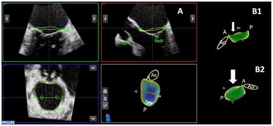
3D Echo Characterization of Proportionate and Disproportionate Functional Mitral Regurgitation before and after Percutaneous Mitral Valve Repair
by Sara Cimino, Luciano Agati, Domenico Filomena, Viviana Maestrini, Sara Monosilio, Lucia Ilaria Birtolo, Michele Mocci, Massimo Mancone, Gennaro Sardella, Paul Grayburn and Francesco Fedele
J. Clin. Med. 2022, 11(3), 645; https://doi.org/10.3390/jcm11030645 - 27 Jan 2022
Cited by 5 | Viewed by 2312
Abstract
Background: The impact of percutaneous mitral valve repair (PMVr) on long-term prognosis in patients with functional mitral regurgitation (FMR) is still unclear. Recently, a new conceptual framework classifying FMR as proportionate (P-MR) and disproportionate (D-MR) was proposed, according to the effective regurgitant orifice [...] Read more.
Background: The impact of percutaneous mitral valve repair (PMVr) on long-term prognosis in patients with functional mitral regurgitation (FMR) is still unclear. Recently, a new conceptual framework classifying FMR as proportionate (P-MR) and disproportionate (D-MR) was proposed, according to the effective regurgitant orifice area/left ventricular end-diastolic volume (EROA/LVEDV) ratio. The aim was to assess its possible influence on PMVr efficacy. Methods: A total of 56 patients were enrolled. MV annulus, LV volumes and function were assessed. Global longitudinal strain (GLS) was also calculated. Patients were divided into two groups, according to the EROA/LVEDV ratio. Echocardiographic follow-up was performed after 6 months, and adverse events were collected after 12 months. Results: D-MR patients (n = 28, 50%) had a significantly more elliptical MV annulus (p = 0.048), lower tenting volume (p = 0.01), higher LV ejection fraction (LVEF: 32 ± 7 vs. 26 ± 5%, p = 0.003), lower LVEDV, LV end-systolic volume (LVESV) and mass (LVEDV/i: 80 ± 20 vs. 126 ± 27 mL, p = 0.001; LVESV/i: 60 ± 20 vs. 94 ± 23 mL, p < 0.001; LV mass: 249 ± 63 vs. 301 ± 69 gr, p = 0.035). GLS was more impaired in P-MR (p = 0.048). After 6 months, P-MR patients showed a higher rate of MR recurrence. After 12 months, the rate of CV death and rehospitalization due to HF was significantly higher in P-MR patients (46% vs. 7%, p < 0.001). P-MR status was strongly associated with CV death/rehospitalization (HR = 3.4, CI 95% = 1.3–8.6, p = 0.009). Conclusions: Patients with P-MR seem to have worse outcomes after PVMr than D-MR patients. Our study confirms the importance of the EROA/LVEDV ratio in defining different subsets of FMR based on the anatomical characteristic of MV and LV. Full article
(This article belongs to the Special Issue The Development and Application of Echocardiography in Heart Disease)
Show Figures

Figure 1

10 pages, 1097 KiB  
Article
Extent of Cardiac Damage and Mortality in Patients Undergoing Transcatheter Aortic Valve Implantation
by Marisa Avvedimento, Anna Franzone, Attilio Leone, Raffaele Piccolo, Domenico Simone Castiello, Federica Ilardi, Andrea Mariani, Roberta Esposito, Cristina Iapicca, Domenico Angellotti, Maria Scalamogna, Ciro Santoro, Luigi Di Serafino, Plinio Cirillo and Giovanni Esposito
J. Clin. Med. 2021, 10(19), 4563; https://doi.org/10.3390/jcm10194563 - 30 Sep 2021
Cited by 18 | Viewed by 2249
Abstract
(1) Aims: We sought to assess the impact of the extent of cardiac damage on survival among real-world patients with severe aortic stenosis (AS) undergoing transcatheter aortic valve implantation (TAVI). (2) Methods: A staging classification was applied to 262 patients from the EffecTAVI [...] Read more.
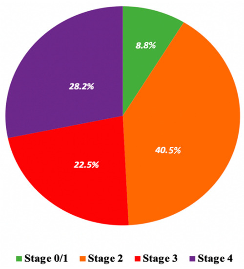
(1) Aims: We sought to assess the impact of the extent of cardiac damage on survival among real-world patients with severe aortic stenosis (AS) undergoing transcatheter aortic valve implantation (TAVI). (2) Methods: A staging classification was applied to 262 patients from the EffecTAVI Registry at baseline and re-assessed within 30-days after TAVI. The primary endpoint of the study was all-cause mortality at 1-year. Secondary endpoints included cerebrovascular accident, myocardial infarction, permanent pacemaker implantation, endocarditis, and re-hospitalization for all causes. (3) Results: At baseline, 23 (8.7%) patients were in Stage 0/1 (no cardiac damage/left ventricular damage), 106 (40.4%) in Stage 2 (left atrial or mitral valve damage), 59 (22.5%) in Stage 3 (pulmonary vasculature or tricuspid valve damage) and 74 (28.3%) in Stage 4 (right ventricular damage). At 30-days after TAVI, a lower prevalence of advanced stages of cardiac damage than baseline, mainly driven by a significant improvement in left ventricular diastolic parameters and right ventricular function, was reported. At 1-year, a stepwise increase in mortality rates was observed according to staging at baseline: 4.3% in Stage 0/1, 6.6% in Stage 2, 18.6% in Stage 3 and 21.6% in Stage 4 (p = 0.08). No differences were found in secondary endpoints. (4) Conclusions: TAVI has an early beneficial impact on the left ventricular diastolic and right ventricular function. However, the extent of cardiac damage at baseline significantly affects the risk of mortality at 1-year after the procedure. Full article
(This article belongs to the Special Issue The Development and Application of Echocardiography in Heart Disease)
Show Figures

Figure 1

Review

Jump to: Research

15 pages, 2054 KiB  
Review
Clinical Usefulness of Right Ventricle–Pulmonary Artery Coupling in Cardiovascular Disease
by Qing He, Yixia Lin, Ye Zhu, Lang Gao, Mengmeng Ji, Li Zhang, Mingxing Xie and Yuman Li
J. Clin. Med. 2023, 12(7), 2526; https://doi.org/10.3390/jcm12072526 - 27 Mar 2023
Cited by 18 | Viewed by 3837
Abstract
Right ventricular–pulmonary artery coupling (RV-PA coupling) refers to the relationship between RV contractility and RV afterload. Normal RV-PA coupling is maintained only when RV function and pulmonary vascular resistance are appropriately matched. RV-PA uncoupling occurs when RV contractility cannot increase to match RV [...] Read more.
Right ventricular–pulmonary artery coupling (RV-PA coupling) refers to the relationship between RV contractility and RV afterload. Normal RV-PA coupling is maintained only when RV function and pulmonary vascular resistance are appropriately matched. RV-PA uncoupling occurs when RV contractility cannot increase to match RV afterload, resulting in RV dysfunction and right heart failure. RV-PA coupling plays an important role in the pathophysiology and progression of cardiovascular diseases. Therefore, early and accurate evaluation of RV-PA coupling is of great significance for a patient’s condition assessment, clinical decision making, risk stratification, and prognosis judgment. RV-PA coupling can be assessed by using invasive or noninvasive approaches. The aim of this review was to summarize the pathological mechanism and evaluation methods of RV-PA coupling, the advantages and disadvantages of each method, and the application value of RV-PA coupling in various cardiovascular diseases. Full article
(This article belongs to the Special Issue The Development and Application of Echocardiography in Heart Disease)
Show Figures

Figure 1

12 pages, 1769 KiB  
Review
Echocardiographic Features of Cardiac Involvement in Myotonic Dystrophy 1: Prevalence and Prognostic Value
by Vincenzo Russo, Antonio Capolongo, Roberta Bottino, Andreina Carbone, Alberto Palladino, Biagio Liccardo, Gerardo Nigro, Michał Marchel, Paolo Golino and Antonello D’Andrea
J. Clin. Med. 2023, 12(5), 1947; https://doi.org/10.3390/jcm12051947 - 1 Mar 2023
Cited by 4 | Viewed by 2387
Abstract
Myotonic dystrophy type 1 (DM1) is the most common muscular dystrophy in adults. Cardiac involvement is reported in 80% of cases and includes conduction disturbances, arrhythmias, subclinical diastolic and systolic dysfunction in the early stage of the disease; in contrast, severe ventricular systolic [...] Read more.
Myotonic dystrophy type 1 (DM1) is the most common muscular dystrophy in adults. Cardiac involvement is reported in 80% of cases and includes conduction disturbances, arrhythmias, subclinical diastolic and systolic dysfunction in the early stage of the disease; in contrast, severe ventricular systolic dysfunction occurs in the late stage of the disease. Echocardiography is recommended at the time of diagnosis with periodic revaluation in DM1 patients, regardless of the presence or absence of symptoms. Data regarding the echocardiographic findings in DM1 patients are few and conflicting. This narrative review aimed to describe the echocardiographic features of DM1 patients and their prognostic role as predictors of cardiac arrhythmias and sudden death. Full article
(This article belongs to the Special Issue The Development and Application of Echocardiography in Heart Disease)
Show Figures

Figure 1

21 pages, 13218 KiB  
Review
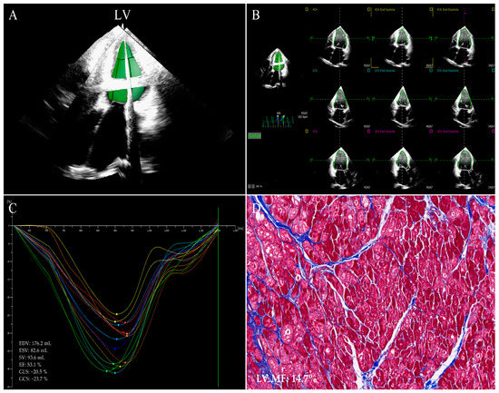
Clinical Utility of Strain Imaging in Assessment of Myocardial Fibrosis
by Lang Gao, Li Zhang, Zisang Zhang, Yixia Lin, Mengmeng Ji, Qing He, Mingxing Xie and Yuman Li
J. Clin. Med. 2023, 12(3), 743; https://doi.org/10.3390/jcm12030743 - 17 Jan 2023
Cited by 5 | Viewed by 2588
Abstract
Myocardial fibrosis (MF) is a non-reversible process that occurs following acute or chronic myocardial damage. MF worsens myocardial deformation, remodels the heart and raises myocardial stiffness, and is a crucial pathological manifestation in patients with end-stage cardiovascular diseases and closely related to cardiac [...] Read more.
Myocardial fibrosis (MF) is a non-reversible process that occurs following acute or chronic myocardial damage. MF worsens myocardial deformation, remodels the heart and raises myocardial stiffness, and is a crucial pathological manifestation in patients with end-stage cardiovascular diseases and closely related to cardiac adverse events. Therefore, early quantitative analysis of MF plays an important role in risk stratification, clinical decision, and improvement in prognosis. With the advent and development of strain imaging modalities in recent years, MF may be detected early in cardiovascular diseases. This review summarizes the clinical usefulness of strain imaging techniques in the non-invasive assessment of MF. Full article
(This article belongs to the Special Issue The Development and Application of Echocardiography in Heart Disease)
Show Figures

Figure 1

12 pages, 696 KiB  
Review
Post-Acute Sequelae of COVID-19: The Potential Role of Exercise Therapy in Treating Patients and Athletes Returning to Play
by Luna Cavigli, Chiara Fusi, Marta Focardi, Giulia Elena Mandoli, Maria Concetta Pastore, Matteo Cameli, Serafina Valente, Alessandro Zorzi, Marco Bonifazi, Antonello D’Andrea and Flavio D’Ascenzi
J. Clin. Med. 2023, 12(1), 288; https://doi.org/10.3390/jcm12010288 - 30 Dec 2022
Cited by 7 | Viewed by 3935
Abstract
Post-acute sequelae of coronavirus disease 19 (COVID-19) (PASC) describe a wide range of symptoms and signs involving multiple organ systems occurring after severe acute respiratory syndrome coronavirus 2 (SARS-CoV-2) infection, representing a growing health problem also in the world of sport and the [...] Read more.
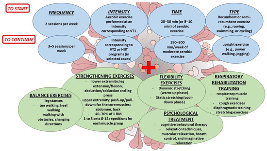
Post-acute sequelae of coronavirus disease 19 (COVID-19) (PASC) describe a wide range of symptoms and signs involving multiple organ systems occurring after severe acute respiratory syndrome coronavirus 2 (SARS-CoV-2) infection, representing a growing health problem also in the world of sport and the athletic population. Patients with PASC have new, returning, or persisting symptoms four or more weeks after the infection. Among the most frequent symptoms, patients complain of fatigue, dyspnea, exercise intolerance, and reduced functional capacity that interfere with everyday life activity. The role of exercise programs in PASC patients will be identified, and upcoming studies will establish the magnitude of their benefits. However, the benefits of exercise to counteract these symptoms are well known, and an improvement in cardiopulmonary fitness, functional status, deconditioning, and quality of life can be obtained in these patients, as demonstrated in similar settings. Based on this background, this review aims to summarise the current evidence about the PASC syndrome and the benefit of exercise in these patients and to provide a practical guide for the exercise prescription in PASC patients to help them to resume their functional status, exercise tolerance, prior activity levels, and quality of life, also considering the athletic population and their return to play and sports competitions. Full article
(This article belongs to the Special Issue The Development and Application of Echocardiography in Heart Disease)
Show Figures

Figure 1

16 pages, 5110 KiB  
Review
Bedside Ultrasound for Hemodynamic Monitoring in Cardiac Intensive Care Unit
by Maria Concetta Pastore, Federica Ilardi, Andrea Stefanini, Giulia Elena Mandoli, Stefano Palermi, Francesco Bandera, Giovanni Benfari, Roberta Esposito, Matteo Lisi, Annalisa Pasquini, Ciro Santoro, Serafina Valente, Antonello D’Andrea and Matteo Cameli
J. Clin. Med. 2022, 11(24), 7538; https://doi.org/10.3390/jcm11247538 - 19 Dec 2022
Cited by 6 | Viewed by 4339
Abstract
Thanks to the advances in medical therapy and assist devices, the management of patients hospitalized in cardiac intensive care unit (CICU) is becoming increasingly challenging. In fact, Patients in the cardiac intensive care unit are frequently characterized by dynamic and variable diseases, which [...] Read more.
Thanks to the advances in medical therapy and assist devices, the management of patients hospitalized in cardiac intensive care unit (CICU) is becoming increasingly challenging. In fact, Patients in the cardiac intensive care unit are frequently characterized by dynamic and variable diseases, which may evolve into several clinical phenotypes based on underlying etiology and its complexity. Therefore, the use of noninvasive tools in order to provide a personalized approach to these patients, according to their phenotype, may help to optimize the therapeutic strategies towards the underlying etiology. Echocardiography is the most reliable and feasible bedside method to assess cardiac function repeatedly, assisting clinicians not only in characterizing hemodynamic disorders, but also in helping to guide interventions and monitor response to therapies. Beyond basic echocardiographic parameters, its application has been expanded with the introduction of new tools such as lung ultrasound (LUS), the Venous Excess UltraSound (VexUS) grading system, and the assessment of pulmonary hypertension, which is fundamental to guide oxygen therapy. The aim of this review is to provide an overview on the current knowledge about the pathophysiology and echocardiographic evaluation of perfusion and congestion in patients in CICU, and to provide practical indications for the use of echocardiography across clinical phenotypes and new applications in CICU. Full article
(This article belongs to the Special Issue The Development and Application of Echocardiography in Heart Disease)
Show Figures

Figure 1

17 pages, 1774 KiB  
Review
Clinical Utility of Three-Dimensional Speckle-Tracking Echocardiography in Heart Failure
by Lang Gao, Yixia Lin, Mengmeng Ji, Wenqian Wu, He Li, Mingzhu Qian, Li Zhang, Mingxing Xie and Yuman Li
J. Clin. Med. 2022, 11(21), 6307; https://doi.org/10.3390/jcm11216307 - 26 Oct 2022
Cited by 22 | Viewed by 3457
Abstract
Heart failure (HF) is an extremely major health problem with gradually increasing incidence in developed and developing countries. HF may lead to cardiac remodeling; thus, advanced imaging techniques are required to comprehensively evaluate cardiac mechanics. Recently, three-dimensional speckle-tracking echocardiography (3D-STE) has been developed [...] Read more.
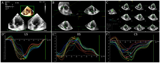
Heart failure (HF) is an extremely major health problem with gradually increasing incidence in developed and developing countries. HF may lead to cardiac remodeling; thus, advanced imaging techniques are required to comprehensively evaluate cardiac mechanics. Recently, three-dimensional speckle-tracking echocardiography (3D-STE) has been developed as a novel imaging technology that is based on the three-dimensional speckle-tracking on the full volume three-dimensional datasets. Three-dimensional speckle-tracking echocardiography allows a more accurate evaluation of global and regional myocardial performance, assessment of cardiac mechanics, detection of subclinical cardiac dysfunction, and prediction of adverse clinical events in a variety of cardiovascular diseases. Therefore, this review summarizes the clinical usefulness of 3D-STE in patients with HF. Full article
(This article belongs to the Special Issue The Development and Application of Echocardiography in Heart Disease)
Show Figures

Figure 1

12 pages, 1290 KiB  
Review
Artificial Intelligence-Enhanced Echocardiography for Systolic Function Assessment
by Zisang Zhang, Ye Zhu, Manwei Liu, Ziming Zhang, Yang Zhao, Xin Yang, Mingxing Xie and Li Zhang
J. Clin. Med. 2022, 11(10), 2893; https://doi.org/10.3390/jcm11102893 - 20 May 2022
Cited by 9 | Viewed by 3271
Abstract
The accurate assessment of left ventricular systolic function is crucial in the diagnosis and treatment of cardiovascular diseases. Left ventricular ejection fraction (LVEF) and global longitudinal strain (GLS) are the most critical indexes of cardiac systolic function. Echocardiography has become the mainstay of [...] Read more.
The accurate assessment of left ventricular systolic function is crucial in the diagnosis and treatment of cardiovascular diseases. Left ventricular ejection fraction (LVEF) and global longitudinal strain (GLS) are the most critical indexes of cardiac systolic function. Echocardiography has become the mainstay of cardiac imaging for measuring LVEF and GLS because it is non-invasive, radiation-free, and allows for bedside operation and real-time processing. However, the human assessment of cardiac function depends on the sonographer’s experience, and despite their years of training, inter-observer variability exists. In addition, GLS requires post-processing, which is time consuming and shows variability across different devices. Researchers have turned to artificial intelligence (AI) to address these challenges. The powerful learning capabilities of AI enable feature extraction, which helps to achieve accurate identification of cardiac structures and reliable estimation of the ventricular volume and myocardial motion. Hence, the automatic output of systolic function indexes can be achieved based on echocardiographic images. This review attempts to thoroughly explain the latest progress of AI in assessing left ventricular systolic function and differential diagnosis of heart diseases by echocardiography and discusses the challenges and promises of this new field. Full article
(This article belongs to the Special Issue The Development and Application of Echocardiography in Heart Disease)
Show Figures

Figure 1

16 pages, 1855 KiB  
Review
Myocardial Work by Echocardiography: Principles and Applications in Clinical Practice
by Federica Ilardi, Antonello D’Andrea, Flavio D’Ascenzi, Francesco Bandera, Giovanni Benfari, Roberta Esposito, Alessandro Malagoli, Giulia Elena Mandoli, Ciro Santoro, Vincenzo Russo, Mario Crisci, Giovanni Esposito, Matteo Cameli and on behalf of the Working Group of Echocardiography of the Italian Society of Cardiology (SIC)
J. Clin. Med. 2021, 10(19), 4521; https://doi.org/10.3390/jcm10194521 - 29 Sep 2021
Cited by 55 | Viewed by 6495
Abstract
Left ventricular (LV) global longitudinal strain (GLS) has established itself in the last decade as a reliable, more objective method for the evaluation of LV systolic function, able to detect subtle abnormalities in LV contraction even in the presence of preserved ejection fraction [...] Read more.
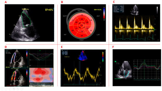
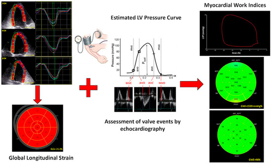
Left ventricular (LV) global longitudinal strain (GLS) has established itself in the last decade as a reliable, more objective method for the evaluation of LV systolic function, able to detect subtle abnormalities in LV contraction even in the presence of preserved ejection fraction (EF). However, recent studies have demonstrated that GLS, similar to LV EF, has important load dependency. Non-invasive myocardial work (MW) quantification has emerged in the last years as an alternative tool for myocardial function assessment. This new method, incorporating measurement of strain and LV pressure, has shown to overcome GLS and LV EF limitations and provide a loading-independent evaluation of myocardial performance. The presence of a commercially available echocardiographic software for the non-invasive MW calculation has allowed the application of this new method in different settings. This review sought to provide an overview on the current knowledge of non-invasive MW estimation, showing its potential applications and possible added value in clinical practice. Full article
(This article belongs to the Special Issue The Development and Application of Echocardiography in Heart Disease)
Show Figures

Figure 1

Back to TopTop