Topical Advisory Panel applications are now closed. Please contact the Editorial Office with any queries.
Journal Description
Journal of Developmental Biology
Journal of Developmental Biology
is an international, peer-reviewed, open access journal on the development of multicellular organisms at the molecule, cell, tissue, organ and whole organism levels published quarterly online by MDPI.
- Open Access— free for readers, with article processing charges (APC) paid by authors or their institutions.
- High Visibility: indexed within Scopus, ESCI (Web of Science), PubMed, PMC, PubAg, CAPlus / SciFinder, and other databases.
- Rapid Publication: manuscripts are peer-reviewed and a first decision is provided to authors approximately 19.6 days after submission; acceptance to publication is undertaken in 4.9 days (median values for papers published in this journal in the first half of 2024).
- Recognition of Reviewers: reviewers who provide timely, thorough peer-review reports receive vouchers entitling them to a discount on the APC of their next publication in any MDPI journal, in appreciation of the work done.
- Testimonials: See what our editors and authors say about Journal of Developmental Biology.
Impact Factor:
2.2 (2023);
5-Year Impact Factor:
2.6 (2023)
Latest Articles
Prosaposin/Saposin Expression in the Developing Rat Olfactory and Vomeronasal Epithelia
J. Dev. Biol. 2024, 12(4), 29; https://doi.org/10.3390/jdb12040029 - 6 Nov 2024
Abstract
►
Show Figures
Prosaposin is a glycoprotein widely conserved in vertebrates, and it acts as a precursor for saposins that accelerate hydrolysis in lysosomes or acts as a neurotrophic factor without being processed into saposins. Neurogenesis in the olfactory neuroepithelia, including the olfactory epithelium (OE) and
[...] Read more.
Prosaposin is a glycoprotein widely conserved in vertebrates, and it acts as a precursor for saposins that accelerate hydrolysis in lysosomes or acts as a neurotrophic factor without being processed into saposins. Neurogenesis in the olfactory neuroepithelia, including the olfactory epithelium (OE) and the vomeronasal epithelium (VNE), is known to occur throughout an animal’s life, and mature olfactory neurons (ORNs) and vomeronasal receptor neurons (VRNs) have recently been revealed to express prosaposin in the adult olfactory organ. In this study, the expression of prosaposin in the rat olfactory organ during postnatal development was examined. In the OE, prosaposin immunoreactivity was observed in mature ORNs labeled using olfactory marker protein (OMP) from postnatal day (P) 0. Immature ORNs showed no prosaposin immunoreactivity throughout the examined period. In the VNE, OMP-positive VRNs were mainly observed in the basal region of the VNE on P10 and showed an adult-like distribution from P20. On the other hand, prosaposin immunoreactivity was observed in VRNs from P0, suggesting that not only mature VRNs but also immature VRNs express prosaposin. This study raises the possibility that prosaposin is required for the normal development of the olfactory organ and has different roles in the OE and the VNE.
Full article
Open AccessReview
How the Oocyte Nucleolus Is Turned into a Karyosphere: The Role of Heterochromatin and Structural Proteins
by
Venera Nikolova, Maya Markova, Ralitsa Zhivkova, Irina Chakarova, Valentina Hadzhinesheva and Stefka Delimitreva
J. Dev. Biol. 2024, 12(4), 28; https://doi.org/10.3390/jdb12040028 - 18 Oct 2024
Abstract
Oocyte meiotic maturation includes large-scale chromatin remodeling as well as cytoskeleton and nuclear envelope rearrangements. This review addresses the dynamics of key cytoskeletal proteins (tubulin, actin, vimentin, and cytokeratins) and nuclear envelope proteins (lamin A/C, lamin B, and the nucleoporin Nup160) in parallel
[...] Read more.
Oocyte meiotic maturation includes large-scale chromatin remodeling as well as cytoskeleton and nuclear envelope rearrangements. This review addresses the dynamics of key cytoskeletal proteins (tubulin, actin, vimentin, and cytokeratins) and nuclear envelope proteins (lamin A/C, lamin B, and the nucleoporin Nup160) in parallel with chromatin reorganization in maturing mouse oocytes. A major feature of this reorganization is the concentration of heterochromatin into a spherical perinucleolar rim called surrounded nucleolus or karyosphere. In early germinal vesicle (GV) oocytes with non-surrounded nucleolus (without karyosphere), lamins and Nup160 are at the nuclear envelope while cytoplasmic cytoskeletal proteins are outside the nucleus. At the beginning of karyosphere formation, lamins and Nup160 follow the heterochromatin relocation assembling a new spherical structure in the GV. In late GV oocytes with surrounded nucleolus (fully formed karyosphere), the nuclear envelope gradually loses its integrity and cytoplasmic cytoskeletal proteins enter the nucleus. At germinal vesicle breakdown, lamin B occupies the karyosphere interior while all the other proteins stay at the karyosphere border or connect to chromatin. In metaphase oocytes, lamin A/C surrounds the spindle, Nup160 localizes to its poles, actin and lamin B are attached to the spindle fibers, and cytoplasmic intermediate filaments associate with both the spindle fibers and the metaphase chromosomes.
Full article
(This article belongs to the Special Issue Feature Papers from Journal of Developmental Biology Reviewers)
►▼
Show Figures

Figure 1
Open AccessReview
Neural Circuit Remodeling: Mechanistic Insights from Invertebrates
by
Samuel Liu, Kellianne D. Alexander and Michael M. Francis
J. Dev. Biol. 2024, 12(4), 27; https://doi.org/10.3390/jdb12040027 - 11 Oct 2024
Abstract
As nervous systems mature, neural circuit connections are reorganized to optimize the performance of specific functions in adults. This reorganization of connections is achieved through a remarkably conserved phase of developmental circuit remodeling that engages neuron-intrinsic and neuron-extrinsic molecular mechanisms to establish mature
[...] Read more.
As nervous systems mature, neural circuit connections are reorganized to optimize the performance of specific functions in adults. This reorganization of connections is achieved through a remarkably conserved phase of developmental circuit remodeling that engages neuron-intrinsic and neuron-extrinsic molecular mechanisms to establish mature circuitry. Abnormalities in circuit remodeling and maturation are broadly linked with a variety of neurodevelopmental disorders, including autism spectrum disorders and schizophrenia. Here, we aim to provide an overview of recent advances in our understanding of the molecular processes that govern neural circuit remodeling and maturation. In particular, we focus on intriguing mechanistic insights gained from invertebrate systems, such as the nematode Caenorhabditis elegans and the fruit fly Drosophila melanogaster. We discuss how transcriptional control mechanisms, synaptic activity, and glial engulfment shape specific aspects of circuit remodeling in worms and flies. Finally, we highlight mechanistic parallels across invertebrate and mammalian systems, and prospects for further advances in each.
Full article
(This article belongs to the Special Issue Feature Papers from Journal of Developmental Biology Reviewers)
►▼
Show Figures

Figure 1
Open AccessArticle
Delayed Blastocyst Formation Reduces the Quality and Hatching Ability of Porcine Parthenogenetic Blastocysts by Increasing DNA Damage, Decreasing Cell Proliferation, and Altering Transcription Factor Expression Patterns
by
Ling Sun, Yan Wang, Mo Yang, Zhuang-Ju Xu, Juan Miao, Ying Bai and Tao Lin
J. Dev. Biol. 2024, 12(4), 26; https://doi.org/10.3390/jdb12040026 - 1 Oct 2024
Abstract
The purpose of this study was to investigate the influence of blastocyst formation timing on the quality of porcine embryos derived from parthenogenetic activation. Newly formed blastocysts at days 6, 7, and 8 of culture [termed formation 6, 7, and 8 blastocysts (F6,
[...] Read more.
The purpose of this study was to investigate the influence of blastocyst formation timing on the quality of porcine embryos derived from parthenogenetic activation. Newly formed blastocysts at days 6, 7, and 8 of culture [termed formation 6, 7, and 8 blastocysts (F6, F7, and F8 blastocysts)] were obtained, and a series of parameters related to the quality of blastocysts, including apoptosis incidents, DNA replication, pluripotent factors, and blastocyst hatching capacity, were assessed. Delayed blastocyst formation (F7 and/or F8 blastocysts) led to increased levels of ROS, DNA damage, and apoptosis while decreasing the mitochondrial membrane potential, DNA replication, Oct4 levels, and numbers of Sox2-positive cells. F7 blastocysts showed a significantly reduced hatching rate compared to F6 blastocysts; however, F8 blastocysts were unable to develop to the hatching stage. Collectively, our findings suggest a negative correlation between delayed blastocyst formation and blastocyst quality.
Full article
(This article belongs to the Special Issue Feature Papers from Journal of Developmental Biology Reviewers)
►▼
Show Figures

Figure 1
Open AccessReview
Myotube Guidance: Shaping up the Musculoskeletal System
by
Aaron N. Johnson
J. Dev. Biol. 2024, 12(3), 25; https://doi.org/10.3390/jdb12030025 - 17 Sep 2024
Abstract
►▼
Show Figures
Myofibers are highly specialized contractile cells of skeletal muscles, and dysregulation of myofiber morphogenesis is emerging as a contributing cause of myopathies and structural birth defects. Myotubes are the myofiber precursors and undergo a dramatic morphological transition into long bipolar myofibers that are
[...] Read more.
Myofibers are highly specialized contractile cells of skeletal muscles, and dysregulation of myofiber morphogenesis is emerging as a contributing cause of myopathies and structural birth defects. Myotubes are the myofiber precursors and undergo a dramatic morphological transition into long bipolar myofibers that are attached to tendons on two ends. Similar to axon growth cones, myotube leading edges navigate toward target cells and form cell–cell connections. The process of myotube guidance connects myotubes with the correct tendons, orients myofiber morphology with the overall body plan, and generates a functional musculoskeletal system. Navigational signaling, addition of mass and volume, and identification of target cells are common events in myotube guidance and axon guidance, but surprisingly, the mechanisms regulating these events are not completely overlapping in myotubes and axons. This review summarizes the strategies that have evolved to direct myotube leading edges to predetermined tendon cells and highlights key differences between myotube guidance and axon guidance. The association of myotube guidance pathways with developmental disorders is also discussed.
Full article

Figure 1
Open AccessReview
Roles of the NR2F Family in the Development, Disease, and Cancer of the Lung
by
Jiaxin Yang, Wenjing Sun and Guizhong Cui
J. Dev. Biol. 2024, 12(3), 24; https://doi.org/10.3390/jdb12030024 - 10 Sep 2024
Abstract
The NR2F family, including NR2F1, NR2F2, and NR2F6, belongs to the nuclear receptor superfamily. NR2F family members function as transcription factors and play essential roles in the development of multiple organs or tissues in mammals, including the central nervous system, veins and arteries,
[...] Read more.
The NR2F family, including NR2F1, NR2F2, and NR2F6, belongs to the nuclear receptor superfamily. NR2F family members function as transcription factors and play essential roles in the development of multiple organs or tissues in mammals, including the central nervous system, veins and arteries, kidneys, uterus, and vasculature. In the central nervous system, NR2F1/2 coordinate with each other to regulate the development of specific brain subregions or cell types. In addition, NR2F family members are associated with various cancers, such as prostate cancer, breast cancer, and esophageal cancer. Nonetheless, the roles of the NR2F family in the development and diseases of the lung have not been systematically summarized. In this review, we mainly focus on the lung, including recent findings regarding the roles of the NR2F family in development, physiological function, and cancer.
Full article
(This article belongs to the Special Issue The 10th Anniversary of JDB: Feature Papers)
►▼
Show Figures

Figure 1
Open AccessArticle
Evolution and Spatiotemporal Expression of ankha and ankhb in Zebrafish
by
Nuwanthika Wathuliyadde, Katherine E. Willmore and Gregory M. Kelly
J. Dev. Biol. 2024, 12(3), 23; https://doi.org/10.3390/jdb12030023 - 9 Sep 2024
Abstract
Craniometaphyseal Dysplasia (CMD) is a rare skeletal disorder that can result from mutations in the ANKH gene. This gene encodes progressive anksylosis (ANK), which is responsible for transporting inorganic pyrophosphate (PPi) and ATP from the intracellular to the extracellular environment, where PPi inhibits
[...] Read more.
Craniometaphyseal Dysplasia (CMD) is a rare skeletal disorder that can result from mutations in the ANKH gene. This gene encodes progressive anksylosis (ANK), which is responsible for transporting inorganic pyrophosphate (PPi) and ATP from the intracellular to the extracellular environment, where PPi inhibits bone mineralization. When ANK is dysfunctional, as in patients with CMD, the passage of PPi to the extracellular environment is reduced, leading to excess mineralization, particularly in bones of the skull. Zebrafish may serve as a promising model to study the mechanistic basis of CMD. Here, we provide a detailed analysis of the zebrafish Ankh paralogs, Ankha and Ankhb, in terms of their phylogenic relationship with ANK in other vertebrates as well as their spatiotemporal expression patterns during zebrafish development. We found that a closer evolutionary relationship exists between the zebrafish Ankhb protein and its human and other vertebrate counterparts, and stronger promoter activity was predicted for ankhb compared to ankha. Furthermore, we noted distinct temporal expression patterns, with ankha more prominently expressed in early development stages, and both paralogs also being expressed at larval growth stages. Whole-mount in situ hybridization was used to compare the spatial expression patterns of each paralog during bone development, and both showed strong expression in the craniofacial region as well as the notochord and somites. Given the substantial overlap in spatiotemporal expression but only subtle patterning differences, the exact roles of these genes remain speculative. In silico analyses predicted that Ankha and Ankhb have the same function in transporting PPi across the membrane. Nevertheless, this study lays the groundwork for functional analyses of each ankh paralog and highlights the potential of using zebrafish to find possible targeted therapies for CMD.
Full article
(This article belongs to the Special Issue The 10th Anniversary of JDB: Feature Papers)
►▼
Show Figures

Figure 1
Open AccessReview
From Germ Cells to Implantation: The Role of Extracellular Vesicles
by
Anna Fazzio, Angela Caponnetto, Carmen Ferrara, Michele Purrello, Cinzia Di Pietro and Rosalia Battaglia
J. Dev. Biol. 2024, 12(3), 22; https://doi.org/10.3390/jdb12030022 - 23 Aug 2024
Abstract
►▼
Show Figures
Extracellular vesicles represent a large heterogeneous class of near and long-distance intercellular communication mediators, released by both prokaryotic and eukaryotic cells. Specifically, the scientific community has shown growing interest in exosomes, which are nano-sized vesicles with an endosomal origin. Not so long ago,
[...] Read more.
Extracellular vesicles represent a large heterogeneous class of near and long-distance intercellular communication mediators, released by both prokaryotic and eukaryotic cells. Specifically, the scientific community has shown growing interest in exosomes, which are nano-sized vesicles with an endosomal origin. Not so long ago, the physiological goal of exosome generation was largely unknown and required more investigation; at first, it was hypothesized that exosomes are able to remove excess, reject and unnecessary constituents from cells to preserve cellular homeostasis. However, thanks to recent studies, the central role of exosomes in regulating cellular communication has emerged. Exosomes act as vectors in cell–cell signaling by their cargo, proteins, lipids, and nucleic acids, and influence physiological and pathological processes. The findings on exosomes are widespread in a large spectrum of biomedical applications from diagnosis and prognosis to therapies. In this review, we describe exosome biogenesis and the current methods for their isolation and characterization, emphasizing the role of their cargo in female reproductive processes, from gametogenesis to implantation, and the potential involvement in human female disorders.
Full article

Graphical abstract
Open AccessArticle
Lowered GnT-I Activity Decreases Complex-Type N-Glycan Amounts and Results in an Aberrant Primary Motor Neuron Structure in the Spinal Cord
by
Cody J. Hatchett, M. Kristen Hall, Abel R. Messer and Ruth A. Schwalbe
J. Dev. Biol. 2024, 12(3), 21; https://doi.org/10.3390/jdb12030021 - 16 Aug 2024
Abstract
►▼
Show Figures
The attachment of sugar to proteins and lipids is a basic modification needed for organismal survival, and perturbations in glycosylation cause severe developmental and neurological difficulties. Here, we investigated the neurological consequences of N-glycan populations in the spinal cord of Wt AB and
[...] Read more.
The attachment of sugar to proteins and lipids is a basic modification needed for organismal survival, and perturbations in glycosylation cause severe developmental and neurological difficulties. Here, we investigated the neurological consequences of N-glycan populations in the spinal cord of Wt AB and mgat1b mutant zebrafish. Mutant fish have reduced N-acetylglucosaminyltransferase-I (GnT-I) activity as mgat1a remains intact. GnT-I converts oligomannose N-glycans to hybrid N-glycans, which is needed for complex N-glycan production. MALDI-TOF MS profiles identified N-glycans in the spinal cord for the first time and revealed reduced amounts of complex N-glycans in mutant fish, supporting a lesion in mgat1b. Further lectin blotting showed that oligomannose N-glycans were more prevalent in the spinal cord, skeletal muscle, heart, swim bladder, skin, and testis in mutant fish relative to WT AB, supporting lowered GnT- I activity in a global manner. Developmental delays were noted in hatching and in the swim bladder. Microscopic images of caudal primary (CaP) motor neurons of the spinal cord transiently expressing EGFP in mutant fish were abnormal with significant reductions in collateral branches. Further motor coordination skills were impaired in mutant fish. We conclude that identifying the neurological consequences of aberrant N-glycan processing will enhance our understanding of the role of complex N-glycans in development and nervous system health.
Full article

Figure 1
Open AccessReview
Canonical and Non-Canonical Wnt Signaling Generates Molecular and Cellular Asymmetries to Establish Embryonic Axes
by
De-Li Shi
J. Dev. Biol. 2024, 12(3), 20; https://doi.org/10.3390/jdb12030020 - 2 Aug 2024
Abstract
The formation of embryonic axes is a critical step during animal development, which contributes to establishing the basic body plan in each particular organism. Wnt signaling pathways play pivotal roles in this fundamental process. Canonical Wnt signaling that is dependent on β-catenin regulates
[...] Read more.
The formation of embryonic axes is a critical step during animal development, which contributes to establishing the basic body plan in each particular organism. Wnt signaling pathways play pivotal roles in this fundamental process. Canonical Wnt signaling that is dependent on β-catenin regulates the patterning of dorsoventral, anteroposterior, and left–right axes. Non-canonical Wnt signaling that is independent of β-catenin modulates cytoskeletal organization to coordinate cell polarity changes and asymmetric cell movements. It is now well documented that components of these Wnt pathways biochemically and functionally interact to mediate cell–cell communications and instruct cellular polarization in breaking the embryonic symmetry. The dysfunction of Wnt signaling disrupts embryonic axis specification and proper tissue morphogenesis, and mutations of Wnt pathway genes are associated with birth defects in humans. This review discusses the regulatory roles of Wnt pathway components in embryonic axis formation by focusing on vertebrate models. It highlights current progress in decoding conserved mechanisms underlying the establishment of asymmetry along the three primary body axes. By providing an in-depth analysis of canonical and non-canonical pathways in regulating cell fates and cellular behaviors, this work offers insights into the intricate processes that contribute to setting up the basic body plan in vertebrate embryos.
Full article
(This article belongs to the Special Issue Feature Papers from Journal of Developmental Biology Reviewers)
►▼
Show Figures

Figure 1
Open AccessArticle
Genes Related to Frontonasal Malformations Are Regulated by miR-338-5p, miR-653-5p, and miR-374-5p in O9-1 Cells
by
Chihiro Iwaya, Sunny Yu and Junichi Iwata
J. Dev. Biol. 2024, 12(3), 19; https://doi.org/10.3390/jdb12030019 - 6 Jul 2024
Abstract
Frontonasal malformations are caused by a failure in the growth of the frontonasal prominence during development. Although genetic studies have identified genes that are crucial for frontonasal development, it remains largely unknown how these genes are regulated during this process. Here, we show
[...] Read more.
Frontonasal malformations are caused by a failure in the growth of the frontonasal prominence during development. Although genetic studies have identified genes that are crucial for frontonasal development, it remains largely unknown how these genes are regulated during this process. Here, we show that microRNAs, which are short non-coding RNAs capable of targeting their target mRNAs for degradation or silencing their expression, play a crucial role in the regulation of genes related to frontonasal development in mice. Using the Mouse Genome Informatics (MGI) database, we curated a total of 25 mouse genes related to frontonasal malformations, including frontonasal hypoplasia, frontonasal dysplasia, and hypotelorism. MicroRNAs regulating the expression of these genes were predicted through bioinformatic analysis. We then experimentally evaluated the top three candidate miRNAs (miR-338-5p, miR-653-5p, and miR-374c-5p) for their effect on cell proliferation and target gene regulation in O9-1 cells, a neural crest cell line. Overexpression of these miRNAs significantly inhibited cell proliferation, and the genes related to frontonasal malformations (Alx1, Lrp2, and Sirt1 for miR-338-5p; Alx1, Cdc42, Sirt1, and Zic2 for miR-374c-5p; and Fgfr2, Pgap1, Rdh10, Sirt1, and Zic2 for miR-653-5p) were directly regulated by these miRNAs in a dose-dependent manner. Taken together, our results highlight miR-338-5p, miR-653-5p, and miR-374c-5p as pathogenic miRNAs related to the development of frontonasal malformations.
Full article
(This article belongs to the Special Issue Feature Papers from Journal of Developmental Biology Reviewers)
►▼
Show Figures

Figure 1
Open AccessReview
Getting to the Core: Exploring the Embryonic Development from Notochord to Nucleus Pulposus
by
Luca Ambrosio, Jordy Schol, Clara Ruiz-Fernández, Shota Tamagawa, Kieran Joyce, Akira Nomura, Elisabetta de Rinaldis, Daisuke Sakai, Rocco Papalia, Gianluca Vadalà and Vincenzo Denaro
J. Dev. Biol. 2024, 12(3), 18; https://doi.org/10.3390/jdb12030018 - 3 Jul 2024
Cited by 1
Abstract
►▼
Show Figures
The intervertebral disc (IVD) is the largest avascular organ of the human body and plays a fundamental role in providing the spine with its unique structural and biomechanical functions. The inner part of the IVD contains the nucleus pulposus (NP), a gel-like tissue
[...] Read more.
The intervertebral disc (IVD) is the largest avascular organ of the human body and plays a fundamental role in providing the spine with its unique structural and biomechanical functions. The inner part of the IVD contains the nucleus pulposus (NP), a gel-like tissue characterized by a high content of type II collagen and proteoglycans, which is crucial for the disc’s load-bearing and shock-absorbing properties. With aging and IVD degeneration (IDD), the NP gradually loses its physiological characteristics, leading to low back pain and additional sequelae. In contrast to surrounding spinal tissues, the NP presents a distinctive embryonic development since it directly derives from the notochord. This review aims to explore the embryology of the NP, emphasizing the pivotal roles of key transcription factors, which guide the differentiation and maintenance of the NP cellular components from the notochord and surrounding sclerotome. Through an understanding of NP development, we sought to investigate the implications of the critical developmental aspects in IVD-related pathologies, such as IDD and the rare malignant chordomas. Moreover, this review discusses the therapeutic strategies targeting these pathways, including the novel regenerative approaches leveraging insights from NP development and embryology to potentially guide future treatments.
Full article

Figure 1
Open AccessArticle
Rho-Associated Protein Kinase Activity Is Required for Tissue Homeostasis in the Xenopus laevis Ciliated Epithelium
by
Fayhaa Khan, Lenore Pitstick, Jessica Lara and Rosa Ventrella
J. Dev. Biol. 2024, 12(2), 17; https://doi.org/10.3390/jdb12020017 - 11 Jun 2024
Abstract
►▼
Show Figures
Lung epithelial development relies on the proper balance of cell proliferation and differentiation to maintain homeostasis. When this balance is disturbed, it can lead to diseases like cancer, where cells undergo hyperproliferation and then can undergo migration and metastasis. Lung cancer is one
[...] Read more.
Lung epithelial development relies on the proper balance of cell proliferation and differentiation to maintain homeostasis. When this balance is disturbed, it can lead to diseases like cancer, where cells undergo hyperproliferation and then can undergo migration and metastasis. Lung cancer is one of the deadliest cancers, and even though there are a variety of therapeutic approaches, there are cases where treatment remains elusive. The rho-associated protein kinase (ROCK) has been thought to be an ideal molecular target due to its role in activating oncogenic signaling pathways. However, in a variety of cases, inhibition of ROCK has been shown to have the opposite outcome. Here, we show that ROCK inhibition with y-27632 causes abnormal epithelial tissue development in Xenopus laevis embryonic skin, which is an ideal model for studying lung cancer development. We found that treatment with y-27632 caused an increase in proliferation and the formation of ciliated epithelial outgrowths along the tail edge. Our results suggest that, in certain cases, ROCK inhibition can disturb tissue homeostasis. We anticipate that these findings could provide insight into possible mechanisms to overcome instances when ROCK inhibition results in heightened proliferation. Also, these findings are significant because y-27632 is a common pharmacological inhibitor used to study ROCK signaling, so it is important to know that in certain in vivo developmental models and conditions, this treatment can enhance proliferation rather than lead to cell cycle suppression.
Full article

Figure 1
Open AccessArticle
Harderian Gland Development and Degeneration in the Fgf10-Deficient Heterozygous Mouse
by
Shiori Ikeda, Keita Sato, Hirofumi Fujita, Hitomi Ono-Minagi, Satoru Miyaishi, Tsutomu Nohno and Hideyo Ohuchi
J. Dev. Biol. 2024, 12(2), 16; https://doi.org/10.3390/jdb12020016 - 3 Jun 2024
Abstract
The mouse Harderian gland (HG) is a secretory gland that covers the posterior portion of the eyeball, opening at the base of the nictitating membrane. The HG serves to protect the eye surface from infection with its secretions. Mice open their eyelids at
[...] Read more.
The mouse Harderian gland (HG) is a secretory gland that covers the posterior portion of the eyeball, opening at the base of the nictitating membrane. The HG serves to protect the eye surface from infection with its secretions. Mice open their eyelids at about 2 weeks of age, and the development of the HG primordium mechanically opens the eye by pushing the eyeball from its rear. Therefore, when HG formation is disturbed, the eye exhibits enophthalmos (the slit-eye phenotype), and a line of Fgf10+/− heterozygous loss-of-function mice exhibits slit-eye due to the HG atrophy. However, it has not been clarified how and when HGs degenerate and atrophy in Fgf10+/− mice. In this study, we observed the HGs in embryonic (E13.5 to E19), postnatal (P0.5 to P18) and 74-week-old Fgf10+/− mice. We found that more than half of the Fgf10+/− mice had markedly degenerated HGs, often unilaterally. The degenerated HG tissue had a melanized appearance and was replaced by connective tissue, which was observed by P10. The development of HGs was delayed or disrupted in the similar proportion of Fgf10+/− embryos, as revealed via histology and the loss of HG-marker expression. In situ hybridization showed Fgf10 expression was observed in the Harderian mesenchyme in wild-type as well as in the HG-lacking heterozygote at E19. These results show that the Fgf10 haploinsufficiency causes delayed or defective HG development, often unilaterally from the unexpectedly early neonatal period.
Full article
(This article belongs to the Special Issue The 10th Anniversary of JDB: Feature Papers)
►▼
Show Figures

Figure 1
Open AccessEditorial
Drosophila—A Model System for Developmental Biology
by
Nicholas S. Tolwinski
J. Dev. Biol. 2024, 12(2), 15; https://doi.org/10.3390/jdb12020015 - 21 May 2024
Abstract
In this Special Issue, titled “Drosophila—A Model System for Developmental Biology”, we present a series of articles and reviews looking at the diverse ways that researchers are using the humble fruit fly, also known as the vinegar fly, to tackle the
[...] Read more.
In this Special Issue, titled “Drosophila—A Model System for Developmental Biology”, we present a series of articles and reviews looking at the diverse ways that researchers are using the humble fruit fly, also known as the vinegar fly, to tackle the many aspects of development and homeostasis [...]
Full article
(This article belongs to the Collection Drosophila - A Model System for Developmental Biology)
Open AccessReview
Emerging Contributions of Pluripotent Stem Cells to Reproductive Technologies in Veterinary Medicine
by
Raiane Cristina Fratini de Castro, Tiago William Buranello, Kaiana Recchia, Aline Fernanda de Souza, Naira Caroline Godoy Pieri and Fabiana Fernandes Bressan
J. Dev. Biol. 2024, 12(2), 14; https://doi.org/10.3390/jdb12020014 - 7 May 2024
Cited by 1
Abstract
The generation of mature gametes and competent embryos in vitro from pluripotent stem cells has been successfully achieved in a few species, mainly in mice, with recent advances in humans and scarce preliminary reports in other domestic species. These biotechnologies are very attractive
[...] Read more.
The generation of mature gametes and competent embryos in vitro from pluripotent stem cells has been successfully achieved in a few species, mainly in mice, with recent advances in humans and scarce preliminary reports in other domestic species. These biotechnologies are very attractive as they facilitate the understanding of developmental mechanisms and stages that are generally inaccessible during early embryogenesis, thus enabling advanced reproductive technologies and contributing to the generation of animals of high genetic merit in a short period. Studies on the production of in vitro embryos in pigs and cattle are currently used as study models for humans since they present more similar characteristics when compared to rodents in both the initial embryo development and adult life. This review discusses the most relevant biotechnologies used in veterinary medicine, focusing on the generation of germ-cell-like cells in vitro through the acquisition of totipotent status and the production of embryos in vitro from pluripotent stem cells, thus highlighting the main uses of pluripotent stem cells in livestock species and reproductive medicine.
Full article
(This article belongs to the Special Issue Cellular Reprogramming and Differentiation)
►▼
Show Figures

Figure 1
Open AccessFeature PaperArticle
Characterization of Angiogenic, Matrix Remodeling, and Antimicrobial Factors in Preterm and Full-Term Human Umbilical Cords
by
Kaiva Zile Zarina and Mara Pilmane
J. Dev. Biol. 2024, 12(2), 13; https://doi.org/10.3390/jdb12020013 - 1 May 2024
Abstract
Background: Little is known about morphogenetic changes in the umbilical cord during the maturation process. Extracellular matrix remodeling, angiogenesis, progenitor activity, and immunomodulation are represented by specific markers; therefore, the aim of this study was to determine the expression of matrix metalloproteinase-2 (MMP2),
[...] Read more.
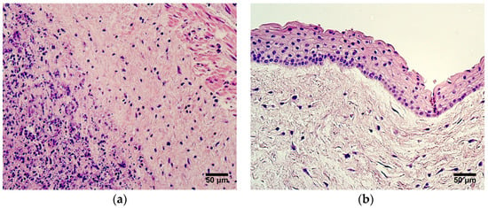
Background: Little is known about morphogenetic changes in the umbilical cord during the maturation process. Extracellular matrix remodeling, angiogenesis, progenitor activity, and immunomodulation are represented by specific markers; therefore, the aim of this study was to determine the expression of matrix metalloproteinase-2 (MMP2), tissue inhibitor of metalloproteinases-2 (TIMP2), CD34, vascular endothelial growth factor (VEGF), and human β-defensin 2 (HBD2) in preterm and full-term human umbilical cord tissue. Methods: Samples of umbilical cord tissue were obtained from 17 patients and divided into two groups: very preterm and moderate preterm birth umbilical cords; late preterm birth and full-term birth umbilical cords. Routine histology examination was conducted. Marker-positive cells were detected using the immunohistochemistry method. The number of positive structures was counted semi-quantitatively using microscopy. Statistical analysis was carried out using the SPSS Statistics 29 program. Results: Extraembryonic mesenchyme cells are the most active cell producers, expressing MMP2, TIMP2, VEGF, and HBD2 at notable levels in preterm and full-term umbilical cord tissue. Statistically significant differences in the expression of CD34, MMP2, and TIMP2 between the two patient groups were found. The expression of VEGF was similar in both patient groups, with the highest number of VEGF-positive cells seen in the extraembryonic mesenchyme. The expression of HBD2 was the highest in the extraembryonic mesenchyme and the amniotic epithelium, where mostly moderate numbers of HBD2-positive cells were detected. Conclusions: Extracellular matrix remodeling in preterm and term umbilical cords is strongly regulated, and tissue factors MMP2 and TIMP2 take part in this process. The expression of VEGF is not affected by the umbilical cord’s age; however, individual patient factors can affect the production of VEGF. Numerous CD34-positive cells in the endothelium of the umbilical arteries suggest a significant role of progenitor cells in very preterm and moderate preterm birth umbilical cords. Antimicrobial activity provided by HBD2 is essential and constant in preterm and full-term umbilical cords.
Full article
(This article belongs to the Special Issue 10th Anniversary Special Issue of JDB—Advances in Developmental Blood Vessel Growth)
►▼
Show Figures

Figure 1
Open AccessReview
Planar Cell Polarity Signaling: Coordinated Crosstalk for Cell Orientation
by
Sandeep Kacker, Varuneshwar Parsad, Naveen Singh, Daria Hordiichuk, Stacy Alvarez, Mahnoor Gohar, Anshu Kacker and Sunil Kumar Rai
J. Dev. Biol. 2024, 12(2), 12; https://doi.org/10.3390/jdb12020012 - 29 Apr 2024
Abstract
►▼
Show Figures
The planar cell polarity (PCP) system is essential for positioning cells in 3D networks to establish the proper morphogenesis, structure, and function of organs during embryonic development. The PCP system uses inter- and intracellular feedback interactions between components of the core PCP, characterized
[...] Read more.
The planar cell polarity (PCP) system is essential for positioning cells in 3D networks to establish the proper morphogenesis, structure, and function of organs during embryonic development. The PCP system uses inter- and intracellular feedback interactions between components of the core PCP, characterized by coordinated planar polarization and asymmetric distribution of cell populations inside the cells. PCP signaling connects the anterior–posterior to left–right embryonic plane polarity through the polarization of cilia in the Kupffer’s vesicle/node in vertebrates. Experimental investigations on various genetic ablation-based models demonstrated the functions of PCP in planar polarization and associated genetic disorders. This review paper aims to provide a comprehensive overview of PCP signaling history, core components of the PCP signaling pathway, molecular mechanisms underlying PCP signaling, interactions with other signaling pathways, and the role of PCP in organ and embryonic development. Moreover, we will delve into the negative feedback regulation of PCP to maintain polarity, human genetic disorders associated with PCP defects, as well as challenges associated with PCP.
Full article

Figure 1
Open AccessArticle
A Residual N-Terminal Peptide Enhances Signaling of Depalmitoylated Hedgehog to the Patched Receptor
by
Sophia F. Ehlers, Dominique Manikowski, Georg Steffes, Kristina Ehring, Fabian Gude and Kay Grobe
J. Dev. Biol. 2024, 12(2), 11; https://doi.org/10.3390/jdb12020011 - 9 Apr 2024
Abstract
During their biosynthesis, Sonic hedgehog (Shh) morphogens are covalently modified by cholesterol at the C-terminus and palmitate at the N-terminus. Although both lipids initially anchor Shh to the plasma membrane of producing cells, it later translocates to the extracellular compartment to direct developmental
[...] Read more.
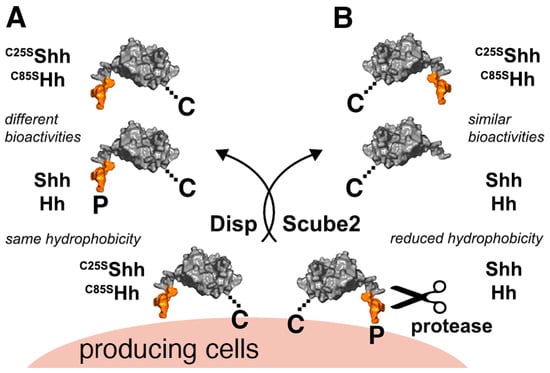
During their biosynthesis, Sonic hedgehog (Shh) morphogens are covalently modified by cholesterol at the C-terminus and palmitate at the N-terminus. Although both lipids initially anchor Shh to the plasma membrane of producing cells, it later translocates to the extracellular compartment to direct developmental fates in cells expressing the Patched (Ptch) receptor. Possible release mechanisms for dually lipidated Hh/Shh into the extracellular compartment are currently under intense debate. In this paper, we describe the serum-dependent conversion of the dually lipidated cellular precursor into a soluble cholesteroylated variant (ShhC) during its release. Although ShhC is formed in a Dispatched- and Scube2-dependent manner, suggesting the physiological relevance of the protein, the depalmitoylation of ShhC during release is inconsistent with the previously postulated function of N-palmitate in Ptch receptor binding and signaling. Therefore, we analyzed the potency of ShhC to induce Ptch-controlled target cell transcription and differentiation in Hh-sensitive reporter cells and in the Drosophila eye. In both experimental systems, we found that ShhC was highly bioactive despite the absence of the N-palmitate. We also found that the artificial removal of N-terminal peptides longer than eight amino acids inactivated the depalmitoylated soluble proteins in vitro and in the developing Drosophila eye. These results demonstrate that N-depalmitoylated ShhC requires an N-peptide of a defined minimum length for its signaling function to Ptch.
Full article
(This article belongs to the Special Issue The 10th Anniversary of JDB: Feature Papers)
►▼
Show Figures

Figure 1
Open AccessArticle
Effect of Cyclic Adenosine Monophosphate on Connexin 37 Expression in Sheep Cumulus-Oocyte Complexes
by
Mengyao Zhao, Gerile Subudeng, Yufen Zhao, Shaoyu Hao and Haijun Li
J. Dev. Biol. 2024, 12(2), 10; https://doi.org/10.3390/jdb12020010 - 27 Mar 2024
Abstract
Gap junctional connection (GJC) in the cumulus–oocyte complex (COC) provides necessary support for message communication and nutrient transmission required for mammalian oocyte maturation. Cyclic adenosine monophosphate (cAMP) is not only a prerequisite for regulating oocyte meiosis, but also the key intercellular factor for
[...] Read more.
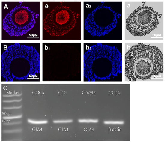
Gap junctional connection (GJC) in the cumulus–oocyte complex (COC) provides necessary support for message communication and nutrient transmission required for mammalian oocyte maturation. Cyclic adenosine monophosphate (cAMP) is not only a prerequisite for regulating oocyte meiosis, but also the key intercellular factor for affecting GJC function in COCs. However, there are no reports on whether cAMP regulates connexin 37 (Cx37) expression, one of the main connexin proteins, in sheep COCs. In this study, the expression of Cx37 protein and gene in immature sheep COC was detected using immunohistochemistry and PCR. Subsequently, the effect of cAMP on Cx37 expression in sheep COCs cultured in a gonadotropin-free culture system for 10 min or 60 min was evaluated using competitive ELISA, real-time fluorescent quantitative PCR (RT-qPCR), and Western blot. The results showed that the Cx37 protein was present in sheep oocytes and cumulus cells; the same results were found with respect to GJA4 gene expression. In the gonadotropin-free culture system, compared to the control, significantly higher levels of cAMP as well as Cx37 gene and protein expression were found in sheep COCs following treatment in vitro with Forskolin and IBMX (100 μM and 500 μM)) for 10 min (p < 0.05). Compared to the controls (at 10 or 60 min), cAMP levels in sheep COCs were significantly elevated as a result of Forskolin and IBMX treatment (p < 0.05). Following culturing in vitro for 10 min or 60 min, Forskolin and IBMX treatment can significantly promote Cx37 expression in sheep COCs (p < 0.05), a phenomenon which can be counteracted when the culture media is supplemented with RP-cAMP, a cAMP-specific competitive inhibitor operating through suppression of the protein kinase A (PKA). In summary, this study reports the preliminary regulatory mechanism of cAMP involved in Cx37 expression for the first time, and provides a novel explanation for the interaction between cAMP and GJC communication during sheep COC culturing in vitro.
Full article
(This article belongs to the Special Issue The 10th Anniversary of JDB: Feature Papers)
►▼
Show Figures

Figure 1
Highly Accessed Articles
Latest Books
E-Mail Alert
News
Topics

Conferences
Special Issues
Special Issue in
JDB
Feature Papers from Journal of Developmental Biology Reviewers
Guest Editors: Junichi Iwata, Christopher A. JohnstonDeadline: 20 November 2024
Special Issue in
JDB
Skin Wound Healing and Regeneration in Vertebrates
Guest Editor: Lorenzo AlibardiDeadline: 25 November 2024
Special Issue in
JDB
Epigenetic Programming in Development: Mechanisms and Consequences
Guest Editors: Lon J. van Winkle, Philip Iannaccone, Rebecca Jean RyznarDeadline: 15 December 2024
Special Issue in
JDB
In Vitro Modeling of the Craniofacial Disorders Using iPSCs/Organoids: Deciphering the Molecular and Genetic Mechanisms of Craniofacial Development
Guest Editors: Md Shaifur Rahman, Quenten P. SchwarzDeadline: 15 December 2024
Topical Collections
Topical Collection in
JDB
Hedgehog Signaling in Embryogenesis
Collection Editors: Henk Roelink, Kay Grobe
Topical Collection in
JDB
Drosophila - A Model System for Developmental Biology
Collection Editor: Nicholas Tolwinski


