


NeuroSci 2025, 6(4), 109; https://doi.org/10.3390/neurosci6040109 - 30 Oct 2025
Abstract
►
Show Figures
Background: Cognitive impairment after stroke often reduces independence and quality of life. Cognitive rehabilitation is therefore essential, and recent research on computer-based interventions has shown promising results. This proof-of-concept study investigated the effects of additional self-administered cognitive training using an electronic device, compared
[...] Read more.
Background: Cognitive impairment after stroke often reduces independence and quality of life. Cognitive rehabilitation is therefore essential, and recent research on computer-based interventions has shown promising results. This proof-of-concept study investigated the effects of additional self-administered cognitive training using an electronic device, compared with traditional paper-and-pencil methods, on attentional functions in individuals with subacute stroke. Methods: Participants were randomly assigned to an experimental group or a control group. For two consecutive weeks, both groups received forty-five-minute, face-to-face cognitive therapy sessions each morning, delivered via an electronic device. In addition, the experimental group engaged in sixty minutes of self-administered cognitive training using the same device, while the control group completed conventional exercises with paper-and-pencil tools. Neuropsychological assessments were conducted before and after the intervention. Results: Twenty-three participants were included (experimental group: eleven; control group: twelve). No significant differences in safety or attentional performance were observed between groups. Within-group analyses showed improvements in the experimental group in attentional shifting, inhibitory control, visuospatial planning, and problem-solving, while the control group improved in visuospatial planning and problem-solving. Conclusions: These preliminary findings suggest that self-administered electronic cognitive training may be a feasible approach to support attentional recovery in individuals with subacute stroke.
Full article
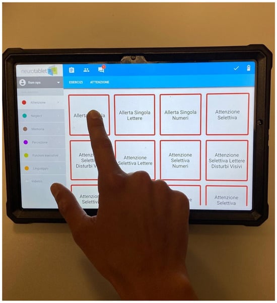
Figure 1


