Toxic and Pharmacological Effect of Plant Toxins

A topical collection in Toxins (ISSN 2072-6651). This collection belongs to the section "Plant Toxins".

Viewed by 118643

Editor


E-Mail Website
Collection Editor
Department for Life Quality Studies, Alma Mater Studiorum-University of Bologna, Corso d’Augusto 237, 47921 Rimini, Italy
Interests: anticancer pharmacology; natural products; in vitro studies; apoptosis; cell death; non-canonical cell death; genotoxicity
Special Issues, Collections and Topics in MDPI journals

Topical Collection Information

Dear Colleagues,

Plant toxins are secondary plant metabolites that naturally occur in the marine and terrestrial plants of several families, some of which commonly consumed as food. The chemical diversity is tremendous. Phytotoxins can evoke many different effects on living organisms, functioning as: nutrients, useful for growth and other body processes; inerts, such as fiber; toxicants, chemicals which have an effect on metabolic or physiological processes; or drugs, chemicals useful for the prevention or treatment of many diseases. Many plant toxins possess the characteristic of a “Janus” compound, i.e., they exert both significant toxic and pharmacological effects, depending on the concentrations used in the test systems applied. They can also be modified to improve affinity and efficacy for health endorsement. I hope that this collection of Toxins entitled “Toxic and Pharmacological Effect of Plant Toxins” will provide an outline of different plant toxins, their mechanisms of action, and the relevance of their toxic and/or pharmacological effects for human health.

Prof. Dr. Carmela Fimognari
Guest Editor

Manuscript Submission Information

Manuscripts should be submitted online at www.mdpi.com by registering and logging in to this website. Once you are registered, click here to go to the submission form. Manuscripts can be submitted until the deadline. All submissions that pass pre-check are peer-reviewed. Accepted papers will be published continuously in the journal (as soon as accepted) and will be listed together on the collection website. Research articles, review articles as well as short communications are invited. For planned papers, a title and short abstract (about 100 words) can be sent to the Editorial Office for announcement on this website.

Submitted manuscripts should not have been published previously, nor be under consideration for publication elsewhere (except conference proceedings papers). All manuscripts are thoroughly refereed through a double-blind peer-review process. A guide for authors and other relevant information for submission of manuscripts is available on the Instructions for Authors page. Toxins is an international peer-reviewed open access monthly journal published by MDPI.

Please visit the Instructions for Authors page before submitting a manuscript. The Article Processing Charge (APC) for publication in this open access journal is 2700 CHF (Swiss Francs). Submitted papers should be well formatted and use good English. Authors may use MDPI's English editing service prior to publication or during author revisions.

Keywords

  • terrestrial phytotoxins
  • marine phytotoxins
  • dietetic phytotoxins
  • non-dietetic phytotoxins
  • toxicological mechanisms
  • pharmacological mechanisms
  • disease prevention
  • disease treatment
Published Papers (24 papers)

2024

Jump to: 2023, 2022, 2021, 2020, 2019

21 pages, 360 KiB  
Review
Grass–Endophyte Interactions and Their Associated Alkaloids as a Potential Management Strategy for Plant Parasitic Nematodes
by Nyambura G. Mwangi, Mark Stevens, Alistair J. D. Wright, Simon G. Edwards, Martin C. Hare and Matthew A. Back
Toxins 2024, 16(6), 274; https://doi.org/10.3390/toxins16060274 - 15 Jun 2024
Viewed by 1418
Abstract
Claviceptaceous endophytic fungi in the genus Epichloë mostly form a symbiotic relationship with cool-season grasses. Epichloë spp. are capable of producing bioactive alkaloids such as peramines, lolines, ergot alkaloids, and indole-diterpenes, which protect the host plant from herbivory by animals, insects, and nematodes. [...] Read more.
Claviceptaceous endophytic fungi in the genus Epichloë mostly form a symbiotic relationship with cool-season grasses. Epichloë spp. are capable of producing bioactive alkaloids such as peramines, lolines, ergot alkaloids, and indole-diterpenes, which protect the host plant from herbivory by animals, insects, and nematodes. The host also benefits from enhanced tolerance to abiotic stresses, such as salt, drought, waterlogging, cold, heavy metals, and low nitrogen stress. The bioactive alkaloids produced can have both direct and indirect effects towards plant parasitic nematodes. Direct interaction with nematodes’ motile stages can cause paralysis (nematostatic effect) or death (nematicidal effect). Indirectly, the metabolites may induce host immunity which inhibits feeding and subsequent nematode development. This review highlights the different mechanisms through which this interaction and the metabolites produced have been explored in the suppression of plant parasitic nematodes and also how the specific interactions between different grass genotypes and endophyte strains result in variable suppression of different nematode species. An understanding of the different grass–endophyte interactions and their successes and failures in suppressing various nematode species is essential to enable the proper selection of grass–endophyte combinations to identify the alkaloids produced, concentrations required, and determine which nematodes are sensitive to which specific alkaloids. Full article

2023

Jump to: 2024, 2022, 2021, 2020, 2019

21 pages, 4585 KiB  
Review
Antifungal and Antibacterial Activities of Isolated Marine Compounds
by Amin Mahmood Thawabteh, Zain Swaileh, Marwa Ammar, Weam Jaghama, Mai Yousef, Rafik Karaman, Sabino A. Bufo and Laura Scrano
Toxins 2023, 15(2), 93; https://doi.org/10.3390/toxins15020093 - 18 Jan 2023
Cited by 18 | Viewed by 5367
Abstract
To combat the ineffectiveness of currently available pharmaceutical medications, caused by the emergence of increasingly resistant bacterial and fungal strains, novel antibacterial and antifungal medications are urgently needed. Novel natural compounds with antimicrobial activities can be obtained by exploring underexplored habitats such as [...] Read more.
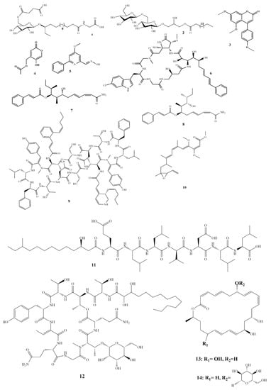
To combat the ineffectiveness of currently available pharmaceutical medications, caused by the emergence of increasingly resistant bacterial and fungal strains, novel antibacterial and antifungal medications are urgently needed. Novel natural compounds with antimicrobial activities can be obtained by exploring underexplored habitats such as the world’s oceans. The oceans represent the largest ecosystem on earth, with a high diversity of organisms. Oceans have received some attention in the past few years, and promising compounds with antimicrobial activities were isolated from marine organisms such as bacteria, fungi, algae, sea cucumbers, sea sponges, etc. This review covers 56 antifungal and 40 antibacterial compounds from marine organisms. These compounds are categorized according to their chemical structure groups, including polyketides, alkaloids, ribosomal peptides, and terpenes, and their organismal origin. The review provides the minimum inhibitory concentration MIC values and the bacterial/fungal strains against which these chemical compounds show activity. This study shows strong potential for witnessing the development of new novel antimicrobial drugs from these natural compounds isolated and evaluated for their antimicrobial activities. Full article
Show Figures

Scheme 1

2022

Jump to: 2024, 2023, 2021, 2020, 2019

14 pages, 2009 KiB  
Article
Cytotoxic Effects of Cannabidiol on Neonatal Rat Cortical Neurons and Astrocytes: Potential Danger to Brain Development
by Damijana Mojca Jurič, Klara Bulc Rozman, Metoda Lipnik-Štangelj, Dušan Šuput and Miran Brvar
Toxins 2022, 14(10), 720; https://doi.org/10.3390/toxins14100720 - 21 Oct 2022
Cited by 6 | Viewed by 4317
Abstract
The influence of cannabidiol (CBD) on brain development is inadequately understood. Since CBD is considered a non-intoxicating drug, it has attracted great interest concerning its potential medical applicability, including in pregnant women and children. Here, we elucidated the response of perinatal rat cortical [...] Read more.
The influence of cannabidiol (CBD) on brain development is inadequately understood. Since CBD is considered a non-intoxicating drug, it has attracted great interest concerning its potential medical applicability, including in pregnant women and children. Here, we elucidated the response of perinatal rat cortical neurons and astrocytes to CBD at submicromolar (0.1, 0.5, 1, 5 µM) concentrations attainable in humans. The effect of CBD was concentration- and time-dependent and cell-specific. In neurons, 0.1 µM CBD induced an early and transient change in mitochondrial membrane potential (ΔΨm), ATP depletion, and caspase-8 activation, followed by rapid ATP recovery and progressive activation of caspase-9 and caspase-3/7, resulting in early apoptotic cell death with reduction and shortening of dendrites, cell shrinkage, and chromatin condensation. The decrease in neuronal viability, ATP depletion, and caspase activation due to CBD exposure was prevented by transient receptor potential vanilloid 1 (TRPV1) antagonist. In astrocytes, 0.5 µM CBD caused an immediate short-term dysregulation of ΔΨm, followed by ATP depletion with transient activation of caspase-8 and progressive activation of caspase-9 and caspase-3/7, leading to early apoptosis and subsequent necroptosis. In astrocytes, both TRPV1 and cannabinoid receptor 1 (CB1) antagonists protected viability and prevented apoptosis. Given that CBD is a non-intoxicating drug, our results clearly show that this is not the case during critical periods of brain development when it can significantly interfere with the endogenous cannabinoid system. Full article
Show Figures

Figure 1

19 pages, 3711 KiB  
Article
Study on the Mechanism of Mesaconitine-Induced Hepatotoxicity in Rats Based on Metabonomics and Toxicology Network
by Qian Chen, Kai Zhang, Mingjie Jiao, Jiakang Jiao, Dongling Chen, Yihui Yin, Jia Zhang and Fei Li
Toxins 2022, 14(7), 486; https://doi.org/10.3390/toxins14070486 - 14 Jul 2022
Cited by 12 | Viewed by 2840
Abstract
Mesaconitine (MA), one of the main diterpenoid alkaloids in Aconitum, has a variety of pharmacological effects, such as analgesia, anti-inflammation and relaxation of rat aorta. However, MA is a highly toxic ingredient. At present, studies on its toxicity are mainly focused on the [...] Read more.
Mesaconitine (MA), one of the main diterpenoid alkaloids in Aconitum, has a variety of pharmacological effects, such as analgesia, anti-inflammation and relaxation of rat aorta. However, MA is a highly toxic ingredient. At present, studies on its toxicity are mainly focused on the heart and central nervous system, and there are few reports on the hepatotoxic mechanism of MA. Therefore, we evaluated the effects of MA administration on liver. SD rats were randomly divided into a normal saline (NS) group, a low-dose MA group (0.8 mg/kg/day) and a high-dose MA group (1.2 mg/kg/day). After 6 days of administration, the toxicity of MA on the liver was observed. Metabolomic and network toxicology methods were combined to explore the effect of MA on the liver of SD rats and the mechanism of hepatotoxicity in this study. Through metabonomics study, the differential metabolites of MA, such as L-phenylalanine, retinyl ester, L-proline and 5-hydroxyindole acetaldehyde, were obtained, which involved amino acid metabolism, vitamin metabolism, glucose metabolism and lipid metabolism. Based on network toxicological analysis, MA can affect HIF-1 signal pathway, MAPK signal pathway, PI3K-Akt signal pathway and FoxO signal pathway by regulating ALB, AKT1, CASP3, IL2 and other targets. Western blot results showed that protein expression of HMOX1, IL2 and caspase-3 in liver significantly increased after MA administration (p < 0.05). Combined with the results of metabonomics and network toxicology, it is suggested that MA may induce hepatotoxicity by activating oxidative stress, initiating inflammatory reaction and inducing apoptosis. Full article
Show Figures

Graphical abstract

28 pages, 3768 KiB  
Article
Mentha pulegium L. (Pennyroyal, Lamiaceae) Extracts Impose Abortion or Fetal-Mediated Toxicity in Pregnant Rats; Evidenced by the Modulation of Pregnancy Hormones, MiR-520, MiR-146a, TIMP-1 and MMP-9 Protein Expressions, Inflammatory State, Certain Related Signaling Pathways, and Metabolite Profiling via UPLC-ESI-TOF-MS
by Amira A. El-Gazar, Ayat M. Emad, Ghada M. Ragab and Dalia M. Rasheed
Toxins 2022, 14(5), 347; https://doi.org/10.3390/toxins14050347 - 16 May 2022
Cited by 11 | Viewed by 3967
Abstract
Pregnant women usually turn to natural products to relieve pregnancy-related ailments which might pose health risks. Mentha pulegium L. (MP, Lamiaceae) is a common insect repellent, and the present work validates its abortifacient capacity, targeting morphological anomalies, biological, and behavioral consequences, compared to [...] Read more.
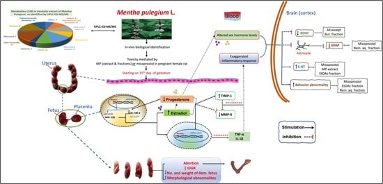
Pregnant women usually turn to natural products to relieve pregnancy-related ailments which might pose health risks. Mentha pulegium L. (MP, Lamiaceae) is a common insect repellent, and the present work validates its abortifacient capacity, targeting morphological anomalies, biological, and behavioral consequences, compared to misoprostol. The study also includes untargeted metabolite profiling of MP extract and fractions thereof viz. methylene chloride (MecH), ethyl acetate (EtOAc), butanol (But), and the remaining liquor (Rem. Aq.) by UPLC-ESI-MS-TOF, to unravel the constituents provoking abortion. Administration of MP extract/fractions, for three days starting from day 15th of gestation, affected fetal development by disrupting the uterine and placental tissues, or even caused pregnancy termination. These effects also entailed biochemical changes where they decreased progesterone and increased estradiol serum levels, modulated placental gene expressions of both MiR-(146a and 520), decreased uterine MMP-9, and up-regulated TIMP-1 protein expression, and empathized inflammatory responses (TNF-α, IL-1β). In addition, these alterations affected the brain's GFAP, BDNF, and 5-HT content and some of the behavioral parameters escorted by the open field test. All these incidences were also perceived in the misoprostol-treated group. A total of 128 metabolites were identified in the alcoholic extract of MP, including hydroxycinnamates, flavonoid conjugates, quinones, iridoids, and terpenes. MP extract was successful in terminating the pregnancy with minimal behavioral abnormalities and low toxicity margins. Full article
Show Figures

Graphical abstract

2021

Jump to: 2024, 2023, 2022, 2020, 2019

24 pages, 14490 KiB  
Article
Ageratina adenophora Disrupts the Intestinal Structure and Immune Barrier Integrity in Rats
by Yujing Cui, Samuel Kumi Okyere, Pei Gao, Juan Wen, Suizhong Cao, Ya Wang, Junliang Deng and Yanchun Hu
Toxins 2021, 13(9), 651; https://doi.org/10.3390/toxins13090651 - 15 Sep 2021
Cited by 21 | Viewed by 3121
Abstract
The aim of this study was to investigate the effects of Ageratina adenophora on the intestines morphology and integrity in rat. Rats were randomly divided into two groups and were fed with 10 g/100 g body weight (BW) basal diet and 10 g/100 [...] Read more.
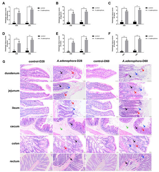
The aim of this study was to investigate the effects of Ageratina adenophora on the intestines morphology and integrity in rat. Rats were randomly divided into two groups and were fed with 10 g/100 g body weight (BW) basal diet and 10 g/100 g BW experimental diet, which was a mixture of A. adenophora powder and basal diet in a 3:7 ratio. The feeding experiment lasted for 60 days. At days 28 and 60 of the experiment, eight rats/group/timepoint were randomly selected, weighed, and sacrificed, then blood and intestinal tissues were collected and stored for further analysis. The results showed that Ageratina adenophora caused pathological changes and injury in the intestine, elevated serum diamine oxidase (DAO), D-lactate (D-LA), and secretory immunoglobulin A (sIgA) levels, reduced occludin levels in intestinal tissues, as well as increased the count of intraepithelial leukocytes (IELs) and lamina propria leukocytes (LPLs) in the intestine (p < 0.05 or p < 0.01). In addition, the mRNA and protein (ELISA) expressions of pro-inflammation cytokines (IL-1β, IL-2, TNF-α, and IFN-ϒ) were elevated in the Ageratina adenophora treatment groups, whereas anti-inflammatory cytokines such as IL-4 and IL-10 were reduced (p < 0.01 or p < 0.05). Therefore, the results obtained in this study indicated that Ageratina adenophora impaired intestinal function in rats by damaging the intestine structure and integrity, and also triggered an inflammation immune response that led to intestinal immune barrier dysfunction. Full article
Show Figures

Figure 1

13 pages, 2172 KiB  
Article
Ageratina adenophora Inhibits Spleen Immune Function in Rats via the Loss of the FRC Network and Th1–Th2 Cell Ratio Elevation
by Zhihua Ren, Pei Gao, Samuel Kumi Okyere, Yujing Cui, Juan Wen, Bo Jing, Junliang Deng and Yanchun Hu
Toxins 2021, 13(5), 309; https://doi.org/10.3390/toxins13050309 - 26 Apr 2021
Cited by 15 | Viewed by 2987
Abstract
The objective of this study was to determine the impact of Ageratina adenophora (A. adenophora) on splenic immune function in a rat model. Rats were fed with 10 g/100 g normal feed and an experimental feed, which was composed of 3:7 A. adenophora [...] Read more.
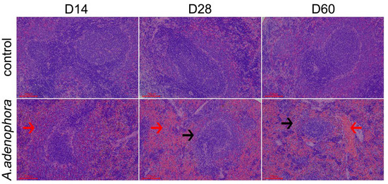
The objective of this study was to determine the impact of Ageratina adenophora (A. adenophora) on splenic immune function in a rat model. Rats were fed with 10 g/100 g normal feed and an experimental feed, which was composed of 3:7 A. adenophora powder and normal feed for 60 days. On days 14, 28, and 60, subsets of rats (n = 8 rats/group/time point) were selected for blood and spleen tissue sample collection. The results showed that the proportion of CD3+ T cells in the spleen was decreased at day 60 (vs. control). Also, mRNA and protein expression of chemokines CCL21 and CCL19 and functional protein gp38 in spleen decreased significantly versus the control at day 60. In addition, ER-TR7 antigen protein expression was also decreased at day 60. Levels of T-helper (Th)1 cells significantly increased, whereas those of Th2 cells decreased significantly versus the control at day 60 in spleen. The finding revealed that A. adenophora could affect splenic immune function in rats by altering the fibroblast reticulocyte (FRC) network, as well as by causing an imbalance in Th1/Th2 cell ratios. This research provides new insights into potential mechanisms of spleen immunotoxicity due to exposures to A. Adenophora. Full article
Show Figures

Figure 1

15 pages, 3244 KiB  
Article
Bergamottin and 5-Geranyloxy-7-methoxycoumarin Cooperate in the Cytotoxic Effect of Citrus bergamia (Bergamot) Essential Oil in Human Neuroblastoma SH-SY5Y Cell Line
by Alessandro Maugeri, Giovanni Enrico Lombardo, Laura Musumeci, Caterina Russo, Sebastiano Gangemi, Gioacchino Calapai, Santa Cirmi and Michele Navarra
Toxins 2021, 13(4), 275; https://doi.org/10.3390/toxins13040275 - 10 Apr 2021
Cited by 22 | Viewed by 3370
Abstract
The plant kingdom has always been a treasure trove for valuable bioactive compounds, and Citrus fruits stand out among the others. Bergamottin (BRG) and 5-geranyloxy-7-methoxycoumarin (5-G-7-MOC) are two coumarins found in different Citrus species with well-acknowledged pharmacological properties. Previously, they have been claimed [...] Read more.
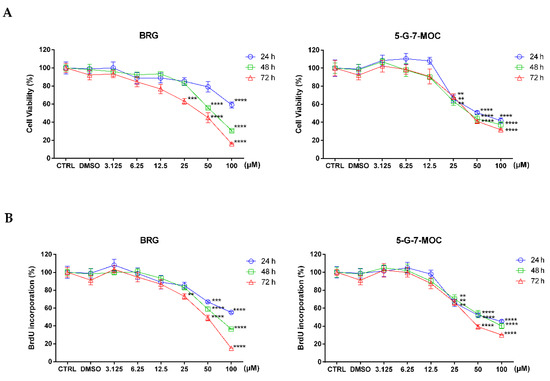
The plant kingdom has always been a treasure trove for valuable bioactive compounds, and Citrus fruits stand out among the others. Bergamottin (BRG) and 5-geranyloxy-7-methoxycoumarin (5-G-7-MOC) are two coumarins found in different Citrus species with well-acknowledged pharmacological properties. Previously, they have been claimed to be relevant in the anti-proliferative effects exerted by bergamot essential oil (BEO) in the SH-SY5Y human neuroblastoma cells. This study was designed to verify this assumption and to assess the mechanisms underlying the anti-proliferative effect of both compounds. Our results demonstrate that BRG and 5-G-7-MOC are able to reduce the proliferation of SH-SY5Y cells, inducing apoptosis and increasing cell population in sub-G0/G1 phase. Moreover, we demonstrated the pro-oxidant activity of the two coumarins that increased reactive oxygen species and impaired mitochondrial membrane potential. From a molecular point of view, BRG and 5-G-7-MOC were able to modulate apoptosis related factors at both protein and gene levels. Lastly, we evaluated the synergistic effect of their combination, finding that the highest synergy was observed at a concentration ratio similar to that occurring in the BEO, supporting our initial hypothesis. Taken together, our results deepen the knowledge regarding the effect of BRG and 5-G-7-MOC in SH-SY5Y cells, emphasizing the relevance of their cooperation in achieving this effect. Full article
Show Figures

Figure 1

14 pages, 2798 KiB  
Article
4′-O-methylpyridoxine: Preparation from Ginkgo biloba Seeds and Cytotoxicity in GES-1 Cells
by Jin-Peng Zhu, Hao Gong, Cai-E Wu, Gong-Jian Fan, Ting-Ting Li and Jia-Hong Wang
Toxins 2021, 13(2), 95; https://doi.org/10.3390/toxins13020095 - 26 Jan 2021
Cited by 5 | Viewed by 3135
Abstract
Ginkgo biloba seeds are wildly used in the food and medicine industry. It has been found that 4′-O-methylpyridoxine (MPN) is responsible for the poisoning caused by G. biloba seeds. The objective of this study was to explore and optimize the extraction [...] Read more.
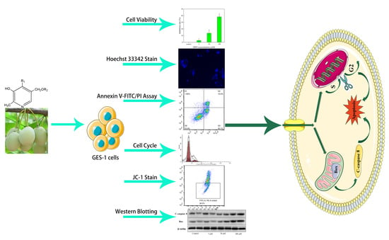
Ginkgo biloba seeds are wildly used in the food and medicine industry. It has been found that 4′-O-methylpyridoxine (MPN) is responsible for the poisoning caused by G. biloba seeds. The objective of this study was to explore and optimize the extraction method of MPN from G. biloba seeds, and investigate its toxic effect on human gastric epithelial cells (GES-1) and the potential related mechanisms. The results showed that the extraction amount of MPN was 1.933 μg/mg, when extracted at 40 °C for 100 min, with the solid–liquid ratio at 1:10. MPN inhibited the proliferation of GES-1 cells, for which the inhibition rate was 38.27% when the concentration of MPN was 100 μM, and the IC50 value was 127.80 μM; meanwhile, the cell cycle was arrested in G2 phase. High concentration of MPN (100 μM) had significant effects on the nucleus of GES-1 cells, and the proportion of apoptotic cells reached 43.80%. Furthermore, the Western blotting analysis showed that MPN could reduce mitochondrial membrane potential by increasing the expression levels of apoptotic proteins Caspase 8 and Bax in GES-1 cells. In conclusion, MPN may induce apoptosis in GES-1 cells, which leads to toxicity in the human body. Full article
Show Figures

Graphical abstract

2020

Jump to: 2024, 2023, 2022, 2021, 2019

33 pages, 915 KiB  
Review
Overview of the Anticancer Potential of the “King of Spices” Piper nigrum and Its Main Constituent Piperine
by Eleonora Turrini, Piero Sestili and Carmela Fimognari
Toxins 2020, 12(12), 747; https://doi.org/10.3390/toxins12120747 - 26 Nov 2020
Cited by 33 | Viewed by 8937
Abstract
The main limits of current anticancer therapy are relapses, chemoresistance, and toxic effects resulting from its poor selectivity towards cancer cells that severely impair a patient’s quality of life. Therefore, the discovery of new anticancer drugs remains an urgent challenge. Natural products represent [...] Read more.
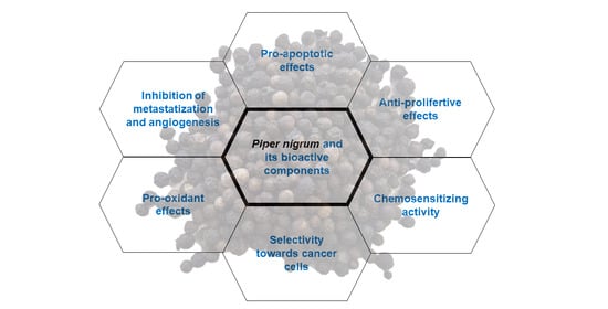
The main limits of current anticancer therapy are relapses, chemoresistance, and toxic effects resulting from its poor selectivity towards cancer cells that severely impair a patient’s quality of life. Therefore, the discovery of new anticancer drugs remains an urgent challenge. Natural products represent an excellent opportunity due to their ability to target heterogenous populations of cancer cells and regulate several key pathways involved in cancer development, and their favorable toxicological profile. Piper nigrum is one of the most popular spices in the world, with growing fame as a source of bioactive molecules with pharmacological properties. The present review aims to provide a comprehensive overview of the anticancer potential of Piper nigrum and its major active constituents—not limited to the well-known piperine—whose undeniable anticancer properties have been reported for different cancer cell lines and animal models. Moreover, the chemosensitizing effects of Piper nigrum in association with traditional anticancer drugs are depicted and its toxicological profile is outlined. Despite the promising results, human studies are missing, which are crucial for supporting the efficacy and safety of Piper nigrum and its single components in cancer patients. Full article
Show Figures

Graphical abstract

26 pages, 1038 KiB  
Review
Natural Sources and Bioactivities of 2,4-Di-Tert-Butylphenol and Its Analogs
by Fuqiang Zhao, Ping Wang, Rima D. Lucardi, Zushang Su and Shiyou Li
Toxins 2020, 12(1), 35; https://doi.org/10.3390/toxins12010035 - 6 Jan 2020
Cited by 179 | Viewed by 12301
Abstract
2,4-Di-tert-butylphenol or 2,4-bis(1,1-dimethylethyl)-phenol (2,4-DTBP) is a common toxic secondary metabolite produced by various groups of organisms. The biosources and bioactivities of 2,4-DTBP have been well investigated, but the phenol has not been systematically reviewed. This article provides a comprehensive review of 2,4-DTBP and [...] Read more.
2,4-Di-tert-butylphenol or 2,4-bis(1,1-dimethylethyl)-phenol (2,4-DTBP) is a common toxic secondary metabolite produced by various groups of organisms. The biosources and bioactivities of 2,4-DTBP have been well investigated, but the phenol has not been systematically reviewed. This article provides a comprehensive review of 2,4-DTBP and its analogs with emphasis on natural sources and bioactivities. 2,4-DTBP has been found in at least 169 species of bacteria (16 species, 10 families), fungi (11 species, eight families), diatom (one species, one family), liverwort (one species, one family), pteridiphyta (two species, two families), gymnosperms (four species, one family), dicots (107 species, 58 families), monocots (22 species, eight families), and animals (five species, five families). 2,4-DTBP is often a major component of violate or essential oils and it exhibits potent toxicity against almost all testing organisms, including the producers; however, it is not clear why organisms produce autotoxic 2,4-DTBP and its analogs. The accumulating evidence indicates that the endocidal regulation seems to be the primary function of the phenols in the producing organisms. Full article
Show Figures

Figure 1

2019

Jump to: 2024, 2023, 2022, 2021, 2020

21 pages, 8289 KiB  
Article
Investigation of the In-Vivo Cytotoxicity and the In Silico-Prediction of MDM2-p53 Inhibitor Potential of Euphorbia peplus Methanolic Extract in Rats
by Yasmina M. Abd-Elhakim, Mohamed Abdo Nassan, Gamal A. Salem, Abdelkarim Sasi, Adil Aldhahrani, Khaled Ben Issa and Amany Abdel-Rahman Mohamed
Toxins 2019, 11(11), 642; https://doi.org/10.3390/toxins11110642 - 4 Nov 2019
Cited by 8 | Viewed by 4988
Abstract
This study explored the probable in vivo cardiac and renal toxicities together with in silico approaches for predicting the apoptogenic potential of Euphorbia peplus methanolic extract (EPME) in rats. Cardiac and renal injury biomarkers were estimated with histopathological and immunohistochemical evaluations of both [...] Read more.
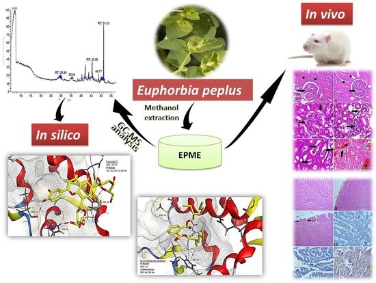
This study explored the probable in vivo cardiac and renal toxicities together with in silico approaches for predicting the apoptogenic potential of Euphorbia peplus methanolic extract (EPME) in rats. Cardiac and renal injury biomarkers were estimated with histopathological and immunohistochemical evaluations of both kidney and heart. The probable underlying mechanism of E. peplus compounds to potentiate p53 activity is examined using Molecular Operating Environment (MOE) docking software and validated experimentally by immunohistochemical localization of p53 protein in the kidney and heart tissues. The gas chromatography/mass spectrometry analysis of E. peplus revealed the presence of nine different compounds dominated by di-(2-ethylhexyl) phthalate (DEHP). Significant elevations of troponin, creatine phosphokinase, creatine kinase–myocardium bound, lactate dehydrogenase, aspartate transaminase, alkaline phosphatase, urea, creatinine, and uric acid were evident in the EPME treated rats. The EPME treated rats showed strong renal and cardiac p53 expression and moderate cardiac TNF-α expression. Further, our in silico results predicted the higher affinity and good inhibition of DEHP, glyceryl linolenate, and lucenin 2 to the MDM2-p53 interface compared to the standard reference 15 a compound. Conclusively, EPME long-term exposure could adversely affect the cardiac and renal tissues probably due to their inflammatory and apoptotic activity. Moreover, the in silico study hypothesizes that EPME inhibits MDM2-mediated degradation of p53 suggesting possible anticancer potentials which confirmed experimental by strong p53 expression in renal and cardiac tissues. Full article
Show Figures

Graphical abstract

18 pages, 4179 KiB  
Article
Annona cherimola Seed Extract Activates Extrinsic and Intrinsic Apoptotic Pathways in Leukemic Cells
by Tony Haykal, Peter Nasr, Mohammad H. Hodroj, Robin I. Taleb, Rita Sarkis, Marvy Nadine El. Moujabber and Sandra Rizk
Toxins 2019, 11(9), 506; https://doi.org/10.3390/toxins11090506 - 30 Aug 2019
Cited by 26 | Viewed by 10857
Abstract
Annona cherimola Mill is a large green fruit with black seeds widely known to possess toxic properties due to the presence of Annonaceous acetogenins. The present study investigates the anti-cancer properties of an Annona cherimola Mill ethanolic seed extract on Acute Myeloid Leukemia [...] Read more.
Annona cherimola Mill is a large green fruit with black seeds widely known to possess toxic properties due to the presence of Annonaceous acetogenins. The present study investigates the anti-cancer properties of an Annona cherimola Mill ethanolic seed extract on Acute Myeloid Leukemia (AML) cell lines in vitro and elucidates the underlying cellular mechanism. The anti-proliferative effects of the extract on various AML cell lines and normal mesenchymal cells (MSCs) were assessed using WST-1 viability reagent. The pro-apoptotic effect of the extract was evaluated using Annexin V/PI staining and Cell Death ELISA. The underlying mechanism was deciphered by analyzing the expression of various proteins using western blots. Treatment with an A. cherimola seed ethanolic extract promotes a dose- and time-dependent inhibition of the proliferation of various AML cell lines, but not MSCs. Positive Annexin V staining, as well as DNA fragmentation, confirm an increase in apoptotic cell death by upregulating the expression of pro-apoptotic proteins which control both intrinsic and extrinsic pathways of apoptosis. GC/MS analysis revealed the presence of phytosterols, in addition to other bioactive compounds. In conclusion, Annona cherimola Mill seed extract, previously known to possess a potent toxic activity, induces apoptosis in AML cell lines by the activation of both the extrinsic and the intrinsic pathways. Full article
Show Figures

Graphical abstract

17 pages, 4410 KiB  
Article
Fusarubin and Anhydrofusarubin Isolated from A Cladosporium Species Inhibit Cell Growth in Human Cancer Cell Lines
by Sabrina Adorisio, Alessandra Fierabracci, Isabella Muscari, Anna Marina Liberati, Lorenza Cannarile, Trinh Thi Thuy, Tran Van Sung, Hossain Sohrab, Choudhury Mahmood Hasan, Emira Ayroldi, Carlo Riccardi, Abdul Mazid and Domenico V. Delfino
Toxins 2019, 11(9), 503; https://doi.org/10.3390/toxins11090503 - 29 Aug 2019
Cited by 33 | Viewed by 5230
Abstract
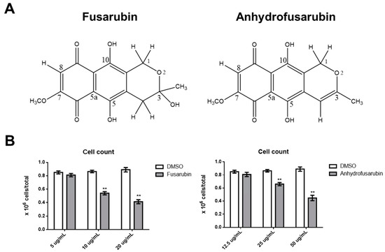
Cladosporium species are endophytic fungi that grow on organic matter and are considered food contaminants. The anti-microbial and anti-tumor naphthoquinones fusarubin (FUS) and anhydrofusarubin (AFU) were isolated using column chromatography from a Cladosporium species residing inside Rauwolfia leaves. The impact of FUS and [...] Read more.
Cladosporium species are endophytic fungi that grow on organic matter and are considered food contaminants. The anti-microbial and anti-tumor naphthoquinones fusarubin (FUS) and anhydrofusarubin (AFU) were isolated using column chromatography from a Cladosporium species residing inside Rauwolfia leaves. The impact of FUS and AFU on cell growth was assessed in acute myeloid leukemia (OCI-AML3) and other hematologic tumor cell lines (HL-60, U937, and Jurkat). Treatment with FUS or AFU reduced the number of OCI-AML3 cells as evaluated by a hemocytometer. Flow cytometry analyses showed that this effect was accompanied by diverse impairments in cell cycle progression. Specifically, FUS (20 or 10 μg/mL significantly decreased the percentage of cells in S phase and increased the percentage of cells in G2/M phase, whereas AFU increased the percentage of cells in G0/G1 phase (50 and 25 μg/mL) and decreased the percentage of cells in S (50 μg/mL) and G2/M (50 and 25 μg/mL) phases. Both substances significantly increased apoptosis at higher concentrations. The effects of FUS were more potent than those of AFU, with FUS up-regulating p21 expression in a p53-dependent manner, as detected by Western blot analyses, likely the consequence of decreased ERK phosphorylation and increased p38 expression (both of which increase p21 stability). FUS also decreased Akt phosphorylation and resulted in increased Fas ligand production and caspase-8/3-dependent apoptosis. These results suggest that FUS and AFU inhibit proliferation and increase apoptosis in cell lines derived from hematological cancers. Full article
Show Figures

Figure 1

9 pages, 2355 KiB  
Article
A Probable Fatal Case of Oleander (Nerium oleander) Poisoning on a Cattle Farm: A New Method of Detection and Quantification of the Oleandrin Toxin in Rumen
by Silva Rubini, Sabina Strano Rossi, Serena Mestria, Sara Odoardi, Sara Chendi, Andrea Poli, Giuseppe Merialdi, Giuseppina Andreoli, Paolo Frisoni, Rosa Maria Gaudio, Anna Baldisserotto, Piergiacomo Buso, Stefano Manfredini, Guido Govoni, Stefania Barbieri, Cinzia Centelleghe, Giorgia Corazzola, Sandro Mazzariol and Carlo Alessandro Locatelli
Toxins 2019, 11(8), 442; https://doi.org/10.3390/toxins11080442 - 25 Jul 2019
Cited by 15 | Viewed by 7729
Abstract
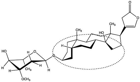
Oleander (Nerium oleander) is an ornamental plant common in tropical and sub-tropical regions that is becoming increasingly widespread, even in temperate regions. Oleander poisoning may occur in animals and humans. The main active components contained in the plant are cardiac glycosides [...] Read more.
Oleander (Nerium oleander) is an ornamental plant common in tropical and sub-tropical regions that is becoming increasingly widespread, even in temperate regions. Oleander poisoning may occur in animals and humans. The main active components contained in the plant are cardiac glycosides belonging to the class of cardenolides that are toxic to many species, from human to insects. This work describes a case of oleander poisoning that occurred on a small cattle farm and resulted in the fatality of all six resident animals. Furthermore, the investigation of the poisonous agent is described, with particular focus on the characterization of the oleandrin toxin that was recovered from the forage and rumen contents. The innovation of this study is the first description of the detection and quantification of the oleandrin toxin by liquid chromatography-high resolution mass spectrometry (LC-HRMS) in rumen. Full article
Show Figures

Figure 1

11 pages, 1889 KiB  
Article
Garcinol A Novel Inhibitor of Platelet Activation and Apoptosis
by Hang Cao, Abdulla Al Mamun Bhuyan, Anja T. Umbach, Ke Ma, Oliver Borst, Meinrad Gawaz, Shaqiu Zhang, Bernd Nürnberg and Florian Lang
Toxins 2019, 11(7), 382; https://doi.org/10.3390/toxins11070382 - 1 Jul 2019
Cited by 5 | Viewed by 3981
Abstract
Garcinol, an anti-inflammatory and anti-carcinogenic polyisoprenylated benzophenone isolated from Garcinia plants, stimulates tumor cell apoptosis and suicidal erythrocyte death, but supports the survival of hepatocytes and neurons. The present study explored whether the substance influences platelet function and/or apoptosis. To this end, we [...] Read more.
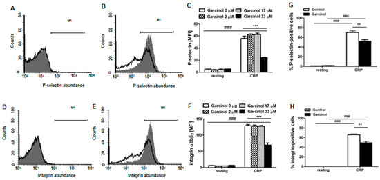
Garcinol, an anti-inflammatory and anti-carcinogenic polyisoprenylated benzophenone isolated from Garcinia plants, stimulates tumor cell apoptosis and suicidal erythrocyte death, but supports the survival of hepatocytes and neurons. The present study explored whether the substance influences platelet function and/or apoptosis. To this end, we exposed murine blood platelets to garcinol (33 µM, 30 min) without and with activation by collagen-related peptide (CRP) (2–5 µg/mL) or thrombin (0.01 U/mL); flow cytometry was employed to estimate cytosolic Ca2+-activity ([Ca2+]i) from Fluo-3 fluorescence, platelet degranulation from P-selectin abundance, integrin activation from αIIbβ3 integrin abundance, caspase activity utilizing an Active Caspase-3 Staining kit, phosphatidylserine abundance from annexin-V-binding, relative platelet volume from forward scatter, and aggregation utilizing staining with CD9-APC and CD9-PE. As a result, in the absence of CRP and thrombin, the exposure of the platelets to garcinol did not significantly modify [Ca2+]i, P-selectin abundance, activated αIIbβ3 integrin, annexin-V-binding, cell volume, caspase activity, and aggregation. Exposure of platelets to CRP or thrombin was followed by a significant increase of [Ca2+]i, P-selectin abundance, αIIbβ3 integrin activity, annexin-V-binding, caspase activity, and aggregation, as well as significant cell shrinkage. All effects of CRP were strong and significant; those of thrombin were only partially and slightly blunted in the presence of garcinol. In conclusion, garcinol blunts CRP-induced platelet activity, apoptosis and aggregation. Full article
Show Figures

Figure 1

21 pages, 3616 KiB  
Article
Intramuscular Ricin Poisoning of Mice Leads to Widespread Damage in the Heart, Spleen, and Bone Marrow
by Anita Sapoznikov, Amir Rosner, Reut Falach, Yoav Gal, Moshe Aftalion, Yentl Evgy, Ofir Israeli, Tamar Sabo and Chanoch Kronman
Toxins 2019, 11(6), 344; https://doi.org/10.3390/toxins11060344 - 16 Jun 2019
Cited by 13 | Viewed by 4802
Abstract
Ricin, a lethal toxin derived from castor oil beans, is a potential bio-threat due to its high availability and simplicity of preparation. Ricin is prepared according to simple recipes available on the internet, and was recently considered in terrorist, suicide, or homicide attempts [...] Read more.
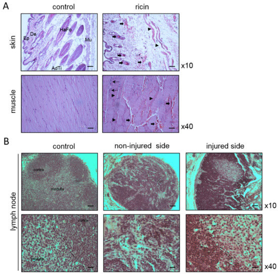
Ricin, a lethal toxin derived from castor oil beans, is a potential bio-threat due to its high availability and simplicity of preparation. Ricin is prepared according to simple recipes available on the internet, and was recently considered in terrorist, suicide, or homicide attempts involving the parenteral route of exposure. In-depth study of the morbidity developing from parenteral ricin poisoning is mandatory for tailoring appropriate therapeutic measures to mitigate ricin toxicity in such instances. The present study applies various biochemical, hematological, histopathological, molecular, and functional approaches to broadly investigate the systemic effects of parenteral intoxication by a lethal dose of ricin in a murine model. Along with prompt coagulopathy, multi-organ hemorrhages, and thrombocytopenia, ricin induced profound morpho-pathological and functional damage in the spleen, bone marrow, and cardiovascular system. In the heart, diffuse hemorrhages, myocyte necrosis, collagen deposition, and induction in fibrinogen were observed. Severe functional impairment was manifested by marked thickening of the left ventricular wall, decreased ventricular volume, and a significant reduction in stroke volume and cardiac output. Unexpectedly, the differential severity of the ricin-induced damage did not correlate with the respective ricin-dependent catalytic activity measured in the various organs. These findings emphasize the complexity of ricin toxicity and stress the importance of developing novel therapeutic strategies that will combine not only anti-ricin specific therapy, but also will target ricin-induced indirect disturbances. Full article
Show Figures

Figure 1

16 pages, 3102 KiB  
Article
Xanthium strumarium Fruit Extract Inhibits ATG4B and Diminishes the Proliferation and Metastatic Characteristics of Colorectal Cancer Cells
by Hsueh-Wei Chang, Pei-Feng Liu, Wei-Lun Tsai, Wan-Hsiang Hu, Yu-Chang Hu, Hsiu-Chen Yang, Wei-Yu Lin, Jing-Ru Weng and Chih-Wen Shu
Toxins 2019, 11(6), 313; https://doi.org/10.3390/toxins11060313 - 2 Jun 2019
Cited by 29 | Viewed by 4649
Abstract
Autophagy is an evolutionarily conserved pathway to degrade damaged proteins and organelles for subsequent recycling in cells during times of nutrient deprivation. This process plays an important role in tumor development and progression, allowing cancer cells to survive in nutrient-poor environments. The plant [...] Read more.
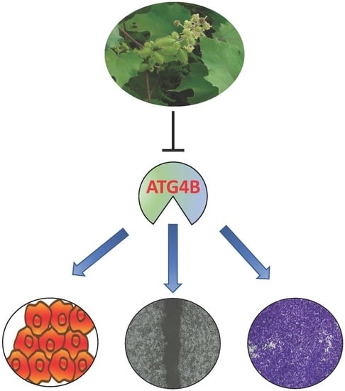
Autophagy is an evolutionarily conserved pathway to degrade damaged proteins and organelles for subsequent recycling in cells during times of nutrient deprivation. This process plays an important role in tumor development and progression, allowing cancer cells to survive in nutrient-poor environments. The plant kingdom provides a powerful source for new drug development to treat cancer. Several plant extracts induce autophagy in cancer cells. However, little is known about the role of plant extracts in autophagy inhibition, particularly autophagy-related (ATG) proteins. In this study, we employed S-tagged gamma-aminobutyric acid receptor associated protein like 2 (GABARAPL2) as a reporter to screen 48 plant extracts for their effects on the activity of autophagy protease ATG4B. Xanthium strumarium and Tribulus terrestris fruit extracts were validated as potential ATG4B inhibitors by another reporter substrate MAP1LC3B-PLA2. The inhibitory effects of the extracts on cellular ATG4B and autophagic flux were further confirmed. Moreover, the plant extracts significantly reduced colorectal cancer cell viability and sensitized cancer cells to starvation conditions. The fruit extract of X. strumarium consistently diminished cancer cell migration and invasion. Taken together, the results showed that the fruit of X. strumarium may have an active ingredient to inhibit ATG4B and suppress the proliferation and metastatic characteristics of colorectal cancer cells. Full article
Show Figures

Graphical abstract

11 pages, 2396 KiB  
Article
Epoxyscillirosidine Induced Cytotoxicity and Ultrastructural Changes in a Rat Embryonic Cardiomyocyte (H9c2) Cell Line
by Hamza Ibrahim Isa, Gezina Catharina Helena Ferreira, Jan Ernst Crafford and Christoffel Jacobus Botha
Toxins 2019, 11(5), 284; https://doi.org/10.3390/toxins11050284 - 21 May 2019
Cited by 2 | Viewed by 4463
Abstract
Moraea pallida Bak. (yellow tulp) poisoning is the most important cardiac glycoside-induced intoxication in ruminants in South Africa. The toxic principle, 1α, 2α-epoxyscillirosidine, is a bufadienolide. To replace the use of sentient animals in toxicity testing, the aim of this study was to [...] Read more.
Moraea pallida Bak. (yellow tulp) poisoning is the most important cardiac glycoside-induced intoxication in ruminants in South Africa. The toxic principle, 1α, 2α-epoxyscillirosidine, is a bufadienolide. To replace the use of sentient animals in toxicity testing, the aim of this study was to evaluate the cytotoxic effects of epoxyscillirosidine on rat embryonic cardiomyocytes (H9c2 cell line). This in vitro cell model can then be used in future toxin neutralization or toxico-therapy studies. Cell viability, evaluated with the methyl blue thiazol tetrazolium (MTT) assay, indicated a hormetic dose/concentration response, characterized by a biphasic low dose stimulation and high dose inhibition. Increased cell membrane permeability and leakage, as expected with necrotic cells, were demonstrated with the lactate dehydrogenase (LDH) assay. The LC50 was 382.68, 132.28 and 289.23 μM for 24, 48, and 72 h respectively. Numerous cytoplasmic vacuoles, karyolysis and damage to the cell membrane, indicative of necrosis, were observed at higher doses. Ultra-structural changes suggested that the cause of H9c2 cell death, subsequent to epoxyscillirosidine exposure, is necrosis, which is consistent with myocardial necrosis observed at necropsy. Based on the toxicity observed, and supported by ultra-structural findings, the H9c2 cell line could be a suitable in vitro model to evaluate epoxyscillirosidine neutralization or other therapeutic interventions in the future. Full article
Show Figures

Figure 1

16 pages, 2505 KiB  
Article
Whole-Cell Multiparameter Assay for Ricin and Abrin Activity-Based Digital Holographic Microscopy
by Efi Makdasi, Orly Laskar, Elad Milrot, Ofir Schuster, Shlomo Shmaya and Shmuel Yitzhaki
Toxins 2019, 11(3), 174; https://doi.org/10.3390/toxins11030174 - 22 Mar 2019
Cited by 12 | Viewed by 5106
Abstract
Ricin and abrin are ribosome-inactivating proteins leading to inhibition of protein synthesis and cell death. These toxins are considered some of the most potent and lethal toxins against which there is no available antidote. Digital holographic microscopy (DHM) is a time-lapse, label-free, and [...] Read more.
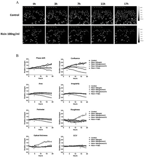
Ricin and abrin are ribosome-inactivating proteins leading to inhibition of protein synthesis and cell death. These toxins are considered some of the most potent and lethal toxins against which there is no available antidote. Digital holographic microscopy (DHM) is a time-lapse, label-free, and noninvasive imaging technique that can provide phase information on morphological features of cells. In this study, we employed DHM to evaluate the morphological changes of cell lines during ricin and abrin intoxication. We showed that the effect of these toxins is characterized by a decrease in cell confluence and changes in morphological parameters such as cell area, perimeter, irregularity, and roughness. In addition, changes in optical parameters such as phase-shift, optical thickness, and effective-calculated volume were observed. These effects were completely inhibited by specific neutralizing antibodies. An enhanced intoxication effect was observed for preadherent compared to adherent cells, as was detected in early morphology changes and confirmed by annexin V/propidium iodide (PI) apoptosis assay. Detection of the dynamic changes in cell morphology at initial stages of cell intoxication by DHM emphasizes the highly sensitive and rapid nature of this method, allowing the early detection of active toxins. Full article
Show Figures

Figure 1

15 pages, 4279 KiB  
Article
Evaluation of the Liver Toxicity of Pterocephalus hookeri Extract via Triggering Necrosis
by Rui Wang, Zhaoyue Dong, Xiaolong Zhang, Jingxin Mao, Fancheng Meng, Xiaozhong Lan, Zhihua Liao and Min Chen
Toxins 2019, 11(3), 142; https://doi.org/10.3390/toxins11030142 - 2 Mar 2019
Cited by 11 | Viewed by 3281
Abstract
Pterocephalus hookeri (C. B. Clarke) Höeck, recorded in the Chinese Pharmacopoeia (2015 version) as a Tibetan medicine for the treatment of various diseases, especially rheumatoid arthritis, was believed to possess a slight toxicity. However, hardly any research has been carried out about it. [...] Read more.
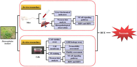
Pterocephalus hookeri (C. B. Clarke) Höeck, recorded in the Chinese Pharmacopoeia (2015 version) as a Tibetan medicine for the treatment of various diseases, especially rheumatoid arthritis, was believed to possess a slight toxicity. However, hardly any research has been carried out about it. The present study aimed to evaluate the toxicity in vivo and in vitro. Toxicity was observed by the evaluation of mice weight loss and histopathological changes in the liver. Then, the comparison research between ethyl acetate extract (EAE) and n-butanol extract (BUE) suggested that liver toxicity was mainly induced by BUE. The mechanical study suggested that BUE-induced liver toxicity was closely associated with necrosis detected by MTT and propidium iodide (PI) staining, via releasing lactate dehydrogenase (LDH), reducing the fluidity, and increasing the permeability of the cell membrane. Western blot analysis confirmed that the necrosis occurred molecularly by the up-regulation of receptor-interacting protein kinase 1 (RIP1) and receptor-interacting protein kinase 3 (RIP3), as well as the activation of the nuclear factor-kappa-gene binding (NF-κB) signaling pathway in vivo and in vitro. This finding indicated that the liver toxicity induced by BUE from P. hookeri was mainly caused by necrosis, which provides an important theoretical support for further evaluation of the safety of this folk medicine. Full article
Show Figures

Graphical abstract

9 pages, 1549 KiB  
Communication
The Inhibitory Effect of Celangulin V on the ATP Hydrolytic Activity of the Complex of V-ATPase Subunits A and B in the Midgut of Mythimna separata
by Liwen Ding, Zongxin Guo, Hang Xu, Tie Li, Yuanyuan Wang and Hu Tao
Toxins 2019, 11(2), 130; https://doi.org/10.3390/toxins11020130 - 22 Feb 2019
Cited by 5 | Viewed by 2801
Abstract
Celangulin V (CV) is a compound isolated from Celastrus angulatus Max that has a toxic activity against agricultural insect pests. CV can bind to subunits a, H, and B of the vacuolar ATPase (V-ATPase) in the midgut epithelial cells of insects. However, the [...] Read more.
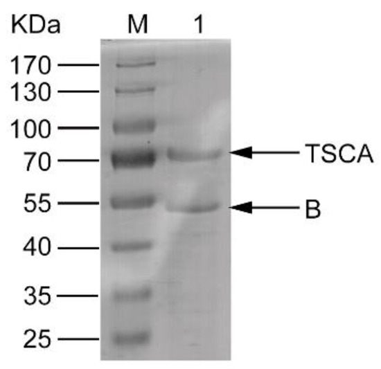
Celangulin V (CV) is a compound isolated from Celastrus angulatus Max that has a toxic activity against agricultural insect pests. CV can bind to subunits a, H, and B of the vacuolar ATPase (V-ATPase) in the midgut epithelial cells of insects. However, the mechanism of action of CV is still unclear. In this study, the soluble complex of the V-ATPase A subunit mutant TSCA which avoids the feedback inhibition by the hydrolysate ADP and V-ATPase B subunit were obtained and then purified using affinity chromatography. The H+K+-ATPase activity of the complex and the inhibitory activity of CV on ATP hydrolysis were determined. The results suggest that CV inhibits the ATP hydrolysis, resulting in an insecticidal effect. Additionally, the homology modeling of the AB complex and molecular docking results indicate that CV can competitively bind to the AB complex at the ATP binding site, which inhibits ATP hydrolysis. These findings suggest that the AB subunits complex is one of the potential targets for CV and is important for understanding the mechanism of interaction between CV and V-ATPase. Full article
Show Figures

Figure 1

21 pages, 9611 KiB  
Article
Augmentation of Saporin-Based Immunotoxins for Human Leukaemia and Lymphoma Cells by Triterpenoid Saponins: The Modifying Effects of Small Molecule Pharmacological Agents
by Wendy S. Smith, David A. Johnston, Suzanne E. Holmes, Harrison J. Wensley, Sopsamorn U. Flavell and David J. Flavell
Toxins 2019, 11(2), 127; https://doi.org/10.3390/toxins11020127 - 20 Feb 2019
Cited by 8 | Viewed by 3286
Abstract
Triterpenoid saponins from Saponinum album (SA) significantly augment the cytotoxicity of saporin-based immunotoxins but the mechanism of augmentation is not fully understood. We investigated the effects of six small molecule pharmacological agents, which interfere with endocytic and other processes, on SA-mediated augmentation of [...] Read more.
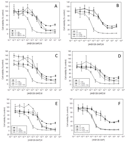
Triterpenoid saponins from Saponinum album (SA) significantly augment the cytotoxicity of saporin-based immunotoxins but the mechanism of augmentation is not fully understood. We investigated the effects of six small molecule pharmacological agents, which interfere with endocytic and other processes, on SA-mediated augmentation of saporin and saporin-based immunotoxins (ITs) directed against CD7, CD19, CD22 and CD38 on human lymphoma and leukaemia cell lines. Inhibition of clathrin-mediated endocytosis or endosomal acidification abolished the SA augmentation of saporin and of all four immunotoxins tested but the cytotoxicity of each IT or saporin alone was largely unaffected. The data support the hypothesis that endocytic processes are involved in the augmentative action of SA for saporin ITs targeted against a range of antigens expressed by leukaemia and lymphoma cells. In addition, the reactive oxygen species (ROS) scavenger tiron reduced the cytotoxicity of BU12-SAP and OKT10-SAP but had no effect on 4KB128-SAP or saporin cytotoxicity. Tiron also had no effect on SA-mediated augmentation of the saporin-based ITs or unconjugated saporin. These results suggest that ROS are not involved in the augmentation of saporin ITs and that ROS induction is target antigen-dependent and not directly due to the cytotoxic action of the toxin moiety. Full article
Show Figures

Figure 1

12 pages, 9389 KiB  
Article
In Vitro Cytotoxicity Induced by the Bufadienolides 1α,2α-Epoxyscillirosidine and Lanceotoxin B on Rat Myocardial and Mouse Neuroblastoma Cell Lines
by Danielle Henn, Annette Venter and Christo Botha
Toxins 2019, 11(1), 14; https://doi.org/10.3390/toxins11010014 - 2 Jan 2019
Cited by 7 | Viewed by 2902
Abstract
Consumption of bufadienolide-containing plants are responsible for many livestock mortalities annually. Bufadienolides are divided into two groups; non-cumulative bufadienolides and cumulative bufadienolides. Cumulative bufadienolides are referred to as neurotoxic, as the chronic intoxication with this type of bufadienolide results in a paretic/paralytic syndrome [...] Read more.
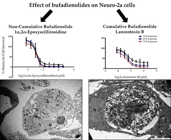
Consumption of bufadienolide-containing plants are responsible for many livestock mortalities annually. Bufadienolides are divided into two groups; non-cumulative bufadienolides and cumulative bufadienolides. Cumulative bufadienolides are referred to as neurotoxic, as the chronic intoxication with this type of bufadienolide results in a paretic/paralytic syndrome known as ‘krimpsiekte’. The in vitro cytotoxicity of a non-cumulative bufadienolide, 1α,2α-epoxyscillirosidine, and a cumulative bufadienolide, lanceotoxin B, were compared using the MTT ((3-(4,5-dimethylthiazol-2-yl)-2,5-diphenyltetrazolium bromide reduction) assay after exposing rat myocardial (H9c2) and mouse neuroblastoma (Neuro-2a) cell lines. The effect of these two bufadienolides on cell ultrastructure was also investigated using transmission electron microscopy (TEM). H9c2 cells exhibited greater cytotoxicity when exposed to 1α,2α-epoxyscillirosidine, compared to lanceotoxin B. In contrast, Neuro-2a cells were more susceptible to lanceotoxin B. The EC50 (half maximal effective concentration) of lanceotoxin B exposure of Neuro-2a cells for 24–72 h ranged from 4.4–5.5 µM compared to EC50s of 35.7–37.6 µM for 1α,2α-epoxyscillirosidine exposure of Neuro-2a cells over the same period. 1α,2α-Epoxyscillirosidine induced extensive vacuolization in both cell types, with swollen RER (rough endoplasmic reticulum) and perinuclear spaces. Lanceotoxin B caused swelling of the mitochondria and sequestration of cytoplasmic material within autophagic vesicles. These results corroborate the notion that cumulative bufadienolides are neurotoxic. Full article
Show Figures

Graphical abstract

Back to TopTop