1. Introduction
Genetic selection for highly prolific sows has been found to largely increase litter size [
1]. However, it is challenging for sows to nurse a number of piglets that exceeds the number of functional teats, which results in higher pre-weaning mortality and poor pig uniformity at weaning [
2]. Milk replacer is recommended as an effective dietary strategy when sow milk is unavailable or insufficient to satisfy piglets’ nutritional needs [
3]. Studies on optimizing milk replacer using functional additives, such as oligosaccharide [
4,
5], lactoferrin [
6] and β-glucan [
7], have been reported, but limited attention has been given to protein sources. Protein is a crucial nutrient for neonates, serving as a vital source of essential amino acids and fundamental bioactive substances for early-stage development [
8]. Meanwhile, the digestion and absorption of protein are closely associated with nutritional status and intestinal health [
9].
As a dairy–based protein, whey protein concentrate (WPC) has been widely used as a major protein source in milk replacer [
10,
11]. It has been reported that milk replacer with WPC contributes to the intestinal maturation and health of preterm piglets [
12]. As an expensive component, however, dairy-based protein has been partially substituted with wheat or soy proteins in milk replacers for calves [
13]. And soy protein–based formula has been reported to maintain the normal growth and development of infants and reduce gastrointestinal symptoms in infants who are intolerant to cows’ milk [
14,
15,
16]. Although soy protein isolate (SPI) is considered an ideal plant–based protein in formula milk, there are concerns regarding the potential negative effects of soy–based protein on digestibility and immune and nervous system development due to insufficient processing for anti-nutritive factors [
17,
18,
19]. To our knowledge, however, very limited data are available on the effects of soy–based protein as a substitute for dairy–based protein in milk replacer on the growth and development of piglets. In contrast, functional proteins could also be considered for improving neonatal growth and maturity. Spray–dried porcine plasma (SDPP) is a high–quality animal–based protein, and is widely used to improve the intestinal health, immunity and growth of weaning piglets due to its enrichment in fibrinogen, immunoglobulins and albumin [
20,
21,
22]. However, the feeding effect of SDPP as a protein source to partially substitute WPC in milk replacer for piglets has not been determined.
In this study, therefore, we aimed to investigate the effects of partially substituting the dairy–based protein WPC in milk replacer with the animal–based protein SDPP or the plant–based protein SPI on the growth performance, intestinal morphology, activity of digestive enzymes, immunity–related parameters, intestinal microbiota composition and metabolites in neonatal piglets.
2. Materials and Methods
2.1. Experimental Design, Animals and Growth Performance
Twenty–four Landrace/Yorkshire crossbred piglets (2 days old) with an average initial body weight of 1.55 ± 0.23 kg, including a mix of females and males, were selected for this experiment. After birth, piglets were allowed to suckle from their respective sows for 48 h prior to being transported to a nursery room, and were housed individually in stainless steel metabolic crates (0.6 × 0.9 × 0.6 m). Piglets were randomly divided into three treatment groups receiving experimental diets, including a milk replacer with 17.70% WPC (WPC group,
n = 8), a milk replacer with 6% SDPP isonitrogenously substituting WPC (SDPP group,
n = 8), and a milk replacer with 5.13% SPI isonitrogenously substituting WPC (SPI group,
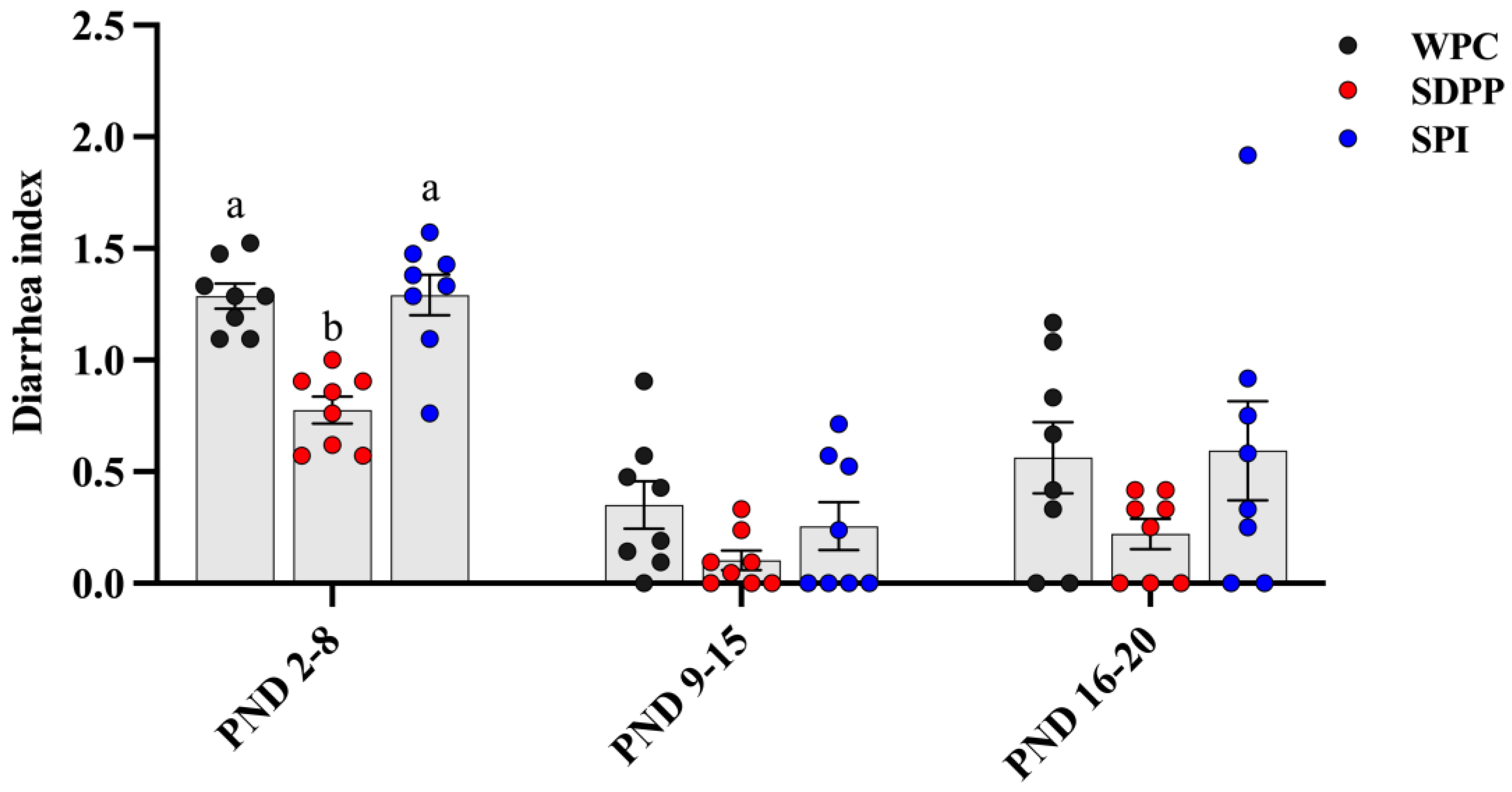
n = 8). The nursery room temperature was controlled at 28~31 °C. The intake of milk replacer was converted into dry matter intake using a dry matter–to–water ratio of 1:4, and piglets were weighed individually at the start and end of the study to calculate the average daily feed intake (ADFI), average daily gain (ADG) and ratio of ADFI to ADG (F/G). Visual diarrhea scores were assessed three times daily and the diarrhea index was calculated [
23].
2.2. Milk Replacer
The milk replacer’s ingredients and nutrient levels are described in
Table 1, and it was formulated to mimic the macronutrient composition of sow milk [
24,
25,
26]. The milk replacer was mixed with 40 °C water at a ratio of 1:4 and added to a milk bucket for storage. It was added 7 times a day to ensure that there was surplus milk in the bucket. The milk was transported from the bucket to the nipple on the metabolism cage, and the piglets could freely drink milk through the nipple until sacrifice on PND 21.
2.3. Sample Collection
Prior to slaughter, milk replacer and water were withheld from piglets for 12 h. Once their final body weight was recorded, blood was collected from the anterior vena cava. Plasma samples were obtained by centrifuging blood samples stored in heparin anticoagulated tubes at 3000× g for 15 min at 4 °C. In order to conduct histological analysis, piglets were anesthetized intramuscularly using 0.5 mL of xylazine and midazolam. Following slaughter, the intestines of piglets were isolated and a 2 cm length piece of the ileum was stored in a 4% paraformaldehyde solution. Tissue samples from the jejunum and ileum were collected and washed in cold saline solution (NaCl 9 g/L, 4 °C). Similarly, chyme samples from the colon were collected, frozen in liquid nitrogen and stored at −80 °C for 16S rRNA sequencing to assess the microbial community composition.
2.4. Histomorphology
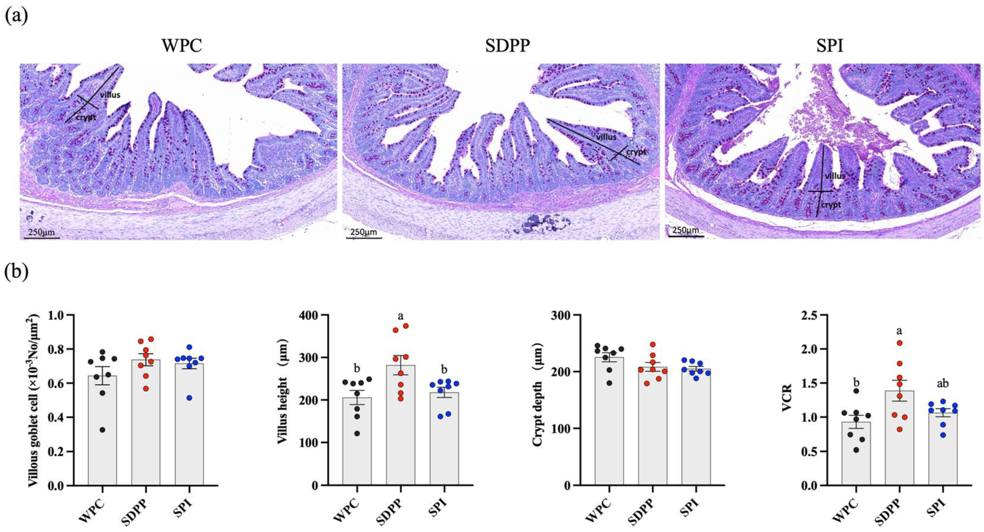
Pieces of ileum were fixed in 4% paraformaldehyde solution for histomorphometric analysis. Ileum samples were dehydrated and embedded in paraffin. Sections 5 μm thick, cut using a Leica RM2235 microtome (Leica Microsystems Ltd., Shanghai, China), were stained using the periodic acid–Schiff (PAS) method. Image Pro Plus 6.0 software (Media Cybernetics, Rockville, MD, USA) was used to acquire images and to measure the villus height, the depth of crypts and the area of goblet cells in the tissue.
2.5. Digestive Enzyme Activity
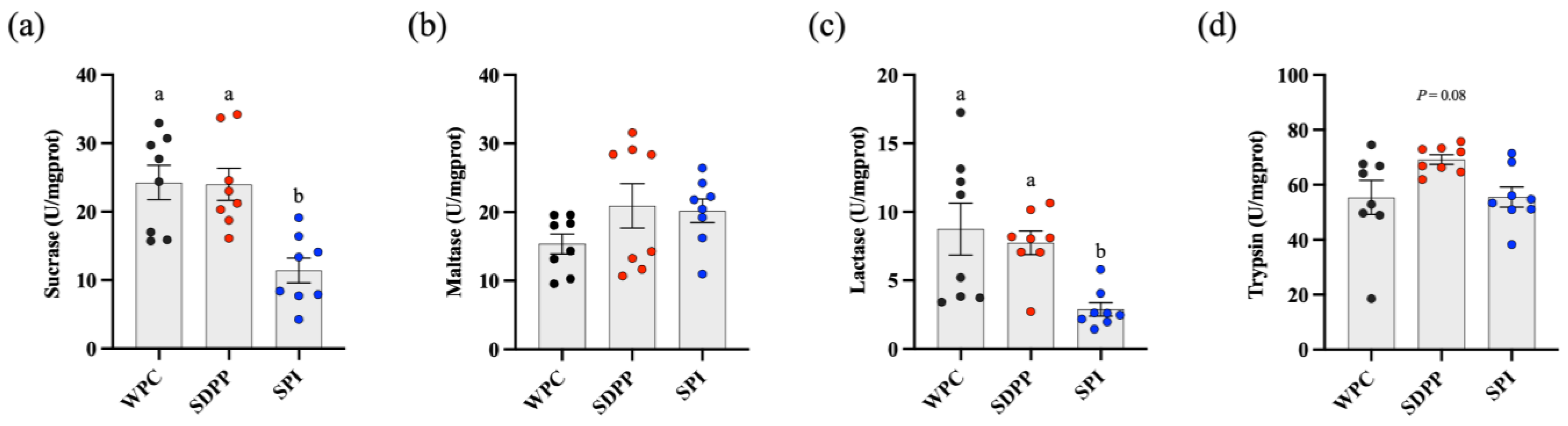
Jejunum samples were homogenized using 4 °C pre–cooled 0.9% saline for further analysis. The protein content of the samples was determined using the Coomassie brilliant blue method, and the test kit was purchased from Nanjing Jiancheng (Nanjing Jiancheng Bioengineering Institute, Nanjing, China). The activities of sucrase, maltase, lactase and trypsin were determined according to the instructions for the Nanjing Jiancheng kit (Nanjing Jiancheng Bioengineering Institute, Nanjing, China). The optical density values of disaccharidase and trypsin were measured at 505 nm and 253 nm, respectively, using a SpectraMax190 microplate reader (Molecular Devices Corporation, San Jose, CA, USA); then, the activities of the enzymes were calculated according to the change in absorbance.
2.6. Gene Expressions
Extracted ileal RNA with a concentration of 500~1000 ng/μL was used to synthesize cDNA, according to the instructions for the reverse transcription kit (Vazyme) (RNase-free ddH
2O, 4× gDNA Wiper Mix, 5× HiScript III qRT SuperMix). Quantitative PCR (2× ChamQ Universal SYBR qPCR Master Mix, Primer, Template DNA/cDNA, ddH
2O) was carried out using the QuantStudio5 instrument (Thermo Fisher Scientific, Waltham, MA, USA). Data were normalized using β-actin and the relative expression was calculated using the 2
−ΔΔCt method [
27]. The primers used in this experiment are listed in
Table 2.
2.7. 16S rRNA Amplicon Sequencing
Colonic chyme samples were used for 16S rRNA amplicon (Novogene Technology Co., Ltd., Beijing, China). Genomic DNA was extracted using either the CTAB or SDS method. Specific primers with barcodes, the Phusion® High–Fidelity PCR Master Mix with GC buffer (New England Biolabs) and high–efficiency high–fidelity enzymes were used to perform PCR. The NEBNext® Ultra™ IIDNA Library Prep Kit (New England Biolabs, Ipswich, MA, USA) was utilized for library construction. Then, the constructed library was qualified and sequenced using the NovaSeq6000 instrument (Illumina, San Diego, CA, USA). After careful processing and analysis, the effective tags were obtained and denoised using QIIME2 software (2019.1) to generate ASVs (Amplicon Sequence Variants) and feature tables. The species information for each ASV was determined by comparing it to a database. Finally, QIIME2 software was used to conduct alpha and beta diversity analysis.
2.8. SCFA Analysis
Briefly, 0.5 g of colonic chyme sample and 1.2 mL of ultrapure water were added to a centrifuge tube after thawing, allowed to stand for 30 min and centrifuged at 10,000× g for 15 min. After centrifugation, 0.2 mL of 25% (w/v) metaphosphoric acid and 23.3 μL of 210 mmol/L crotonic acid were added to 1 mL of supernatant, mixed well, and left to stand at 4 °C for 30 min, and then, centrifuged at 8000× g for 10 min. After centrifugation, 0.9 mL of chromatographic methanol (1:3 dilution) was added to 0.3 mL of supernatant, and mixed well. Finally, the supernatant was filtered using a 0.22 μm filter membrane into a 1.5 mL EP tube for later use. SCFA concentrations were measured using GC CP3800 gas chromatography (Varian Medical Systems, Palo Alto, CA, USA).
2.9. Statistical Analysis
The Shapiro–Wilk test and UNIVARIATE procedures of SAS 9.4 (SAS Institute, Inc., Cary, NC, USA) were used to analyze variance homogeneity and normality, respectively. Data were analyzed using one–way analysis of variance (ANOVA), and the LSD method was used for multiple comparisons. Data are presented as mean ± pooled standard error (SEM) and considered significant at p < 0.05 and a tendency at p < 0.10.
4. Discussion
Whey protein concentrate (WPC) is a commonly used dairy–based protein in milk replacer for piglets [
28]. However, the efficacy of the partial substitution of WPC with animal–based or plant–based protein on the growth and intestinal function of piglets needs to be determined.
In the present study, the higher final BW and ADG, and the lower F/G, in SDPP–fed piglets indicated the crucial role of SDPP in improving the growth performance of piglets, which is consistent with previous results from studies that include SDPP in weaning diets in which it increased post–weaning growth rate [
29,
30,
31]. Also, calves fed a milk replacer using SDPP substituting 20% of the WPC had significantly increased feed intake and feed conversion ratios [
32]. Meanwhile, the diarrhea index from PND 2 to PND 8 was lower in SDPP–fed piglets, which is consistent with a previous study in which the fecal scores of calves tended to be lower when SDPP was included in the milk replacer [
32]. The improvements in SDPP on growth rate and diarrhea index could be associated with improved intestinal function. The intestinal mucosa plays a crucial role in nutrient absorption [
33]. Our results showed that SDPP–fed piglets had significantly increased ileal villus height and VCR compared with WPC– and SPI–fed piglets, indicating an expansion of the intestinal epithelial surface area [
34], and an improvement in epithelial cell renewal [
35,
36]. Consistently, the weaned piglets fed an SDPP diet had significantly increased ileal villus height relative to those fed a soybean protein concentrate (SPC) diet [
37]. Intestinal enzyme activity can be altered in response to diet as early as the second day after birth [
38]. Piglets fed colostrum-containing plasma showed increased activities of sucrase and maltase (50% and 200%, respectively) compared with colostrum-fed piglets [
39]. In this study, moreover, SDPP–fed piglets had increased activities of sucrase, lactase and trypsin in the jejunum compared with WPC– and SPI–fed piglets, which suggested that SDPP–fed piglets were favorable for protein and carbohydrate digestion.
In contrast, SPI–fed piglets had the lowest growth rate, which may be associated with poor digestive capability, immunity and bacterial structure. Our study found that SPI–fed piglets had higher plasma urea, indicating a potential increase in protein breakdown or lower protein utilization, because urea is the main end product of amino acid and protein metabolism, which is negatively correlated with amino acid utilization and protein retention [
40]. Immunity is crucial for the intestinal health of neonates. The dynamic changes in levels of pro–inflammatory factor
IL–6 and anti-inflammatory factor
IL–10 play an important role in regulating the inflammatory response system. In this study, both
IL–6 and
IL–10 were over-expressed in the ileum of SPI–fed piglets. This finding seems paradoxical considering the anti–inflammatory effects of
IL–10. However, previous research has found that an increased level of
IL–10 is a protective mechanism for the body against the release of a large number of pro–inflammatory cytokines [
41]. In addition, elevated
IL–6 can also stimulate the release of
IL–10 in local tissues [
42]. Therefore, we speculated that feeding milk replacer including SPI may pose a challenge to the intestinal immunity of neonatal piglets.
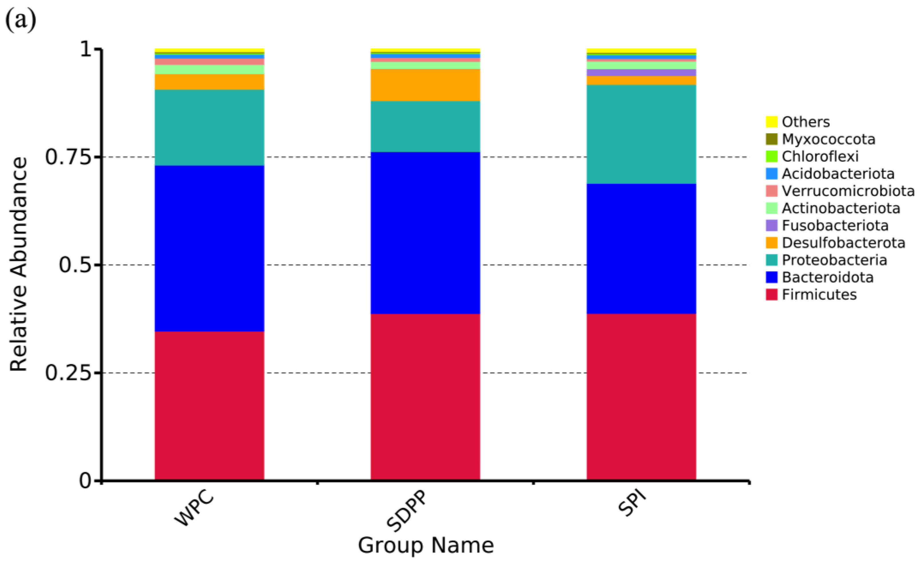
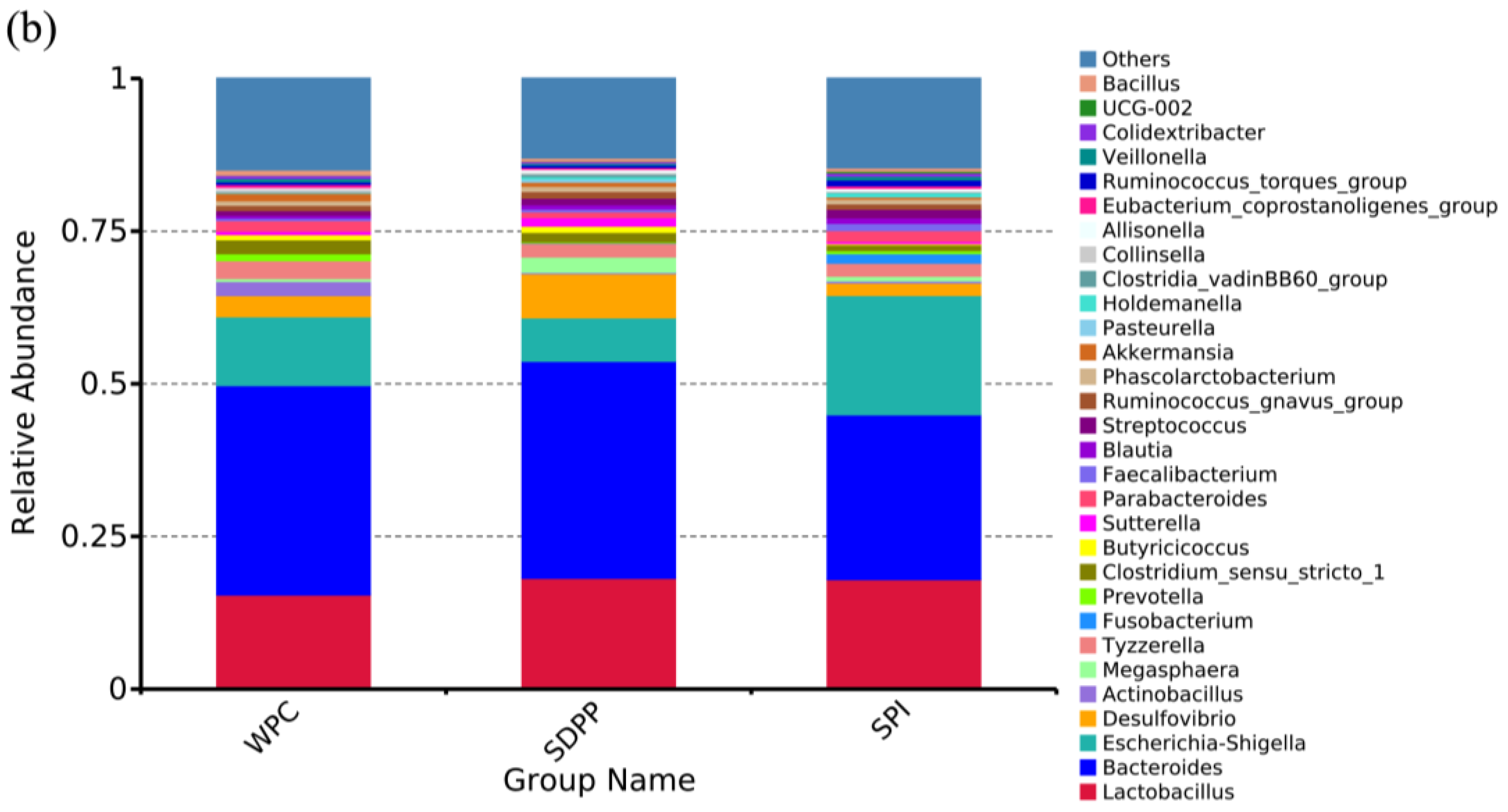
The normal intestinal microbiota is essential for intestinal homeostasis [
43]. Dietary protein is the preferred substrate for the intestinal microbiota and affects the composition and metabolic activity of microbiota [
44]. In this study, LEfSe analysis found that microbiota composition was significantly different across the WPC, SDPP and SPI groups. At the phylum level,
Firmicutes,
Bacteroidetes and
Proteobacteria were the most abundant bacterial phyla in the colonic chyme. SDPP-fed piglets had lower
Proteobacteria levels compared with WPC– or SPI–fed piglets. Interestingly, previous research demonstrated that breast–fed infants had lower
Proteobacteria levels (3.29%) compared with formula–fed infants (13.85%) [
45]. Furthermore,
Proteobacteria has been linked to inflammation and intestinal dysbiosis [
46,
47]. In this study, moreover, the abundance of
Butyricicoccus, a high butyrate–producing probiotic [
48], was increased in SDPP–fed piglets, which also explains the increased concentration of butyrate in SDPP–fed piglets. As a short–chain fatty acid, butyrate not only provides energy for colonic cells, but also inhibits the expression of pro–inflammatory cytokines [
49]. In addition, the abundance of some pathogenic bacteria, such as
Marinifilaceae,
Fusobacterium and
Enterococcus, was increased in the colonic chyme of WPC– and SPI–fed piglets. It has been reported that
Marinifilaceae was enriched in the lumen of colitis and infection animal models [
50,
51]. One member of
Fusobacterium,
F. nucleatum has been recognized as an opportunistic pathogen that plays an important role in intestinal diseases, which promotes the inflammatory response and induces the release of the pro–inflammatory cytokines
IL–6 and
IL–8 [
52,
53,
54]. This finding may explain the over-expression of
IL–6 in SPI–fed piglets.


 ,
, 


