Article

Relative Leukocyte Telomere Length and Telomerase Complex Regulatory Markers Association with Leber’s Hereditary Optic Neuropathy

by
Rasa Liutkeviciene
1,2,
Rasa Mikalauskaite
1,
Greta Gedvilaite
2,*,
Brigita Glebauskiene
1,
Loresa Kriauciuniene
1,2 and
Reda Žemaitienė
1
1
Department of Ophthalmology, Lithuanian University of Health Sciences, 44307 Kaunas, Lithuania
2
Neuroscience Institute, Lithuanian University of Health Sciences, 44307 Kaunas, Lithuania
*
Author to whom correspondence should be addressed.
Medicina 2022, 58(9), 1240; https://doi.org/10.3390/medicina58091240
Submission received: 22 June 2022 / Revised: 21 August 2022 / Accepted: 5 September 2022 / Published: 7 September 2022
(This article belongs to the Section Ophthalmology)

Abstract

:
Background and Objectives: To evaluate the association of relative leukocyte telomere length (RLTL) and telomerase complex regulatory markers with Leber’s hereditary optic neuropathy (LHON). Material and Methods: A case-control study was performed in patients with LHON (≥18 years) and healthy subjects. The diagnosis of LHON was based on a genetic blood test (next-generation sequencing with Illumina MiSeq, computer analysis: BWA2.1 Illumina BaseSpace, Alamut, and mtDNA Variant analyzer 1000 were performed) and diagnostic criteria approved by the LHON disease protocol. Statistical analysis was performed using the standard statistical software package, IBM SPSS Statistics 27. Statistically significant results were considered when p < 0.05. Results: Significantly longer RLTL was observed in LHON patients than in healthy controls (p < 0.001). RLTL was significantly longer in women and men with LOHN than in healthy women and men in the control group (p < 0.001 and p = 0.003, respectively). In the elderly group (>32 years), RLTL was statistically significantly longer in LHON patients compared with healthy subjects (p < 0.001). The GG genotype of the TERC rs12696304 polymorphism was found to be statistically significantly higher in the LHON group (p = 0.041), and the C allele in the TERC rs12696304 polymorphism was found to be statistically significantly less common in the LHON group (p < 0.001). The RLTL of LHON patients was found to be statistically significantly longer in the TERC rs12696304 polymorphism in all tested genotypes (CC, p = 0.005; CG, p = 0.008; GG, p = 0.025), TEP1 rs1760904 polymorphism in the GA genotype (p < 0.001), and TEP1 gene rs1713418 in the AA and AG genotypes (p = 0.011 and p < 0.001, respectively). Conclusions: The RLTL in LHON patients was found to be longer than in healthy subjects regardless of treatment with idebenone. The TERC rs12696304 polymorphism, of all studied polymorphisms, was the most significantly associated with changes in LHON and telomere length.

1. Introduction

Leber’s hereditary optic neuropathy (LHON) is one of the most common diseases caused by mutations in mitochondrial deoxyribonucleic acid (mtDNA) [1,2]. Symptomatic LHON disease leads to progressive, irreversible loss of visual acuity, including total blindness, and dramatically impacts the quality of life of affected patients [3,4,5,6]. The first symptoms of the disease usually appear in young adult males in the second to third decade of life, and 90% of affected individuals lose their vision before the age of 50 [2,7,8,9,10]. LHON is caused by a genetic mutation in the mitochondrial genome. The most common mutations are m.11778G > A, m.3460G > A, and m.14484T > C. In LHON, mutations in mtDNA in the retinal ganglion cells (RGC) lead to a decrease in adenosine triphosphate (ATF) synthesis, increased oxidative stress, and degeneration and apoptosis of these cells [11]. The definitive diagnosis of LHON is made when ophthalmologic features characteristic of the disease appear and/or a pathogenic variant of mtDNA is detected by molecular genetic testing. Evaluation of visual acuity is the primary goal in the treatment of this disease, and to date, the only approved drug for the treatment of LHON is idebenone. Idebenone, an analogue of coenzyme Q10, helps to restore impaired ATF production in retinal cells and activate the function of still viable but inactive cells [12,13]. Patients treated with idebenone have noted improvement in vision, but blindness due to optic nerve cell death remains a pharmacological challenge [2,14].
Telomeres are nucleoprotein complexes at the ends of eukaryotic chromosomes. The length of telomeres has been associated with a variety of diseases and pathologies [15,16]. Evidence suggests that relative leukocyte telomere length (RLTL) is related to age, sex, various oncologic and chronic diseases, diet and lifestyle, changes in telomere lengths associated with mitochondrial dysfunction, and longer telomere lengths in optic nerve and inflammatory eye diseases (Birdshot uveitis) [17]. Repeated telomere sequences at the ends of chromosomes are synthesized by telomerase [18]. Regulatory proteins inhibit their activity. Therefore, polymorphisms in the TERC gene rs12696304, rs35073794, and TEP1 gene rs1713418, rs1760904 are thought to affect telomerase activity and closely related to changes in RLTL [19,20,21].
In studying the pathogenesis of LHON, the most important changes occur in mitochondria, whose dysfunction can be associated with altered RLTL. The pathophysiology of the disease is complex, and selective damage to RGCs associated with LHON-specific mitochondrial dysfunction has not been fully elucidated [5]. Given the lack of studies examining the relationship between telomere length and damage to ocular structures, we decided to establish links between RLTL and LHON. It is important to continue the study of this disease at the molecular level, as the rapid development of research promotes the search for genetic markers that could potentially contribute to the discovery and development of treatments, and a better understanding of the pathogenesis mechanism would lead to earlier diagnosis.

2. Materials and Methods

2.1. Ethics Statement

All subjects have signed an agreement according to the Declaration of Helsinki (Nr-BE-2-102, approved date: 6 September 2019). The study was managed in the Department of Ophthalmology, Lithuanian University of Health Sciences. The control group included healthy people with no ocular pathology at an examination.
The exclusion criteria for Leber’s hereditary optic neuropathy (LHON) patients were:
  • Under the age of 18;
  • Systemic diseases (e.g., malignant tumors, systemic connective tissue diseases, diabetes mellitus, chronic infections, or conditions following organ or tissue transplantation).

2.2. Leber’s Hereditary Optic Neuropathy (LHON) Diagnostics

Individuals with LHON mutations are generally divided into asymptomatic mutation carriers and symptomatic patients with ocular lesions. The reason some carriers never experience symptoms is still unknown, but genetic factors and environmental influences, alcohol, and smoking are thought to play an important role [22].
The classic clinical manifestation of LHON is subacute, painless loss of central vision in one eye followed by loss of vision in the other eye over days, weeks, or months. It is essential to pay attention to macular and papillomacular retinal ganglion cells (RGC) due to the degeneration of these cells by optical coherence tomography (OCT). The common findings in asymptomatic mutation carriers and early subacute stage are optic nerve disc (OND) telangiectasia microangiopathy and OND swelling (pseudoedema) [5,23,24]. As the disease progresses, rapid axonal loss occurs first, followed by diffuse visual atrophy. The chronic stage of the disease is reached approximately 1 year after onset [5,23,24,25].
The final diagnosis of LHON is made when the probe exhibits ophthalmological features characteristic of the disease and/or one of the pathogenic variants of mtDNA is detected by molecular genetic testing.
A neuro-ophthalmology examination and next-generation sequencing (NGS) with Illumina MiSeq, computer analysis: BWA2.1 Illumina BaseSpace, Alamut, mtDNA Variant analyser 1000 were performed. Tested genes: MT-ND1, MT-ND2, MT-ND3, MT-ND4, MT-ND4L, MT-NDS, MT-ND6, MT-CYB, MT-CO1, MT-CO2, MT-CO3, MT-ATP6, MT-ATP8, MT-RNRI, MT-RNR2, MT-TA, MT-TT, MT-TP, MT-TE, MT-TL2, MT-TS2, MT-TH, MT-TR, MT-TG, MT-TK, MT-TD, MT-TS1, MT-TC, MT-TY, MT-TN, MT-TW, MT-TM, MT-TI, MT-TQ, MT-TL1, MT-TF, MT-TV for all the patients.

2.3. DNA Extraction, RLTL Measurement, and Genotyping

DNA extraction, relative telomere leukocyte length measurement, and analysis of TEP1 rs1760904, rs1713418, TERC rs12696304, and rs35073794 were carried out at the Laboratory of Ophthalmology, Neuroscience Institute, Lithuanian University of Health Sciences. DNA was extracted using the DNA salting-out method from venous blood samples.
SNPs were determined using TaqMan® genotyping assays (Thermo Scientific, Pleasanton, CA, USA). RLTL measurement and TEP1 rs1760904, rs1713418, TERC rs12696304, and rs35073794 genotyping were conducted using real-time PCR according to the manufacturer’s recommendations using a Step One Plus real-time PCR system (Applied Biosystems, Foster City, CA, USA).

2.4. Statistical Analysis

Statistical analysis was conducted with SPSS/W 27.0 software (Statistical Package for the Social Sciences for Windows, Inc., Chicago, IL, USA). Gender distribution was presented as absolute numbers with percentages and compared with the χ2 test. Data, which are not ordinally distributed between the two groups or subgroups, were measured with the Mann-Whitney U test.
We conducted a Hardy-Weinberg analysis to analyze the observed and expected frequencies of TEP1 rs1760904, rs1713418, TERC rs12696304, and rs35073794 with the χ2 test in the control group. The frequencies of TEP1 rs1760904, rs1713418, TERC rs12696304, and rs35073794 between groups were analyzed with the χ2 test. In addition, binary logistic regression analysis was performed to assess the influence of genotypes on the development of PA. The odds ratios (OR) and 95% confidence intervals (CI) are reported. The Akaike Information Criterion (AIC) selected the best genetic model. Statistically significant differences were considered when p < 0.05. We also presented an altered significance threshold for multiple comparisons, alpha = 0.017 (0.05/3) since we assessed three SNPs after excluding TERC rs35073794.

3. Results

The study included 144 subjects who were divided into two groups: a control group (n = 130) and a group of patients with Leber’s hereditary optic neuropathy (LHON) (n = 14). Genotyping of TEP1 rs1760904, rs1713418, TERC rs12696304, and rs35073794 polymorphisms was performed after the formation of the study groups. The LHON group of patients consisted of 14 subjects: 10 men (71.4%) and 4 women (28.6%). The mean age of the patients was 40.6 years. The control group consisted of 130 subjects: 61 men (46.9%) and 69 women (53.1%). The average age of the control group was 39.9 years. The study groups did not differ when compared by gender and age (p = 0.081, p = 0.843, respectively). The relative leukocyte telomeres length (RLTL) was determined in the subjects, and a comparison showed that the RLTL was statistically significantly longer in patients with LHON than in the control group (p < 0.001). The demographics of the subjects are presented in Table 1.
After estimating the frequencies of the genotypes according to the Hardy-Weinberg equilibrium (HWE), the results showed that TERC rs35073794 deviated from HWE (p < 0.05) (Table 2). This distribution of genotypes and alleles is possible due to the small sample size, so the sample should be increased in future studies [26]. Therefore, SNP was not included in the further analysis.

3.1. Influence of TEP1 rs1760904, rs1713418, TERC rs12696304 on LHON Development

The genotype and allele analysis revealed that the TERC rs12696304 GG genotype was more frequent in the LHON group than in the control group (21.4% vs. 6.2%, p = 0.041). In addition, the C allele was found to be statistically significantly less frequent in the LHON group compared with the control group (36.0% vs. 73.1%, p < 0.001) (Table 3).

3.2. Differences in Relative Leukocyte Telomere Length (RLTL) between LHON and Healthy Subjects

Analyzing the relative length of leukocyte telomere length, we found statistically significant differences between LHON and the control groups (median (IQR): 4.123 (9.039) vs. 0.613 (0.582), p < 0.001). The results are shown in Figure 1.
A comparison of RLTLs between different genotypes for all polymorphisms studied was also performed. RLTL was found to be statistically significantly longer in LHON than in controls with the TEP1 rs1760904 GA genotype (median (IQR): 4.505 (15.405) vs. 0.696 (0.549), p < 0.001). It was also statistically significantly longer in LHON than in controls with TEP1 rs1713418 AA (median (IQR): 3.474 (16.635) vs. 0.506 (0.682), p = 0.011) and AG genotypes (median (IQR): 7.940 (12.964) vs. 0.644 (0.565), p < 0.001). RLTL was also found to be statistically significantly longer in LHON than in controls with TERC rs12696304 CC (median (IQR): 3.097 (3.780) vs. 0.456 (0.652), p = 0.005), CG (median (IQR): 12.105 (16.187) vs. 0.694 (0.573), p = 0.008) and GG genotypes (median (IQR): 9.764 (-) vs. 0.982 (1.529), p = 0.025) (Table 4).

4. Discussion

In Lithuania, LHON disease has been diagnosed in 19 individuals since 2018, of whom 17 patients (13 men and 4 women) are monitored and treated at the Department of Ophthalmology, Lithuanian University of Health Sciences. LHON patients’ ages in Lithuania range from 9 to 61 years depending on the onset time (our study includes patients aged 19 to 61 years), and the literature describes cases in which the age varies from 2 to 87 years [27,28]. The mean age of LHON patients in our study was 38.93 years (SD = 14.69), but we included only adult patients older than 18 years, and the mean age of all patients currently treated in Kaunas clinics was 21.64 years. In the literature, an age of 22–25 years has been reported for the onset of LHON [27,28].
The male-to-female ratio was 2.75:1 in our study and 3.25:1 for all patients monitored at the eye clinic LUHS KC, with the majority of patients being male. These results are not significantly different from studies in Denmark (3.72:1) and the northwest of England (3.32:1) [27]. In our LHON study group, the proportion of males was 73.3 percent (76.5% of all patients treated for LUHS in that year), and the research data indicate that approximately 90% of patients are men [8]. This may be due to the fact that in Lithuania, LHON has only been diagnosed since 2018 and not all patients with LHON have been identified yet.
According to the data of this study, the diversity and prevalence of mutations in our country are slightly different from the three most common mtDNA mutations reported in the literature. The literature emphasizes that more than 90% of all patients with LHON (about 70% in Northern Europe) have one of the following three pathogenic variants: m.11778G > A (MT-ND4), m.14484T > C (MT-ND6), or m.3460G > A (MT-ND1) [1,6,24,29,30,31,32,33,34], but in our study in Lithuania, in patients with LHON > 18 years, these main mutations were detected only in 33.4%. (41.2% of all patients treated in LUHS KC in that year). From the available data, the most common LHON-causing mutation, m.11778G > A (MT-ND4), was diagnosed in 5 out of 17 patients (29.4%) in LUHS, which is in contrast to global statistics, in which this mutation is found in about 60% of diseased Northern Europeans and 90% of diseased Asians [7,30,31,34]. It is important to note that a relatively large variety of other (non-primary) mtDNA mutations leading to LHON disease were observed during the study survey. As many as four-tenths (40%) of the mutations identified in the study have not yet been included in the official list of pathogenic mutations in MitoMap, but have been described in studies as mutations associated with LHON (https://www.mitomap.org (accessed on 16 January 2022)). The following mutations are not included in the list: m.3394T > C (MT-ND1), m.3866T > C (MT-ND1), m.8836A > G (MT-ND4), and an autosomal recessive mutation in the DNAJC30 gene that was first described in the literature only in 2021 [35].
One of the main tasks of our study was to link RLTL to LHON. Prior to this study, shorter RLTL lengths were expected to be found in patients with primary mitochondrial disorders (e.g., diseases caused by mutations in the mitochondrial genome) and shorter telomeres compared with the healthy population, as described in the literature [36]. The literature also mentions that telomeres are sensitive to oxidative damage, leading to an increase in the rate of telomere shortening [37,38]. A study by Rovcanin et al. found that LHON patients have an increase in total oxidant status (TOS) and a decrease in total antioxidant status (TAS) levels, which suggests the existence of substantial oxidative stress [39]. As both telomeres and LHON patients are sensitive to oxidative stress, it could be an essential factor in the pathogenesis of the disease. One of the mechanisms in the pathogenesis of LHON is electron transport chain (ETC) dysfunction caused by primary mtDNA mutations, resulting in decreased ATF production, excessive secretion of ROS, and death of RGC [40]. However, this theory could not be confirmed in our study, and a statistically significant higher incidence of RLTL was observed in LHON patients than in healthy controls. This prompted a more detailed analysis of the literature, as no studies on the association between RLTL and LHON disease have been published to date. In the literature, an association between a genetically determined longer telomere length and an increased risk of certain oncological processes was observed [41], with lower stress levels, physical activity, a high-quality diet (e.g., with free ω-3 fatty acids, intake of some antioxidants, and low consumption of processed red meat), and adequate sleep associated with longer telomeres [42,43]. Studies of ocular structures showed that telomeres were longer in people with uveitis than in healthy controls (in cases of chronic inflammation) [17]. Long telomeres were observed in the neurosensory part of the retina (longer telomere lengths may be related to the fact that metabolic processes are most active in this part of the retina) [44]. In the pathogenesis of LHON, retinal ganglion cells are the most vulnerable, and therefore a longer RLTL was observed in patients with LHON, which stimulates discussion, and a larger sample of studies should be investigated in the future.
In our study, patients were categorized by sex and age, and the literature emphasizes the dependence of RLTL length on these two parameters. Previous studies showed that women’s telomeres are generally longer than those of men, and this phenomenon increases with age [45]. However, in our study, no statistically significant association was found between RLTL in relation to sex within the LHON or control groups; and statistically significantly longer RLTLs were found between LHON men and women compared with healthy control men and women, respectively.
The length of telomeres and their attrition rate were proposed in the literature as biological markers of age-related disease risk [46]; therefore, the data obtained in this study were disaggregated by age group. There was no statistically significant difference in RLTL between LHON and control subjects in the younger group (<32 years), but RLTL was statistically significantly longer in LHON patients compared with controls in the older group (>32 years). A study of ocular structures investigated whether chronological aging is associated with the shortening of telomere length in the human retina. However, no significant differences were found, suggesting that age does not affect telomere length in retinal neuronal cells [44]. Another important question in the study was whether treatment with idebenone, a synthetic coenzyme Q10 analogue, could affect RLTL. In the literature, idebenone was reported to act as an antioxidant and inhibit the formation of excess ROS, and the use of some antioxidants was associated with longer telomeres [41,42]. Therefore, in this study, we wanted to determine whether there was a statistically significant difference in the RLTL of LHON patients who received blood before treatment with idebenone compared with other LHON patients treated with idebenone, but no statistically significant difference was found. This suggests that a statistically longer duration of treatment with idebenone was observed in patients with RLTL compared with the controls.
To our knowledge, no studies have been performed to analyze the association of the TEP1 rs1760904, rs1713418, and TERC rs12696304, rs35073794 gene polymorphisms with LHON. It should be noted that genetic variants affecting telomere length, telomerase activation, telomeric protein configuration, and telomere-related gene SNPs can cause functional alterations responsible for the risk of developing cancer and a wide range of other telomere and oxidative stress-related pathologies [47,48,49,50,51,52].
In our study, we found that TERC rs12696304 can influence the occurrence of LHON. The GG genotype of the polymorphism is statistically significantly higher in the LHON group compared to that of the control group (p = 0.041). In addition, the C allele of the TERC polymorphism rs12696304 was found to be statistically significantly lower in the LHON group than in the control group (p < 0.001). It is important to mention that in the study of RLTL in LHON of this polymorphism, statistically significantly longer telomeres were found in all studied genotypes compared to healthy controls (CC p = 0.005; CG p = 0.008; GG p = 0.025). This is consistent with the exceptional relationship between SNPs and telomere lengths reported in the literature for this particular polymorphism (TERC rs12696304) [53].
In our study, we also found an association between other polymorphisms and RLTL in individuals with LHON. The GA-genotype of the TEP1 gene rs1760904 showed a statistically significant longer RLTL in LHON patients compared with controls (p < 0.001) and the AA- and AG-genotypes of the TEP1 gene rs1713418 (p = 0.011 and p < 0.001, respectively).
The regulatory markers of the telomerase complex included in the study (TEP1 rs1760904, rs1713418; TERC rs12696304) are useful to explain the factors leading to changes in RLTL, and the association of these polymorphisms with LHON has not been previously studied in the literature. The crucial changes in RLTL of these polymorphisms have been largely associated with tumor processes. It would be useful to conduct further scientific research studies in the context of various pathologies for which statistically significant changes in RLTL have been identified. Telomere length is measured in adults (>18 years) and provides only partial information about the dynamics of RLTL. Repeated lifetime measurements that take into account genetic factors and the effects of early childhood would provide a better understanding of telomere dynamics in adulthood, although such data collection and analysis would be very complex [54]. Blood samples were collected from our subjects at a specific age and different stages of LHON disease to measure RLTL. The literature emphasizes that telomere lengths are dynamic and vary due to a variety of factors as mentioned above, and patients with LHON should be the subject of further RLTL-related studies to monitor disease-related and treatment-related changes in RLTLs as a function of age, the onset of LHON, duration of treatment, and efficacy; and enroll as many subjects as possible in the study.

Author Contributions

Conceptualization R.L. and G.G.; methodology R.M. and G.G.; software G.G. and L.K.; validation R.Ž.; formal analysis B.G.; investigation R.L. and R.M.; resources R.Ž.; data curation L.K.; writing—original draft preparation R.L., G.G. and R.M.; writing—review and editing B.G. and L.K.; visualization; supervision, R.L. All authors have read and agreed to the published version of the manuscript.

Funding

This research received no external funding.

Institutional Review Board Statement

The study was conducted according to the guidelines of the Declaration of Helsinki and approved by the Ethics Committee of the Lithuanian University of Health Sciences (No. No. BE–2–10, approved date: 6 September 2019).

Informed Consent Statement

Informed consent was obtained from all subjects involved in the study.

Data Availability Statement

Data will be provided if a request is made by editors, reviewers, or scientists.

Conflicts of Interest

The authors declare no conflict of interest.

References

  1. Wallace, D.C.; Singh, G.; Lott, M.T.; Hodge, J.A.; Schurr, T.G.; Lezza, A.M.; Elsas, L.J., II; Nikoskelainen, E.K. Mitochondrial DNA mutation associated with Leber’s hereditary optic neuropathy. Science 1988, 242, 1427–1430. [Google Scholar] [CrossRef] [PubMed]
  2. Bi, R.; Logan, I.; Yao, Y.-G. Leber Hereditary Optic Neuropathy: A Mitochondrial Disease Unique in Many Ways. Handb. Exp. Pharmacol. 2017, 240, 309–336. [Google Scholar] [PubMed]
  3. Mackey, D.A.; Oostra, R.-J.; Rosenberg, T.; Nikoskelainen, E.; Bronte-Stewart, J.; Poulton, J.; Harding, A.E.; Govan, G.; Bolhuis, P.A.; Norby, S. Primary pathogenic mtDNA mutations in multigeneration pedigrees with Leber hereditary optic neuropathy. Am. J. Hum. Genet. 1996, 59, 481–485. [Google Scholar] [PubMed]
  4. Fraser, J.A.; Biousse, V.; Newman, N.J. The neuro-ophthalmology of mitochondrial disease. Surv. Ophthalmol. 2010, 55, 299–334. [Google Scholar] [CrossRef]
  5. Yu-Wai-Man, P.; Griffiths, P.G.; Chinnery, P.F. Mitochondrial optic neuropathies: Disease mechanisms and therapeutic strategies. Prog. Retin. Eye Res. 2011, 30, 81–114. [Google Scholar] [CrossRef]
  6. Sadun, A.A.; La Morgia, C.; Carelli, V. Leber’s hereditary optic neuropathy. Curr. Treat. Options Neurol. 2011, 13, 109–117. [Google Scholar] [CrossRef]
  7. Nikoskelainen, E.K.; Huoponen, K.; Juvonen, V.; Lamminen, T.; Nummelin, K.; Savontaus, M.-L. Ophthalmologic Findings in Leber Hereditary Optic Neuropathy, with Special Reference to mtDNA Mutations. Ophthalmology 1996, 103, 504–514. [Google Scholar] [CrossRef]
  8. Yu-Wai-Man, P.; Griffiths, P.G.; Hudson, G.; Chinnery, P.F. Inherited mitochondrial optic neuropathies. J. Med. Genet. 2009, 46, 145–158. [Google Scholar] [CrossRef]
  9. Jancic, J.; Samardzic, J.; Stojanovic, S.; Stojanovic, A.; Milanovic, A.M.; Nikolić, B.; Ivancevic, N.; Kostic, V. Leber’s Hereditary Optic Neuropathy: Novel Views and Persisting Challenges. CNS Neurol. Disord. Drug Targets 2017, 16, 927–935. [Google Scholar] [CrossRef]
  10. Yu-Wai-Man, P.; Chinnery, P.F. Leber Hereditary Optic Neuropathy. GeneReviews 2000, 1, 1–20. [Google Scholar]
  11. Tonska, K.; Kodron, A.; Bartnik, E. Genotype-phenotype correlationsin Leber hereditary optic neuropathy. Biochim. Biophys. Acta. Bioenerg. 2010, 1797, 1119–1123. [Google Scholar] [CrossRef] [PubMed] [Green Version]
  12. Heitz, F.D.; Erb, M.; Anklin, C.; Robay, D.; Pernet, V.; Gueven, N. Idebenone protects against retinal damage and loss of vision in a mouse mode of Leber’s hereditary optic neuropathy. PLoS ONE. 2012, 7, e45182. [Google Scholar] [CrossRef] [PubMed]
  13. Carelli, V.; La Morgia, C.; Valentino, M.L.; Rizzo, G.; Carbonelli, M.; De Negri, A.M.; Sadun, F.; Carta, A.; Guerriero, S.; Simonelli, F.; et al. Idebenone treatment in Leber’s hereditary optic neuropathy. Brain. 2011, 134, e188. [Google Scholar] [CrossRef] [PubMed]
  14. Karanjia, R.; Chahal, J.; Ammar, M.; Sadun, A.A. Treatment of Leber’s hereditary optic neuropathy. Curr. Pharm. Des. 2017, 23, 624–628. [Google Scholar]
  15. Shay, J.W. Role of telomeres and telomerase in aging and cancer. Cancer Discov. 2016, 6, 584–593. [Google Scholar] [CrossRef]
  16. Ludlow, A.T.; Ludlow, L.W.; Roth, S.M. Do Telomeres Adapt to Physiological Stress? Exploring the Effect of Exercise on Telomere Length and Telomere-Related Proteins. Biomed. Res. Int. 2013, 2013, 601368. [Google Scholar] [CrossRef] [PubMed]
  17. Vazirpanah, N.; Verhagen, F.H.; Rothova, A.; Missotten, T.O.A.R.; van Velthoven, M.; Den Hollander, A.I.; Hoyng, C.B.; Radstake, T.R.D.J.; Broen, J.C.A.; Kuiper, J.J.W. Kuiper, Aberrant leukocyte telomere length in Birdshot Uveitis. PLoS ONE 2017, 12, e0176175. [Google Scholar] [CrossRef]
  18. Giardini, M.A.; Segatto, M.; da Silva, M.S.; Nunes, V.S.; Cano, M.I. Telomere and telomerase biology. Prog. Mol. Biol. Transl. Sci. 2014, 125, 1–40. [Google Scholar]
  19. Terada, K.; Miyake, K.; Yamaguchi, H.; Miyake, N.; Yamanaka, K.; Kojima, S.; Ito, E.; Inokuchi, K.; Okada, T. TERT and TERC mutations detected in cryptic dyskeratosis congenita suppress telomerase activity. Int. J. Lab. Hematology. 2020, 42, 316–321. [Google Scholar] [CrossRef]
  20. Laudadio, I.; Orso, F.; Azzalin, G.; Calabrò, C.; Berardinelli, F.; Coluzzi, E.; Gioiosa, S.; Taverna, D.; Sgura, A.; Carissimi, C.; et al. AGO2 promotes telomerase activity and interaction between the telomerase components TERT and TERC. EMBO Rep. 2019, 20, e45969. [Google Scholar] [CrossRef]
  21. Cimino-Reale, G.; Gandellini, P.; Santambrogio, F.; Recagni, M.; Zaffaroni, N.; Folini, M. miR-380-5p-mediated repression of TEP1 and TSPYL5 interferes with telomerase activity and favours the emergence of an “ALT-like” phenotype in diffuse malignant peritoneal mesothelioma cells. J. Hematol. Oncol. 2017, 10, 140. [Google Scholar] [CrossRef]
  22. Kirkman, M.A.; Yu-Wai-Man, P.; Korsten, A.; Leonhardt, M.; Dimitriadis, K.; De Coo, I.F.; Klopstock, T.; Chinnery, P.F. Gene– environment interactions in Leber hereditary optic neuropathy. Brain 2009, 132, 2317–2326. [Google Scholar] [CrossRef]
  23. Carelli, V.; Ross-Cisneros, F.N.; Sadun, A.A. Mitochondrial dysfunction as a cause of optic neuropathies. Prog. Retin. Eye Res. 2004, 23, 53–89. [Google Scholar] [CrossRef] [PubMed]
  24. La Morgia, C.; Carbonelli, M.; Barboni, P.; Sadun, A.A.; Carelli, V. Medical management of hereditary optic neuropathies. Front. Neurol. 2014, 5, 141. [Google Scholar] [CrossRef] [PubMed]
  25. Carelli, V.; Carbonelli, M.; de Coo, I.F.; Kawasaki, A.; Klopstock, T.; Lagreze, W.A.; La Morgia, C.; Newman, N.J.; Orssaud, C.; Pott, J.W.R.; et al. International consensus statement on the clinical and therapeutic management of leber hereditary optic neuropathy. J. Neuro-Ophthalmol. 2017, 37, 371–381. [Google Scholar] [CrossRef] [PubMed]
  26. Turner, S.; Armstrong, L.L.; Bradford, Y.; Carlson, C.S.; Crawford, D.C.; Crenshaw, A.T.; de Andrade, M.; Doheny, K.F.; Haines, J.L.; Hayes, G. Quality control procedures for genome-wide association studies. Curr. Protoc. Hum. Genet. 2011, 68, 1–19. [Google Scholar] [CrossRef] [PubMed]
  27. Yu-Wai-Man, P.; Griffiths, P.G.; Brown, D.T.; Howell, N.; Turnbull, D.M.; Chinnery, P.F. The epidemiology of leber hereditary optic neuropathy in the north east of England. Am. J. Hum. Genet. 2003, 72, 333–339. [Google Scholar] [CrossRef]
  28. Rosenberg, T.; Nørby, S.; Schwartz, M.; Saillard, J.; Magalhães, P.J.; Leroy, D.; Kann, E.C.; Duno, M. Prevalence and genetics of Leber hereditary optic neuropathy in the Danish population. Investig. Ophthalmol. Vis. Sci. 2016, 57, 1370–1375. [Google Scholar] [CrossRef]
  29. Barboni, P.; Savini, G.; Valentino, M.L.; Montagna, P.; Cortelli, P.; De Negri, A.M.; Sadun, F.; Bianchi, S.; Longanesi, L.; Zanini, M. Retinal nerve fiber layer evaluation by optical coherence tomography in leber’s hereditary optic neuropathy. Ophthalmology 2005, 112, 120–126. [Google Scholar] [CrossRef]
  30. Barboni, P.; Carbonelli, M.; Savini, G.; Ramos, C.D.V.; Carta, A.; Berezovsky, A.; Salomao, S.R.; Carelli, V.; Sadun, A.A. Natural history of leber’s hereditary optic neuropathy: Longitudinal analysis of the retinal nerve fiber layer by optical coherence tomography. Ophthalmology 2010, 117, 623–627. [Google Scholar] [CrossRef]
  31. Balducci, N.; Savini, G.; Cascavilla, M.L.; La Morgia, C.; Triolo, G.; Giglio, R.; Carbonelli, M.; Parisi, V.; Sadun, A.A.; Bandello, F.; et al. Macular nerve fibre and ganglion cell layer changes in acute leber’s hereditary optic neuropathy. Br. J. Ophthalmol. 2016, 100, 1232–1237. [Google Scholar] [CrossRef] [PubMed]
  32. Wallace, D.C.; Lott, M.T. Leber hereditary optic neuropathy: Exemplar of an mtDNA disease. Handb. Exp. Pharmacol. 2017, 240, 339–376. [Google Scholar] [PubMed]
  33. Dimitriadis, K.; Leonhardt, M.; Yu-Wai-Man, P.; Kirkman, M.A.; Korsten, A.; De Coo, I.F.; Chinnery, P.F.; Klopstock, T. Leber’s hereditary optic neuropathy with late disease onset: Clinical and molecular characteristics of 20 patients. Orphanet J. Rare Dis. 2014, 9, 158. [Google Scholar] [CrossRef] [PubMed] [Green Version]
  34. Mashima, Y.; Yamada, K.; Wakakura, M.; Kigasawa, K.; Kudoh, J.; Shimizu, N.; Oguchi, Y. Spectrum of pathogenic mitochondrial DNA mutations and clinical features in Japanese families with Leber’s hereditary optic neuropathy. Curr Eye Res. 1998, 17, 403–408. [Google Scholar] [CrossRef] [PubMed]
  35. Wiggs, J.L. DNAJC30 biallelic mutations extend mitochondrial complex I-deficient phenotypes to include recessive Leber’s hereditary optic neuropathy. J. Clin. Investig. 2021, 131, e147734. [Google Scholar] [CrossRef]
  36. Gonzales-Ebsen, A.C.; Gregersen, N.; Olsen, R.K. Linking telomere loss and mitochondrial dysfunction in chronic disease. Front. Biosci. 2017, 22, 117–127. [Google Scholar] [CrossRef]
  37. Von Zglinicki, T.; Saretzki, G.; Docke, W.; Lotze, C. Mild hyperoxia shortens telomeres and inhibits proliferation of fibroblasts: A model for senescence? Exp. Cell Res. 1995, 220, 186–193. [Google Scholar] [CrossRef]
  38. Oikawa, S.; Kawanishi, S. Site-specific DNA damage at GGG sequence by oxidative stress may accelerate telomere shortening. FEBS Lett. 1999, 453, 365–368. [Google Scholar] [CrossRef]
  39. Rovcanin, B.; Jancic, J.; Pajic, J.; Rovcanin, M.; Samardzic, J.; Djuric, V.; Nikolic, B.; Ivancevic, N.; Novakovic, I.; Kostic, V. Oxidative Stress Profile in Genetically Confirmed Cases of Leber’s Hereditary Optic Neuropathy. J. Mol. Neurosci. 2021, 71, 1070–1081. [Google Scholar] [CrossRef]
  40. Bahr, T.; Welburn, K.; Donnelly, J.; Bai, Y. Emerging model systems and treatment approaches for Leber’s hereditary optic neuropathy: Challenges and opportunities. Biochim. Biophys. Acta Mol. Basis. Dis. 2020, 1866, 165743. [Google Scholar] [CrossRef]
  41. Telomeres Mendelian Randomization Collaboration; Haycock, P.C.; Burgess, S.; Nounu, A.; Zheng, J.; Okoli, G.N.; Bowden, J.; Wade, K.H.; Timpson, N.J.; Evans, D.M. Association Between Telomere Length and Risk of Cancer and Non-Neoplastic Diseases: A Mendelian Randomization Study. JAMA Oncol. 2017, 3, 636–651. [Google Scholar] [CrossRef] [PubMed]
  42. Farzaneh-Far, R.; Lin, J.; Epel, E.S.; Harris, W.S.; Blackburn, E.H.; Whooley, M.A. Association of marine omega-3 fatty acid levels with telomeric aging in patients with coronary heart disease. JAMA 2010, 303, 250–257. [Google Scholar] [CrossRef] [PubMed]
  43. Ornish, D.; Lin, J.; Daubenmier, J.; Weidner, G.; Epel, E.; Kemp, C.; Magbanua, M.J.M.; Marlin, R.; Yglecias, L.; Carroll, P.R.; et al. Increased telomerase activity and comprehensive lifestyle changes: A pilot study. Lancet Oncol. 2008, 9, 1048–1057. [Google Scholar] [CrossRef]
  44. Desgarnier, M.C.D.; Zinflou, C.; Mallet, J.D.; Gendron, S.P.; Méthot, S.J.; Rochette, P.J. Telomere Length Measurement in Different Ocular Structures: A Potential Implication in Corneal Endothelium Pathogenesis. Investig. Ophthalmol. Vis. Sci. 2016, 57, 5547–5555. [Google Scholar] [CrossRef] [PubMed]
  45. Gardner, M.; Bann, D.; Wiley, L.; Cooper, R.; Hardy, R.; Nitsch, D.; Martin-Ruiz, C.; Shiels, P.; Sayer, A.A.; Barbieri, M.; et al. Gender and telomere length: Systematic review and meta- analysis. Exp. Gerontol. 2014, 51, 15–27. [Google Scholar] [CrossRef] [PubMed]
  46. Blackburn, E.H.; Epel, E.S.; Lin, J. Human telomere biology: A contributory and interactive factor in aging, disease risks, and protection. Science 2015, 350, 1193–1198. [Google Scholar] [CrossRef]
  47. Polat, F.; Yilmaz, M.; Budak Diler, S. The Association of MYNN and TERC Gene Polymorphisms and Bladder Cancer in a Turkish Population. Urol. J. 2019, 16, 50–55. [Google Scholar]
  48. Jones, A.M.; Beggs, A.D.; Carvajal-Carmona, L.; Farrington, S.; Tenesa, A.; Walker, M.; Howarth, K.; Ballereau, S.; Hodgson, S.V.; Zauber, A.; et al. TERC polymorphisms are associated both with susceptibility to colorectal cancer and with longer telomeres. Gut 2012, 61, 248–254. [Google Scholar] [CrossRef]
  49. Li, Y.; Cheang, I.; Zhang, Z.; Yao, W.; Zhou, Y.; Zhang, H.; Liu, Y.; Zuo, X.; Li, X.; Cao, Q. Prognostic Association of TERC, TERT Gene Polymorphism, and Leukocyte Telomere Length in Acute Heart Failure: A Prospective Study. Front Endocrinol. 2021, 12, 650922. [Google Scholar] [CrossRef]
  50. Gu, C.; Li, Q.; Zhu, Y.; Qu, Y.; Zhang, G.; Wang, M.; Yang, Y.; Wang, J.; Jin, L.; Wei, Q.; et al. Genetic variants in the TEP1 gene are associated with prostate cancer risk and recurrence. Prostate Cancer Prostatic Dis. 2015, 18, 310–316. [Google Scholar] [CrossRef]
  51. Yan, L.; Wu, S.; Zhang, S.; Ji, G.; Gu, A. Genetic variants in telomerase reverse transcriptase (TERT) and telomerase-associated protein 1 (TEP1) and the risk of male infertility. Gene 2014, 534, 139–143. [Google Scholar] [CrossRef] [PubMed]
  52. Duan, X.; Wang, H.; Yang, Y.; Wang, P.; Zhang, H.; Liu, B.; Wei, W.; Yao, W.; Zhou, X.; Zhao, J.; et al. Genetic variants in telomerase-associated protein 1 are associated with telomere damage in PAH- exposed workers. Ecotoxicol. Environ. Saf. 2021, 223, 112558. [Google Scholar] [CrossRef] [PubMed]
  53. Codd, V.; Mangino, M.; van der Harst, P.; Braund, P.S.; Kaiser, M.; Beveridge, A.J.; Rafelt, S.; Moore, J.; Nelson, C.; Soranzo, N.; et al. Common variants near TERC are associated with mean telomere length. Nat. Genet. 2010, 42, 197–199. [Google Scholar] [CrossRef] [PubMed]
  54. Müezzinler, U.; Mons, A.K.; Dieffenbach, K.; Butterbach, K.-U.; Saum, M.; Schick, H.; Stammer, P.; Boukamp, B.; Holleczek, C.; Stegmaier, H.; et al. Smoking habits and leukocyte telomere length dynamics among older adults: Results from the ESTHER cohort. Exp. Gerontol. 2015, 70, 18–25. [Google Scholar] [CrossRef]
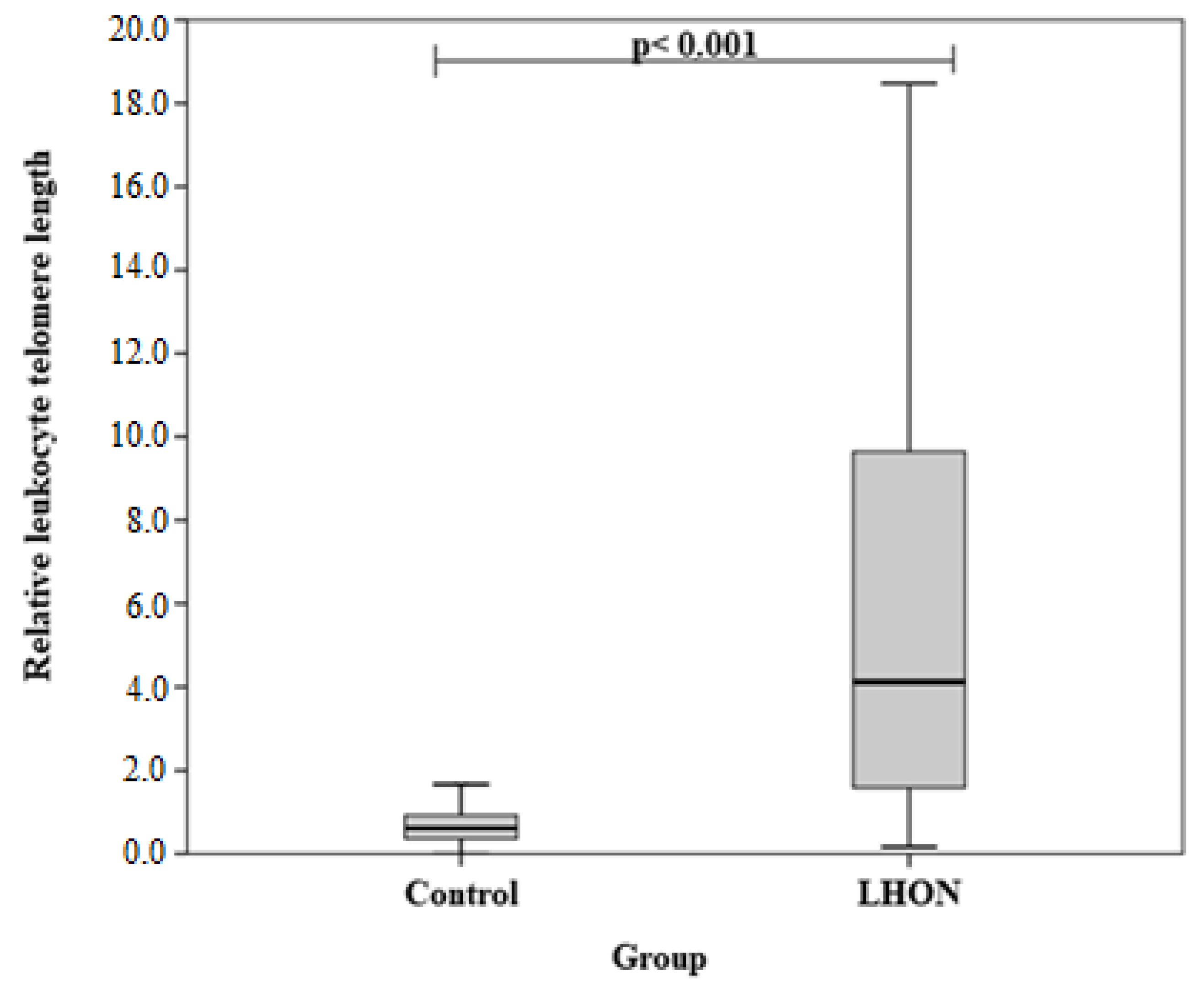
Figure 1. Relative length of leukocyte telomeres in LHON and control groups. The Mann-Whitney U test was used.
Medicina 58 01240 g001
Table 1. Demographics.
CharacteristicsGroupp-Value *
LHON n (%)Control n (%)
GenderMales10 (71.4)61 (46.9)0.081
Females4 (28.6)69 (53.1)
Age, mean (IQR)40.6 (13.1)39.9 (17.9)0.843
RLTL, mean (SD)7.31 (7.28)0.77 (0.75)<0.001
* p-significance level and Bonferroni-corrected significance level when p = 0.05/3.
Table 2. Hardy-Weinberg equilibrium analysis.
Gene, SNPAllele FrequenciesGenotype Distributionp-Value *
TEP1 rs17609040.49 (A)0.51 (G)30/69/310.482
TEP1 rs17134180.43 (G)0.57 (A)21/71/380.204
TERC rs126963040.27 (G)0.73 (C)8/54/680.526
TERC rs350737940.19 (A)0.81 (G)0/49/81<0.05
* p-significance level and Bonferroni-corrected significance level when p = 0.05/3.
Table 3. Influence of TEP1 rs1760904, rs1713418, TERC rs12696304 on LHON development.
ModelGenotype/AlleleOR (95% CI)p-ValueAIC
TEP1 rs1760904
CodominantAG vs. GG
AA vs. GG
1.647 (0.429–6.323)
0.000 (0.000–)
0.467
0.998
88.358
DominantAG+AA vs. GG1.148 (0.301–4.380)0.84093.812
RecessiveAA vs. GG+AG0.000 (0.000–)0.99886.926
OverdominantAG vs. AA+GG3.242 (0.864–12.162)0.08190.283
AdditiveA0.622 (0.266–1.454)0.27492.626
TEP1 rs1713418
CodominantAG vs. AA
GG vs. AA
1.070 (0.303–3.786)
0.905 (0.153–5.360)
0.916
0.912
95.809
DominantAG+GG vs. AA1.033 (0.305–3.496)0.95993.851
RecessiveGG vs. AA+AG0.865 (0.180–4.150)0.85693.820
OverdominantAG vs. AA+GG1.108 (0.364–3.373)0.85793.821
AdditiveG0.972 (0.421–2.248)0.94892.849
TERC rs12696304
CodominantCG vs. CC
GG vs. CC
0.720 (0.200–2.586)
3.643 (0.782–16.961)
0.6140.10092.529
DominantCG+GG vs. CC1.097 (0.364–3.304)0.87093.827
RecessiveGG vs. CC+CG4.159 (0.963–17.969)0.05690.789
OverdominantCG vs. CC+GG0.563 (0.168–1.890)0.35292.935
AdditiveC1.513 (0.662–3.458)0.32692.917
Table 4. Distribution of relative leukocyte telomere lengths by genotypes.
GenotypeRLTLp-Value *
LHON Group
Median (IQR)
Control Group Median (IQR)
TEP1 rs1760904
GG0.725 (-)0.441 (0.511)0.738
GA4.505 (15.405)0.696 (0.549)<0.001
AA-0.512 (0.858)NA
TEP1 rs1713418
AA3.474 (16.635)0.506 (0.682)0.011
AG7.940 (12.964)0.644 (0.565)<0.001
GG1.629 (-)0.695 (0.573)0.913
TERC rs12696304
CC3.097 (3.780)0.456 (0.652)0.005
CG12.105 (16.187)0.694 (0.573)0.008
GG9.764 (-)0.982 (1.529)0.025
* Mann-Whitney U test was used.
Publisher’s Note: MDPI stays neutral with regard to jurisdictional claims in published maps and institutional affiliations.