Apolar Annonaceous Acetogenins from the Fruit Pulp of Annona muricata

Abstract
:Introduction
Results and Discussion
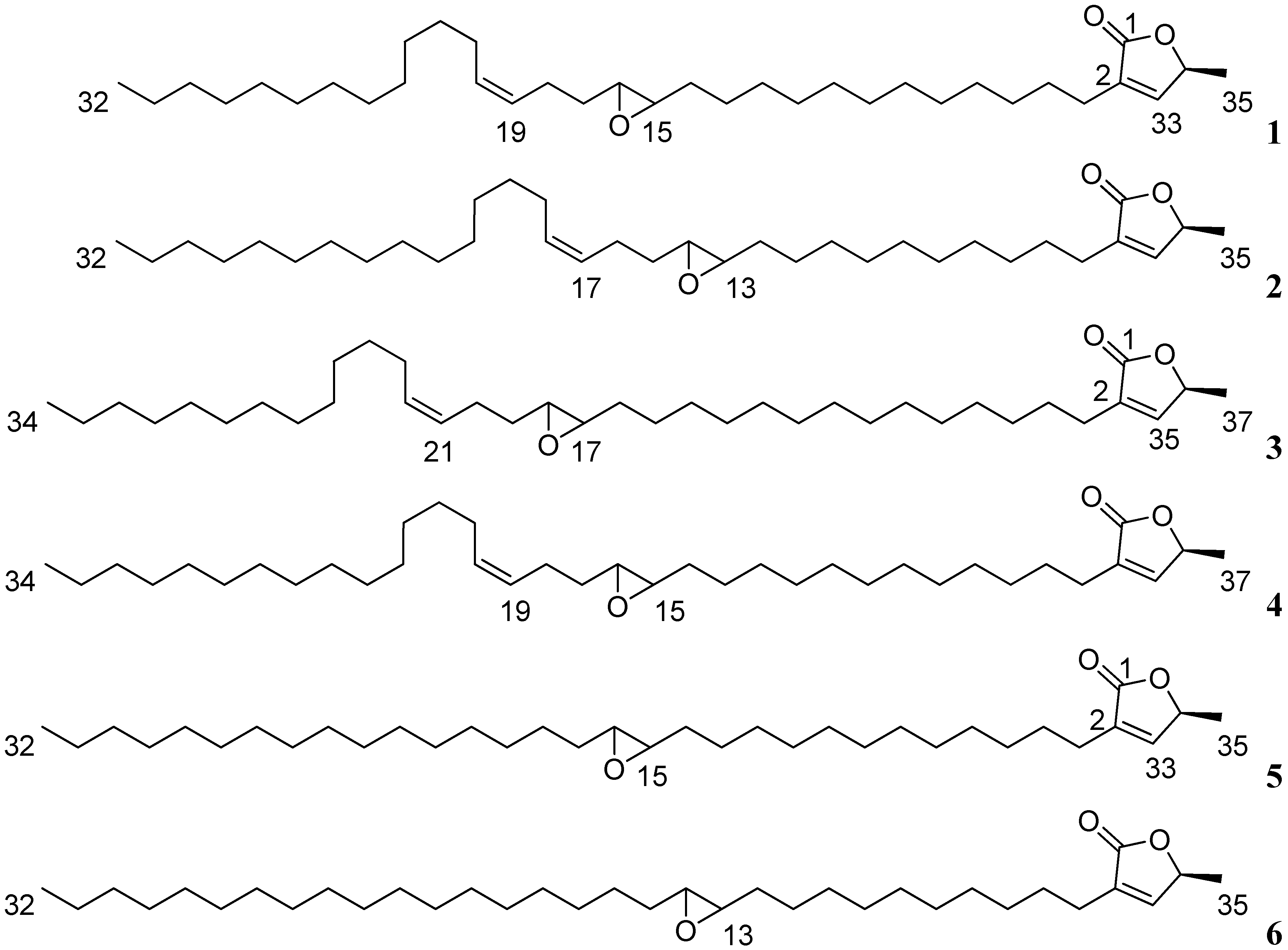
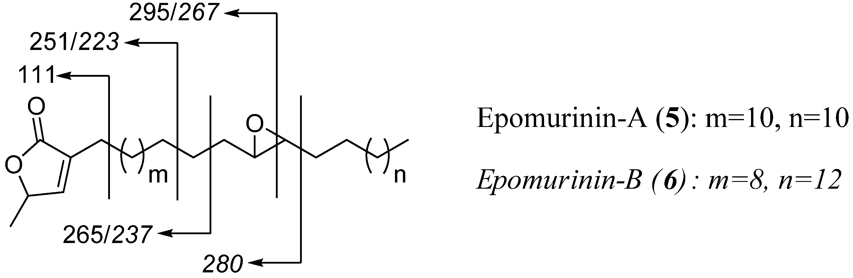
Isolation and structural determination


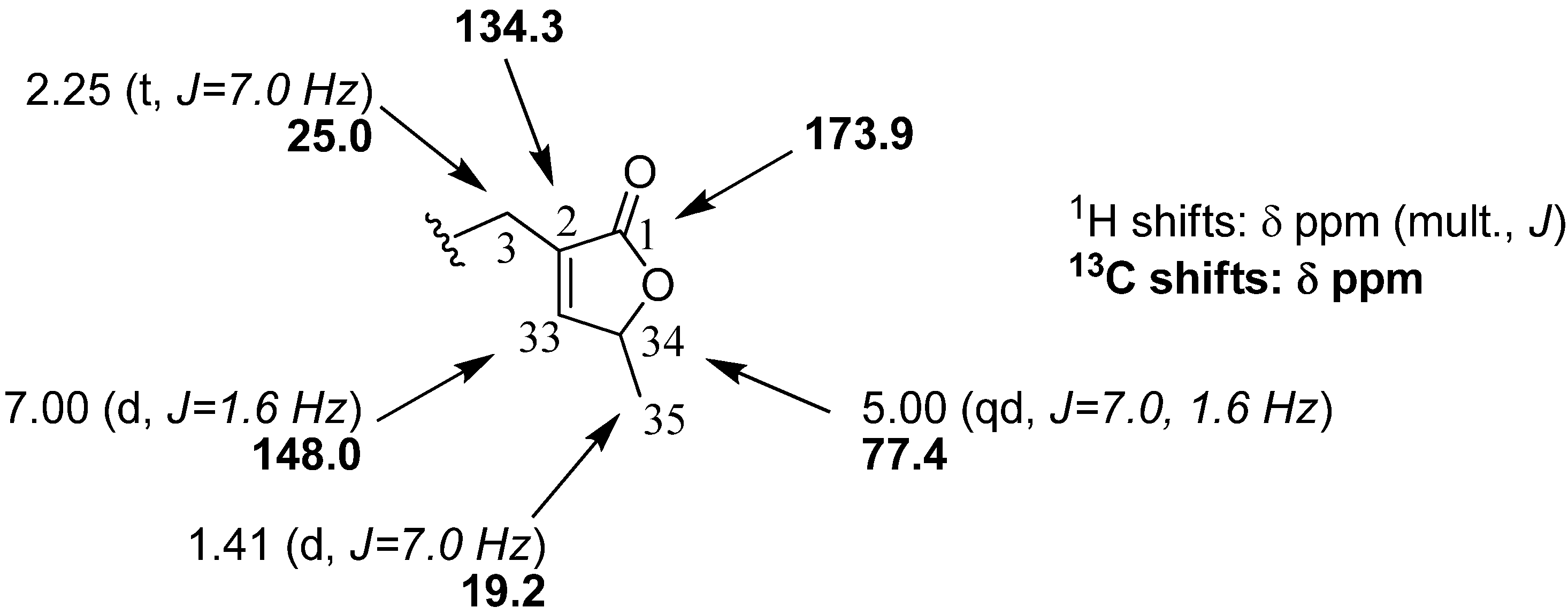
| Position (5) | Position (6) | δH, mult., ( J) | Position (5) | Position (6) | δH, mult., ( J) |
|---|---|---|---|---|---|
| 3 | 3 | 2.26, t (7.0) | 18 to 31 | 16 to 31 | 1.25-1.19, m |
| 4 | 4 | 1.55, m | 32 | 32 | 0.87, t (6.6) |
| 5 to 13 | 5 to 11 | 1.25-1.29, m | 33 | 33 | 6.99, d (1.6) |
| 14 | 12 | 1.32, m | 34 | 34 | 5.01, dq (7.0; 1.6) |
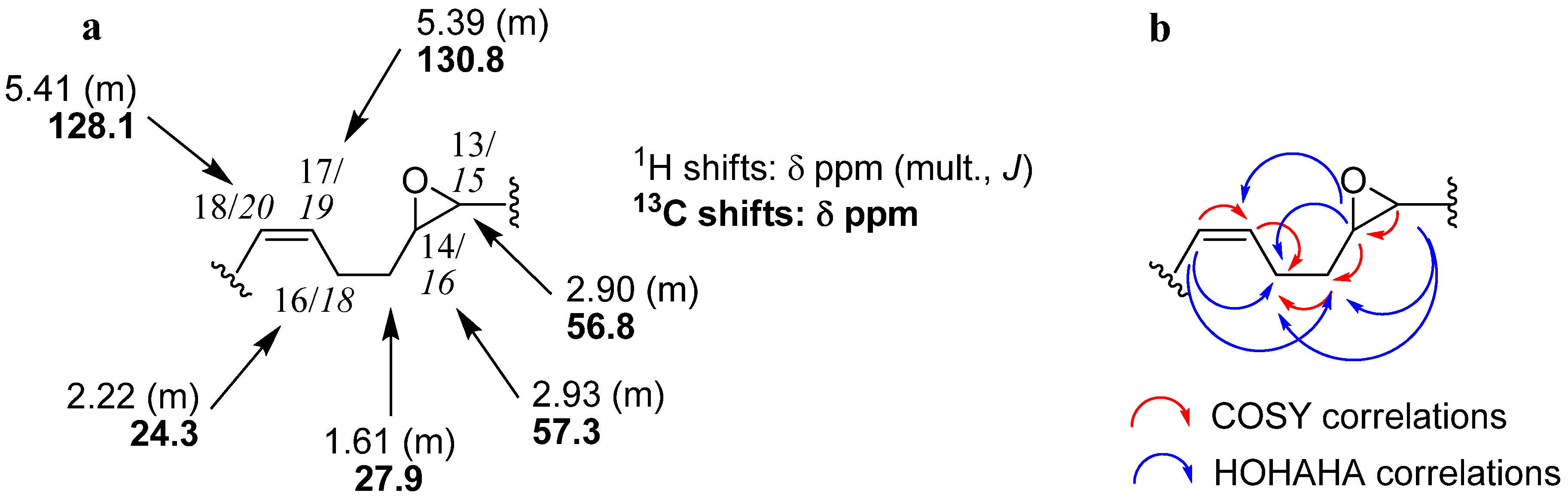
| 15, 16 | 13, 14 | 2.92, m | |||
| 17 | 15 | 1.61, m | 35 | 35 | 1,41, d (7.0) |

Experimental
General
Plant material
Extraction and isolation
Spectral data
Conclusion

Abbreviations
Acknowledgements
References and Notes
- Höglinger, G.U.; Michel, P.P.; Champy, P.; Féger, J.; Hirsch, E.C.; Ruberg, M.; Lannuzel, A. Experimental evidence for a toxic etiology of tropical parkinsonism. Mov. Disord. 2005, 20, 118–119. [Google Scholar]
- Champy, P; Guérineau, V.; Laprévote, O. MALDI-TOF MS profiling of Annonaceous acetogenins in Annona muricata products of human consumption. 2009; Submitted. [Google Scholar]
- Cavé, A.; Figadère, B.; Laurens, A.; Cortes, D. Acetogenins from Annonaceae. In Progress in the Chemistry of Organic Natural Products; Herz, W., Kirby, G.W., Moore, R.E., Steglich, W., Tamm, C., Eds.; Springer: New York, NY, USA, 1997; pp. 81–288. [Google Scholar]
- Bermejo, A.; Figadère, B.; Zafra-Polo, M.C.; Barrachina, I.; Estornell, E.; Cortes, D. Acetogenins from annonceae: Recent progress in isolation, synthesis and mechanisms of action. Nat. Prod. Rep. 2005, 22, 269–303. [Google Scholar]
- McLaughlin, J.L. Paw Paw and Cancer: Annonaceous acetogenins from discovery to commercial products. J. Nat. Prod. 2008, 71, 1311–1321. [Google Scholar] [CrossRef]
- Champy, P.; Melot, A.; Guérineau, V.; Gleye, C.; Höglinger, G.U.; Ruberg, M.; Lannuzel, A.; Laprévote, O.; Laurens, A.; Hocquemiller, R. Quantification of acetogenins in Annona muricata linked to atypical parkinsonism in Guadeloupe. Mov. Disord. 2005, 20, 1629–1633. [Google Scholar] [CrossRef]
- Abe, M.; Kenmochi, A.; Ichimaru, N.; Hamada, T.; Nishioka, T.; Miyoshi, H. Essential structural features of acetogenins: role of hydroxy groups adjacent to the bis-THF rings. Bioorg. Med. Chem. Lett. 2004, 14, 779–782. [Google Scholar] [CrossRef]
- Konno, H.; Hiura, N.; Makabe, H.; Abe, M.; Miyoshi, H. Synthesis and mitochondrial complex I inhibition of dihydroxy-cohibin A, non-THF Annonaceous acetogenin analogue. Bioorg. Med. Chem. Lett. 2004, 14, 629–632. [Google Scholar] [CrossRef]
- Höllerhage, M.; Matusch, A.; Champy, P.; Lombès, A.; Ruberg, M.; Oertel, W.H.; Höglinger, G.U. Natural lipophilic inhibitors of mitochondrial complex I are candidate toxins for sporadic tau pathologies. Exp. Neurol. 2009, in press. [Google Scholar]
- Roblot, F.; Laugel, T.; Lebœuf, M.; Cavé, A.; Laprévote, O. Two acetogenins from Annona muricata seeds. Phytochemistry 1993, 34, 281–285. [Google Scholar]
- Zeng, L.; Zhang, Y.; McLaughlin, J.L. Gigantransenin A, B, and C, novel mono-THF acetogenins bearing trans double bonds from Goniothalamus giganteus (Annonaceae). Tetrahedron Lett. 1996, 37, 5449–5452. [Google Scholar]
- Gleye, C.; Laurens, A.; Hocquemiller, R.; Laprévote, O.; Serani, L.; Cavé, A. Cohibins A and B, acetogenins isolated from roots of Annona muricata L. Phytochemistry 1997, 44, 1541–1545. [Google Scholar] [CrossRef]
- Gleye, C. Acétogénines des racines et des graines d’Annona muricata (Annonaceae): Etude particulière des précurseurs biogénétiques. PhD Thesis, Université Paris-Sud 11, Paris, France, 1998. [Google Scholar]
- Hisham, A.; Sreekala, U.; Pieters, L.; De Bruyne, T.; Van den Heuvel, H.; Claeys, M. Epoxymurins A and B, two biogenetic precursors of Annonaceous acetogenins from Annona muricata. Tetrahedron 1993, 49, 6913–6920. [Google Scholar]
- Chen, Y.Y; Chang, F.R.; Yen, H.F.; Wu, Y.C. Epomusenins A and B, two acetogenins from fruits of Rollinia mucosa. Phytochemistry 1996, 42, 1081–1083. [Google Scholar] [CrossRef]
- Hu, Y.; Cecil, A.R.L.; Franck, X.; Gleye, C.; Figadère, B.; Brown, R.C.D. Natural cis-solamin is a mixture of tetraepimeric diastereoisomers: Biosynthetic implications for Annonaceous acetogenins. Org. Biomol. Chem. 2006, 4, 1217–1219. [Google Scholar] [CrossRef]
- Gleye, C.; Laurens, A.; Laprévote, O.; Serani, L.; Hocquemiller, R. Isolation and structure elucidation of sabadelin, an acetogenin from roots of Annona muricata. Phytochemistry 1999, 52, 1403–1408. [Google Scholar] [CrossRef]
- Bajin ba Ndob, I.; Champy, P.; Gleye, C.; Lewin, G.; Akendengué, B. Annonaceous acetogenins: Precursors from the seeds of Annona squamosa. Phytochemistry Lett. 2009, 2, 72–76. [Google Scholar]
- Sample Availability: Samples of the compounds 1+2 and 3+4 are available from the authors.
© 2009 by the authors; licensee Molecular Diversity Preservation International, Basel, Switzerland. This article is an open access article distributed under the terms and conditions of the Creative Commons Attribution license ( http://creativecommons.org/licenses/by/3.0/).
Share and Cite
Melot, A.; Fall, D.; Gleye, C.; Champy, P. Apolar Annonaceous Acetogenins from the Fruit Pulp of Annona muricata. Molecules 2009, 14, 4387-4395. https://doi.org/10.3390/molecules14114387
Melot A, Fall D, Gleye C, Champy P. Apolar Annonaceous Acetogenins from the Fruit Pulp of Annona muricata. Molecules. 2009; 14(11):4387-4395. https://doi.org/10.3390/molecules14114387
Chicago/Turabian StyleMelot, Alice, Djibril Fall, Christophe Gleye, and Pierre Champy. 2009. "Apolar Annonaceous Acetogenins from the Fruit Pulp of Annona muricata" Molecules 14, no. 11: 4387-4395. https://doi.org/10.3390/molecules14114387
APA StyleMelot, A., Fall, D., Gleye, C., & Champy, P. (2009). Apolar Annonaceous Acetogenins from the Fruit Pulp of Annona muricata. Molecules, 14(11), 4387-4395. https://doi.org/10.3390/molecules14114387