Investigating the Spectrum of Biological Activity of Ring-Substituted Salicylanilides and Carbamoylphenylcarbamates

Abstract
:1. Introduction
2. Results and Discussion
2.1. Chemistry
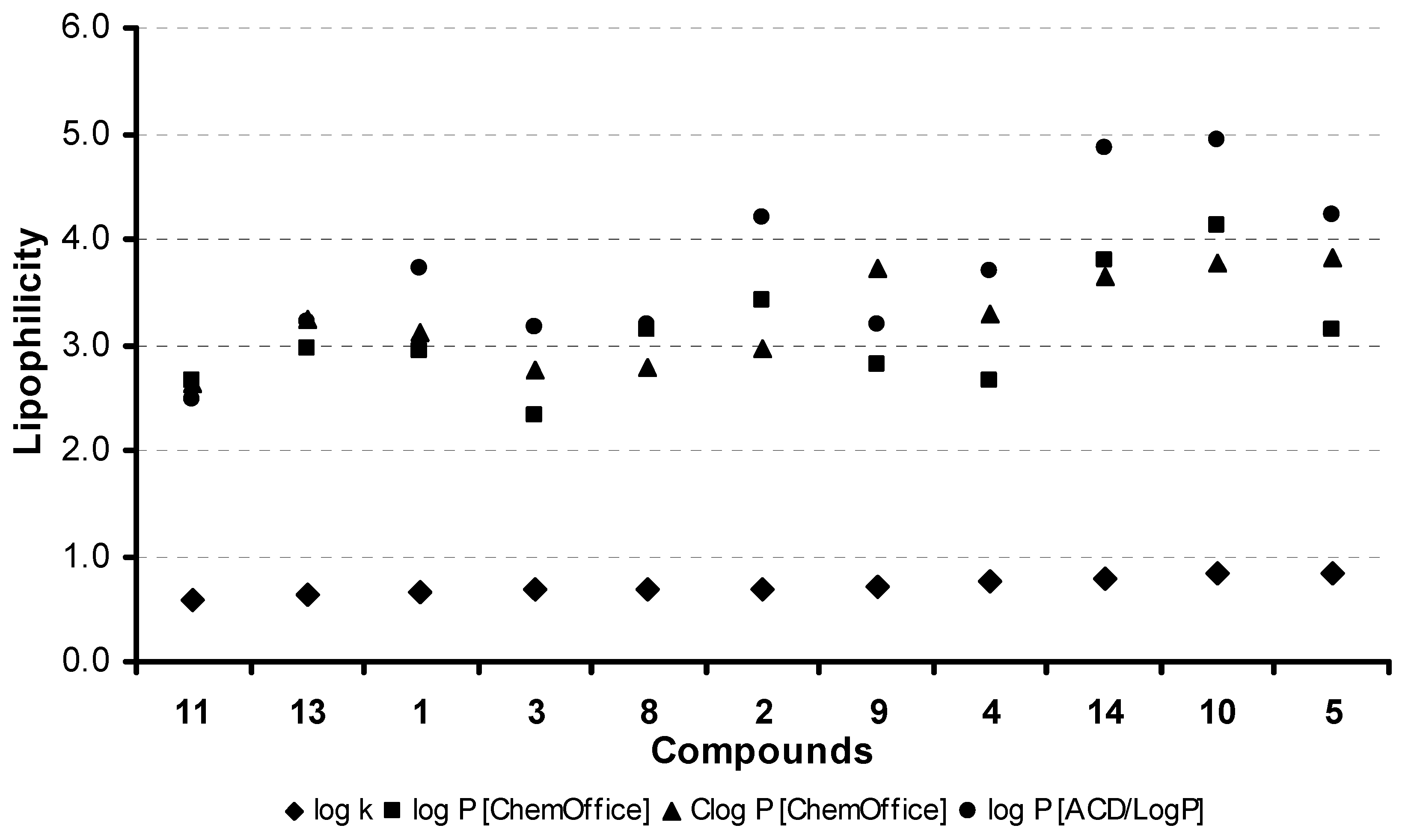
2.2. Lipophilicity
2.3. Biological activities
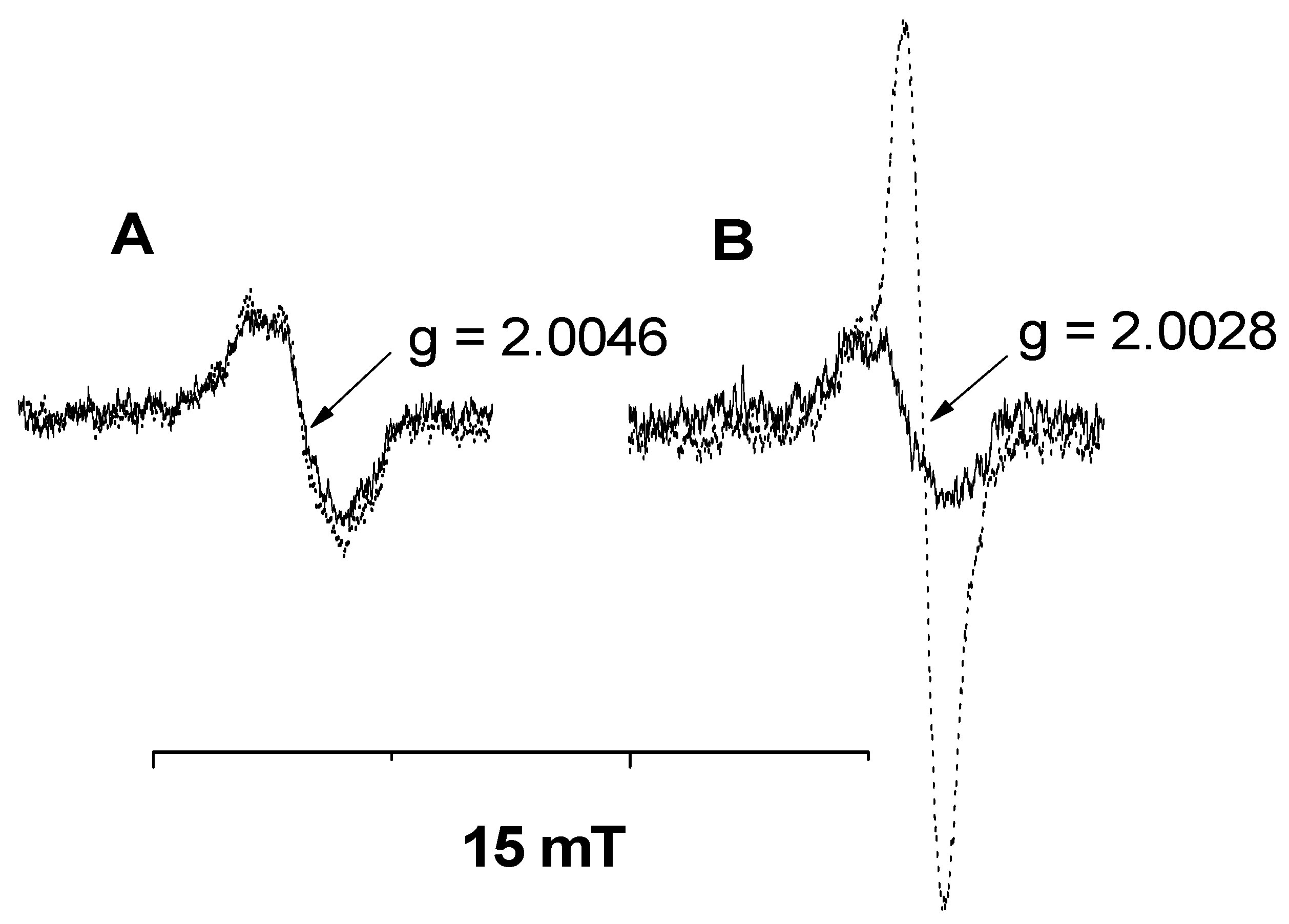
2.3.1. Inhibition of photosynthetic electron transport (PET) in spinach chloroplasts
2.3.2. In vitro anti-fungal and anti-bacterial susceptibility testing
2.3.3. In vitro antimycobacterial evaluation
2.3.4. In vitro anti-proliferative activity
3. Experimental
3.1. General
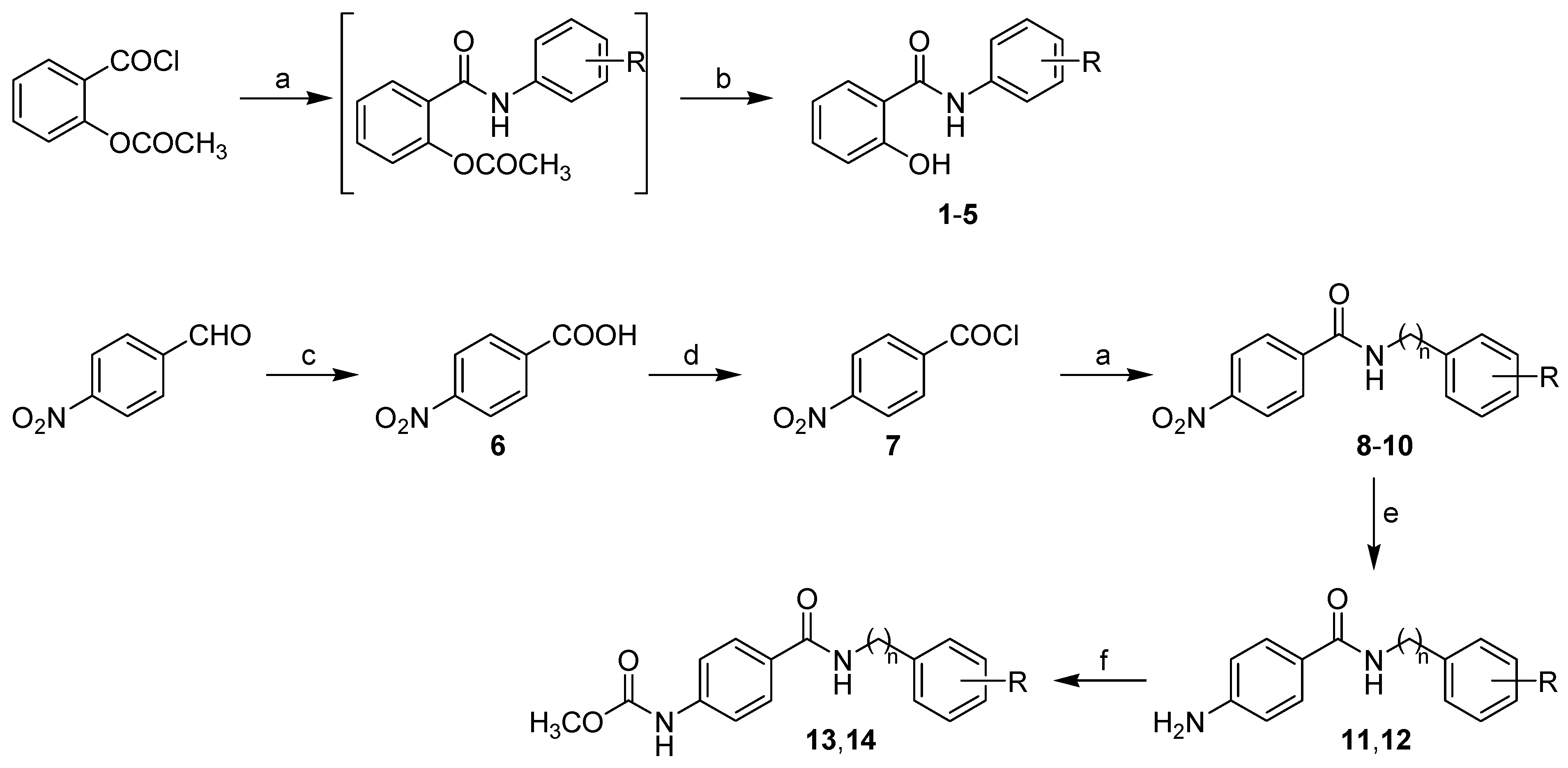
3.2. Synthesis
3.2.1. General procedure for synthesis of carboxamide derivatives 1-5
3.2.2. General procedure for synthesis of carboxamide derivatives 8-10
3.3. Lipophilicity determination by HPLC (capacity factor k/calculated log k)
3.4. Lipophilicity calculations
3.5. Study of inhibition photosynthetic electron transport (PET) in spinach chloroplasts
3.6. In vitro anti-fungal susceptibility testing
3.7. In vitro anti-bacterial susceptibility testing
3.8. In vitro anti-mycobacterial evaluation
3.9. Cell culture and in vitro antiproliferative activity
4. Conclusions
Acknowledgements
References
- Vinsova, J.; Imramovsky, A. Salicylanilides: Still a topical potential antibacterially active group. Ces. Slov. Farm. 2004, 53, 294–299. [Google Scholar]
- De la Fuente, R.; Sonawane, N.D.; Arumainayagam, D.; Verkman, A.S. Small molecules with antimicrobial activity against E. coli and P. aeruginosa identified by high-throughput screening. Br. J. Pharmacol. 2006, 149, 551–559. [Google Scholar] [CrossRef] [PubMed]
- Dahlgren, M.K.; Kauppi, A.M.; Olsson, I.M.; Linusson, A.; Elofsson, M. Design, synthesis, and multivariate quantitative structure–activity relationship of salicylanilidess–potent inhibitors of type III secretion in Yersinia. J. Med. Chem. 2007, 50, 6177–6188. [Google Scholar] [CrossRef] [PubMed]
- Stephenson, K.; Yamaguchi, Y.; Hoch, J.A. The mechanism of action of inhibitors of bacterial two-component signal transduction systems. J. Biol. Chem. 2000, 275, 38900–38904. [Google Scholar] [CrossRef] [PubMed]
- Vinsova, J.; Imramovsky, A.; Buchta, V.; Ceckova, M.; Dolezal, M.; Staud, F.; Jampilek, J.; Kaustova, J. Salicylanilide acetates: Synthesis and antibacterial evaluation. Molecules 2007, 12, 1–12. [Google Scholar] [CrossRef] [PubMed]
- Imramovsky, A.; Vinsova, J.; Ferriz, J.M.; Dolezal, R.; Jampilek, J.; Kaustova, J.; Kunc, F. New antituberculotics originated from salicylanilides with promising in vitro activity against atypical mycobacterial strains. Bioorg. Med. Chem. 2009, 17, 3572–3579. [Google Scholar] [CrossRef] [PubMed]
- Imramovsky, A.; Vinsova, J.; Ferriz, J.M.; Buchta, V.; Jampilek, J. Salicylanilide esters of N-protected amino acids as novel antimicrobial agents. Bioorg. Med. Chem. Lett. 2009, 19, 348–351. [Google Scholar] [CrossRef] [PubMed]
- Hassan, G.S.; Hegazy, G.H.; Safwat, H.M. Synthesis of Furo-salicylanilides and their heterocyclic derivatives with anticipated molluscicidal activity. Arch. Pharm. Chem. Life Sci. 2006, 339, 448–455. [Google Scholar] [CrossRef] [PubMed]
- Daidone, G.; Raffa, D.; Plescia, S.; Matera, M.; Caruso, A.; Leone, V.; Amico-Roxas, M. Synthesis and evaluation of the analgesic and antiinflammatory activities of N-substituted salicylamides. Farmaco 1989, 44, 465–473. [Google Scholar] [PubMed]
- Brown, M.E.; Fitzner, J.N.; Stevens, T.; Chin, W.; Wright, C.D.; Boyce, J.P. Salicylanilides: Selective inhibitors of interleukin-12p40 production. Bioorg. Med. Chem. 2008, 16, 8760–8764. [Google Scholar] [CrossRef] [PubMed]
- Liechti, C.; Sequin, U.; Bold, G.; Furet, P.; Meyer, T.; Traxler, P. Salicylanilides as inhibitors of the protein tyrosine kinase epidermal growth factor receptor. Eur. J. Med. Chem. 2004, 39, 11–26. [Google Scholar] [CrossRef] [PubMed]
- Deng, W.; Guo, Z.; Guo, Y.; Feng, Z.; Jiang, Y.; Chu, F. Acryolylamino-salicylanilides as EGFR PTK inhibitors. Bioorg. Med. Chem. Lett. 2006, 16, 469–472. [Google Scholar] [CrossRef] [PubMed]
- Kamath, S.; Buolamwini, J.K. Targeting EGFR and HER-2 receptor tyrosine kinases for cancer drug discovery and development. Med. Res. Rev. 2006, 26, 569–594. [Google Scholar] [CrossRef] [PubMed]
- Ray, S.; Pathak, S.R.; Chaturvedi, D. Organic carbamates in drug development. Part II: antimicrobial agents - Recent reports. Drugs Future 2005, 30, 161–180. [Google Scholar] [CrossRef]
- Ferriz, J.M.; Vavrova, K.; Kunc, F.; Imramovsky, A.; Stolarikova, J.; Vavrikova, E.; Vinsova, J. Salicylanilide carbamates: Antitubercular agents active against multidrug-resistant Mycobacterium tuberculosis strains. Bioorg. Med. Chem. 2010, 18, 1054–1061. [Google Scholar] [CrossRef] [PubMed]
- Agouridas, C.; Denis, A.; Auger, J.M.; Benedetti, Y.; Bonnefoy, A.; Bretin, F.; Chantot, J.F.; Dussarat, A.; Fromentin, C.; D‘Ambrieres, S.G.; Lachaud, S.; Laurin, P.; Le Martret, O.; Loyau, V.; Tessot, N. Synthesis and antibacterial activity of ketolides (6-O-methyl-3-oxoerythromycin derivatives): A new class of antibacterials highly potent against macrolide-resistant and -susceptible respiratory pathogens. J. Med. Chem. 1998, 41, 4080–4100. [Google Scholar] [CrossRef] [PubMed]
- Meng, Q.; Luo, H.; Liu, Y.; Li, W.; Zhang, W.; Yao, Q. Synthesis and evaluation of carbamate prodrugs of SQ109 as antituberculosis agents. Bioorg. Med. Chem. 2009, 19, 2808–2810. [Google Scholar] [CrossRef] [PubMed]
- Thorberg, S.; Berg, S.; Lundstrom, L.; Pettersson, B.; Wijkstrom, A.; Sanchez, D.; Lindberg, P.; Nilsson, J.G. Carbamate ester derivatives as potential prodrugs of the presynaptic dopamine autoreceptor agonist (-)-3-(3-hydroxyphenyl)-N-propylpiperidine. J. Med. Chem. 1987, 30, 2008–2012. [Google Scholar] [CrossRef] [PubMed]
- Good, N.E. Inhibitors of the Hill reaction. Plant Physiol. 1961, 36, 788–803. [Google Scholar] [CrossRef] [PubMed]
- Kralova, K.; Sersen, F.; Cizmarik, J. Inhibitory effect of piperidinoethylesters of alkoxyphenylcarbamic acids on photosynthesis. Gen. Physiol. Biophys. 1992, 11, 261–267. [Google Scholar]
- Kralova, K.; Sersen, F.; Kubicova, L.; Waisser, K. Inhibitory effects of substituted benzanilides on photosynthetic electron transport in spinach chloroplasts. Chem. Pap. 1999, 53, 328–331. [Google Scholar] [CrossRef]
- Kralova, K.; Sersen, F.; Kubicova, L.; Waisser, K. Inhibition of photosynthetic electron transport in spinach chloroplasts by 3- and 4-halogeno substituted benzanilides and thiobenzanilides. J. Trace Microprobe Technol. 2000, 18, 251–256. [Google Scholar]
- Kubicova, L.; Kralova, K.; Sersen, F.; Gregor, J.; Waisser, K. Effects of substituted salicylanilides on the photosynthetic apparatus of spinach chloroplasts. Folia Pharm. Univ. Carol. 2000, 25, 89–96. [Google Scholar]
- Pravda, M.; Hrnciarova, D.; Kralova, K. 3-Methylthiosalicylanilides – inhibitors of Hill reaction. Chem. Listy 2003, 97, 1122–1123. [Google Scholar]
- Black, C.C. Photosynthetic phosphorylation and associated reactions in the presence of a new group of uncouplers: Salicylanilides. Biochim. Biophys. Acta 1968, 162, 294–296. [Google Scholar] [CrossRef]
- Govindjee, S. Sixty-three years since Kautsky: Chlorophyll a fluorescence. Aust. J. Plant Physiol. 1995, 22, 131–160. [Google Scholar] [CrossRef]
- Jegerschold, C.; Styring, S. Fast oxygen-independent degradation of D1 reaction center protein in photosystem II. FEBS Lett. 1991, 280, 87–90. [Google Scholar] [CrossRef]
- Kralova, K.; Kubicova, L.; Sersen, F.; Waisser, K. Inhibition of Hill reaction in spinach chloroplasts by 5-bromo- and 3,5-dibromosalicylanilides. Proceedings of 51st Congress of Chemical Societies, Nitra, Slovakia, 6-9 September 1999. [Google Scholar]
- Kubicova, L.; Kissova, K.; Waisser, K. Inhibition of chlorophyll production in Chlorella vulgaris by substituted salicylanilides. Folia Pharm. Univ. Carol. 2000, 25, 67–72. [Google Scholar]
- Pravda, M.; Sustr, M.; Hrnciarova, D.; Kubicova, L.; Kralova, K. Effects of 3-methylthiosalicylanilides on chlorophyll content in freshwater alga Chlorella vulgaris. In Proceedings of ECOpole’03, Opole, Poland, 16–18 October 2003; Society of Ecological Chemistry and Engineering: Opole, Poland, 2003; pp. 105–108. [Google Scholar]
- Jampilek, J.; Dolezal, M.; Kunes, J.; Buchta, V.; Kralova, K. Quinaldine derivatives: Preparation and biological activity. Med. Chem. 2005, 1, 591–599. [Google Scholar] [CrossRef] [PubMed]
- Dolezal, M.; Palek, L.; Vinsova, J.; Buchta, V.; Jampilek, J.; Kralova, K. Substituted pyrazinecarboxamides: Synthesis and biological evaluation. Molecules 2006, 11, 242–256. [Google Scholar] [CrossRef] [PubMed]
- Musiol, R.; Jampilek, J.; Kralova, K.; Richardson, D.R.; Kalinowski, D.; Podeszwa, B.; Finster, J.; Niedbala, H.; Palka, A.; Polanski, J. Investigating biological activity spectrum for novel quinoline analogues. Bioorg. Med. Chem. 2007, 15, 1280–1288. [Google Scholar] [CrossRef] [PubMed]
- Musiol, R.; Tabak, D.; Niedbala, H.; Podeszwa, B.; Jampilek, J.; Kralova, K.; Dohnal, J.; Finster, J.; Mencel, A.; Polanski, J. Investigating biological activity spectrum for novel quinoline analogues 2: Hydroxyquinolinecarboxamides with photosynthesis inhibiting activity. Bioorg. Med. Chem. 2008, 16, 4490–4499. [Google Scholar] [CrossRef] [PubMed]
- Dolezal, M.; Cmedlova, P.; Palek, L.; Vinsova, J.; Kunes, J.; Buchta, V.; Jampilek, J.; Kralova, K. Synthesis and antimycobacterial evaluation of substituted pyrazinecarboxamides. Eur. J. Med. Chem. 2008, 43, 1105–1113. [Google Scholar] [CrossRef] [PubMed]
- Jampilek, J.; Musiol, R.; Pesko, M.; Kralova, K.; Vejsova, M.; Carroll, J.; Coffey, A.; Finster, J.; Tabak, D.; Niedbala, H.; Kozik, V.; Polanski, J.; Csollei, J.; Dohnal, J. Ring-substituted 4-hydroxy-1H-quinolin-2-ones: Preparation and biological activity. Molecules 2009, 14, 1145–1159. [Google Scholar] [CrossRef] [PubMed]
- Jampilek, J.; Musiol, R.; Finster, J.; Pesko, M.; Carroll, J.; Kralova, K.; Vejsova, M.; O’Mahony, J.; Coffey, A.; Dohnal, J.; Polanski, J. Investigating biological activity spectrum for novel styrylquinazoline analogues. Molecules 2009, 14, 4246–4265. [Google Scholar] [CrossRef] [PubMed]
- Musiol, R.; Jampilek, J.; Nycz, J.E.; Pesko, M.; Carroll, J.; Kralova, K.; Vejsova, M.; O’Mahony, J.; Coffey, A.; Mrozek, A.; Polanski, J. Investigating the activity spectrum for ring-substituted 8-hydroxyquinolines. Molecules 2010, 15, 288–304. [Google Scholar] [CrossRef] [PubMed]
- Josuu, R.M.; Patel, M.M. Chelation ion-exchange properties of salicylic acid-urea-formaldehyde copolymers. J. Chem. Sci. 1982, 91, 351–358. [Google Scholar]
- Mahmoud, M.E.; Soliman, E.M. Study of the selective extraction of iron (III) by silica-immobilized 5-Formyl-3-Arylazo-salicylic acid derivatives. Talanta 1997, 44, 1063–1071. [Google Scholar] [CrossRef]
- Rho, H.S.; Baek, H.S.; You, J.W.; Kim, S.J.; Kim, M.K.; Kim, D.H.; Chang, I.S. Biological activities of 3,5-dihydroxy-N-(4-hydroxyphenyl)benzamide: A mimic compound of trans-resveratrol. Bull. Korean Chem. Soc. 2007, 28, 837–839. [Google Scholar]
- Althuis, T.H.; Hess, H.J. Synthesis and identification of the major metabolites of prazosin formed in dog and rat. J. Med. Chem. 1977, 20, 146–149. [Google Scholar] [CrossRef] [PubMed]
- Brown, R.K.; Nelson, N.A. 6-Aminoindole. J. Am. Chem. Soc. 1954, 76, 5149–5150. [Google Scholar] [CrossRef]
- Fellows, I.M.; Kaelin, D.E.; Martin, S.F. Application of ring-closing metathesis to the formal total synthesis of (+)−FR900482. J. Am. Chem. Soc. 2000, 122, 10781–10787. [Google Scholar] [CrossRef]
- Hiraj, K.; Yano, T.; Matsukawa, T.; Ugai, S.; Nagato, S.; Hori, M. Synthesis and herbicidal activity of new oxazolidinedione derivates. J. Pestic. Sci. 1999, 24(2), 156–169. [Google Scholar]
- Kerns, E.H.; Li, D. Drug-like Properties: Concept, Structure Design and Methods; Elsevier: San Diego, CA, USA, 2008. [Google Scholar]
- Norrington, F.E.; Hyde, R.M.; Williams, S.G.; Wotton, R. Physicochemical-activity relations in practice. 1. Rational and self-consistent data bank. J. Med. Chem. 1975, 18, 604–607. [Google Scholar] [CrossRef] [PubMed]
- Svensson, B.; Vass, I.; Styring, S. Sequence analysis of D1 and D2 reaction center proteins of photosystem II. Z. Naturforsch C. 1991, 46c, 765–776. [Google Scholar] [CrossRef]
- Noren, G.H.; Barry, B.A. The YF161D1 mutant of synechocystis 6803 exhibits an EPR signal from a light-induced photosystem II radical. Biochemistry 1992, 31, 3335–3342. [Google Scholar] [CrossRef] [PubMed]
- Hoff, A.J. Application of ESR in photosynthesis. Phys. Rep. 1979, 54, 75–200. [Google Scholar] [CrossRef]
- Izawa, S. Acceptors and Donors for Chloroplast Electron Transport; San Pietro, A., Ed.; Academic Press: London, UK, 1980; Volume 69, pp. 413–434. [Google Scholar]
- Kalinowski, D.S.; Richardson, D.R. The evolution of iron chelators for the treatment of iron overload disease and cancer. Pharmacol. Rev. 2005, 57, 547–583. [Google Scholar] [CrossRef] [PubMed]
- Richardson, D.R.; Sharpe, P.C.; Lovejoy, D.B.; Senaratne, D.; Kalinowski, D.S.; Islam, M.; Bernhardt, P.V. Dipyridyl thiosemicarbazone chelators with potent and selective antitumor activity form iron complexes with redox activity. J. Med. Chem. 2006, 49, 6510–6521. [Google Scholar] [CrossRef] [PubMed]
- Kalinowski, D.S.; Yu, Y.; Sharpe, P.C.; Islam, M.; Liao, Y.T.; Lovejoy, D.B.; Kumar, N.; Bernhardt, P.V.; Richardson, D.R. Design, synthesis, and characterization of novel iron chelators: Structure−activity relationships of the 2-benzoylpyridine thiosemicarbazone series and their 3-nitrobenzoyl analogues as potent antitumor agents. J. Med. Chem. 2007, 50, 3716–3729. [Google Scholar] [CrossRef] [PubMed]
- Wagner, G.; Singer, D.; Weuffen, W. Untersuchungen uber 2-hydroxythiobenzamide und 2-hydroxythiobenzanilide. Pharmazie 1966, 21, 161–166. [Google Scholar] [PubMed]
- Bahrami, K.; Khodaei, M.M.; Farrokhi, A. H2O2/SOCl2: A useful reagent system for the conversion of thiocarbonyls to carbonyl compounds. Tetrahedron 2009, 65, 7658–7661. [Google Scholar] [CrossRef]
- Masarovicova, E.; Kralova, K. Approaches to measuring plant photosynthesis activity. In Handbook of Photosynthesis, 2nd ed.; Pessarakli, M., Ed.; Taylor & Francis Group: Boca Raton, FL, USA, 2005; pp. 617–656. [Google Scholar]
- Kralova, K.; Sersen, F.; Sidoova, E. Photosynthesis inhibition produced by 2-alkylthio-6-R-benzothiazoles. Chem. Pap. 1992, 46, 348–350. [Google Scholar]
- Fedke, C. Biochemistry and Physiology of Herbicide Action; Springer Verlag: New York, NY, USA, 1982. [Google Scholar]
- National Committee for Clinical Laboratory Standards. Method for Antifungal Disk Diffusion Susceptibility Testing of Yeasts: Approved Guideline M44-A; National Committee for Clinical Laboratory Standards: Wayne, PA, USA, 2004. [Google Scholar]
- Carroll, J.; Douarre, P.; Coffey, A.; Buckley, J.; Cashman, B.; O’Farrell, K.; O’Mahony, J. Optimization of a rapid viability assay for Mycobacterium avium paratuberculosis by using alamarBlue. Appl. Environ. Microbiol. 2009, 75, 7870–7872. [Google Scholar] [CrossRef] [PubMed]
- Richardson, D.R.; Tran, E.H.; Ponka, P. The potential of iron chelators of the pyridoxal isonicotinoyl hydrazone class as effective antiproliferative agents. Blood 1995, 86, 4295–4306. [Google Scholar] [PubMed]
Sample Availability: Samples of the compounds are available from the authors. |


 | |||||||
| Comp. | R1 | R2 | log k | log P/Clog P ChemOffice | log P ACD/LogP | σ [47] | MR [47] |
| 1 | 2-OH |  | 0.6661 | 2.94 / 3.1212 | 3.73 ± 0.37 | 0.10 | 4.7 |
| 2 | 2-OH |  | 0.6807 | 3.42 / 2.9702 | 4.19 ± 0.38 | 0.20 | 9.4 |
| 3 | 2-OH |  | 0.6770 | 2.32 / 2.7576 | 3.17 ± 0.39 | 0.00 | 6.5 |
| 4 | 2-OH |  | 0.7515 | 2.66 / 3.2866 | 3.70 ± 0.39 | 0.02 | 11.3 |
| 5 | 2-OH |  | 0.8401 | 3.15 / 3.8156 | 4.23 ± 0.39 | NF | 15.9 |
| 8 | 4-NO2 |  | 0.6782 | 3.15 / 2.7782 | 3.19 ± 0.34 | 0.67 | 4.7 |
| 9 | 4-NO2 |  | 0.6966 | 2.82 / 3.7170 | 3.19 ± 0.35 | 0.67 | 4.7 |
| 10 | 4-NO2 |  | 0.8269 | 4.13 / 3.7726 | 4.94 ± 0.43 | 1.10 | 8.7 |
| 11 | 4-NH2 |  | 0.5886 | 2.66 / 2.6350 | 2.47 ± 0.39 | 0.67 | 4.7 |
| 12 | 4-NH2 |  | ND | 3.52 / 3.0299 | 4.12 ± 0.42 | 1.10 | 8.7 |
| 13 | 4-CH3OCONH |  | 0.6345 | 2.96 / 3.2510 | 3.21 ± 0.43 | 0.67 | 4.7 |
| 14 | 4-CH3OCONH |  | 0.7782 | 3.81 / 3.6459 | 4.86 ± 0.50 | 1.10 | 8.7 |
| Comp. | PET inhibition IC50 [μmol/L] | MIC [µmol/L] | MIC/IC90 [µg/mL] | |||||
|---|---|---|---|---|---|---|---|---|
| ACa | TMa | MRSAb | SEb | MAC | MAP | MK | ||
| 24 h 48 h | 72 h 120 h | 24 h 48 h | 24 h 48 h | |||||
| 1 | 2.7 | 62.50 250 | 62.50 125 | 125 250 | 125 250 | 250 | 250 | 125 |
| 2 | 331.4 | >250 >250 | 250 500 | >500 >500 | >500 >500 | NA | NA | NA |
| 3 | 1.6 | 125 125 | 125 125 | 500 >500 | >500 >500 | NA | NA | NA |
| 4 | 15.0 | 62.50 125 | 62.50 62.50 | 125 125 | 125 125 | NA | NA | NA |
| 5 | 44.8 | 31.25 31.25 | 31.25 31.25 | 31.25 31.25 | 31.25 31.25 | NA | NA | NA |
| 10 | 50.1 | NA | NA | NA | NA | NA | NA | NA |
| 11 | 398.4 | NA | NA | NA | NA | NA | NA | NA |
| 13 | 1.6 | NA | NA | NA | NA | NA | NA | NA |
| 14 | 1.0 | NA | NA | NA | NA | NA | NA | NA |
| DCMU | 1.9 | – | – | – | – | – | – | – |
| FLU | – | >125 >125 | 1.95 3.91 | – | – | – | – | – |
| PEN | – | – | – | 125 125 | 31.62 125 | |||
| CPF | – | – | – | 500 500 | 250 250 | |||
| INH | – | – | – | – | – | <10 | >250 | <10 |
© 2010 by the authors; licensee MDPI, Basel, Switzerland. This article is an open access article distributed under the terms and conditions of the Creative Commons Attribution license (http://creativecommons.org/licenses/by/3.0/).
Share and Cite
Otevrel, J.; Mandelova, Z.; Pesko, M.; Guo, J.; Kralova, K.; Sersen, F.; Vejsova, M.; Kalinowski, D.S.; Kovacevic, Z.; Coffey, A.; et al. Investigating the Spectrum of Biological Activity of Ring-Substituted Salicylanilides and Carbamoylphenylcarbamates. Molecules 2010, 15, 8122-8142. https://doi.org/10.3390/molecules15118122
Otevrel J, Mandelova Z, Pesko M, Guo J, Kralova K, Sersen F, Vejsova M, Kalinowski DS, Kovacevic Z, Coffey A, et al. Investigating the Spectrum of Biological Activity of Ring-Substituted Salicylanilides and Carbamoylphenylcarbamates. Molecules. 2010; 15(11):8122-8142. https://doi.org/10.3390/molecules15118122
Chicago/Turabian StyleOtevrel, Jan, Zuzana Mandelova, Matus Pesko, Jiahui Guo, Katarina Kralova, Frantisek Sersen, Marcela Vejsova, Danuta S. Kalinowski, Zaklina Kovacevic, Aidan Coffey, and et al. 2010. "Investigating the Spectrum of Biological Activity of Ring-Substituted Salicylanilides and Carbamoylphenylcarbamates" Molecules 15, no. 11: 8122-8142. https://doi.org/10.3390/molecules15118122