Article

Antioxidant Tannins from Stem Bark and Fine Root of Casuarina equisetifolia

1
Key Laboratory of the Ministry of Education for Coastal and Wetland Ecosystems, School of Life Sciences, Xiamen University, Xiamen 361005, China
2
Fujian Academy of Forestry, Fuzhou 350012, China
*
Author to whom correspondence should be addressed.
Molecules 2010, 15(8), 5658-5670; https://doi.org/10.3390/molecules15085658
Submission received: 21 July 2010 / Revised: 10 August 2010 / Accepted: 13 August 2010 / Published: 16 August 2010
(This article belongs to the Special Issue Tannins)

Abstract

:
Structures of condensed tannins from the stem bark and fine root of Casuarina equisetifolia were identified using MALDI-TOF MS and HPLC analyses. The condensed tannins from stem bark and fine root consist predominantly of procyanidin combined with prodelphinidin and propelargonidin, and epicatechin is the main extension unit. The condensed tannins had different polymer chain lengths, varying from trimers to tridecamer for stem bark and to pentadecamer for fine root. The antioxidant activities were measured by two models: 1,1-diphenyl-2- picrylhydrazyl (DPPH) radical scavenging activity and ferric reducing/ antioxidant power (FRAP). The condensed tannins extracted from C. equisetifolia showed very good DPPH radical scavenging activity and ferric reducing/ antioxidant power, suggesting that these extracts may be considered as new sources of natural antioxidants for food and nutraceutical products.

1. Introduction

Condensed tannins are a class of secondary metabolites with pronounced biological activities found in many plants [1]. Condensed tannins are formed of flavan-3-ol units, which are linked together through C4–C6 or C4–C8 bonds to oligomers and high molecular weight polymers [2,3,4,5]. The diversity of condensed tannins is given by the structural variability of the monomer units: different hydroxylation patterns of the aromatic rings A and B, different stereochemistry at the chiral centers C2 and C3, and the distinct location and stereochemistry of the interflavanoid bond [2,3,4,5].
Condensed tannins, a major group with antioxidant properties, and act against allergies, ulcers, tumours, platelet aggregation, cardiovascular diseases and can reduce the risk of cancer [6]. The bioactivity capacity of plant tannins is generally recognized to be largely dependent on their structure and particularly the degree of polymerization [7]. However, tannins are diverse compounds with great variation in structure and concentration within and among plant species. Due to the diversity and structural complexity of highly polymerized tannins, the analysis and characterization of condensed tannins is a difficult task, and less is known regarding structure-activity relationships [8,9]. Various techniques including NMR, acid-catalyzed depolymerization of the polymers in the presence of nucleophilic reagents, and MALDI-TOF MS have been used to characterize condensed tannins [10,11,12,13,14,15].
Casuarina equisetifolia is traditionally used as a medicinal plant [16,17]. The phenolic compounds from branchlets (leaf) and bark showed the significant antioxidant activity [17]. Therefore, this plant might be a good candidate for further development as a nutraceutical or for its antioxidant remedies. However, the structures of the condensed tannins from C. equisetifolia were rarely studied [18], and detailed information on the condensed tannins profiles, especially with respect to polymer chain length, chemical constitution of individual chains, and the sequential succession of monomer units in individual chains present in C. equisetifolia is currently lacking. In this study, contents of total phenolics and extractable condensed tannins of stem bark and fine root of C. equisetifolia were determined, and the structures of condensed tannins from stem bark and fine root were characterized by MALDI-TOF MS and HPLC analyses. The free radical scavenging capacities and ferric reducing power of condensed tannins from stem bark and fine root were also evaluated.

2. Results and Discussion

2.1. Content of Total Phenolics and Extractable Condensed Tannins

Total phenolic contents in the stem bark and fine root of C. equisetifolia were 110.83 ± 3.65 mg/g and 106.23 ± 11.28 mg/g, respectively (Table 1). Extractable condensed tannin contents in the stem bark and fine root of C. equisetifolia were 112.69 ± 6.67 mg/g and 116.33 ± 10.65 mg/g, respectively. Stem bark and fine root of C. equisetifolia thus have the same tannin levels (Table 1).
It is well-known that diets rich in fruit and vegetables are protective against cardiovascular disease and certain forms of cancer [19]. These protective effects have been attributed, in large part, to the antioxidants including plant phenolics such as the tannins [20]. Phenols are very important plant constituents because of their radical scavenging ability due to their hydroxyl groups. The phenolic content may contribute directly to the antioxidative action [21]. In the present study, the stem bark and fine root of C. equisetifolia show relatively high phenolics levels, which may be potential sources of natural antioxidants.
Table 1. Contents of total phenolics and extractable condensed tannins in stem bark and fine root of C. equisetifolia (n = 3).
SamplesTotal phenolics aExtractable condensed b
(mg/g dry weight)tannins (mg/g dry weight)
Stem bark110.83 ± 3.65a112.69 ± 6.67a
Fine root106.23 ± 11.28a116.33 ± 10.65a
a Using tannic acid as the standards; b Using respective purified tannins from stem bark and fine root as the standards. Different letters in the same column show significant differences from each other at P < 0.05 level.

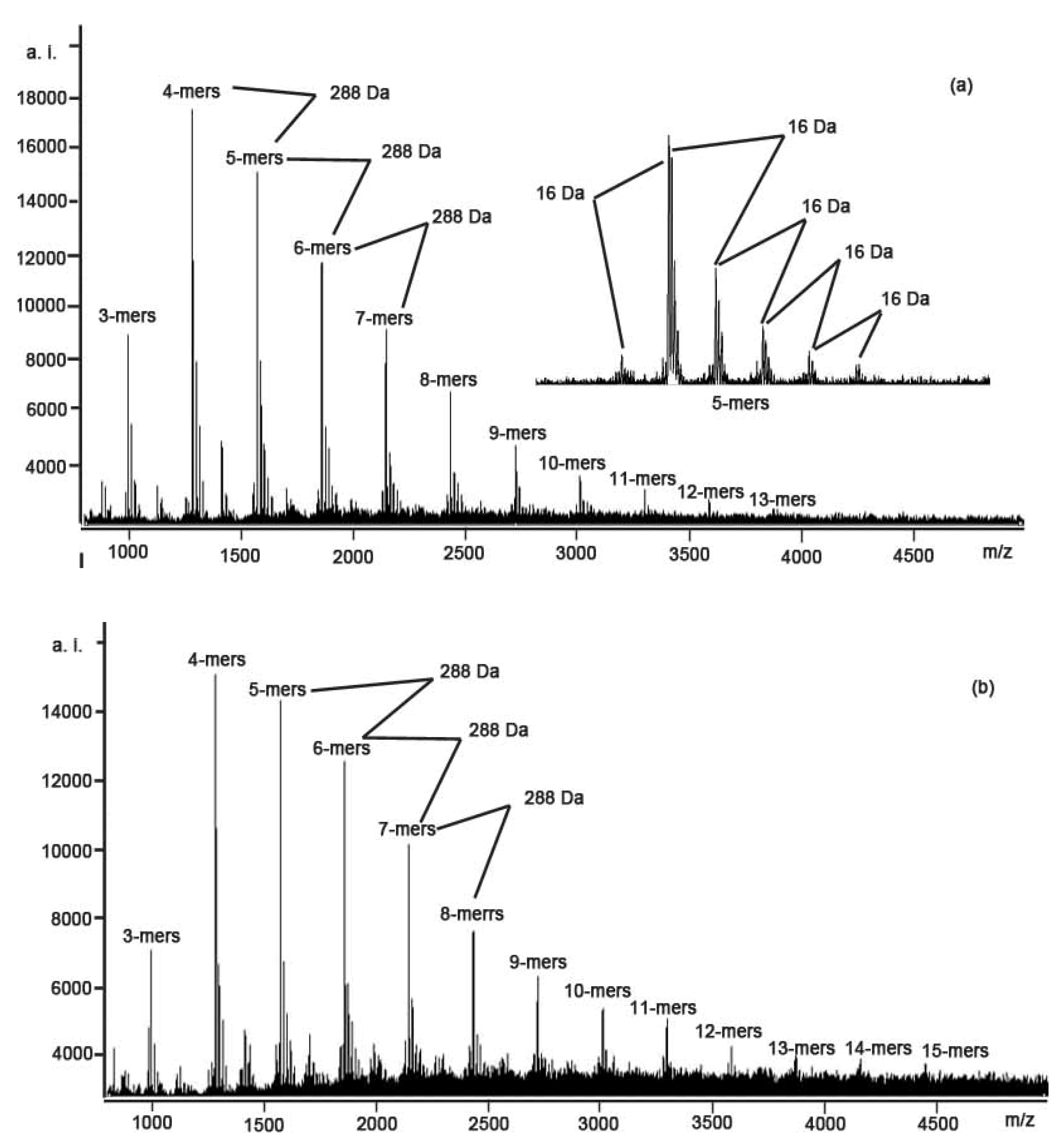
2.2. MALDI-TOF MS Analysis

Many previous studies have shown that MALDI-TOF MS is a powerful technique for the characterization of both synthetic and natural polymers (including condensed tannins) [22,23,24,25,26]. In our study, according to the findings of Xiang et al. [27], the CS+ was used as the cationization reagent. This resulted in the best conditions for their MALDI-TOF analysis and a relatively simple MALDI-TOF spectrum.
Figure 1 shows the MALDI-TOF mass spectra of the polymeric condensed tannins mixture of the stem bark and fine root of C. equisetifolia, recorded as CS+ adducts in the positive ion reflectron mode. The polymeric character is reflected by the periodic peak series representing different chain lengths. The results indicate that condensed tannins isolated from the stem bark and fine root are characterized by mass spectra with a series of peaks with distances of 288 Da, corresponding to one catechin/ epicatechin monomer (Table 2). The condensed tannins from the stem bark and fine root of C. equisetifolia had different polymer chain length varying from trimers to tridecamer for stem bark and to pentadecamer for fine root.
Figure 1. MALDI-TOF positive reflectron mode mass spectra of the condensed tannins from (a) stem bark and (b) fine root of C. equisetifolia.
Molecules 15 05658 g001
In addition to the predicted homopolyflavan-3-ol mass series mentioned above, each DP had a subset of masses 16 Da higher were also detected, which can be explained by heteropolymers of repeating flavan-3-ol units containing an additional hydroxyl group at the position 5' of the B-ring. Each DP had a subset of masses 16 Da lower in the spectra of condensed tannins from stem bark and fine root (Figure 1 and Table 2). These masses indicated the polymer chains containing monomers with only one hydroxyl group (16 Da) at the aromatic ring B. Given the absolute masses corresponding to each peak, it was further suggested that the condensed tannins from stem bark and fine root contain procyanidin, prodelphindin and propelargonidin, both with the procyanidin dominating. The structures of condensed tannins from stem bark and fine roots of C. equisetifolia are successfully characterized using MALDI-TOF MS for the first time.
Table 2. Summary of peaks with the highest intensities in MALDI-TOF MS of the condensed tannins from stem bark and fine root of C. equisetifolia.
Polymern1n2n3CalculatedObserved [M + Cs]+
[M + Cs]+Stem barkFine root
Trimer030999999.27999.24
120983983.11983.25
02110151015.241015.25
01210311031.241031.24
00310471047.221046.83
Tetramer04012871287.301287.28
13012711271.311271.25
03113031303.301303.23
02213191319.291319.30
01313351335.491335.20
00413511351.391351.23
Pentamer05015751575.341575.30
14015591559.321559.30
04115911591.481591.33
03216071607.491607.32
02316231623.321623.27
01416391640.481639.30
Hexamer06018631863.341863.34
15018471847.371847.47
05118791879.361879.34
04218951895.391896.45
03319111911.34--
02419271928.68--
Heptamer07021512151.412152.36
16021352135.392135.51
06121672167.352167.36
05221832183.382183.31
04321992199.362200.33
03422152215.942215.49
Octamer08024392440.402439.48
17024232423.472423.51
07124552456.402456.28
06224712472.512471.52
05324872488.402489.49
04425032504.422503.56
Nonamer09027272728.392728.42
18027112712.392711.06
08127432744.392744.31
07227592760.382759.47
06327752775.422776.33
Decamer010030153016.443016.51
19029993001.363000.16
09130313032.923032.29
08230473048.483049.47
07330633064.37--
06430793079.48--
Undecamer011033033304.193305.48
11003287--3289.40
010133193320.753319.19
09233353335.953335.78
08333513351.45--
Dodecamer012035913592.753592.98
11103575--3577.91
Tridecamer013038793880.243880.90
01213895--3896.31
Tetradecamer01404167--4169.66
01314183--4184.57
Pentadecamer01504455 4456.93
n1: Number of afzelechin/epiafzelechin unit; n2: Number of catechin/epicatechin unit; n3: Number of gallocatechin/epigallocatechin unit; “--” means no observed peaks corresponding to those calculated ones.

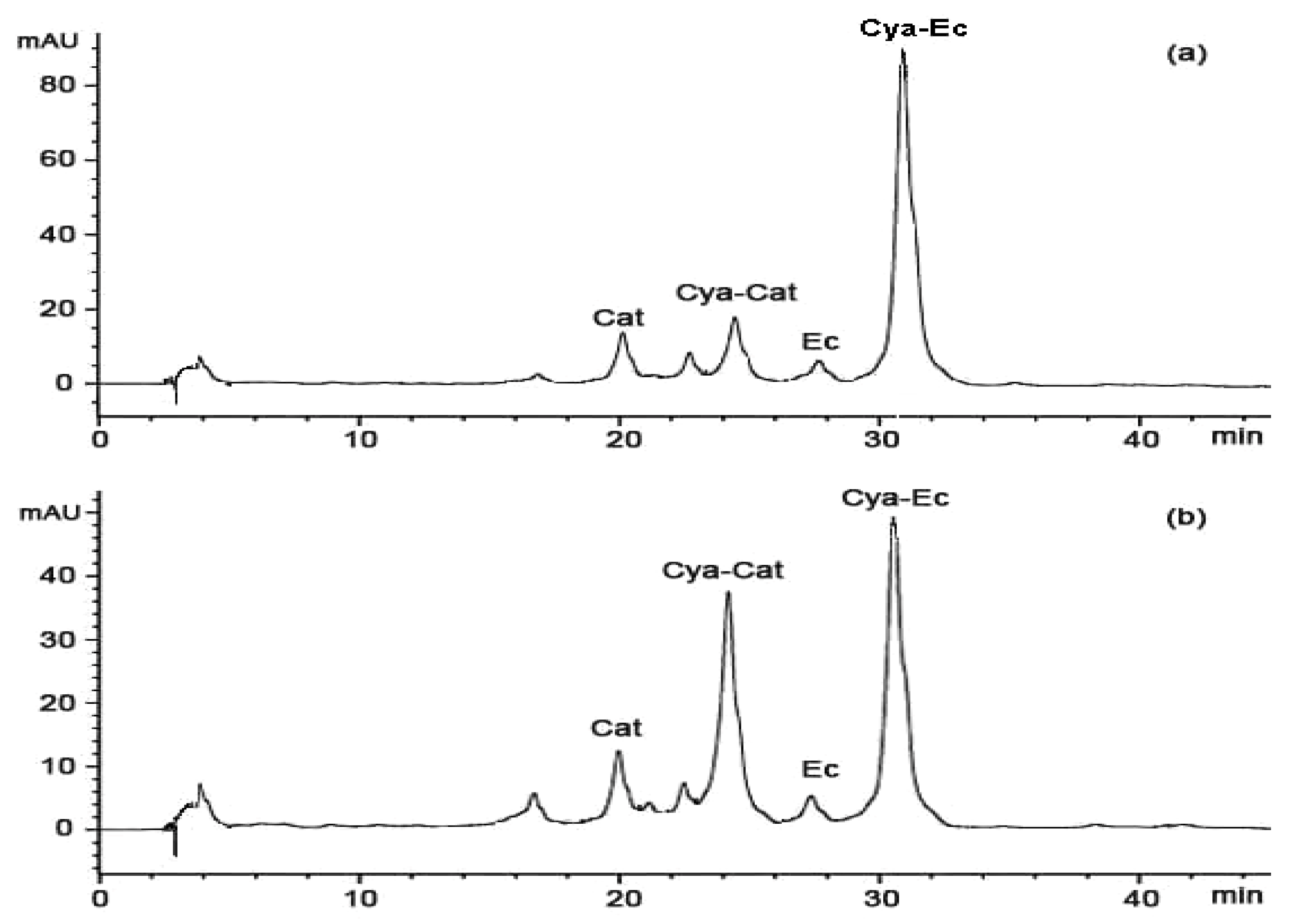
2.3. Thiolysis with Cysteamine Followed by RP-HPLC Analysis

To further investigate whether the condensed tannins from stem bark and fine root are composed of catechin and epicatechin, depolymerization through thiolysis was carried out by following standard conditions using cysteamine, which was preferred to toluene-α-thiol for being more user-friendly and much less toxic [28]. The reaction mixture was analysed by HPLC (Figure 2). In agreement with the MALDI-TOF MS results, the condensed tannins from stem bark and fine root consists primarily of procyanidins. The major product observed was the 4β-(2-aminoethylthio) epicatechin (Cya-EC) along with the small amount of (+)-catechin (Cat), (-)-epicatechin (EC), and 4β-(2-aminoethylthio) catechin (Cya-Cat). This result suggested that there are significant amounts of epicatechin extension units in condensed tannins from stem bark and fine root.
Figure 2. Reversed phase HPLC chromatograms of the condensed tannins from: (a) stem bark and (b) fine root degraded in the presence of cysteamine; Cat, (+)-catechin; EC, (-)-epicatechin; Cya-Cat, 4β-(2-aminoethylthio)catechin; Cya-EC, 4β-(2-aminoethylthio)-epicatechin.
Molecules 15 05658 g002

2.4. DPPH Radical Scavenging Activity

The relatively stable organic radical DPPH has been widely used in the determination of antioxidant activity of single compounds as well as the different plant extracts [29]. The reduction capability of DPPH radical is determined by the decrease in absorbance at 517 nm induced by antioxidants [30]. Great decreases in a concentration-dependent manner of remaining DPPH indicated that condensed tannins from stem bark and fine root of C. equisetifolia possess potent free radical-scavenging activity (Table 3).
Table 3. Concentration effect of condensed tannins from stem bark and fine root of C. equisetifolia on their free radical scavenging activity (DPPH) and reducing capacity (FRAP) (n = 3).
Concentration (μg/mL)DPPH aFRAP b
Stem barkFine rootStem barkFine root
15.6391.48 ± 0.23d89.45 ± 0.42d0.13 ± 0.01a0.14 ± 0.00a
31.2583.33 ± 0.78c80.38 ± 0.84c0.26 ± 0.01b0.26 ± 0.00b
62.566.42 ± 1.00b62.73 ± 0.65b0.51 ± 0.00c0.53 ± 0.00c
12539.98 ± 1.08a31.92 ± 0.09a0.99 ± 0.00d1.01 ± 0.01d
a Data are presented as the percentage of remaining DPPH; b Data are presented as the absorbance at 593 nm; Different letters on the same column show significant differences from each other at P < 0.05; Statistical analysis was done by Duncan’s multiple range tests.
The quality of the antioxidants about the condensed tannins from stem bark and fine root of C. equisetifolia was determined by the IC50 values (the concentration with scavenging activity of 50%). A lower value of IC50 indicates greater antioxidant activity. The IC50 values of the stem bark (101.69 ± 2.24 µg/mL) and fine root (89.32 ± 0.21 µg/mL) were significantly lower than the postive compounds ascorbic acid (110.87 ± 0.88 µg/mL) and BHA (115.66 ± 2.13 µg/mL), indicating the condensed tannins of stem bark and fine root exhibited the higher radical scavenging effect than them. The scavenging effect on the DPPH radical decreased in the order: fine root > stem bark > ascorbic acid > BHA (Table 4).
Table 4. Antioxidant activities of the condensed tannins from stem bark and fine root of C. equisetifolia using the (DPPH) free radical-scavenging assay and the (FRAP) ferric-reducing antioxidant power assay (n = 3).
SamplesAntioxidant activity
IC50/DPPH (µg/mL) aFRAP (mmol AAE/g) b
Stem bark101.69 ± 2.24b5.70 ± 0.03b
Fine root89.32 ± 0.21a5.87 ± 0.04c
BHA115.66 ± 2.13d5.21 ± 0.04a
Ascorbic acid110.87 ± 0.88c--
a The antioxidant activity was evaluated as the concentration of the test sample required to decrease the absorbance at 517 nm by 50% in comparison to the control; b FRAP values are expressed in mmol ascorbic acid equivalent/g sample in dry weight; Different letters on the same column show significant differences from each other at P < 0.05; Statistical analysis was done by Duncan’s multiple range tests.

2.5. Ferric Reducing Antioxidant Power (FRAP)

The FRAP assay is based on the redox reaction of ferric ion in the presence of reducer. The reduction capacity of a compound may serve as a significant indicator of its potential antioxidant activity [31]. A higher absorbance corresponds to a higher ferric reducing power. All tannins showed increased ferric reducing power with the increasing concentration (Table 3). The FRAP value, used to determine the antioxidant ability of stem bark and fine root of C. equisetifolia in present study, was expressed in ascorbic acid equivalents. The FRAP values for stem bark and fine root are 5.70±0.03 and 5.87±0.04 mmol AAE/g dried tannins, and were both significantly higher than that of BHA (5.21±0.04 mmol AAE/g dried sample, Table 4).

3. Experimental

3.1. Chemicals and Materials

All solvents used were of analytical reagent (AR) purity grade. 1,1-Diphenyl-2-picrylhydrazyl (DPPH), 2,4,6-tripyridyl-s-triazine (TPTZ), ascorbic acid, butylated hydroxyanisole (BHA), and cesium chloride were purchased from Sigma-Aldrich (USA). Sephadex LH-20 was purchased from Amersham (USA). Stem bark and fine root of C. equisetifolia were collected from Chihu Forestry Center of Huian County (23°45′N, 118°55′E), Fujian Province, China. The C. equisetifolia plantations were artificial, pure forests planted in 1988, with the coverage of dense forest 0.8, tree density 1535 tree/ha, canopy height 13.5 m and DBH (diameter at breast height) 17.32 ± 2.5 cm. Thirty trees with similar height and growth conditions were chosen and divided into three groups (10 trees in a group). About 50 g/tree stem bark and fine root were collected, washed, freeze-dried (48 h) and then ground to pass through the 40-mesh sieve. The samples were stored at -20 °C prior to analyses.

3.2. Extraction and Purification of the Condensed Tannins

Extraction and purification of the condensed tannins were described by Lin et al. [32]. Briefly, freeze-dried stem bark and fine root powder (each 25 g) were extracted thrice with 7:3 (v/v) acetone/water solutions at room temperature. Each extract was filtered and pooled, and the solvent was removed under reduced pressure by use a rotary evaporator at 38 °C. The remaining aqueous fraction was extracted thrice with hexane in order to remove chlorophyll and lipophilic compounds. The remaining crude tannin fraction was chromatographed on an LH-20 column (Pharmacia Biotech, Uppsala, Sweden) which was first eluted with methanol-water (50:50, v/v) and then with acetone- water (7:3, v/v). The last fraction purified tannins were freezed-dried and stored at -20 °C before analysis by MALDI-TOF mass spectrometry.

3.3. Determination of Total Phenolics and Extractable Condensed Tannins

The amount of total phenolics was determined using the Folin-Ciocalteu method [33]. Briefly, an aliquot of extract (0.2 mL) was added to a 10 mL volumetric flask containing distilled H2O (0.3 mL). Folin-Ciocalteu reagent (0.5 mL) and 20% Na2CO3 solution (2.5 mL) were added to the mixture and shaken. After incubation for 40 min at room temperature, the absorbance versus a blank was determined at 725 nm. Total phenolics contents of extracts were expressed as mg tannic acid equivalents (TAE)/g extract. Extractable condensed tannins were assayed by the butanol-HCl method [34], using respective purified condensed tannins as the standards. All samples were analyzed in three replications.

3.4. MALDI-TOF MS Analysis

The MALDI-TOF MS spectra were recorded on a Bruker Reflex III instrument (Germany). The irradiation source was a pulsed nitrogen laser with a wavelength of 337 nm, and the duration of the laser pulse was 3 ns. In the positive reflectron mode, an accelerating voltage of 20.0 kV and a reflectron voltage of 23.0 kV were used. 2,5-Dihydroxybenzoic acid (DHB, 10 mg/mL 30% acetone solution) was used as the matrix. The sample solutions (10 mg/mL 30% acetone solution) were mixed with the matrix solution at a volumetric ratio of 1:3. The mixture (1 µL) was spotted to the steel target. Amberlite IRP-64 cation-exchange resins (Sigma-Aldrich, USA), equilibrated in deionized water, and was used to deionize the analyte-matrix solution thrice. Cesium trifluoroacetate (1.52 mg/mL) was mixed with the analyte-matrix solution (1:3, v/v) to promote the formation of a single type of ion adduct ([M+Cs]+) [35].

3.5. Thiolysis of the Condensed Tannins for HPLC Analysis

Thiolysis was carried out according to the method of Torres and Lozano [36]. A condensed tannin solution (4 mg/mL in methanol) was prepared. A sub-sample (50 µL) was placed in a vial and to this was added hydrochloric acid in methanol (3.3%, v/v; 50 µL) and cysteamine hydrochloride in methanol (50 mg/mL, 100 µL). The solution was heated at 40°C for 30 min, and cooled to room temperature. The size and composition of the condensed tannins were estimated from the RP-HPLC analysis of the depolymerised fractions [28]. Briefly, the terminal flavan-3-ols units were released as such by acid cleavage in the presence of cysteamine whereas the extension moieties were released as the C4 cysteamine derivatives. Thiolysis reaction media (20 µL) filtrated through a membrane filter with an aperture size of 0.45 µm was analyzed by RP-HPLC.
The high performance liquid chromatograph was an Agilent 1200 system (Palo, Alto, CA, USA) equipped with a diode array detector and a quaternary pump. The thiolysis media were further analyzed using LC/MS (QTRAP 3200, USA) with a Hypersil ODS column (4.6 mm × 250 mm, 2.5 μm) (China). The mobile phase was composed of solvent A (0.5% v/v trifluoroacetic acid (TFA) in water) and solvent B (0.5% v/v TFA in acetonitrile). The gradient condition was: 0-5 min, 3% B (isocratic); 5-15 min, 3%-9% B (linear gradient); 15-45 min, 9%-16% B (linear gradient), 45-60 min, 16%-60% B (linear gradient). The column temperature was ambient and the flow-rate was set at 1 mL/min. Detection was at 280 nm and the UV spectra were acquired between 200-600 nm. Degradation products were identified on chromatograms according to their relative retention times and their UV-visible spectra. Each sample analysis was repeated three times.

3.6. DPPH Radical Scavenging Activity

The free radical scavenging activities of the purified condensed tannins on the DPPH radical were measured using the method described by Braca et al. [37]. A 0.1 mL sample of various concentrations of each freeze-dried purified condensed tannins at different concentration (15.63-250 µg/mL) was added to DPPH solution (3 mL, 0.1 mM in methanolic solution). An equal amount of methanol and DPPH served as control. After the mixture was shaken and left at room temperature for 30 min, the absorbance at 517 nm was measured. Lower absorbance of the reaction mixture indicates higher free radical scavenging activity. The IC50 value, defined as the amount of antioxidant necessary to decrease the initial DPPH concentration by 50%, was calculated from the results and used for comparison. The capability to scavenge the DPPH radical was calculated by using the following equation:
DPPH scavenging effect (%) = [(A1–A2)/A1] ×100
where A1 = the absorbance of the control reaction; A2 = the absorbance in the presence of the sample. BHA and ascorbic acid were used as standards.

3.7. Ferric Reducing/Antioxidant Power (FRAP) Assay

FRAP assay is a simple and reliable colorimetric method commonly used for measuring the total antioxidant capacity [38]. In brief, prepared freshly FRAP reagent (3 mL) was mixed with test sample (0.1 mL) or methanol (0.1 mL, for the reagent blank). The FRAP reagent was prepared from 300 mmol/L acetate buffer (pH 3.6), 20 mmol/L ferric chloride and 10 mmol/L TPTZ made up in 40 mmol/L hydrochloric acid. All the above three solutions were mixed together in the ratio of 25:2.5:2.5 (v/v/v). The absorbance of reaction mixture at 593 nm was measured spectrophotometrically after incubation at 25 °C for 10 min. The FRAP values, expressed in mmol ascorbic acid equivalents (AAE)/g dried tannins, were derived from a standard curve.

3.8. Statistical Analysis

All data were expressed as means ± standard deviation of three independent determinations. One-way analysis of variance (ANOVA) was used, and the differences were considered to be significant at P < 0.05. All statistical analyses were performed with SPSS 13.0 for Windows.

4. Conclusions

Structures of condensed tannins from stem bark and fine root of C. equisetifolia characterized by MALDI-TOF MS analysis showed that the condensed tannins consist predominantly of procyanidin combined with prodelphinidin and propelargonidin. The HPLC analysis revealed that the epicatechin is the main extension unit of condensed tannins from stem bark and fine root. These condensed tannins had different polymer chain length varying from trimers to tridecamer for stem bark and to pentadecamer for fine root. The condensed tannins extracted from the stem bark and fine root all showed a very good DPPH radical scavenging activity and ferric reducing power, suggesting that these extracts may be considered as new sources of natural antioxidants for food and nutraceutical products.

Acknowledgements

This work was supported by the Special Prophase Project on National Basic Research Program of China (973 Program) (No. 2009CB426306), the National Eleventh-Five Year Key Project (NO. 2009BADB2B0302), and by Program for New Century Excellent Talents in University (NCET-07-0725).

References

  1. Kraus, T.E.C.; Dahlgren, R.A.; Zasoski, R.J. Tannins in nutrient dynamics of forest ecosystems—a review. Plant Soil 2003, 256, 41–66. [Google Scholar] [CrossRef]
  2. Hemingway, R.W.; Karchesy, J.J. Chemistry and significance of condensed tannins; Plenum: New York, NY, USA, 1989. [Google Scholar]
  3. Porter, L.J. The Flavanoids: Advances in Research Since 1980; Harborne, J.B., Ed.; Chapman and Hall: New York, NY, USA, 1988; pp. 21–62. [Google Scholar]
  4. Porter, L.J. The Flavanoids: Advances in Research Since 1986; Harborne, J.B., Ed.; Chapman and Hall: London, UK, 1994; pp. 23–54. [Google Scholar]
  5. Waterman, P.G.; Mole, S. Analysis of Phenolic Plant Metabolites; Blackwell Scientific Publications: Oxford, UK, 1994. [Google Scholar]
  6. Santos-Buelga, C.; Scalbert, A. Proantocyanidins and tannin-like compounds: nature, occurrence dietary intake and effects on nutrition and health. J. Sci. Food Agri. 2000, 80, 1094–1117. [Google Scholar] [CrossRef]
  7. Svedström, U.; Vuorela, H.; Kostiainen, R.; Huovinen, K.; Laakso, I.; Hiltunen, R. High-performance liquid chromatographic determination of oligomeric procyanidins from dimers up to the hexamer in hawthorn. J. Chromatogr. A. 2002, 968, 53–60. [Google Scholar] [CrossRef]
  8. Hümmer, W.; Schreier, P. Analysis of proanthocyanidins. Mol. Nutr. Food Res. 2008, 52, 1381–1398. [Google Scholar] [CrossRef]
  9. Es-Safi, N.E.; Guyot, S.; Ducrot, P.H. NMR, ESI/MS, and MALDI-TOF/MS Analysis of Pear Juice Polymeric Proanthocyanidins with Potent Free Radical Scavenging Activity. J. Agric. Food Chem. 2006, 54, 6969–6977. [Google Scholar] [CrossRef]
  10. Behrens, A.; Maie, N.; Knicker, H.; Kögel-Knabner, I. MALDI-TOF mass spectrometry and PSD fragmentation as means for the analysis of condensed tannins in plant leaves and needles. Phytochem. 2003, 62, 1159–1170. [Google Scholar]
  11. Chen, Y.; Hagerman, A.E. Characterization of Soluble Non-covalent Complexes between Bovine Serum Albumin and [beta]-1, 2, 3, 4, 6-Penta-O-galloyl-d-glucopyranose by MALDI-TOF MS. J. Agric. Food Chem. 2004, 52, 4008–4011. [Google Scholar] [CrossRef]
  12. Rahim, A.A.; Rocca, E.; Steinmetz, J.; Jain Kassim, M.; Sani Ibrahim, M.; Osman, H. Antioxidant activities of mangrove Rhizophora apiculata bark extracts. Food Chem. 2008, 107, 200–207. [Google Scholar] [CrossRef]
  13. Vivas, N.; Nonier, M.F.; de Gaulejac, N.V.; Absalon, C.; Bertrand, A.; Mirabel, M. Differentiation of proanthocyanidin tannins from seeds, skins and stems of grapes (Vitis vinifera) and heartwood of Quebracho (Schinopsis balansae) by matrix-assisted laser desorption/ionizationtime-of-flightmassspectrometryandthioacidolysis/liquidchromatography/electrosprayionization mass spectrometry. Anal. Chim. Acta 2004, 513, 247–256. [Google Scholar] [CrossRef]
  14. Zhang, L.L.; Lin, Y.M. HPLC, NMR and MALDI-TOF MS analysis of condensed tannins from Lithocarpus glaber leaves with potent free radical scavenging activity. Molecules 2008, 13, 2986–2997. [Google Scholar] [CrossRef]
  15. Zhang, L.L.; Lin, Y.M.; Zhou, H.C.; Wei, S.D.; Chen, J.H. Condensed tannins from mangrove species Kandelia candel and Rhizophora mangle and their antioxidant activity. Molecules 2010, 15, 420–431. [Google Scholar] [CrossRef]
  16. Chen, H.Y. Flora of Hainan; Science Press: Beijing, China, 1997. [Google Scholar]
  17. Prakash, D.; Suri, S.; Upadhyay, G.; Singh, B.N. Total phenol, antioxidant and free radical scavenging activities of some medicinal plants. Int. J. Food Sci. Nutr. 2007, 58, 18–28. [Google Scholar] [CrossRef]
  18. Sun, D.W. Chemistry of Vegetable Tannins; Forestry Press of China: Beijing, China, 1992; Volume 8, p. 378. [Google Scholar]
  19. Block, G. The data support a role for antioxidants in reducing cancer risk. Nutrition reviews (USA) 1992, 50, 207–213. [Google Scholar] [CrossRef]
  20. Rice-Evans, C.A.; Miller, N.J.; Paganga, G. Structure-antioxidant activity relationships of flavonoids and phenolic acids. Free Radic. Biol. Med. 1996, 20, 933–956. [Google Scholar] [CrossRef]
  21. Duh, P.D.; Tu, Y.Y.; Yen, G.C. Antioxidant activity of water extract of Harng Jyur (Chrysanthemum morifolium Ramat). Lebensm-wiss. Technol. 1999, 32, 269–277. [Google Scholar] [CrossRef]
  22. Danis, P.O.; Karr, D.E.; Mayer, F.; Holle, A.; Watson, C.H. The analysis of water-soluble polymers by matrix-assisted laser desorption time-of-flight mass spectrometry. Organic Mass Spect. 1992, 27, 843–846. [Google Scholar] [CrossRef]
  23. Pasch, H.; Pizzi, A.; Rode, K. MALDI-TOF mass spectrometry of polyflavonoid tannins. Polymer 2001, 42, 7531–7539. [Google Scholar]
  24. Jerez, M.; Sineiro, J.; Guitián, E.; Núñez, M.J. Identification of polymeric procyanidins from pine bark by mass spectrometry. Rap. Comm. Mass Spec. 2009, 23, 4013–4018. [Google Scholar]
  25. Oo, C.W.; Pizzi, A.; Pasch, H.; Kassim, M.J. Study on the structure of mangrove polyflavonoid tannins with MALDI-TOF mass spectrometry. J. Appl. Polym. Sci. 2008, 109, 963–967. [Google Scholar]
  26. Montaudo, G.; Montaudo, M.S.; Samperi, F. Martix-assisted laserdesorption/ionizationmass spectrometryofpolymers (MALDI-MS). In Mass Spectrometry of Polymers; Montaudo, G., Lattimer, R.P., Eds.; CRC Press: Boca Raton, FL, USA, 2002; pp. 419–521. [Google Scholar]
  27. Xiang, P.; Lin, Y.; Lin, P.; Xiang, C.; Yang, Z.; Lu, Z. Effect of cationization reagents on the matrix-assisted laser desorption/ionization time-of-flight mass spectrum of Chinese gallotannins. J. Appl. Polym. Sci. 2007, 105, 859–864. [Google Scholar] [CrossRef]
  28. Torres, J.L.; Selga, A. Procyanidin size and composition by thiolysis with cysteamine hydrochloride and chromatography. Chromatographia 2003, 57, 441–445. [Google Scholar] [CrossRef]
  29. Oyaizu, M. Studies on products of browning reactions: antioxidative activities of products of browning reaction prepared from glucosamine. Jpn. J. Nutr. 1986, 44, 307–315. [Google Scholar] [CrossRef]
  30. Kumaran, A.; Karunakaran, J. In vitro antioxidant activities of methanol extracts of five Phyllanthus species from India. Food. Sci. Technol. 2007, 40, 344–352. [Google Scholar]
  31. Meir, S.; Kanner, J.; Akiri, B.; Philosoph-Hadas, S. Determination and involvement of aqueous reducing compounds in oxidative defense systems of various senescing leaves. J. Agric. Food Chem. 1995, 43, 1813–1819. [Google Scholar] [CrossRef]
  32. Lin, Y.M.; Liu, J.W.; Xiang, P.; Lin, P.; Ye, G.F.; Sternberg, L.D.S.L. Tannin dynamics of propagules and leaves of Kandelia candel and Bruguiera gymnorrhiza in the Jiulong River Estuary, Fujian, China. Biogeochemistry 2006, 78, 343–359. [Google Scholar] [CrossRef]
  33. Makkar, H.P.S.; Blümmel, M.; Borowy, N.K.; Becker, K. Gravimetric determination of tannins and their correlations with chemical and protein precipitation methods. J. Sci. Food Agric. 1993, 61, 161–165. [Google Scholar] [CrossRef]
  34. Terrill, T.H.; Rowan, A.M.; Douglas, G.B.; Barry, T.N. Determination of extractable and bound condensed tannin concentrations in forage plants, protein concentrate meals and cereal grains. J. Sci. Food Agric. 1992, 58, 321–329. [Google Scholar] [CrossRef]
  35. Xiang, P.; Lin, Y.M.; Lin, P.; Xiang, C. Effects of adduct ions on matrix-assisted laser desorption/ionization time of flight mass spectrometry of condensed tannins: a prerequisite knowledge. Chin. J. Anal. Chem. 2006, 34, 1019–1022. [Google Scholar]
  36. Torres, J. L.; Lozano, C. Chromatographic characterization of proanthocyanidins after thiolysis with cysteamine. Chromatographia 2001, 54, 523–526. [Google Scholar] [CrossRef]
  37. Braca, A.; De Tommasi, N.; Di Bari, L.; Pizza, C.; Politi, M.; Morelli, I. Antioxidant principles from Bauhinia tarapotensis. J. Nat. Prod. 2001, 64, 892–895. [Google Scholar]
  38. Benzie, I.F.F.; Strain, J.J. The ferric reducing ability of plasma (FRAP) as a measure of “antioxidant power”: The FRAP assay. Anal. Biochem. 1996, 239, 70–76. [Google Scholar]
  • Sample Availability: Samples of the compounds are available from the authors.

