Article

New Catechol Derivatives of Safrole and Their Antiproliferative Activity towards Breast Cancer Cells

1
Departamento de Química, Universidad Técnica Federico Santa María, Av. España N° 1680, Valparaíso, Chile
2
Centro de Investigaciones Biomedicas (CIB), Centro Regional de Estudios en Alimentos Saludables, Escuela de Medicina, Universidad de Valparaíso, Creas, Av. Hontaneda N° 2664, Valparaíso, Chile
3
Departamento de Ciencias Químicas, Universidad Andrés Bello, Campus Viña del Mar, Los Fresnos N° 52, Viña del Mar, Chile
*
Authors to whom correspondence should be addressed.
Molecules 2011, 16(6), 4632-4641; https://doi.org/10.3390/molecules16064632
Submission received: 13 April 2011 / Revised: 31 May 2011 / Accepted: 2 June 2011 / Published: 3 June 2011
(This article belongs to the Section Organic Chemistry)

Abstract

:
Catechols were synthesized from safrole. Nine derivatives were prepared and assessed for antiproliferative effects using different human cell lines. The in vitro growth inhibition assay was based on the sulphorhodamine dye to quantify cell viability. The derivatives 4-allylbenzene-1,2-diol (3), 4 4-[3-(acetyloxy)propyl]-1,2-phenylene diacetate (6) and 4-[3-(acetyloxy)propyl]-5-nitro-1,2-phenylene diacetate (10) showed higher cytotoxicity than the parent compound 2 in tests performed on two breast cancer cell lines (MCF-7 and MDA-MB-231). The IC50 values of 40.2 ± 6.9 μM, 5.9 ± 0.8 μM and 33.8 ± 4.9 μM, respectively, were obtained without toxicity towards dermal human fibroblast (DHF cells).

1. Introduction

Safrole is a colorless or slightly yellow oily liquid that has been commonly used in cosmetics and as a food flavoring, extracted as sassafras oil from the root-bark or the fruit of sassafras (Lauraceae) plants, or synthesized from other compounds linked to methylenedioxy [1,2]. In vitro, a depletion of glutathione (GSH) was observed by the addition of safrole to hepatocytes [3,4]. In 1994, Bolton pointed out that hydroxychavicol could be further biotransformed to o-quinone through a 2-electron oxidation in vitro [4]. This characterization of the GSH conjugates revealed that the o-quinone moiety undergoes regioselective attack by GSH at C6 on the cyclohexadienedione ring. Under neutral or acidic conditions, sulfur nucleophiles add primarily 1,6 to 4-substituted o-benzoquinones. As catechols are readily oxidized, the conversion of l-allyl-3,4-dihydroxybenze (HC) to an o-quinone or p-quinone methide (QM) may contribute to the cytotoxic and/or genotoxic activity of safrole. In previous studies, the effects of safrole oxide derivatives on cellular growth and apoptosis in vascular endothelial cells (VEC) were evaluated [5]. In this study, the authors concluded that safrole oxide induced apoptosis by up-regulating the expressions of transmembrane protein (Fas) and by reducing the activity of reactive oxygen species (ROS). The authors also reported that safrole oxide can inhibit angiogenesis. This is induced by cancer cells in vitro and in vivo [6,7]. It has been recently reported that safrole derivatives (catechols) are more toxic than safrole against MCF-7 and MDA-MB-231 cells [8]. The safrole derivatives proved to be more toxic than safrole itself against the selected cancer cells and not to cause damage in dermal human fibroblast cells. The formation of catechols from safrole with AlCl3 was favoured by the presence of the electron attractor group (NO2) in the molecule. These results suggest that derivatives of safrole bearing a nitro group and a hydroxyl group can present an antiproliferative/cytotoxic effects on cells. The screening in vitro provides preliminary data to select compounds with potential antineoplastic properties. Safrole (1) and its synthetic derivatives 3-11 were tested in vitro for antiproliferative effects on two human tumor breast cancer cell lines (MDA-MB-231, MCF-7), and on one dermal human fibroblast cell line (DHF). The sulforhodamine B assay was used to quantify cell viability.

2. Results and Discussion
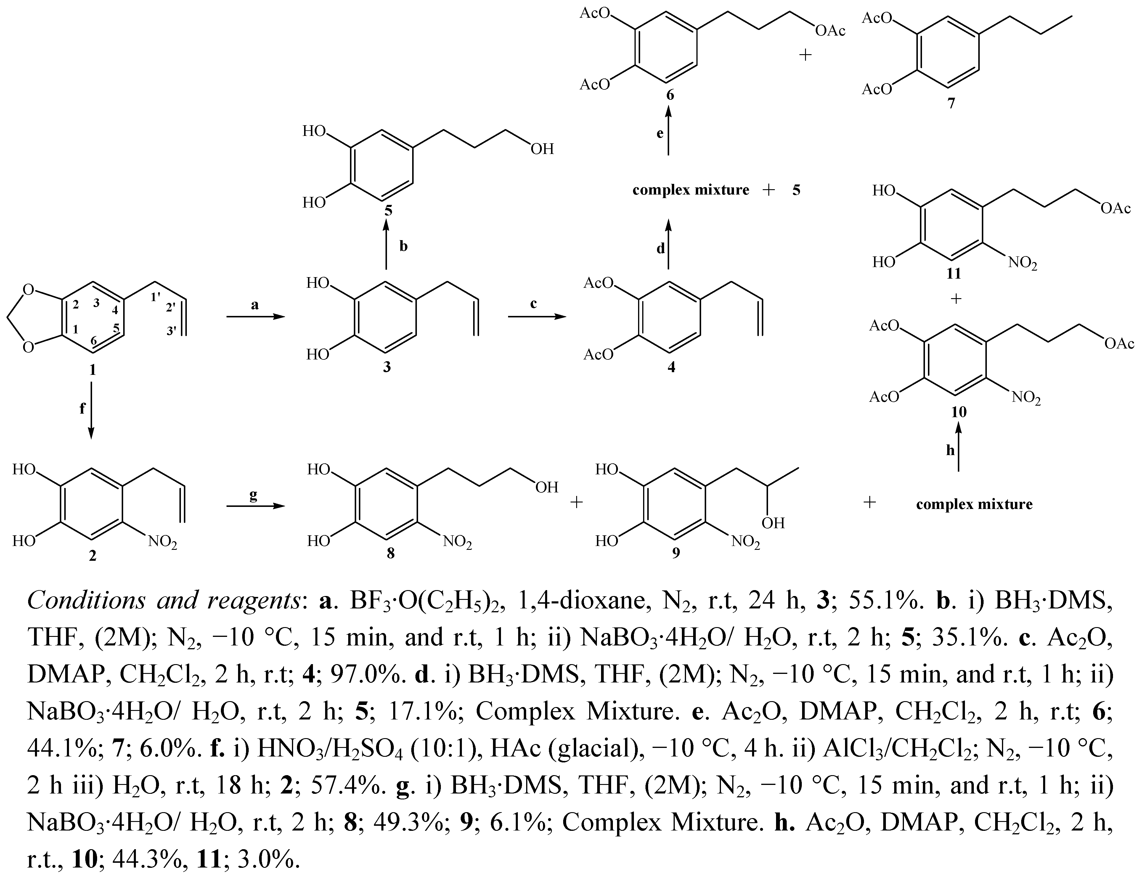
2.1. Chemistry

The catechol 3 was obtained and synthesized by the BF3•O(C2H5)2 solution method [9] in 55.1% yield, higher that the reported previously for compounds 2 [8,10,11,12,13] (Scheme 1). Oxidative hydroboration of the side chain in both catechols led to a hydroxyl group in C-3’. The side chains of the catechols were oxidized using BH3·DMS/THF/NaBO3·4H2O/H2O as previously described for other alkenes and alkynes [14,15,16,17,18,19]. The primary alcohols 5 and 8 were obtained in 35.1% and 49.3% w/w yields and the secondary alcohol 9 with 6.1% yield (Scheme 1). Hydroboration of acetylated catechol 4 produced the triol 5 (a product obtained due to the alkaline pH generated in the oxidation process) [8] plus a polar complex mixture, which after acetylation gave acetylated triol 6 plus the catechol with a saturated side chain 7 (Scheme 1). All the compounds were characterized by NMR, IR and MS spectral data.
Scheme 1. General scheme of synthesis of derivatives of safrole.
Molecules 16 04632 g001
Cleavage of the methylenedioxy ring of safrole took place during the conversion into the catechol 3 (55.1% yield) using the new BF3•O(C2H5)2 solution method [12,13]. In the 1H-NMR spectrum, the absence of the methylenedioxy singlet between 5.90–6.05 ppm confirmed the presence of catechol, where now two broad signals at δ = 5.29 (b.s., 1H) and δ = 5.21 (b.s., 1H) are observed. The MS spectrum of the diacetate derivative 3 showed two peaks at m/z 175 (26.6%) and 116 (24.3%) presumably due to the loss of two acetate groups. The first oxidative hydroboration of the side chain in catechol 2 and the subsequent hydroboration of acetylated catechol 3 was carried out to obtain the C-3’ alcohol 5 with different yields in both cases (35.1% and 17.1%, respectively). Additionally in the hydroboration of 4 a complex mixture was obtained that was derivatized by acetylation, to give triacetate compound 6 and the acetylated catechol 7 with a saturated side chain.
In the IR spectrum of compound 5 the broad absorption at 3398 cm−1 was assigned to a hydroxyl group, whereas in the 1H-NMR spectrum three singlet signals at δ = 7.75 ppm (s, 1H), 7.73 ppm (s, 1H) and 5.69 ppm (b.s, 1H) for three hydroxyl groups were observed. Finally, using the experimental data obtained from the NMR spectra, the structure of triacetyl derivative 6 was confirmed.
On the other hand in the 1H-NMR spectrum of compound 7 three signals at δ = 2.57 ppm (t, 2H, J = 7.7 Hz), δ = 1.63 ppm (m, 2H) and δ = 0.95 ppm (t, 3H, J = 7.7 Hz) were observed, assigned to the hydrogens H-1', H-2' and H-3', respectively, corresponding these three signals to a typical n-propyl group. The 13C-NMR spectrum (in combination with 13C-DEPT-135) showed the presence of two CH3 signals at δ = 20.6 and 13.8 ppm; the first was assigned to the methyl groups of both acetates and second assigned to the terminal CH3 in the n-propyl side chain.
In the hydroboration of compound 2 both isomers, the primary and secondary alcohols, were obtained in 49% and 6.1% yield, respectively. In the 1H-NMR spectrum of compound 8, three signals at δ = 4.39 (t, 2H, J = 6.0 Hz); 2.88 (m, 2H) and 1.79 (m, 2H) were assigned to H-3’, H-1’ and H-2’ in the saturated side chain, respectively. In the 1H NMR spectrum of isomer 9, the presence of signal at δ = 4.12 ppm (m, 1H’) was assigned to a carbinolic hydrogen in the C-2’ position (secondary alcohol). Also the signal at δ = 1.33 (d, 3H, J = 6.6 Hz) was to assigned a terminal methyl group. Compound 11 was finally identified as follows: in IR spectrum the observed bands at 3351, 1739 and 1546 cm−1, were assigned to the OH, carbonyl and NO2 groups. The 1H-NMR spectrum showed a signal at δ = 2.94 ppm (s, 2H) assigned to two OH groups. The structure was confirmed by the 13C-NMR spectrum with two signals δ = 171.0 and 20.7 ppm, corresponding to a single carbonyl carbon and a methyl acetate group, respectively.

2.2. Biological Results

The in vitro cytotoxicity evaluation of compounds 3-11 synthesized from safrole and nitrated catechol 2 (see Scheme 1) indicated that the cell viability expressed as % vs. control vehicle (ethanol 0.1%) for the compounds synthesized was dose-dependant (µM). IC50 values of compounds 3-11, safrole, catechol 2 and the reference compound daunorubicin are summarized in Table 1 as the micromolar concentration that produces 50% cell growth inhibition after 72 hours of drug exposure.
Table 1. Cytotoxic activity of synthesized derivatives safrole compounds against two breast cancer cell lines, MCF-7, MDA-MB-231 and a dermal human fibroblast cells, DHF, expressed as inhibitory concentration IC50.
CompoundIC 50 (μM)
MCF-7MDA-MB-231DHF
255.0 ± 7.1137.5 ± 2.65>100
398.4 ± 11.640.2 ± 6.9>100
4>10054.7 ± 6.6>100
5>10054.3 ± 4.1>100
697.8 ± 12.45.9 ± 0.8>100
7>10041.7 ± 7.4>100
878.2 ± 8.957.3 ± 4.1>100
9>100>100>100
1051.3 ± 2.133.8 ± 4.9>100
1163.8 ± 5.527.1 ± 4.4>100
safrole>100>100>100
Daunorubicin0.19 ± 0.010.38 ± 0.0614.3 ± 1.85
The results indicate that derivative 10 is more toxic than safrole and 2 against MCF-7 cells. Compound 10 also shows higher toxicity against all the cancer cells tested than the parent compound and derivative 2. The activity of compounds 3, 4, 5, 6, 7, 8, 9 and 11 was not significantly different from that of safrole. The compounds 6 and 10 have higher activity in MDA-MB-231 cells than in MCF-7 cells. On the other hand the catechol compounds 2 and 8 have a high activity than compounds 6 and 10. The rest of the compounds have no significant differences with activity of safrole. However, daunorubicin is at least one hundred times more active as an anticancer compound against MDA-MB-231 cells and 100–200 times more effective towards MCF-7 cells than the safrole derivatives 3 and 4. Nevertheless, it is important to emphasize that compound 6 has ten times less cytotoxic than daunorubicin against normal cells, an interesting feature. Safrole has been shown to become cytotoxic to various human cell types at higher concentrations (5–10 mM) [20,21]. Our work suggests that the derivatization of this compound generates potent anti-cancer compounds, as demonstrated by previously synthesized products as 2 and the new modifications as in 6 and 10.

3. Experimental

3.1. General

Safrole was obtained commercially from Sigma-Aldrich and the chemical reagents were purchased (Merck or Aldrich) with the highest purity commercially available and were used without previous purification. IR spectra were obtained by using KBr pellets or thin films in a Nicolet Impact 420 spectrometer. Frequencies are reported in cm−1. Low resolution mass spectra were recorded on a Shimadzu QP-2000 spectrometer at 70 eV ionising voltage and are given as m/z (% rel. int.) 1H, 13C (DEPT 135 and DEPT 90). Some spectra were recorded in CDCl3 solutions and were referenced to the residual peaks of CHCl3, δ = 7.26 ppm and δ = 77.0 ppm for 1H and 13C, respectively; CD3COCD3 solutions were referenced to the residual peaks of CH3COCH3, δ = 2.04 ppm and δ = 29.8 ppm for 1H and 13C, respectively, on a Bruker Avance 400 Digital NMR spectrometer operating at 400.1 MHz for 1H and 100.6 MHz for 13C. Chemical shifts are reported in δ ppm and coupling constants (J) are given in Hz. Silica gel (Merck 200-300 mesh) was used for C.C. and silica gel plates HF-254 for TLC. TLC spots were detected by heating after spraying with 25% H2SO4 in H2O.
Synthesis of 4-allylbenzene-1,2-diol (3) from 1: A BF3•O(C2H5)2 solution (0.3 mL, 2.39 mmol) was slowly added to a solution of safrole (1, 100 mg, 0.62 mmol) in anh.1,4-dioxane (10 mL) under an atmosphere of N2 with gentle stirring. The reaction was continued for 24 h. to r.t. After this the mixture was taken up in water and then extracted with ethyl acetate (3 × 50 mL). Later the layers were separated. The watery layer was discarded and the organic layer was washed to neutrality with a saturated solution of NaHCO3. The organic layer was dried over MgSO4, filtered, evaporated. Then it was absorbed on silica, chromatographed by CC eluting with mixtures of petroleum ether/EtOAc of increasing polarity (17.0:3.0→16.0:4.0) to give an orange oil identified as catechol 3 (51.3 mg, 55.1%); IR (cm−1): 3321 (O-H); 2917 (C-H); 1618 (C=C); 1430 (-CH2); 1045 (-C-O); 809 (-C-H). 1H- NMR: 6.78 (d, 1H, J = 8.0 Hz, H-6); 6.71 (d, 1H, J = 1.9 Hz, H-3); 6.62 (dd, 1H, J = 8.0, 1.9 Hz, H-5); 5.93 (ddt, 1H, J = 16.9, 10.2 and 6.8 Hz, H-2’); 5.29 (b.s., 1H, OH); 5.21 (b.s., 1H, OH); 5.05 (m, 2H, H-3’); 3.27 (d, 2H, J = 6.7 Hz, H-1’). 13C-NMR: 143.5 (C-2); 141.7 (C-1); 137.6 (C-2’); 133.2 (C-4); 121.0 (C-5); 115.7 (C-6); 115.6 (C-3); 115.3 (C-3’); 39.5 (C-1’).
Synthesis of 4-allyl-1,2-phenylene diacetate (4) from 3: To a solution of 3 (0.30 g, 2.00 mmol) in dry CH2Cl2 (60 mL), DMAP (1.00 mg) and Ac2O (0.38 µL, 4.00 mmol) were added and the mixture was stirred at room temperature for 2 h. A cooled solution of 10% KHSO4 (approx. 50 mL) was then added to this mixture. The watery layer was discarded and the organic layer was washed to neutrality with a saturated solution of NaHCO3 and water. Then it was dried over MgSO4, filtered, evaporated and re-dissolved in CH2Cl2 (5 mL). Subsequently, it was absorbed on silica and chromatographed by CC with petroleum ether/EtOAc mixtures of increasing polarity (19.8:0.2→16.4:3.6) to give 4, an amber oil (0.45 mg, 97.0); 39.4 (C-1’); 20.6 (2×CH3CO).
Synthesis of 4-(3-hydroxypropyl)benzene-1,2-diol (5) from 3: The compound 3 (150 mg, 1.0 mmol) cooled to −5 °C, under an atmosphere of N2, was slowly hydroborated with a 2.0 M solution of BH3·DMS/THF (0.25 mL) added dropwise at −10 °C over 15 min. Then the mixture was stirred at room temperature for 1 h. The resultant organoborane was oxidized by sodium perborate (0.95 g, 6.2 mmol) and water (100 mL). The mixture was stirred at room temperature for 2 h. Then, it was extracted with portions of ethyl ether (4 × 50 mL) and the layers were separated. The organic layer was dried over MgSO4, filtered, evaporated and re-dissolved in 5 mL of CH2Cl2. It was absorbed on silica, chromatographed by CC, eluting with mixtures of petroleum ether/EtOAc of increasing polarity (18.8:1.2→17.6:2.4) to give a brown oil identified as triol 5 (58.8 mg (35.1%); IR (cm−1): 3398 (O-H); 2931 (C-H); 1643 (C=C); 1467 (-CH2); 1031 (C-O); 890 (-C-H). 1H-NMR: 7.75 (s, 1H, OH); 7.73 (s, 1H, OH); 5.69 (b.s, 1H, OH); 6.68 (m, 2H, H-3 and H-6); 6.51 (dd, 1H, J = 8.1 and J = 1.8 Hz, H-5); 3.56 (t, 2H, J = 6.5 Hz, H-3’); 2.52 (t, 2H, J = 7.7 Hz, H-1’); 1.75 (m, 2H, H-2’). 13C-NMR: 145.2 (C-2); 144.6 (C-1); 134.7 (C-4); 120.3 (C-5); 117.2 (C-6); 116.1 (C-3); 63.1 (C-3’); 32.1 (C-2’); 30.4 (C-1’).
Synthesis of 4-[3-(acetyloxy)propyl]-1,2-phenylene diacetate (6) and 4-allyl-1,2-phenylene diacetate (7) from 4: The compound 4 (250 mg, 1.1 mmol) cooled to −5 °C, under an atmosphere of N2, was slowly hydroborated with a 2.0 M solution of BH3·DMS/THF (0.25 mL) at −10 °C for 15 min was added dropwise. Then the mixture was stirred at room temperature for 1 h. The resultant organoborane was oxidized by sodium perborate (0.95 g, 6.2 mmol) and water (100 mL). The mixture was stirred at room temperature for 2 h. Then, it was extracted with portions of ethyl ether (4 × 50 mL) and the layers were separated. The organic layer was dried over MgSO4, filtered, evaporated and re-dissolved in 5 mL of CH2Cl2. It was absorbed on silica, chromatographed by CC, eluting with mixtures of petroleum ether/EtOAc of increasing polarity (18.8:1.2→17.6:2.4). Then two fractions were obtained: fraction I (4.8 mg, 17.1%) of compound 5 and fraction II (147.4 mg), a mixture of polar products. This mixture was dissolved in dry CH2Cl2 (50 mL), DMAP (1.02 mg) and Ac2O (0.40 mL, 4.23 mmol) were added and the mixture was stirred at room temperature for 2 h. A cooled solution of 10% KHSO4 (approx. 50 mL) was then added to this mixture. The watery layer was discarded and the organic layer was washed to neutrality with a saturated solution of NaHCO3 and water. Then it was dried over MgSO4, filtered, evaporated and re-dissolved in 5 mL of CH2Cl2. Subsequently, it was absorbed on silica and chromatographed by CC with petroleum ether/EtOAc mixtures of increasing polarity (19.8:0.2→17.8:2.2). Two products were isolated identified as the triacetylated 6 (139 mg, 44.1%) and the diacetylated 7 (15 mg, 6.0%). Compound 6; IR (cm−1): 2925 (C-H); 1731 (C=O); 1561 (C=C); 1429 (-CH2); 1214 (C-O); 1076 (C-O); 896 (C-H). M.S. (m/z, %): 295 (25.7); 282 (18.4); 281 (64.2); 267 (11.5); 222 (18.7); 221 (80.6); 207 (33.6); 147 (78.6); 73 (100). 1H-NMR: 7.08 (m, 2H, H-5 and H-6); 7.01 (s, 1H, H-3); 4.10 (t, 2H, J = 6.5 Hz, H-3’); 2.68 (t, 2H, J = 7.8 Hz, H-1’); 2.26 (s, 6H, CH3CO); 2.05 (s, 3H, CH3CO); 1.98 (m, 2H, H-2’). 13C-NMR: 171.1 (CH3CO); 168.4 (CH3CO); 168.3 (CH3CO); 141.9 (C-2); 140.2 (C-1); 140.1 (C-4); 126.5 (C-5); 123.2 (C-3); 123.1 (C-6); 63.6 (C-3’); 31.6 (C-2’); 29.9 (C-1’); 20.9 (CH3); 20.6 (2 × CH3). Compound 7; IR (cm−1): 2943 (C-H); 1707 (C=O); 1429 (-CH2); 1371 (-CH3); 1221 (C-O); 1017 (C-O); 905 (C-H). M.S. (m/z, %): 236 (5.3); 194 (19.3); 153 (11.8); 152 (100); 124 (6.9); 123 (74.5); 109 (3.6); 77 (5.4). 1H-NMR: 7.06 (m, 2H, H-5 and H-6); 6.99 (d, 1H, J = 1.6 Hz, H-3); 2.57 (t, 2H, J = 7.7 Hz, H-1’); 2.28 (s, 6H, CH3CO); 1.63 (m, 2H, H-2’); 0.95 (t, 3H, J = 7.7 Hz, H-3’). 13C-NMR: 168.5 (CH3CO); 168.4 (CH3CO); 141.7 (C-2); 141.6 (C-1); 139.8 (C-4); 126.5 (C-5); 123.1 (C-6); 122.9 (C-3); 37.3 (C-2’); 24.2 (C-2’); 20.6 (2x CH3); 13.8 (CH3).
Synthesis of 4-(3-hydroxypropyl)-5-nitrobenzene-1,2-diol (8), 4-(2-hydroxypropyl)-5-nitrobenzene-1,2-diol (9), 4-[3-(acetyloxy)propyl]-5-nitro-1,2-phenylene diacetate (10) and 3-(4,5-dihydroxy-2-nitrophenyl)propyl acetate (11) from 2: The compound 2 (4-allyl-5-nitrobenzene-1,2-diol previously synthesized [8,9]) (0.7 g, 0.4 mmol), was cooled at −5 °C, under an atmosphere of N2 and slowly hydroborated with a 2.0 M solution of BH3·DMS/THF (0.20 mL) added dropwise at −10 °C for 15 min, and later the mixture was stirred at room temperature for 1 h. The resultant organoborane was oxidized by a solution of sodium perborate (0.10 g, 0.70 mmol) and water (100 mL) and then the mixture was stirred at room temperature for 2 h. Then, it was extracted with portions of ethyl ether (4 × 50 mL) and the layers were separated. The organic layer was dried over MgSO4, filtered, evaporated and re-dissolved in 5 mL of CH2Cl2. It was absorbed on silica, chromatographed by CC eluting with mixtures of petroleum ether/EtOAc of increasing polarity (16.0:4.0→14.0:6.0). Isolated two solids compounds. Solids were recrystallized in a mixture methanol/ether, purifying the product 8 (37.5 mg, 49.3%) and isomer 9 (4.6 mg, 6.1%). Compound 8, dark yellow, p.f.: 98.9–99.8 °C. IR (cm−1): 3458 (O-H); 2921 (=C-H); 1593 (C=C); 1532 (NO2); 1457 (CH2); 1385 (CH3); 1331 (N=O); 880 (-C-H). 1H- NMR: 9.24 (s, 1H, OH); 8.88 (s, 1H, OH); 7.53 (s, 1H, H-6); 6.85 (s, 1H, H-3); 5.89 (b.s, 1H, OH); 4.39 (t, 2H, J = 6,4 Hz, H-3’); 2.88 (m, 2H, H-1’), 1.79 (m, 2H, H-2’). 13C-NMR: 151.3 (C-2); 144.2 (C-1); 141.5 (C-5); 132.2 (C-4); 118.5 (C-3); 112.9 (C-6); 61.8 (C-3’); 34.4 (C-2’); 30.1 (C-1’). Compound 9, dark yellow; p.f.: 104.3–105.8 °C. IR (cm−1): 3520 (O-H); 2923 (C-H); 1599 (C=C); 1530 (NO2); 1458 (-CH2); 1376 (-CH3); 1327 (N=O); 1043 (-C-O); 885(-C-H). 1H-NMR: 9.19 (s, 1H, OH); 8.84 (s, 1H, OH); 7.53 (s, 1H, H-6); 6.84 (s, 1H, H-3); 4.12 (m, 1H, H-2’); 3.73 (b.s., 1H, OH); 3.36 (dd, 1H, J = 13.6, 3.9 Hz, H-1’a); 3.16 (dd, 1H, J = 13.6, 8.4 Hz, H-1’b); 1.33 (d, 3H, J = 6.6 Hz, H-3’). 13C-NMR: 151.1 (C-2); 145.1 (C-1); 144.2 (C-5); 125.8 (C-4); 114.6 (C-3); 108.55 (C-6); 68.4 (C-2’); 39.1 (C-1’), 24.7 (C-3’). Then, this complex mixture (0.15 g) was dissolved in dry CH2Cl2 (50 mL), DMAP (1.08 mg) and Ac2O (0.40 mL, 4.23 mmol) were added and the mixture was stirred at room temperature for 2 h. A cooled solution of 10% KHSO4 (approx. 50 mL) was then added to this mixture. The watery layer was discarded and the organic layer was washed to neutrality with a saturated solution of NaHCO3 and water. Then it was dried over MgSO4, filtered, evaporated and re-dissolved in 5 mL of CH2Cl2. Subsequently, it was absorbed on silica and chromatographed by CC with petroleum ether/EtOAc mixtures of increasing polarity (18.8:1.2→15.6:4.4). Two products were isolated and identified as nitrated triacetyl derivative 10 (139.2 mg, 44.1%), and monoacetylated nitrated compound 11 (7.2 mg, 3.0%). Compound 10, IR (cm−1): 2955 (C-H); 1757 (C=O); 1588 (C=C); 1531 (NO2); 1432 (-CH2); 1368 (C-NO2); 1045 (C-O). M.S. (m/z, %): 339 (2.8); 326 (5.7); 298 (23.3); 267 (8.2); 255 (18.3); 199 (11.3); 143 (25.3); 87 (69.6); 74 (100); 69 (14.1); 55 (22.0). 1H-NMR: 7.89 (s, 1H, H-6); 7.23 (s, 1H, H-3); 4.14 (t, 2H, J = 6.2 Hz, H-3’); 2.98 (t, 2H, J = 7.8 Hz, H-1’); 2.32 (s, 6H, CH3CO); 2.06 (s, 3H, CH3CO); 2.00 (m, 2H, H-2’). 13C-NMR: 171.1 (CH3CO); 167.6 (CH3CO); 167.3 (CH3CO); 145.9 (C-5); 145.6 (C-2); 140.4 (C-1); 135.8 (C-4); 126.4 (C-3); 120.9 (C-6); 63.5 (C-3’); 29.7 (C-2’); 29.2 (C-1’); 20.9 (CH3CO); 20.6 (CH3CO); 20.5 (CH3CO). Compound 11; IR (cm−1): 3351 (O-H); 2978 (C-H); 1739 (C=O); 1633 (C=C); 1546 (NO2); 1425 (CH2); 1253 (C-O). 1H-NMR: 7.56 (s, 1H, H-6); 6.85 (s, 1H, H-3); 4.06 (t, 2H, J = 6.4 Hz, H-3’); 2.94 (b.s., 2H, 2×OH); 2.92 (t, 2H, J = 7.6 Hz, H-3’); 1.99 (s, 3H, CH3CO); 1.91 (m, 2H, H-2’). 13C-NMR: 171.0 (CH3CO); 151.5 (C-2); 144.4 (C-1); 141.4 (C-5); 131.4 (C-4); 118.7 (C-3); 113.2 (C-6); 64.1 (C-3’); 30.3 (C-2’); 29.9 (C-1’); 20.7 (CH3CO).

3.2. Cell Lines

The experimental cell cultures were obtained from American Type Culture Collection (Rockville, MD, USA). MCF-7, MDA-MB-231 cells were grown in DMEM-F12 medium containing 10% FCS, 100 U/mL penicillin, 100 µg/mL streptomycin and 1 mM glutamine. Cells are seeded into 96 well microtiter plates in 100 µL at plating density of 3 × 10³ cells/well. After 24 h incubation at 37 °C (under a humidified 5% carbon dioxide to allow cell attachment) the cells were treated with different concentrations of drugs (safrole and derivatives) and incubated for 72 h under the same conditions. Stock solution of compounds was prepared in ethanol and the final concentration of this solvent was kept constant at 0.1%. Control cultures received 0.1% ethanol alone.

3.3. In vitro Growth Inhibition Assay

The sulforhodamine B assay according to the method of Skehan et al. [22] was used with some modifications [23]. Briefly, the cells were set up 3 × 10³ cells per well of a 96-well, flat-bottomed 200 μL microplate. Cells were incubated at 37 °C in a humidified 5% CO2/95% air mixture and treated with the compounds at different concentrations for 72 hours. At the end of drug exposure, cells were fixed with 50% trichloroacetic acid at 4 °C (TCA final concentration 10%). After washing with destilled water, cells were stained with 0.1% sulforhodamine B (Sigma-Aldrich, St. Louis, MO, USA), dissolved in 1% acetic acid (50 µL/well) for 30 min, and subsequently washed with 1% acetic acid to remove unbound stain. Protein-bound stain was solubilized with 100 µL of 10 mM unbuffered Tris base. The cell density was determined using a fluorescence plate reader (wavelength 540 nm). Values shown are the mean + SD three independent experiments in triplicate.

4. Conclusions

In synthetic terms, the establishment of a new protocol for the deprotection of safrole with BF3 was achieved with good performance and successful hydroboration. In relation to the above method, both protected and free catechols enhanced the reaction in order to resolve the complex mixture. As a subproduct, a catechol with an n-propyl side chain was unexpectedly obtained. The safrole derivatives 6 and 10 proved to be more cytotoxic than safrole and derivative 2 against the selected cancer cells lines. These results indicate that 6 and 10 exert their cytotoxic and selective effects on the breast cancer cells lines MCF-7, MDA-MB-231 over a wide concentration range without effects in non tumoral cell line DFH. We think that the cytotoxic effect of catechol derivatives could be related to changes in intracellular free Ca2+ levels, as reported in safrole by Jan [24]. The authors are currently investigating the mechanisms of action for these catechols.

Acknowledgements

The authors thank Universidad Técnica Federico Santa María (grant DGIP N° 13.08.59, N° 13.09.42, N° 13.11.36, PAC 2010-2011 and PIIC 2010) for financial support.

References and Notes

  1. Miller, E.C.; Swanson, A.B.; Phillips, D.H.; Fletcher, T.L.; Liem, A.; Miller, J.A. Structure-activity studies of the carcinogenicities in the mouse and rat of some naturally occurring and synthetic alkenylbenzene derivatives related to safrole and estragole. Cancer Res. 1983, 43, 1124–1134. [Google Scholar]
  2. Liu, T.Y.; Chen, C.C.; Chen, C.L.; Chi, C.L. Safrole-induced Oxidative Damage in the Liver of Sprague-Dawley Rats. Food Chem. Toxicol. 1999, 37, 697–702. [Google Scholar] [CrossRef]
  3. Bridges, J.W.; Fennell, T.R. The relationship between the metabolism and toxicity of benzodioxole compounds. Adu. Exp. Med. Biol. 1981, 136, 881–893. [Google Scholar]
  4. Bolton, J.L.; Acay, N.M.; Vukomanovic, V. Evidence That 4-Allyl-o-quinones Spontaneously Rearrange to Their More Electrophilic Quinone Methides: Potential Bioactivation Mechanism for the Hepatocarcinogen Safrole. Chem. Res. Toricol. 1994, 7, 443–450. [Google Scholar] [CrossRef]
  5. Miao, J.Y.; Zhao, B.X.; Li, H.H.; Zhang, S.L.; Du, C.Q. Effect of safrole oxide on vascular endothelial cell growth and apoptosis induced by deprivation of fibroblast growth factor. Acta Pharmacol. Sin. 2002, 23, 323–326. [Google Scholar]
  6. Zhao, J.; Zhao, B.; Miao, J.; Zhang, S.; Yin, D. Safrole Oxide Inhibits Angiogenesis by Inducing Apoptosis. Vasc. Pharmacol. 2005, 43, 69–74. [Google Scholar] [CrossRef]
  7. Zhao, J.; Miao, J.; Zhao, B.; Zhang, S. Upregulating of Fas, integrin β4 and P53 and depressing of PC-PLC activity and ROS level in VEC apoptosis by safrole oxide. FEBS Lett. 2005, 25, 5809–5813. [Google Scholar]
  8. Espinoza, L.; Madrid, A.; Taborga, L.; Villena, J.; Cuellar, M.; Carrasco, H. Synthesis of Nine Safrole Derivatives and their Antiproliferative Activity Towards Human Cancer Cells. J. Chil. Chem. Soc. 2010, 55, 219–222. [Google Scholar]
  9. Madrid, A. Synthesis of Compounds Similar Structures of ent-Labdane from Diterpenhydroquinones and Safrole, with possible Biological Activity. PhD Thesis, Joint Doctoral Program Universidad Técnica Federico Santa María- Universidad de Valparaíso, Valparaíso, Chile, 2009. [Google Scholar]
  10. Rosa, F.; Rebelo, R.; Nascimento, M. Synthesis of new indolecarboxylic acids related to the plant hormone indoleacetic acid IAA. J. Braz. Chem. Soc. 2003, 14, 11–15. [Google Scholar]
  11. Khadem, S.; Joseph, R.; Rategar, M.; Leek, D.M.; Outdatchin, K.A.; Arya, P. A Solution- and Solid-Phase approach to Tetrahydroquinoline-derived Polycyclics having a 10-Membered Ring. J. Comb. Chem. 2004, 6, 724–734. [Google Scholar] [CrossRef] [Green Version]
  12. Leite, A.; Peixoto da Silva, K.; de Souza, I.A.; Magali de Araujo, J.; Brondani, D.J. Synthesis, antitumour and antimicrobial activities of new peptidyl derivatives containing the 1,3-benzodioxole system. Eur. J. Med. Chem. 2004, 39, 1059–1065. [Google Scholar] [CrossRef]
  13. Kabalka, G.W.; Shoup, T.M.; Goudgaon, N.M. Sodium perborate: A mild and convenient reagent for efficiently oxidizing organoboranes. J. Org. Chem. 1989, 54, 5930–5933. [Google Scholar] [CrossRef]
  14. Avery, M.A.; Verlander, M.S.; Goodman, M. Synthesis of 6-aminoisoproterenol. J. Org. Chem. 1980, 45, 2750–2753. [Google Scholar] [CrossRef]
  15. Amorim, M.; Da Silva, A.J.; Costa, P.R. The reaction of safrole derivatives with aluminum chloride: Improved procedures for the preparation of catechols or their mono-O-Methylated Derivatives and a mechanistic interpretation. J. Braz. Chem. Soc. 2001, 12, 346–353. [Google Scholar] [CrossRef]
  16. Costa, P.R.R. Safrol E Eugenol: Estudo Da Reatividade Química E Uso Em Síntese De Produtos Naturais Biologicamente Ativos E Seus Derivados. Quím. Nova 2000, 23, 357–369. [Google Scholar] [CrossRef]
  17. Cox, M.; Klass, G. Synthesis by-products from the Wacker oxidation of safrole in methanol using p-benzoquinone and palladium chloride. Forensic Sci. Int. 2006, 164, 138–147. [Google Scholar] [CrossRef]
  18. Araújo, J.X., Jr.; Barreiro, E.J.; Parente, J.P.; Fraga, C. Synthesis of Piperamides from Natural Safrole. Synth. Commun. 1999, 29, 2263–2273. [Google Scholar]
  19. Barreiro, J.; Fraga, C. A utilização do safrol, principal componente químico do óleo de sassafráz, na síntese de substâncias bioativas na cascata do ácido araquidônico: Antiinflamatórios, analgésicos e anti-trombóticos. Quím. Nova 1999, 22, 744–759. [Google Scholar] [CrossRef]
  20. Hung, S.L.; Chen, Y.L. Effects of Safrole on the Defensive Functions of Human Neutrophils. J. Period. Res. 2003, 38, 130–134. [Google Scholar] [CrossRef]
  21. Uhl, M.; Helma, C.; Knasmuller, S. Evaluation of the single cell gel electrophoresis assay with human hepatoma (Hep G2) cells. Mutat. Res. 2000, 468, 213–225. [Google Scholar] [CrossRef]
  22. Skehan, P.; Storeng, R.; Scudiero, D.; Monks, A.; McMahon, J.; Vistica, D.; Warren, J.T.; Bokesch, H.; Kenney, S.; Boyd, M.R. New colorimetric cytotoxicity assay for anticancer-drug screening. J. Natl. Cancer Inst. 1990, 82, 1107–1112. [Google Scholar] [CrossRef]
  23. Vichai, V.; Kirtikara, K. Sulforhodamine B colorimetric assay for cytotoxicity screenin. Nat. Protoc. 2006, 1, 1112–1116. [Google Scholar] [CrossRef]
  24. Chen, W.; Cheng, H.; Lu, Y.H.; Chen, I.; Liu, S.; Hsu, S.; Chang, H.; Huang, H.; Chen, J.; Jan, C. The Carcinogen Safrole Increases Intracellular Free Ca2+ Levels and Causes Death in MDCK Cells. Chin. J. Physiol. 2007, 50, 34–40. [Google Scholar]
  • Sample Availability: Samples of the compounds 1, 2, 3 and 4 are available from the authors.

