The Biological Response of Spermidine Induced by Ionization Radiation


Abstract
:1. Introduction
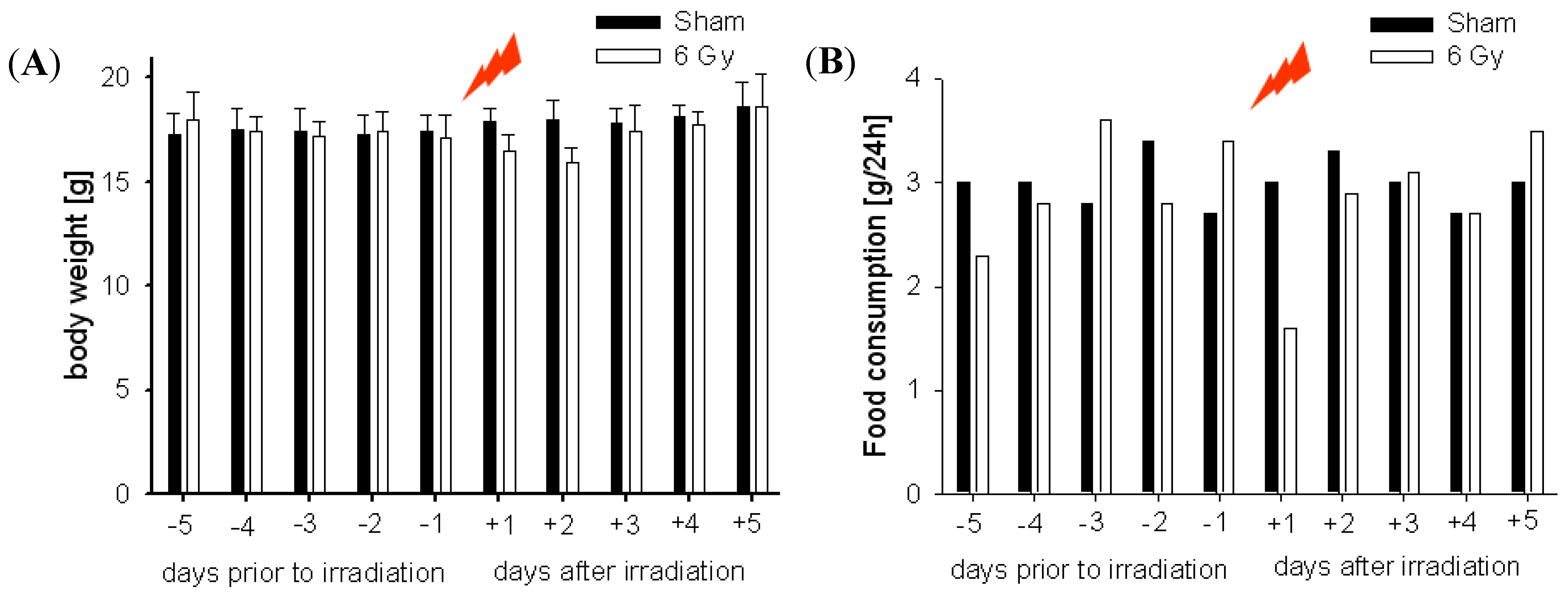
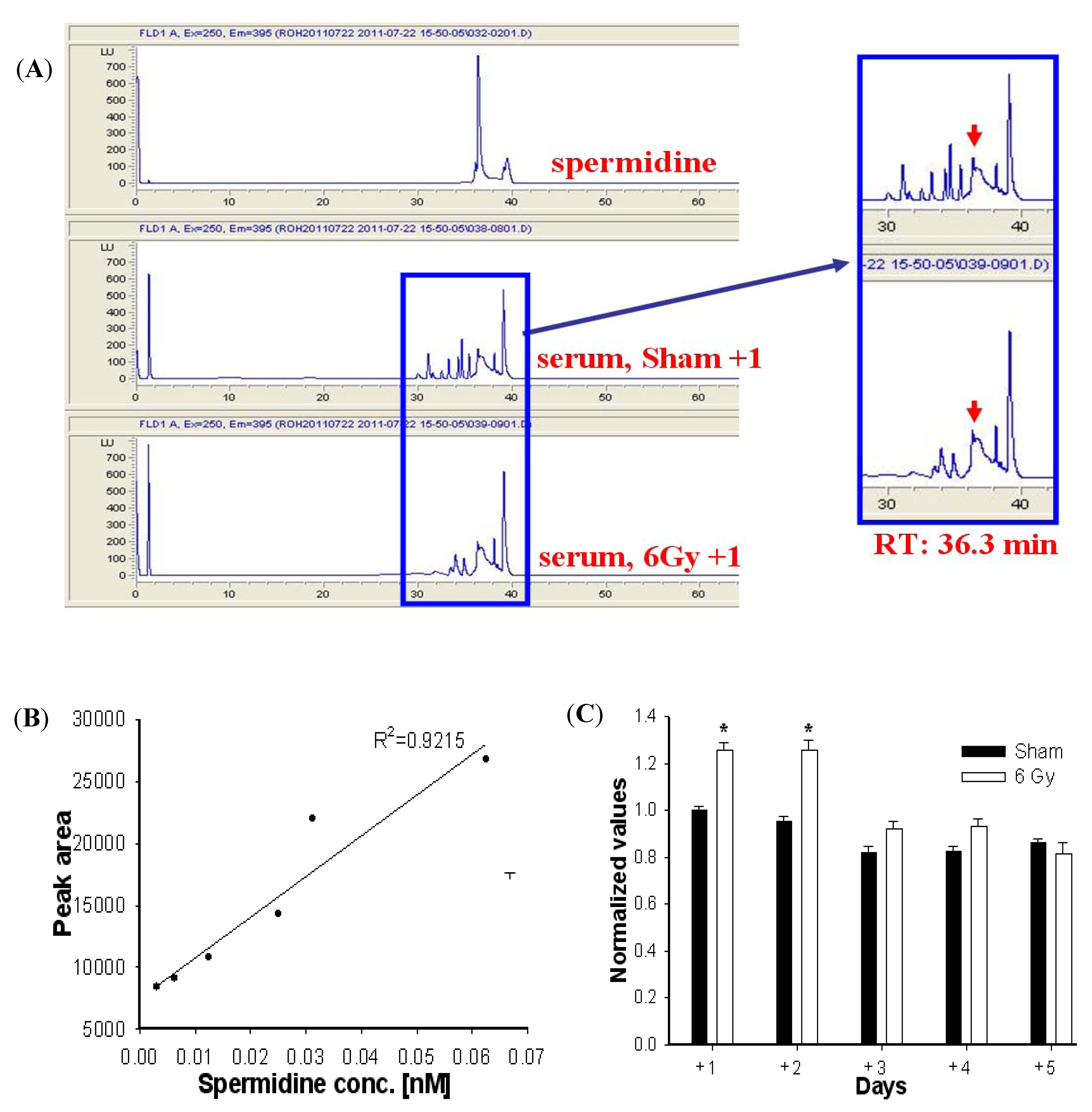
2. Results and Discussion


3. Experimental
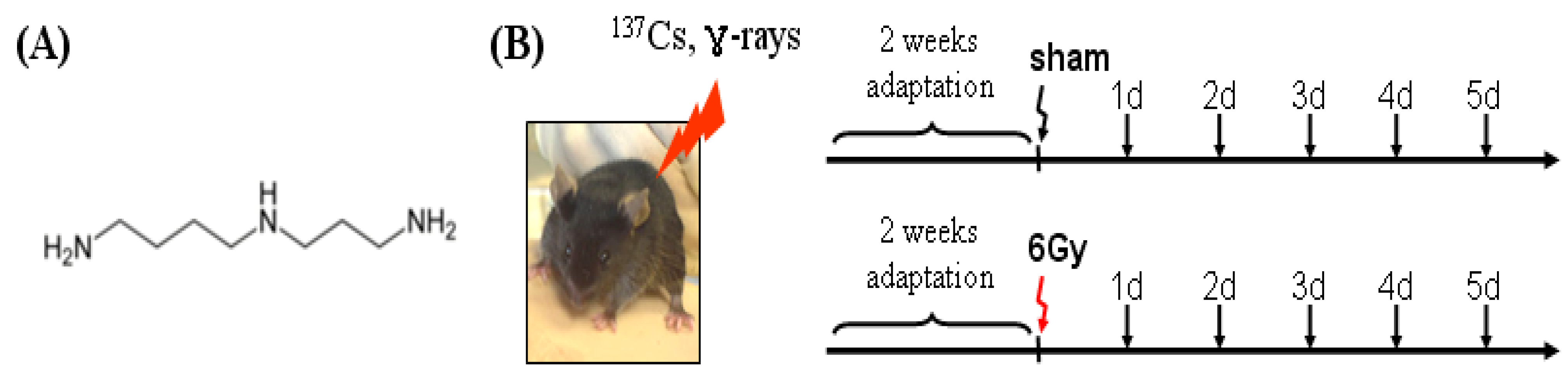
3.1. Mice
3.2. Ionizing Radiation
3.3. Spermidine Analysis by HPLC
3.4. Statistical Analysis
4. Conclusions
Acknowledgments
References and Notes
- Hall, E.J.; Giaccia, A.J. Radiobiology for the Radiologist, 6th ed; Lippincott Williams & Wilkins: Philadephia, PA, USA, 2006. [Google Scholar]
- Chao, N.J. Accidental or intentional exposure to ionizing radiation: Biodosimetry and treatment options. Exp. Hematol. 2007, 35, 24–27. [Google Scholar] [CrossRef]
- Pellmar, T.C.; Rockwell, S. Priority list of research areas for radiological nuclear threat countermeasures. Radiat. Res. 2005, 163, 115–123. [Google Scholar]
- Yatin, M. Polyamines in living organisms. J. Cell Mol. Biol. 2002, 1, 57–67. [Google Scholar]
- Wallace, H.M. Polyamines and their role in human disease—An introduction. Biochem. Soc. Trans. 2003, 31, 354–355. [Google Scholar]
- Janne, J.; Alhonen, L.; Leinonen, P. Polyamines: From molecular biology to clinical applications. Trends Mol. Med. 1991, 23, 241–259. [Google Scholar]
- Wallace, H.M.; Fraser, A.V.; Hughes, A. A perspective of polyamine metabolism. Biochem. J. 2003, 376, 1–14. [Google Scholar]
- Gilmour, S.K.; Birchler, M.; Smith, M.K.; Rayca, K.; Mostochuk, J. Effect of elevated levels of ornithine decarboxylase on cell cycle progression in skin. Cell Growth Differ. 1999, 10, 739–748. [Google Scholar]
- Hebby, O. Role of Polyamines in the control of cell proliferation and differentiation. Differentiation 1981, 19, 1–20. [Google Scholar] [CrossRef]
- Tabor, C.W.; Tabor, H. Polyamines. Annu. Rev. Biochem. 1984, 53, 749–790. [Google Scholar] [CrossRef]
- Seiler, N.; Delcros, J.G.; Moulinoux, J.P. Polyamine transport in mammalian cells. An update. Int. J. Biochem. Cell Biol. 1996, 28, 843–861. [Google Scholar] [CrossRef]
- Sample Availability: Not available.
© 2012 by the authors; licensee MDPI, Basel, Switzerland. This article is an open-access article distributed under the terms and conditions of the Creative Commons Attribution license (http://creativecommons.org/licenses/by/3.0/).
Share and Cite
Roh, C.; Yu, D.-K.; Kim, I.; Jo, S.K. The Biological Response of Spermidine Induced by Ionization Radiation. Molecules 2012, 17, 145-150. https://doi.org/10.3390/molecules17010145
Roh C, Yu D-K, Kim I, Jo SK. The Biological Response of Spermidine Induced by Ionization Radiation. Molecules. 2012; 17(1):145-150. https://doi.org/10.3390/molecules17010145
Chicago/Turabian StyleRoh, Changhyun, Dong-Kyung Yu, Insub Kim, and Sung Kee Jo. 2012. "The Biological Response of Spermidine Induced by Ionization Radiation" Molecules 17, no. 1: 145-150. https://doi.org/10.3390/molecules17010145