2. Results and Discussion
As an important form of ROS, hydroxyl radical (•OH), is generated in the human body via the Fenton reaction (Equation 1):
Since •OH radicals are extremele reactive, they can easily damage proteins, lipids, sugars and DNA. For example, •OH attacks deoxyribose, which is the backbone of DNA, to generate malondialdehyde (MDA) (Equation 2). MDA then combines 2-thiobarbituric acid (TBA) to yield TBARS (thiobarbituric acid reactive substances) which presents a maximum absorbance at 530 nm (Equation 3) [
12].
Hence, the value of A
532nm can reflect the amount of •OH radicals. If an antioxidant sample is added, A
532nm value will decrease, suggesting that some •OH radicals are scavenged by the antioxidant. This is the principle of deoxyribose degradation assay used in the study. Using this deoxyribose degradation assay, we measured the •OH radical-scavenging ability of AM. As shown in
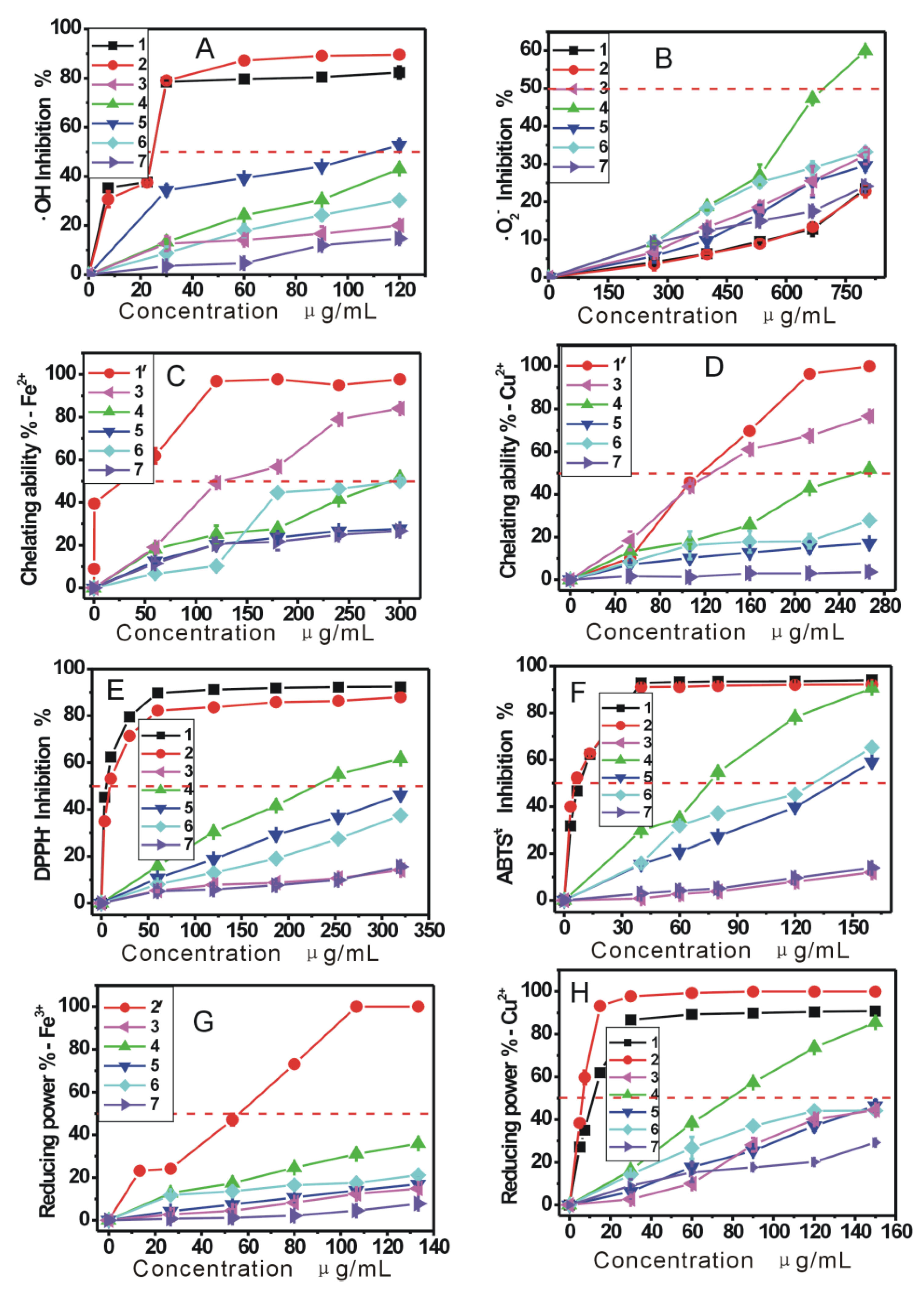
Figure 1A five AM extracts scavenged •OH radicals in a dose-dependent manner.
Figure 1.
The dose response curves of five extracts in the antioxidant assays: (A) •OH scavenging; (B) •O2− scavenging; (C) Fe2+-chelating; (D) Cu2+-chelating; (E) DPPH• scavenging; (F) ABTS+• scavenging; (G) Fe3+-reducing; (H) Cu2+-reducing. 1—Trolox; 1'—Sodium citrate; 2—BHA (butylated hydroxyanisole); 2'—GSH (glutathione); 3—PEAM (petroleum ether extract of Rhizoma Atractylodes macrocephala); 4—EAAM (ethyl acetate extract of Rhizoma Atractylodes macrocephala); 5—AEAM (absolute ethanol extract of Rhizoma Atractylodes macrocephala); 6—95EAM (95% ethanol extract of Rhizoma Atractylodes macrocephala); 7—WAM (water extract of Rhizoma Atractylodes macrocephala).
Besides •OH, superoxide anion (•O
2−) is also regarded as an important form of ROS in living cells. Although •O
2− is much weaker than •OH, however, it is able to directly attack DNA and lipids [
13], or transform into •OH via the Haber-Weiss reaction (Equation 4), to damage biomolecules [
14]:
In the study, the •O
2− -scavenging ability of AM was determined at physiological pH 7.4 [
15].
Figure 1B shows that the •O
2−-scavenging percentages of the five AM extracts increased in a concentration-dependent manner.
As indicated in Equations 1 and 4, transition metals (especially Fe and Cu) can catalyze the generation of •O
2− and •OH radicals. The metal-chelating ability of AM was therefore explored in the study. The dose-response curves (
Figure 1C,D) confirmed that the five AM extracts presented effective metal chelating abilities. The fact that AM can bind the Fe
2+ and Cu
2+, suggests that metal-chelating may be one of mechanisms used to scavenge •OH or •O
2−.
In order to confirm whether AM can directly scavenge radicals, the five AM extracts were further determined using the DPPH and ABTS assays. As seen in
Figure 1E,F, the five extracts can effectively eliminate DPPH• and ABTS•
+ radicals. Since the generation of both these radicals don’t involve the transition metal catalysis, direct radical-scavenging can be considered as another mechanism for AM to scavenge •OH or •O
2−.
The previous studies have demonstrated that DPPH· may be scavenged by an antioxidant through donation of a hydrogen atom (H•) to form a stable DPPH-H molecule [
16,
17,
18]. For example, the mechanism of DPPH• scavenging by ferulic acid can be proposed to occur as shown in Equation 5:
Unlike DPPH• scavenging, ABTS•
+ scavenging is considered an electron (e) transfer reaction [
19]. For example, the proposed reaction of ferulic acid and ABTS·
+ can be briefly illustrated by Equation 6:
In a word, the five AM extracts can effectively scavenge DPPH• and ABTS+• radicals, suggesting that AM exerts radical-scavenging action by donating hydrogen atoms (H•) and electrons (e).
Phytochemical studies indicated that AM mainly contained volatile oil, polysaccharides, flavonoids, saponins, and total phenolics [
20,
21]. From its volatile oil, atractylenolide I, II, and III could be obtained by eluting with petroleum ether-diethyl ether [
22,
23]. From the hot aqueous or alcoholic extracts of AM, various sugars and glycosides were isolated, such as saponins, mannose, fructose, and inulin [
20,
24]. Flavonoids, however, could be extracted from AM by ethanol [
25]. Taken together, the volatile oil and atractylenolides mainly exist in petroleum ether (non or lower polar solvent) extracts, while sugars and glycosides generally occur in water or hot alcohol extracts, and flavonoids could be enriched by ethanol extraction.
As shown in
Figure 1, the results of •O
2− assay, Fe
2+-chelating, Cu
2+-chelating, DPPH• assay, ABTS
+• assay, Fe
3+-reducing, and Cu
2+-reducing assay were generally similar. According to the IC
50 values (
Table 1), the order of antioxidant levels of the five AM extracts was EAAM > AEAM ≈ 95EAM > PEAM > WAM in the five assays. This is because the bioactive components relevant to antioxidant possess mild polarity, so they can be easily extract by mild polarity ethyl acetate. These bioactive components mainly included phenolic acids and flavonoids. In the water extract, the enriched components were saccharides and saponins which possess weak antioxidant properties. Therefore, EAAM exhibited the strongest abilities and WAM presented the weakest abilities in the five antioxidant assays.
Unlike the five antioxidant assays above, the •OH assay did not present so significant differences between the antioxidant levels of the AM extracts (
Figure 1A). In fact, •OH has extremely high reactivity and can react with any substance. Hence, the hydroxyl-scavenging abilities cannot be greatly affected by the types or contents of components, and the difference between the antioxidant levels of the AM extracts was the smallest.
In order to further confirm which chemical component could be responsible for the antioxidant of AM, we quantitatively analyzed the correlations (R values) between antioxidant levels and chemical contents in AM extracts. In the study, a spectrophotometric method was used to measure the relevant chemical contents in the five AM extracts, including total phenolics, total saponins and total sugars (
Table 2 &
Supplementary Information Part 2). On the other hand, since the 1/IC
50 value (not IC
50 value) showed a parallelism with antioxidant activity, 1/IC
50 values were then used for evaluating antioxidant levels in the study (
Supplementary Information Part 3). On this basis, the correlation graphs were plotted (
Supplementary Information Part 6) and the R values were calculated (
Table 3). The results suggested that total phenolics had significant positive correlations between antioxidant levels (R = 0.45~0.90, the average R value was 0.685), while total sugars and total saponins possessed lower or negative correlations (the average values of R were −0.272 and 0.244, respectively). This implies that the antioxidant activity of AM should be attributed to the presence of total phenolics, not of total sugars and total saponins.
Earlier studies have shown that AM polysaccharides also exhibited antioxidant ability [
26,
27]. However, the IC
50 values of polysaccharides in DPPH assay (>8 mg/mL) [
27] far exceeded those of total phenolics (239.42–1444.26 μg/mL,
Table 1), suggesting the antioxidant activity of polysaccharides was actually much weaker than total phenolics. It can be inferred that polysaccharides don’t effectively exert the antioxidant action when they are used along with phenolics in AM or AM extracts.
Table 1.
The IC50 values of five extracts from Rhizoma Atractylodes macrocephala (µg/mL).
Table 1.
The IC50 values of five extracts from Rhizoma Atractylodes macrocephala (µg/mL).
| | PEAM | EAAM | AEAM | 95EAM | WAM | Positive controls |
|---|
| Trolox | BHA |
|---|
| •OH scavenging | 419.62 ± 9.48 g | 140.39 ± 1.26 d | 104.97 ± 2.64c | 193.70 ± 4.71 e | 357.52 ± 4.31 f | 36.67 ± 0.67 b | 27.95 ± 0.39 a |
| •O2− scavenging | 1227.26 ± 41.27 b | 699.29 ± 6.30 a | 1115.68 ± 170.20 b | 1079.10 ± 26.79 b | 2191.70 ± 58.08 c | 2118.20 ± 30.01 c | 2178.82 ± 18.02 c |
| Fe2+-chelating | 142.70 ± 1.92 b | 303.70 ± 4.86 c | 543.86 ± 17.94 d | 305.87 ± 1.33 c | 697.69 ± 36.96 e | 26.32 ± 2.72 *,a | ND |
| Cu2+-chelating | 134.91 ± 3.92 b | 255.94 ± 1.04 c | 1025.38 ± 75.44 e | 507.61 ± 1.98 d | 7047.13 ± 895.31 f | 116.77 ± 1.58 *,a | ND |
| DPPH• scavenging | 1444.26 ± 68.47 g | 239.42 ± 0.64 c | 347.72 ± 0.60 d | 446.84 ± 11.42 e | 1077.70 ± 19.35 f | 4.10 ± 0.21 a | 11.98 ± 0.49 b |
| ABTS+• scavenging | 534.68 ± 11.30 f | 78.25 ± 0.30 c | 140.00 ± 0.16 e | 122.34 ± 0.83 d | 471.41 ± 36.91 f | 8.84 ± 0.14 b | 6.99 ± 0.05 a |
| Fe3+-reducing | 442.25 ± 10.15 c | 197.41 ± 1.53 b | 411.19 ± 9.11 c | 420.12 ± 2.63 c | 667.04 ± 20.58 d | 51.84 ± 0.99 **,a | ND |
| Fe3+-reducing | 137.59 ± 2.18 d | 83.24 ± 0.59 c | 160.86 ± 8.42 e | 133.75 ± 0.28 d | 277.42 ± 19.97 f | 12.44 ± 0.17 b | 6.54 ± 0.25 a |
Table 2.
The chemical contents of five extracts from Rhizoma Atractylodes macrocephala.
Table 2.
The chemical contents of five extracts from Rhizoma Atractylodes macrocephala.
| | PEAM | EAAM | AEAM | 95EAM | WAM |
|---|
| Total phenolics (mg Pyr./g) | 15.32 ± 0.00 d | 21.03 ± 0.052 e | 13.61 ± 0.052 c | 10.01 ± 0.026 b | 2.43 ± 0.045 a |
| Total flavonoids (mg rutin/g) | 3.36 ± 0.0064 d | 2.88 ± 0.011 c | 0.50 ± 0.00 b | 2.09 ± 0.00 c | 0.026 ± 0.064 a |
| Total sugar (mg glucose/g) | 1.10 ± 0.016 a | 1.54 ± 0.0090 a | 4.97 ± 0.00 c | 4.99 ± 0.016 c | 3.56 ± 0.0066 b |
| Total saponin (mg Ole./g) | 14.98 ± 0.021 d | 6.08 ± 0.11 c | 5.08 ± 0.20 c | 2.48 ± 0.19 b | 0.026 ± 0.064 a |
| Caffeic acid (peak area) | 5530.33 ± 684.22 b | 15863.98 ± 1254.02 d | 6466.64 ± 655.75 c | 5990.33 ± 94.99 b | 4333.34 ± 194.72 a |
| Ferulic acid (peak area) | 7768.53 ± 584.85 b | 22358.69 ± 2001.20 d | 8878.73 ± 784.7 c | 7968.53 ± 324.90 b | 5013.33 ± 424.87 a |
| Protocatechuic aicd (peak area) | 0.00 a | 463523 ± 44277.3 c | 0.00 a | 51570.5 ± 714.89 b | 0.00 a |
It was reported that total phenolics in AM generally include flavonoids [
28] and phenolic acids [
29]. Therefore, we estimated the total flavonoids of the five AM extracts. Similarly, the correlation graphs (
Supplementary Information Part 6) between total flavonoids and 1/IC
50 values were drawn to calculate the R values. The average R value (0.479) in
Table 3 revealed that flavonoids might also be the relevant antioxidant components of AM. However, the individual R values varied with the antioxidant assays. In general, the R values in radical-scavenging assays (R = −0.21–0.74) were much lower than those in the chelating assays (R = 0.86 and 0.90). This means that flavonoids may be responsible for the chelating abilities of AM. Despite the fact individual flavonoids have not been isolated from AM so far, however, we believe that these flavonoids may contain
ortho-dihydroxyl groups in their molecules, because chelating ability in antioxidant molecules usually results from the presence of such
ortho-dihydroxyls [
30].
Table 3.
The R (correlation coefficient) values between chemical contents and antioxidant levels.
Table 3.
The R (correlation coefficient) values between chemical contents and antioxidant levels.
| | Total phenolics | Total flavonoids | Total sugars | Total saponins | Caffeic acid | Ferulic acid | PCA |
|---|
| •OH assay | 0.45 | −0.21 | 0.43 | −0.21 | 0.44 | 0.45 | 0.32 |
| •O2− assay | 0.90 | 0.60 | −0.31 | 0.22 | 0.92 | 0.93 | 0.87 |
| Fe2+-chelating | 0.46 | 0.86 | −0.68 | 0.92 | 0.00092 | 0.040 | −0.023 |
| Cu2+-chelating | 0.62 | 0.90 | −0.81 | 0.94 | 0.22 | 0.26 | 0.18 |
| DPPH· assay | 0.66 | 0.14 | 0.066 | −0.20 | 0.85 | 0.85 | 0.80 |
| ABTS+• assay | 0.65 | 0.26 | 0.055 | −0.21 | 0.85 | 0.85 | 0.82 |
| Fe3+-reducing | 0.84 | 0.54 | −0.45 | 0.16 | 0.99 | 1.00 | 0.97 |
| Cu2+-reducing | 0.90 | 0.74 | −0.48 | 0.33 | 0.92 | 0.93 | 0.88 |
| Average | 0.685 | 0.479 | −0.272 | 0.244 | 0.649 | 0.664 | 0.602 |
As for another type of total phenolics, phenolic acidd were also analyzed in the study. The HPLC analysis identified three phenolic acids in AM extracts, including caffeic acid, ferulic acid and protocatechuic acid. Further analysis indicated that three phenolic acids were of higher average R values (0.649, 0.664, and 0.602, respectively) with antioxidant levels, so these three phenolic acids can also be regarded as relevant antioxidant components of AM. However, contrary to the flavonoids, phenolic acids exhibited higher R values with radical-scavenging assays (R = 0.32–1.00), but lower R values with chelating assays (R = −0.023–0.26) (
Table 3). This means that three phenolic acids may act as radical-scavengers and not metal-chelators in AM.
3. Experimental
3.1. Plant Material
Rhizoma Atractylodes macrocephala was purchased from Yanghe Pharmacy of Guangzhou University of Chinese Medicine (Guangzhou, China), and authenticated by Professor Shuhui Tan. A voucher specimen was deposited in our laboratory.
3.2. Chemicals
Trolox (±-6-hydroxyl-2,5,7,8-tetramethlyhromane-2-carboxylic acid), ferrozin [3-(2-pyridyl)-5,6-bis(4-phenylsulfonic acid)-1,2,4-triazine], BHA (butylated hydroxyanisole), DPPH• (1,1-diphenyl-2-picrylhydrazyl radical), neocuproine (2,9-dimethyl-1,10-phenanthroline), pyrogallol, murexide (5,5′-nitrilodibarbituric acid monoammonium salt), and protocatechuic acid were purchased from Sigma Aldrich Trading Co. (Shanghai, China); ABTS diammonium salt [2,2′-azino-bis(3-ethyl-benzothiazoline-6-sulfonic acid diammonium salt)], D-2-deoxyribose, and GSH (glutathione) were obtained from Amresco Co. (Solon, OH, USA); Ferulic acid and caffeic acid were purchased from National Institute for the Control of Pharmaceutical and Biological Products (Beijing, China). Acetonitrile, methanol and water were of HPLC grade. All other chemicals used were analytical grade.
3.4. Hydroxyl (•OH) Radical-Scavenging Assay
The scavenging activity on the hydroxyl radical was investigated by the deoxyribose method [
30], with some modifications. Our preliminary experiments demonstrated that most organic solvents especially alcohol can promote the inhibition percentage value. Hence, the inhibition of the hydroxyl radical was evaluated as follows: all test samples were first dissolved in methanol at 4 mg/mL. An aliquot of sample solution (12–48 μL) was brought to a tube, then the sample solution in tube was evaporated to dryness (water bath, 60 °C). The sample residue was mixed with 400 μL phosphate buffer (0.2 mol/L, pH 7.4). Subsequently, 50 μL deoxyribose (50 mmol/L), 50 μL H
2O
2 (50 mmol/L), 50 μL FeCl
3 (3.2 mmol/L) and 50 μL Na
2EDTA (1 mmol/L) were added. The reaction was initiated by mixing 50 μL ascorbic acid (1.8 mmol/L) and the total volume of the reaction mixture was adjusted to 800 μL with buffer. After incubation at 50 °C for 20 min, the reaction was terminated by 250 μL trichloroacetic acid (10%, w/w).
The color was then developed by addition of 150 μL TBA (1g/20 mL, in 1.25% NaOH aqueous solution) and heated in an oven at 105 °C for 15 min. The mixture was cooled and absorbance was measured at 530 nm against the buffer (as blank). The inhibition percentage for ·OH is expressed as follows:
where,
A0 is the
A532nm of mixture without sample, and
A is the
A532nm of the mixture with sample.
3.5. Superoxide Anion (•O2−) Radical-Scavenging Assay
Measurement of superoxide anion (•O
2−) scavenging activity was based on our method [
15]. Briefly, the sample was dissolved in ethanol at 8 mg/mL. The sample solution (
x μL, where
x = 0, 100, 150, 200, 250, and 300 μL) was mixed with 2920-
x μL Tris-HCl buffer (0.05mol/L, pH 7.4) containing EDTA (1 mmol/L). When 80 μL pyrogallol (60 mmol/L in 1 mmol/L HCl) was added, the mixture was shaken at room temperature immediately. The absorbance at 325 nm of the mixture was measured (Unico 2100, Shanghai, China) against the Tris-HCl buffer as blank every 30 s for 5 min. The •O
2− scavenging ability was calculated as:
where,
ΔA325nm, control is the increase in
A325nm of the mixture without the sample and
ΔA325nm, sample is that with the sample;
T = 5 min. The experiment temperature was 37 °C.
3.6. Fe2+-Chelating Assay
The Fe
2+ chelating activities of five AM extracts were estimated by the method as described by Li [
31]. Briefly,
x μL sample solutions (2 mg/mL,
x = 0, 30, 60, 90, 120, and 150) were added to 100 μL FeCl
2 aqueous solutions (250 μmol/L). The reaction was initiated by the addition of 150 μL ferrozine aqueous solutions (500 μmol/L) and the total volume of the reaction mixture was adjusted to 1,000 μL with methanol. Then, the mixture was shaken vigorously and stood at room temperature for 10 min. Absorbance of the solution was measured spectrophotometrically at 562 nm (Unico 2100). The percentage of chelating effect on Fe
2+ was calculated by the following formula:
where
A0 is the absorbance without sample, and
A is the absorbance with sample.
3.7. Cu2+-Chelating Assay
The Cu
2+-chelating activities of the five AM extracts were measured by a complexometric method using murexide [
31]. Briefly, murexide solution (1.2 mL, 0.25 mmol/L) and CuSO
4 aqueous solution (60 μL, 20 mmol/L) were added to hexamine HCl buffer (pH 5.0, 30 mmol/L) containing 30 mmol/L KCl. After incubation for 1 min at room temperature, 40–200 μL sample solutions (2 mg/mL in methanol) were added. The final volume was adjusted to 1,500 μL with methanol. Then, the mixture was shaken vigorously and left at room temperature for 10 min. Absorbance of the solution was then measured spectrophotometrically at 485 nm and 520 nm (Unico 2100). The absorbance ratio (
A485/A520) reflected the free Cu
2+ content. Therefore, the percentage of cupric chelating effect was calculated by the following formula:
where
![Molecules 17 13457 i010]()
is the absorbance ratio of the sample, while
![Molecules 17 13457 i011]()
is the maximum absorbance ratio and
![Molecules 17 13457 i012]()
is the minimum absorbance ratio in the test.
3.8. DPPH• Radical-Scavenging Assay
DPPH• radical-scavenging activity was determined as described [
32]. Briefly, 1 mL DPPH• ethanolic solutions (0.1 mmol/L) were mixed with 0.5 mL sample alcoholic solutions (0.18–0.96 mg/mL). The mixtures were kept at room temperature for 30 min, and then measured with a spectrophotometer (Unico 2100, Shanghai, China) at 519 nm. The DPPH• inhibition percentages were calculated:
where
A is the absorbance with samples, while
A0 is the absorbance without samples. Trolox and BHA were used as the positive controls.
3.9. ABTS+• Radical-Scavenging Assay
ABTS
+• scavenging activity was measured as described [
32] with some modifications. The ABTS
+• was produced by mixing 0.2 mL ABTS diammonium salt aqueous solution (7.4 mmol/L) with 0.2 mL K
2S
2O
8 aqueous solution (2.6 mmol/L). The mixture was kept in the dark at room temperature for 12 h to allow completion of ABTS
+• generation. Before usage, it was diluted with 95% ethanol (about 1:50) so that its absorbance at 734 nm was 0.70 ± 0.02. Then, 1.2 mL diluted ABTS
+• reagents were brought to 0.3 mL sample ethanolic solutions (0.2–0.8 mg/mL). After incubation for 6 min, the absorbance at 734 nm was read on a spectrophotometer (Unico 2100, Shanghai, China). The percentage inhibition was calculated as:
where
A0 is the absorbance of the mixture without sample,
A is the absorbance of the mixture with sample (or positive control).
3.10. Fe3+-Reducing Power Assay
Ferric cyanide (Fe
3+) reducing power was determined according to the method of Oyaizu [
33] as described by Li
et al. [
32]. In brief,
x μL sample methanolic solution (4 mg/mL,
x = 0, 20, 40, 60, 80, and 100) was mixed with (500 −
x) μL Na
2HPO
4/KH
2PO
4 buffer (0.2 mol/L, pH 6.6) and 250 μL K
3Fe(CN)
6 aqueous solution (1 g/100 mL). After the mixture was incubated at 50 °C for 20 min, it was added by 250 μL trichloroacetic acid (10 g/100 mL in distilled water) then centrifuged at 3500 rpm for 10 min. As soon as 400 μL supernatant was mixed with 400 μL FeCl
3 (0.1 g/100 mL in distilled water), the timer was started. At 90 s, absorbance of the mixture was read at 700 nm (Unico 2100, Shanghai, China). Samples were analyzed in groups of three, and when the analysis of one group has finished, the next group of three samples were mixed with FeCl
3 to avoid oxidization by air. The relative reducing ability of the sample was calculated by using the formula:
Here, Amax is the maximum absorbance and Amin is the minimum absorbance in the test. A is the absorbance of sample.
3.11. Cu2+-Reducing Power Assay
The cupric ions (Cu
2+) reducing capacity was determined by the method [
34], with minor modifications. Briefly, CuSO
4 aqueous solution (125 μL, 0.01 mol/L), neocuproine ethanolic solution (125 μL, 7.5 mmol/L) and (750 −
x) μL CH
3COONH
4 buffer solution (0.1 mol/L, pH 7.5) were brought to test tubes with different volumes of samples (1 mg/mL,
x = 30–150 μL). Then, the total volume was adjusted to 1,000 μL with the buffer and mixed vigorously. Absorbance against a buffer blank was measured at 450 nm after 30 min (Unico 2100). The relative reducing power of the sample as compared with the maximum absorbance, was calculated by the formula:
where,
Amax is the maximum absorbance at 450 nm and
Amin is the minimum absorbance in the test.
A is the absorbance of sample.
3.12. Determination of Total Phenolics
The total phenolics contents of the five AM extracts were determined using a modified Folin-Ciocalteu colorimetric method [
35]. Briefly, methanolic sample solution (0.5 mL, 1 mg/mL) was added to Folin-Ciocalteu reagent (0.5 mL, 0.25 mol/L). The mixture was left standing for 3 min, followed by the addition of Na
2CO
3 aqueous solution (1.0 mL, 15%, w/w). After incubation at ambient temperature for 30 min, the mixture was centrifuged at 3,500 rpm for 3 min. The supernatant was measured using a spectrophotometer (Unico 2100) at 760 nm. The standard curve was prepared using different concentrations of pyrogallol and the results were expressed as pyrogallol equivalents in milligrams per gram extract.
3.13. Determination of Total Flavonoids
Total flavonoids contents of the five AM extracts were measured using the NaNO
2-Al(NO
3)
3 method with minor modifications [
35]. In brief, methanolic sample solution (1 mL, 10 mg/mL) was mixed with NaNO
2 aqueous solution (0.15 mL, 5%, w/w). The mixture stood at ambient temperature for 6 min, followed by the addition of Al(NO
3)
3 aqueous solution (0.15 mL, 10%, w/w). After incubation at ambient temperature for 6 min, NaOH aqueous solution (2 mL, 4%, w/w) was added to the mixture whicj was then adjusted to 5 mL with distilled water. The absorbance was read at 508 nm on a spectrophotometer (Unico 2100). The standard curve was prepared using different concentrations of quercetin and the results were also expressed as quercetin in milligrams per gram extract.
3.14. Determination of Total Sugars
The total sugars content was evaluated according to the phenol-sulfuric acid method [
36]. An aliquot of sample solution (1 mL, 1 mg/mL) was placed in a test tube, and the volume was adjusted to 2 mL with distilled water. Then an aliquot of 5% phenol solution (1 mL) and concentrated sulfuric acid (5 mL) were added. After incubation for 20 min at room temperature, the reaction mixture was measured using a spectrophotometer (Unico 2100) at 490 nm. The standard curve was prepared using different concentrations of glucose and the results were expressed as glucose in milligrams per gram extract.
3.15. Determination of Total Saponins
The total saponins content was measured according to the method described in [
36]. Methanolic sample solution (0.2 mL, 1 mg/mL) was taken in a test tube. After the methanol solvent was removed at 80 °C, an aliquot of vanillin-acetic acid solution (0.1 mL, 0.5%) and perchloric acid (0.4 mL) were added. The reaction mixture was incubated at 70 °C for 15 min, then cooled and diluted with acetic acid (1.25 mL). After 10 min, the absorbance of the diluted solution was measured at 540 nm (Unico 2100) against a blank control, which contained all reagents except for sample. Quantification was based on the standard curve for oleanolic acid (11–69 μg/mL). The results were expressed in milligrams of oleanolic acid equivalents per gram of extract.
3.16. HPLC Analysis
HPLC analysis was performed on a Syltech P510 system (Los Angeles, CA, USA), equipped with a Diamonsil C18 (250 mm × 4.6 mm, 5 μm) column (Dikma Co., Beijing, China). All samples were dissolved in methanol at 4 mg/mL and filtered using 0.45 μm filter.
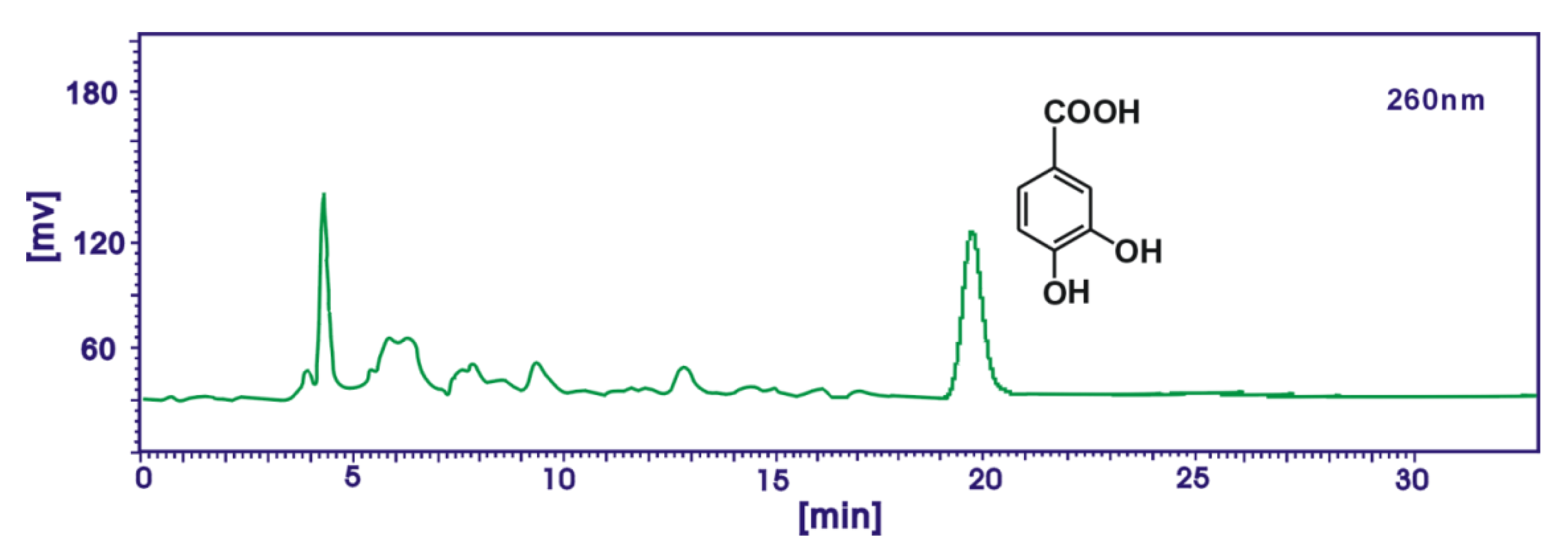
In the analysis for caffeic acid and ferulic acid, the mobile phase was methanol/acetonitrile/5% acetate acid (24:6:70) and the flow rate was 0.5 mL/min, injection volume was 10 μL, detection wavelength was 325 nm. In the analysis for protocatechuic acid, the mobile phase was methanol/acetonitrile/5% acetate acid (12:3:10) and the flow rate was 1.0 mL/min, injection volume was 10 μL, detection wavelength was 260 nm. Caffeic acid, ferulic acid and protocatechuic acid in AM extracts were identified by the retention times and the peak areas were used to characterize the relative contents in the study (
Figure 3 and
Figure 4).
Figure 3.
A typical HPLC profile of protocatechuic acid in the ethyl acetate extract of Rhizoma Atractylodes macrocephala. Syltech P510 system (Los Angeles, CA, USA), Dikma Diamonsil C18 (250 mm × 4.6 mm, 5 μm) column, 1.0 mL/min flow rate, methanol/acetonitrile/5% acetate acid (12:3:10), 10 μL injection, 60 nm wavelength.
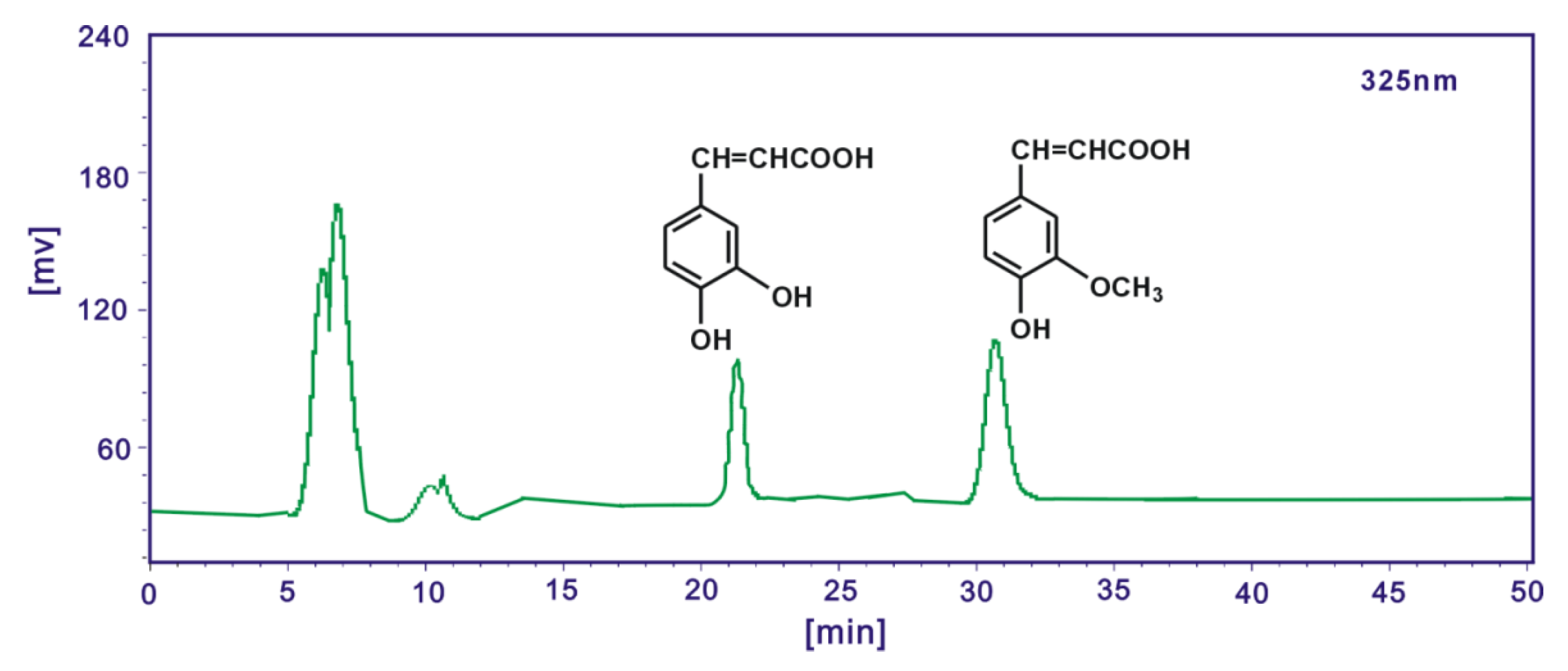
Figure 4.
A typical HPLC profile of caffeic acid and ferulic acid in ethyl acetate extract of Rhizoma Atractylodes macrocephala. Syltech P510 system (Los Angeles, CA, USA), Dikma Diamonsil C18 (250 mm × 4.6 mm, 5 μm) column, methanol/acetonitrile/5% acetate acid (24:6:70), 0.5 mL/min flow rate, 10 μL injection, 325 nm wavelength.
3.17. Statistical Analysis
Data are given as the mean ± SD of three measurements. The IC50 values were calculated by linear regression analysis. All linear regression in this paper was analyzed by Origin 6.0 professional software. Significant differences were performed using the T-test (p < 0.05). The analysis was performed using SPSS software (v.12, SPSS, Chicago, IL, USA).

 ,
, 


 is the absorbance ratio of the sample, while
  is the maximum absorbance ratio and
  is the minimum absorbance ratio in the test.

