Synthesis and Antiproliferative Activity of Ammonium and Imidazolium Ionic Liquids against T98G Brain Cancer Cells


Abstract
:1. Introduction
2. Results and Discussion
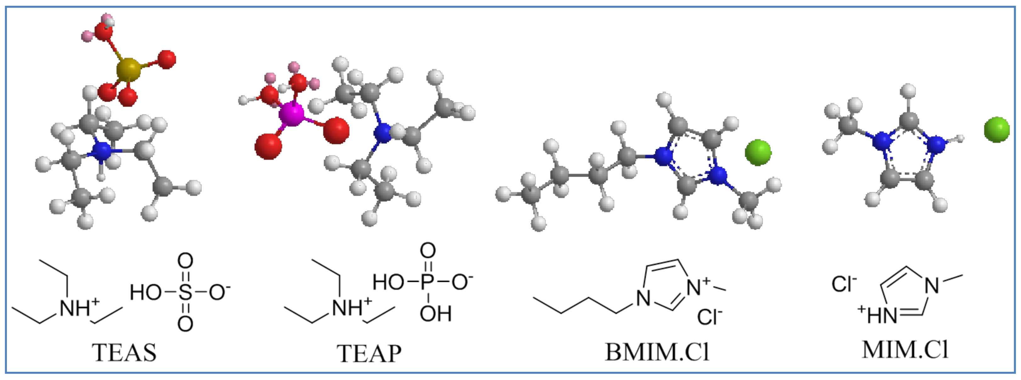
2.1. Synthesis
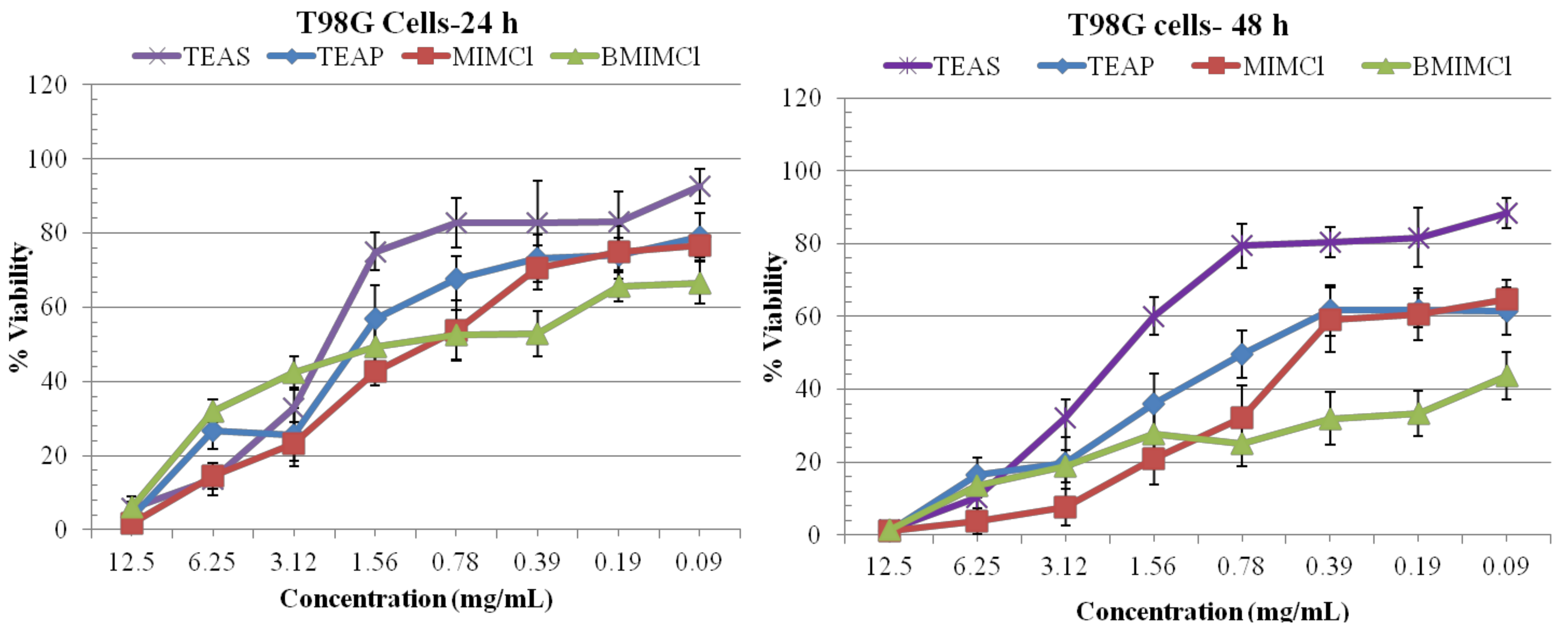
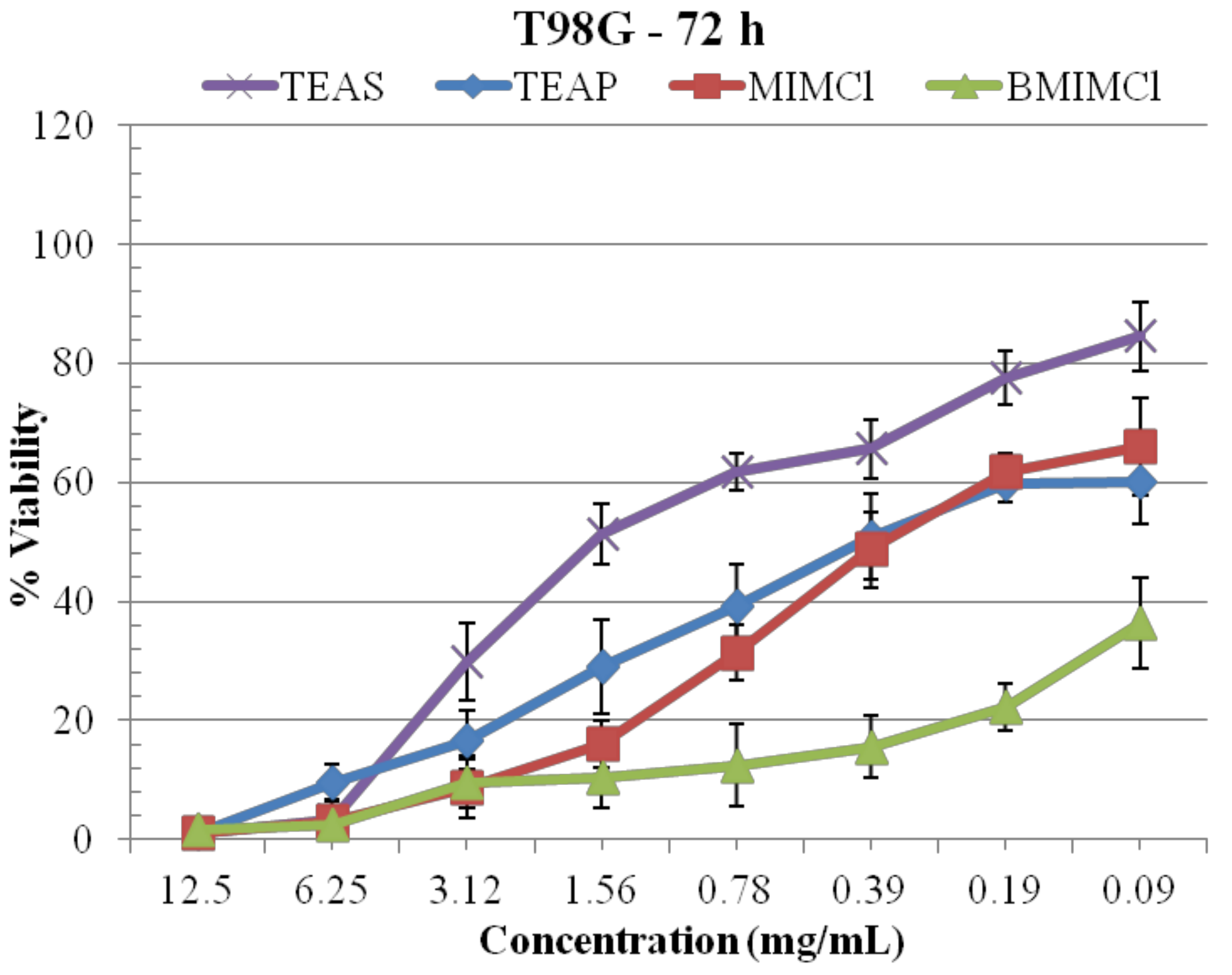
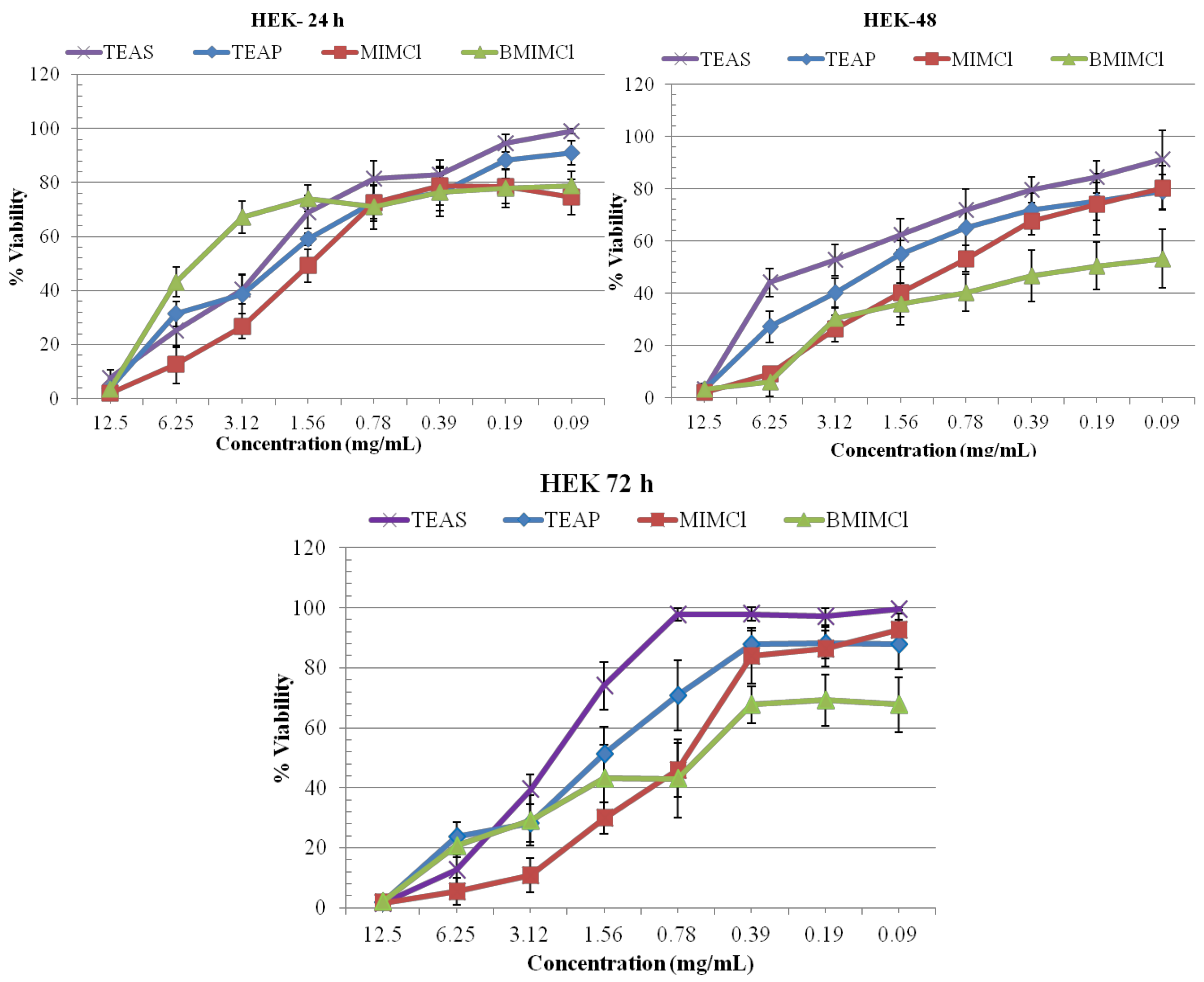
2.2. MTT Assay
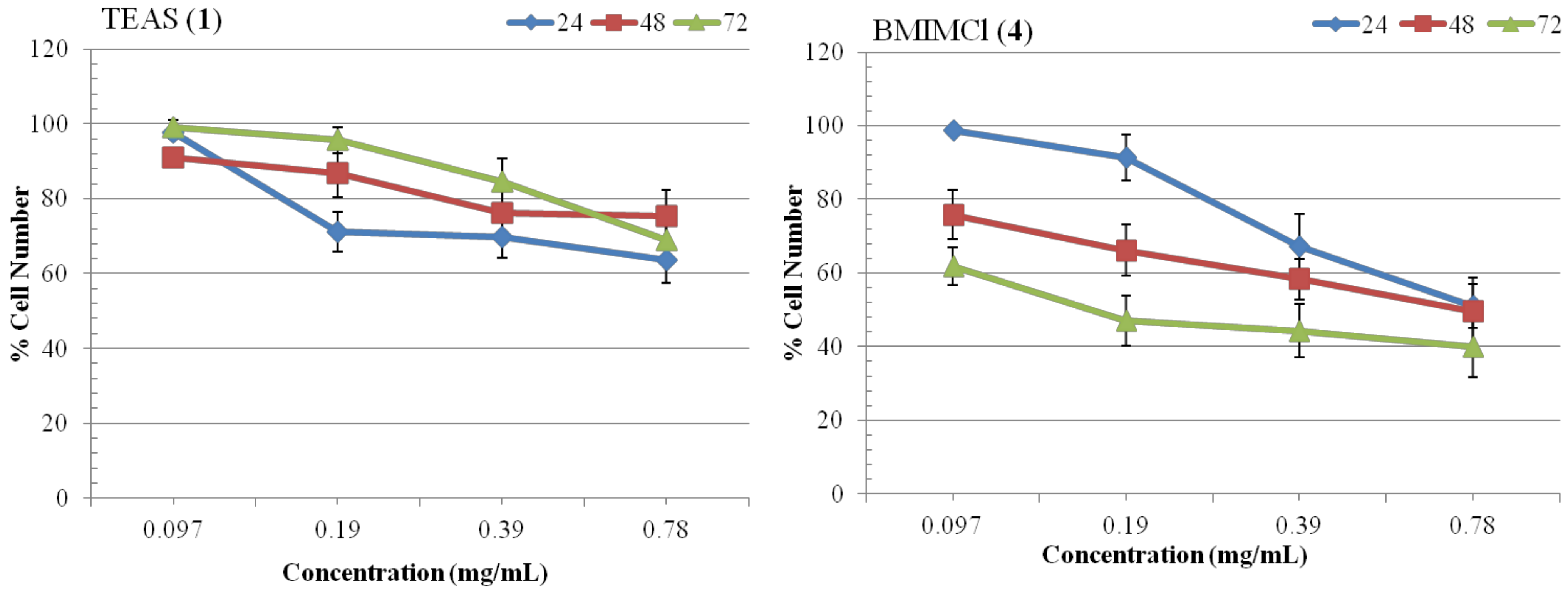
2.3. Growth Kinetics Assay
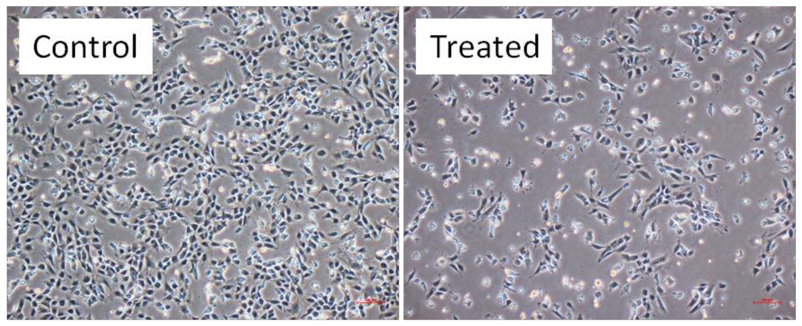
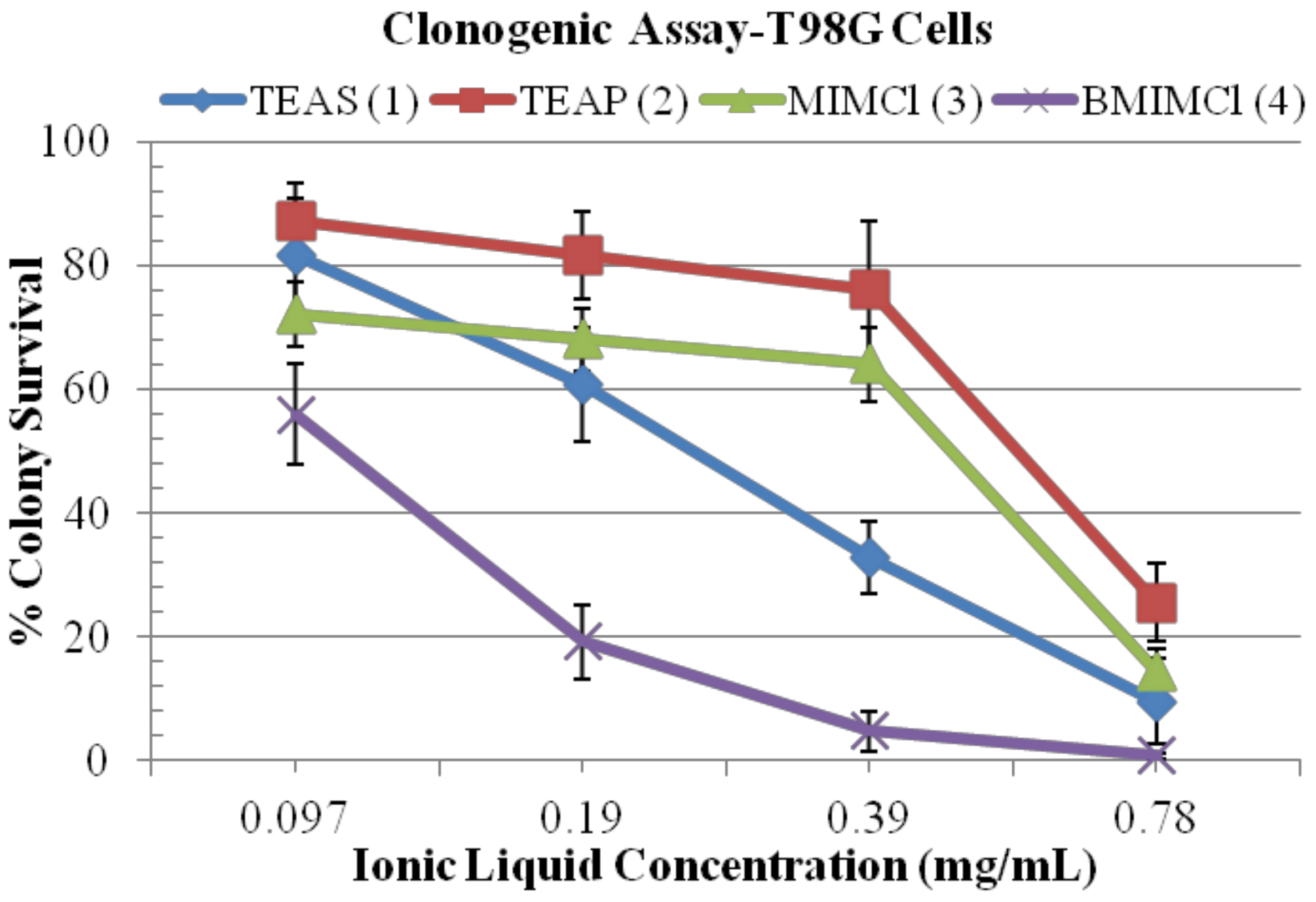
2.4. Clonogenic Assay
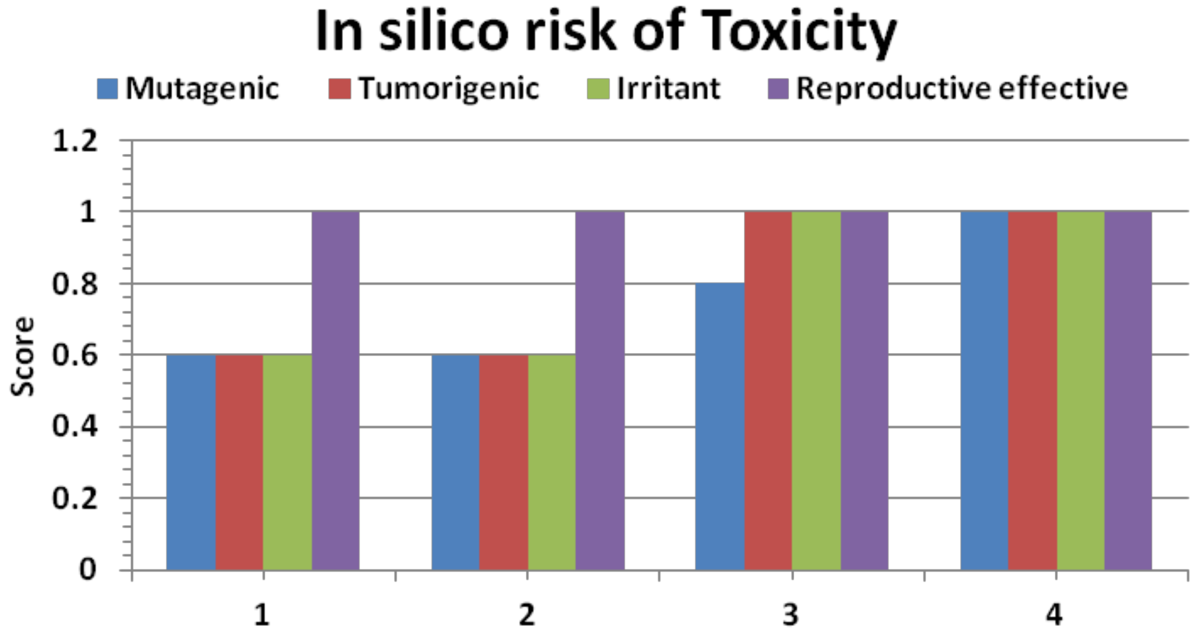
2.5. In Silico Pharmacokinetics
3. Experimental
3.1. General
3.2. Preparation of ILs 1–4
3.3. Human Cell Culture
3.4. In Vitro Metabolic Viability Assay
3.5. Growth Kinetics Assay
3.6. Clonogenic Assay
3.7. In Silico Pharmacokinetic Screening
3.8. Statistical Analysis
4. Conclusions
Acknowledgments
References
- Stoimenovski, J.; MacFarlane, D.R.; Bica, K.; Rogers, R.D. Crystalline vs. ionic liquid salt forms of active pharmaceutical ingredients: A position paper. Pharm. Res. 2010, 27, 521–526. [Google Scholar] [CrossRef] [PubMed]
- Rogers, R.D.; Seddon, K.R. Ionic Liquids: Industrial Applications to Green Chemistry; ACS Symp. Ser.: Washington, DC, USA, 2002. [Google Scholar]
- Hough, W.L.; Smiglak, M.; Rodrıguez, H.; Swatloski, R.P.; Spear, S.K.; Daly, D.T.; Pernak, J.; Grisel, J.E.; Carliss, R.D.; Soutullo, M.D.; et al. The third evolution of ionic liquids: Active pharmaceutical ingredients. New J. Chem. 2007, 31, 1429–1436. [Google Scholar] [CrossRef]
- Yu, M.; Wang, S.-H.; Luo, Y.-R.; Han, Y.-W.; Li, X.-Y.; Zhang, B.-J.; Wang, J.-J. Effects ofthe1-alkyl-3-methylimidazolium bromide ionic liquids on the antioxidant defense system of Daphnia magna. Ecotox. Environ. Saf. 2009, 72, 1798–1804. [Google Scholar] [CrossRef] [PubMed]
- Attri, P.; Venkatesu, P. Influence of protic ionic liquids on the structure and stability of succinylated Con A. Int. J. Biol. Macromol. 2012, 51, 119–128. [Google Scholar] [CrossRef]
- Attri, P.; Venkatesu, P.; Kumar, A. Water and a protic ionic liquid acted as refolding additives for chemically denatured enzymes. Org. Biomol. Chem. 2012, 10, 7441–7636. [Google Scholar] [CrossRef] [PubMed]
- Hough-Troutman, W.L.; Smiglak, M.; Griffin, S.; Reichert, W.M.; Mirska, I.; Jodynis-Liebert, J.; Adamska, T.; Nawrot, J.; Stasiewicz, M.; Rogers, R.D.; et al. Ionic liquids with dual biological function: Sweet and anti-microbial, hydrophobic quaternary ammonium-based salts. New J. Chem. 2009, 33, 26–33. [Google Scholar] [CrossRef]
- Pernak, J.; Goc, I.; Mirska, I. Antimicrobial activities of protic ionic liquids with lactate anion. Green Chem. 2004, 6, 323–329. [Google Scholar] [CrossRef]
- Demberelnyamba, D.; Kim, K.-S.; Choi, S.; Park, S.Y.; Lee, H.; Kim, C.J.; Yoo, I.D. Synthesis and antimicrobial properties of imidazolium and pyrrolidinonium salts. Bioorg. Med. Chem. 2004, 12, 853–857. [Google Scholar] [CrossRef] [PubMed]
- Pernak, J.; Sobaszkiewicz, K.; Mirska, I. Anti-microbial activities of ionic liquids. Green Chem. 2003, 5, 52–56. [Google Scholar] [CrossRef]
- Pernak, J.; Feder-Kubis, J. Synthesis and properties of chiral ammonium-based ionic liquids. Chem. Eur. J. 2005, 11, 4441–4449. [Google Scholar] [CrossRef] [PubMed]
- Pernak, J.; Sobaszkiewicz, K.; Foksowicz-Flaczyk, J. Ionic liquids with symmetrical dialkoxy methyl-substituted imidazolium cations. Chem. Eur. J. 2004, 10, 3479–3485. [Google Scholar] [CrossRef] [PubMed]
- Kumar, V.; Malhotra, S.V. Study on the potential anti-cancer activity of phosphonium and ammonium-based ionic liquids. Bioorg. Med. Chem. Lett. 2009, 19, 4643–4646. [Google Scholar] [CrossRef] [PubMed]
- Stock, F.; Hoffman, J.; Ranke, J.; Ondruschka, B.; Jastorff, B. Effects of ionic liquids on the acetylcholinesterase—A structure–activity relationship consideration. Green Chem. 2004, 6, 286–290. [Google Scholar] [CrossRef]
- Skladanowski, A.C.; Stepnowski, P.; Kleszczynski, K.; Dmochowska, B. AMP deaminase in vitro inhibition by xenobiotics A potential molecular method for risk assessment of synthetic nitro- and polycyclic musks, imidazolium ionic liquids and N-glucopyranosyl ammonium salts. Environ. Toxicol. Pharmacol. 2005, 19, 291–296. [Google Scholar] [PubMed]
- Hosseinzadeh, F.; Mahkam, M.; Galehassadi, M. Synthesis and characterization of ionic liquid functionalized polymers for drug delivery of an anti-inflammatory drug. Des. Monomers Polym. 2012, 15, 379–388. [Google Scholar] [CrossRef]
- Malhotra, S.V.; Kumar, V. A profile of the in-vitro antitumor activity of imidazolium-based ionic liquids. Bioorg. Med. Chem. Lett. 2010, 20, 581–585. [Google Scholar] [CrossRef] [PubMed]
- Fujita, K.; MacFarlane, D.R.; Forsyth, M. Protein solubilising and stabilising ionic liquids. Chem. Commun. 2005, 4804–4806. [Google Scholar] [CrossRef] [PubMed]
- Von Hagen, J.; Michelsen, U. Use of ionic liquids for protein extraction. U.S. Patent US7470778B2 2008. [Google Scholar]
- Fujita, K.; Forsyth, M.; MacFarlane, D.R.; Reid, R.W.; Elliott, G.D. Unexpected improvement in stability and utility of cytochrome c by solution in biocompatible ionic liquids. Biotechnol. Bioeng. 2006, 94, 1209–1213. [Google Scholar] [CrossRef] [PubMed]
- Fujita, K.; MacFarlane, D.R.; Forsyth, M.; Yoshizawa-Fujita, M.; Murata, K.; Nakamura, N.; Ohno, H. Solubility and stability of cytochrome c in hydrated ionic liquids: Effect of Oxo acid residues and kosmotropicity. Biomacromolecules 2007, 8, 2080–2086. [Google Scholar] [CrossRef] [PubMed]
- Schöfer, S.H.; Kaftzik, N.; Wassrescheid, P.; Kragl, U. Enzyme catalysis in ionic liquids: Lipase catalysed kinetic resolution of 1-phenylethanol with improved enantioselectivity. Chem. Commun. 2001, 425–426. [Google Scholar] [CrossRef]
- Ranke, J.; Stolte, S.; Stormann, R.; Arning, J.; Jastorff, B. Design of sustainable chemical products-the example of ionic liquids. Chem. Rev. 2007, 107, 2183–2206. [Google Scholar] [CrossRef] [PubMed]
- Carson, L.; Chau, P.K.W.; Earle, M.J.; Gilea, M.A.; Gilmore, B.F.; Gorman, S.P.; McCann, M.T.; Seddon, K.R. Antibiofilm activities of 1-alkyl-3-methylimidazolium chloride ionic liquids. Green Chem. 2009, 11, 492–497. [Google Scholar] [CrossRef]
- Shaw, G.; Morse, S.; Ararat, M.; Graham, F.L. Preferential transformation of human neuronal cells by human adenoviruses and the origin of HEK 293 cells. FASEB J. 2002, 16, 869–871. [Google Scholar] [CrossRef] [PubMed]
- Attri, P.; Reddy, P.M.; Venkatesu, P.; Kumar, A.; Hofman, T. Measurements and Molecular Interactions for N,N-dimethylformamide with Ionic Liquid Mixed Solvents. J. Phys. Chem. B 2010, 114, 6126–6133. [Google Scholar] [CrossRef] [PubMed]
- Attri, P.; Venkatesu, P.; Kumar, A. Temperature effect on the molecular interactions between ammonium ionic liquids and N,N-dimethylformamide. J. Phys. Chem. B 2010, 114, 13415–13425. [Google Scholar] [CrossRef] [PubMed]
- Attri, P.; Venkatesu, P.; Hofman, T. Temperature dependence measurements and structural characterization of trimethyl ammonium ionic liquids with a highly polar solvent. J. Phys. Chem. B 2011, 115, 10086–10097. [Google Scholar] [CrossRef] [PubMed]
- Kaushik, N.K.; Kim, Y.H.; Han, Y.G.; Choi, E.H. Effect of jet plasma on T98G human brain cancer cells. Curr. Appl. Phys. 2013, 13, 176–180. [Google Scholar] [CrossRef]
- Wahab, R.; Kaushik, N.K.; Verma, A.K.; Mishra, A.; Hwang, I.H.; Yang, Y.B.; Shin, H.S.; Kim, Y.S. Fabrication and growth mechanism of ZnO nanostructures and their cytotoxic effect on human brain tumor U87, cervical cancer HeLa, and normal HEK cells. J. Biol. Inorg. Chem. 2011, 16, 431–442. [Google Scholar] [CrossRef] [PubMed]
- Mishra, A.; Jung, H.; Park, J.W.; Kim, H.K.; Kim, H.; Stang, P.J.; Chi, K.W. Anticancer activity of self-assembled molecular rectangles via arene-ruthenium acceptors and a new unsymmetrical amide ligand. Organometallics 2012, 31, 3519–3526. [Google Scholar] [CrossRef] [PubMed]
- Kaushik, N.K.; Kim, H.S.; Chae, Y.J.; Lee, Y.N.; Kwon, G.C.; Choi, E.H.; Kim, I.T. Synthesis and anticancer activity of di(3-thienyl)methanol and di(3-thienyl)methane. Molecules 2012, 17, 11456–11468. [Google Scholar] [CrossRef] [PubMed]
- Kaushik, N.K.; Uhm, H.S.; Choi, E.H. Micronucleus formation induced by dielectric barrier discharge plasma exposure in brain cancer cells. Appl. Phys. Lett. 2012, 100, 0841021–0841024. [Google Scholar] [CrossRef]
- Organic Chemistry Portal. Available online: http://www.organic-chemistry.org/prog/peo/ (accessed on 6–13 October 2012).
- Tekto, I.V. Computing chemistry on the web. Drug Discov. Today 2005, 10, 1497–1500. [Google Scholar]
- Wenlock, M.C.; Austin, R.P.; Barton, P.; Davis, A.M.; Leeson, P.D. A comparison of physicochemical property profiles of development and marketed oral drugs. J. Med. Chem. 2003, 46, 1250–1256. [Google Scholar] [CrossRef] [PubMed]
- Lipinski, C.A.; Lombardo, F.; Dominy, B.W.; Feeney, P.J. Experimental and computational approaches to estimate solubility and permeability in drug discovery and development settings. Adv. Drug. Del. Rev. 2001, 4, 3–26. [Google Scholar] [CrossRef]
Sample Availability: Samples of all the ILs are available from the authors. |


| ILs | nHba | nHbd | nrotb | MW | cLog P | Druglikeness | Drug Score |
|---|---|---|---|---|---|---|---|
| 1 | 1 | 0 | 3 | 199 | 1.5 | −1.29 | 0.130 |
| 2 | 1 | 0 | 3 | 199 | 1.5 | −1.28 | 0.128 |
| 3 | 2 | 0 | 0 | 118 | 0.12 | −0.35 | 0.560 |
| 4 | 2 | 0 | 3 | 174 | 1.14 | −0.79 | 0.641 |
© 2012 by the authors. licensee MDPI, Basel, Switzerland. This article is an open access article distributed under the terms and conditions of the Creative Commons Attribution license (http://creativecommons.org/licenses/by/3.0/).
Share and Cite
Kaushik, N.K.; Attri, P.; Kaushik, N.; Choi, E.H. Synthesis and Antiproliferative Activity of Ammonium and Imidazolium Ionic Liquids against T98G Brain Cancer Cells. Molecules 2012, 17, 13727-13739. https://doi.org/10.3390/molecules171213727
Kaushik NK, Attri P, Kaushik N, Choi EH. Synthesis and Antiproliferative Activity of Ammonium and Imidazolium Ionic Liquids against T98G Brain Cancer Cells. Molecules. 2012; 17(12):13727-13739. https://doi.org/10.3390/molecules171213727
Chicago/Turabian StyleKaushik, Nagendra Kumar, Pankaj Attri, Neha Kaushik, and Eun Ha Choi. 2012. "Synthesis and Antiproliferative Activity of Ammonium and Imidazolium Ionic Liquids against T98G Brain Cancer Cells" Molecules 17, no. 12: 13727-13739. https://doi.org/10.3390/molecules171213727