Article

Two New Iridoid Glycosides from the Root Barks of Sambucus williamsii Hance

Key Laboratory of Chinese Materia Medica, Heilongjiang University of Chinese Medicine, Ministry of Education, Harbin 150040, China
*
Author to whom correspondence should be addressed.
Molecules 2012, 17(2), 1830-1836; https://doi.org/10.3390/molecules17021830
Submission received: 26 December 2011 / Revised: 9 February 2012 / Accepted: 9 February 2012 / Published: 13 February 2012
(This article belongs to the Section Natural Products Chemistry)

Abstract

:
Two new iridoid glycosides, named williamsoside C (1) and williamsoside D (2) were isolated from the root barks of Sambucus williamsii Hance. Their structures were established on the basis of extensive spectroscopic analysis (1D, 2D NMR and HRESIMS) and chemical studies as α-D-glucopyranosyl (1→2)-β-D-fructofuranosyl (4→6)-β-morroniside (1) and 7β-O-ethyl morroniside-(6′-O-7′′)-β-morroniside (2), respectively.

Graphical Abstract

1. Introduction

Sambucus williamsii Hance is a deciduous shrub or small tree widely distributed in China [1] and used for centuries for the treatment of inflammation [2] and bone fractures and joint diseases [3]. The chemical composition of S. williamsii has been extensively studied. Triterpenoids, flavonoids, lignans and the iridoids were reported [4,5]. In our present work, we investigated fraction of the root barks of S. williamsii obtained from a macroporous resin by elution with 50% ethanol. Our extraction and separation method can greatly enrich fractions in iridoid compounds so trace iridoids can be isolated. In this paper, we present the isolation and structural characterization of the two new iridoid morronisides (in Figure 1) on the basis of the interpretation of their spectral data, including 1D, 2D NMR and HRESIMS data.
Figure 1. Structures of compounds 1 and 2.
Molecules 17 01830 g001

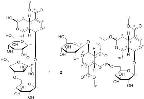
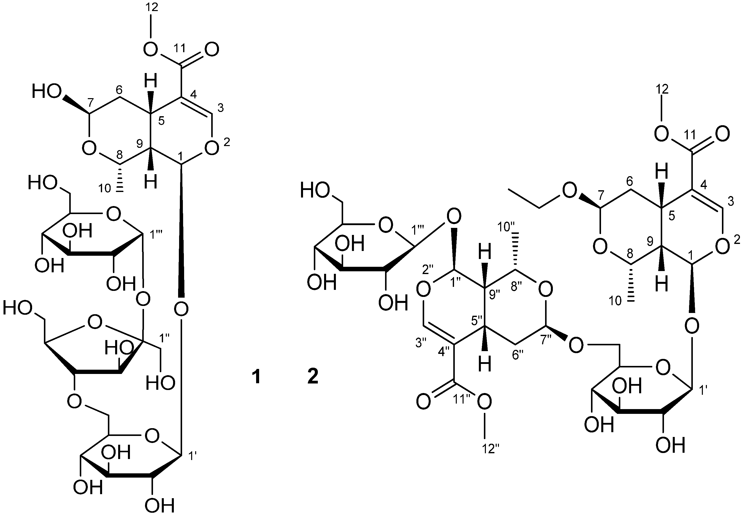
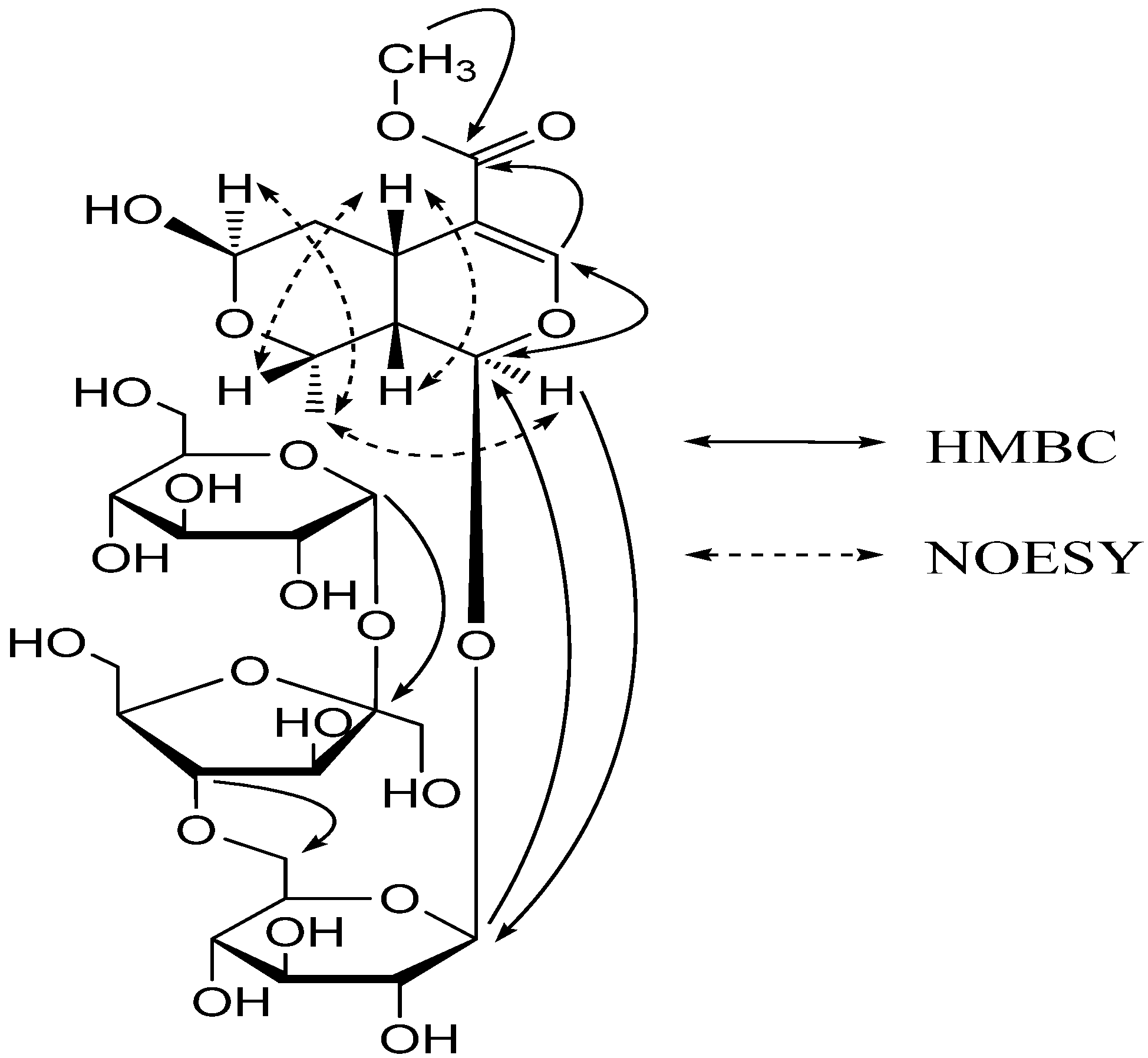
2. Results and Discussion

Compound 1 was obtained as a white amorphous powder and showed positive results for the Molisch reagent. Its molecular formula was determined as C29H46O21 by the positive HRESIMS data. The UV spectrum of the compound displayed an absorption maximum at 239 nm, which is the characteristic of an iridoid skeleton, and intense IR bands at 3,450 and 1,708 cm−1, which indicated the presence of hydroxyl and ester carbonyl functionalities, respectively.
Detailed interpretation of 1D and 2D NMR data of 1 confirmed an iridoid structure, the presence of two glucopyranosyl and a fructofuranosyl moieties. The 1H-NMR spectrum (Table 1) of 1 in CD3OD showed signals diagnostic for an iridoid glycoside at δH 5.89 (1H, d, J = 9.2 Hz, H-1), 7.50 (1H, s, H-3), 4.78 (1H, d, J = 7.6 Hz, H-1′) and 5.43 (1H, d, J = 3.8 Hz, H-1′′′). In addition, the signals at δH 3.68 (3H, s) and 1.33 (3H, d, J = 6.8 Hz) were attributed to Me-12 and Me-10, respectively. The 13C-NMR spectrum of 1 (Table 1) showed resonances for 29 C-atoms, including two quaternary carbons, six methines, one methylenes, and two methyls belonging to the aglycone moiety, two glucopyranosyl groups and a fructofuranosyl group.
Comparison of the NMR data of 1 and β-morroniside [4] indicated the presence of an additional fructofuranosyl and an additional glucopyranosyl unit. In addition, the NMR data of C-6′ of 1 were significantly deshielded by comparison with those of β-morroniside. This indicated that 1 was a β-morroniside derivative with a fructofuranosyl and a glucopyranosyl moieties located at C-6′, which was verified by correlations from H2-6′ to C-4′′, from H-4′′ to C-6′, from H-1′′′ to C-2′′ and from H-2′′ to C-1′′′ in the HMBC spectrum of 1. Acid hydrolysis of 1 afforded glucose and fructose, which were identified by TLC comparison with authentic samples. The glucose and fructose isolated from the hydrolysate gave optical rotations of [α]D20 +35.3 (c 0.05, MeOH) and [α]D20 −90.2 (c 0.025, MeOH), respectively, indicating that they were D-glucose and D-fructose, respectively [6,7,8,9]. The coupling constants of anomeric signals indicated that terminal glucosyl linkage is in α-configuration and inner glucosyl linkage is in β-configuration. Comparison of 13C-NMR chemical shifts with literature data [10,11] indicated D-fructose in 1 is in β-configuration.
The stereochemistry of 1 was established based on the NOESY experiment (Figure 2). The NOESY spectrum showed correlations of H-1/Me-10, Me-10/H-7, H-5/H-8 and H-5/H-9. These data indicated that compound 1 has the same stereochemistry at C-5, C-7, C-8 and C-9 as that of β-morroniside and the hydroxy group bound to C-7 is in the β orientation. Thus, the structure of 1 was identified to be α-D-glucopyranosyl (1→2)-β-D-fructofuranosyl (4→6)-β-morroniside, with the structure shown in Figure 1, and it was named williamsoside C.
Figure 2. Key HMBC correlations of compound 1.
Molecules 17 01830 g002
Compound 2 was obtained as a white amorphous powder and showed positive results for the Molisch reagent. Its molecular formula was established as C36H54O21 by the positive HRESIMS data. The IR absorption bands at 3,355 and 1,692 cm−1, respectively, indicated the presence of hydroxyl groups and a carbonyl group in the molecule.
The 1H- and 13C-NMR spectra (Table 1) of 2 resemble those of β-morroniside [4], but is more complex. Whereas the 1H- and 13C-NMR spectrum of 2 shows two sets of signals, this distribution of signals is consistent with a dimeric structure. The analysis of NMR spectra indicated the presence of two distinct iridoid units, which are hereafter referred to as units A and B. The 1H-NMR spectrum showed signals at δH 7.50 (1H, s), 5.84 (1H, d, J = 9.5 Hz), 1.39 (1H, dt, J = 3.7, 13.6 Hz), 1.85 (1H, dd, J = 4.7, 13.6 Hz), 4.76 (1H, br.d, J = 4.6 Hz) and 1.29 (3H, d, J = 6.9 Hz) which were assigned to H-3, H-1 H-6, H-7 and Me-10 of unit A, based on analysis of the HMQC and HMBC spectra. Analysis of the 13C-NMR spectra revealed signal values similar to those reported for 7β-O-ethyl morroniside [12]. The remaining spectral data revealed a second iridoid unit due to part B of the new iridoid. Signals at δH 7.50 (1H, s), 5.87 (1H, d, J = 9.7 Hz), 1.49 (1H, dt, J = 3.7, 13.6 Hz), 1.97 (1H, dd, J = 4.6, 13.7 Hz), 4.94 (1H, d, J = 3.2 Hz) and 1.34 (3H, d, J = 6.9 Hz) in the 1H-NMR spectrum were assigned to H-3′′, H-1′′, H-6′′, H-7′′ and Me-10′′ of unit B. One significant difference observed for this part was the lack of an ethyoxyl group. The 13C-NMR data of this moiety indicated signals identical to those of β-morroniside [4], which was confirmed by 2D NMR data analysis (Table 1).
Table 1. 1H- and 13C-NMR data for compounds 1 and 2 in CD3OD (400 MHz for 1H and 100 MHz for 13C).
No.1No.2
δCδH (J in Hz)δCδH (J in Hz)
195.75.89 (1H, d, 9.2)196.05.84 (1H, d, 9.5)
3154.57.50 (1H, s)3154.57.50 (1H, s)
4111.7-4111.7-
528.03.06 (1H, dt, 4.4, 12.8)527.83.06 (1H, m)
633.71.51 (1H, dt, 4.8, 13.6)634.01.39 (1H, dt, 3.7, 13.6)
1.97 (1H, dd, 3.6, 13.6) 1.85 (1H, dd, 4.7, 13.6)
792.75.00 (1H, br.d, 3.2)797.84.76 (1H, br.d, 4.6)
866.54.36 (1H, dq, 2.0, 6.8)866.04.26 (1H, dq, 2.1, 6.9)
940.31.83 (1H, m)940.41.79 (1H, m)
1019.71.33 (3H, d, 6.8)1019.71.29 (3H, d, 6.9)
11168.7-11168.6-
1251.83.68 (3H, s)1251.83.68 (3H, s)
1′100.14.78 (1H, d, 7.6)-OCH2CH363.63.41 (1H, o)
2′75.03.21 (1H, m) 3.64 (1H, o)
3′78.03.36 (1H, m)-OCH2CH315.41.19 (3H, t, 7.1)
4′71.63.26 (1H, m)1′100.44.78 (1H, d, 7.9)
5′78.63.06 (1H, dt, 4.4, 12.8)2′75.43.20 (1H, m)
6′69.43.84 (2H, o)3′78.03.37 (1H, m)
1′′63.83.62 (2H, s)4′71.83.34 (1H, m)
2′′105.6-5′76.83.47 (1H, m)
3′′78.74.10 (1H, d, 8.4)6′68.03.62 (1H, o)
4′′76.33.99 (1H, t, 8.2) 3.96 (1H, dd, 1.8, 12.0)
5′′82.13.90 (1H, o)1′′95.75.87 (1H, d, 9.7)
6′′62.33.80 (1H, o)3′′154.57.50 (1H, s)
3.72 (1H, o)4′′111.8-
1′′′93.45.43 (1H, d, 3.8)5′′28.03.06 (1H, m)
2′′′73.33.41 (1H, m)6′′34.01.49 (1H, dt, 3.7, 13.6)
3′′′74.83.68 (1H, m) 1.97 (1H, dd, 4.6, 13.7)
4′′′71.43.34 (1H, m)7′′99.34.94 (1H, d, 3.2)
5′′′74.23.84 (1H, m)8′′66.44.30 (1H, dq, 2.1, 7.0)
6′′′62.83.67 (1H, dd, 4.8, 11.8)9′′40.31.79 (1H, m)
3.86 (1H, dd, 2.0, 11.8)10′′19.81.34 (3H, d, 6.9)
11′′168.7-
12′′51.83.66 (3H, s)
1′′′100.14.78 (1H, d, 7.9)
2′′′75.03.20 (1H, m)
3′′′78.03.37 (1H, m)
4′′′71.63.28 (1H, m)
5′′′78.53.28 (1H, m)
6′′′62.83.63 (1H, o)
3.86 (1H, dd, 2.0, 11.6)
The absence of the ethyoxyl of unit B, when compared to β-morroniside, and observation of deshielding of C-6′ (δC 68.0), when compared to the 7β-O-ethyl morroniside (C-6′, δC 62.8), was the first indication for the attachment of units A and B between C-6′ and C-7′′. The partial structures A and B were reasonably connected to each other by HMBC correlations. The correlations from H2-6′ with C-7′′ and from H-7′′ with C-6′ in HMBC strongly indicated the connection between unit A and B through an ether linkage between C-6′ and C-7′′.
The stereo configuration of the substituent group at C-7 and C-7′′ was determined to be the β-orientation on the basis of the obvious NOESY correlations between H-7/Me-10, Me-10/H-1, H-7′′/Me-10′′ and Me-10′′/H-1′′. On the basis of above data, the structure of 2 was identified to be 7β-O-ethyl morroniside-(6′-O-7′′)-β-morroniside, with the structure shown in Figure 1, and it was named williamsoside D.

3. Experimental

3.1. General

Optical rotations were measured with a PE-241 digital polarimeter. UV spectra were recorded on a Shimadzu UV-1601 spectrometer. IR spectra were recorded on a Shimadzu FTIR-8400S spectrometer. NMR spectra were recorded on a Bruker DPX 400 NMR instrument (at 400 MHz for 1H-NMR and 100 MHz for 13C-NMR). Chemical shifts are given as δ values with reference to tetramethylsilane (TMS) used as internal standard, and coupling constants are given in Hz. HRESIMS were carried out on Waters Xevo QTOF mass spectrometer. Preparative HPLC (Waters, Delta 600-2487) was performed on a Hypersil-ODS II column (10 μm, 20 × 300 mm, Yilite, Dalian, China).

3.2. Plant Material

The root barks of S. williansii were collected in August 2008 from the Fangzheng district, Heilongjiang Province, China, and identified by the author Zhen-Yue Wang. A voucher specimen (20080079) has been deposited at Heilongjiang University of Chinese Medicine, Harbin, China.

3.3. Extraction and Isolation

The dried root barks (5.0 kg) were extracted with 95% EtOH (2 × 10 L) for 2 h. The EtOH extracts was concentrated under reduced pressure and fractioned on an AB-8 macroporous resin column (8 × 60 cm) with H2O, 50% and 95% EtOH-H2O to give three fractions (H2O fraction, 50% EtOH-H2O fraction, 95% EtOH-H2O fraction). The 50% EtOH-H2O fraction (52.0 g) was repeatedly column chromatographed on silica gel with a gradient of CHCl3/MeOH (15:1→1:1) solvents as eluents to afford 10 fractions: Fraction 1–10. Fraction 4 (20 g) continues silica gel chromatography elution with CHCl3/MeOH (10:1 to 5:1) to afford a number of sub-fractions A1–A4. Compound 1 (45.5 mg) was obtained by prep. HPLC chromatography of the sub-fraction A2 (3 g) and elution with MeOH/H2O (2:5). A3 (7 g) was separated on ODS-A column with MeOH/H2O (1:4 to 1:0) as eluent, to produce five sub-fractions (B1–B5). The sub-fraction B3 (2 g) was purified by prep. HPLC with MeOH/H2O (3:10) to afford 2 (35.1 mg).
Williamsoside C (1): White amorphous powder. [α]D20 −16.5 (c 0.075, MeOH). UV (MeOH) λmax (log ε) nm: 239 (3.10). IR (KBr): ν = 3,450, 2,500, 1,708, 1,632 cm−1. HRESIMS (positive): m/z = 731.2619 (calc. for C29H47O21, 731.2610, [M+H]+), 1H- and 13C-NMR: see Table 1.
Williamsoside D (2): White amorphous powder. [α]D20 −12.5 (c 0.05, MeOH). UV (MeOH) λmax (log ε) nm: 241 (3.05). IR (KBr): ν = 3,355, 2,450, 1,692, 1,637 cm−1. HRESIMS (positive): m/z = 823.3230 (calc. for C36H55O21, 823.3236, [M+H]+), 1H and 13C-NMR: see Table 1.
Acid Hydrolysis of 1 and 2. To a solution of 1 and 2 (each, 15 mg) in MeOH (5 mL) was added 5% H2SO4 (5 mL) and the mixture was refluxed for 8 h. Each reaction mixture was then neutralized with saturated sodium carbonate and extracted with ethyl acetate (EtOAc, 2 × 10 mL) to give an aqueous fraction containing sugars and an EtOAc fraction containing the aglycone part. The aqueous phase was dried by using a N2 stream. The residues were separately subjected to CC over silica gel with MeCN-H2O (8:1) as the eluent to yield glucose and fructose from 1, and glucose from 2, respectively. The solvent systems Me2CO/H2O/CHCl3/MeOH (15:1:2:2) and CHCl3/MeOH/water (6:4:1) [6,7] were used for TLC identification of glucose and fructose.

4. Conclusions

As a part of our chemical investigation on S. williamsii, two new iridoid glycosides, α-D-glucopyranosyl (1→2)-β-D-fructofuranosyl (4→6)-β-morroniside (1) and 7β-O-ethyl morroniside-(6′-O-7′′)-β-morroniside (2) were isolated. Their structures were established on the basis of spectroscopic evidence. The discovery of compounds 1 and 2 represents a further addition to number and diversity of iridoid glycoside compounds.

Acknowledgments

Our work was supported by the State Key Creative New Drug Project of 11th Five-year Plan of China (2009ZX09103-383), the National Natural Science Foundation of China (No. 81173501) and Heilongjiang University of Chinese Medicine Doctoral Innovation Foundation (No.B201003).
  • Sample Availability: Samples of williamsosides C and D are available from the authors.

References and Notes

  1. Hance, H.F. Sambucus williamsii Hance. Ann. Sci. Nat. Bot. 1866, 5. Available online: http://www.tropicos.org/Name/6000520/ (accessed on 22 December 2011). Series 4. Missouri Botanical Garden.
  2. Liu, W.; Wu, C.F.; Guo, Y.Y.; Yu, Q.H. Anti-inflammatory activity of aqueous extract of the root of Sambucus williamsii. Fitoterapia 1991, 62, 83–85. [Google Scholar]
  3. Han, H.; Yan, X.Y.; Kuang, H.X.; Dong, P.L. Advances in research of Sambucus williamsii hance. Inf. Tradit. Chin. Med. 2008, 25, 14–16. [Google Scholar]
  4. Ouyang, F.; Liu, Y.; Li, R.; Li, L.; Wang, N.L.; Yao, X.S. Five lignans and an iridoid from Sambucus williamsii. Chin. J. Nat. Med. 2011, 9, 26–29. [Google Scholar]
  5. Wang, Z.Y.; Han, H.; Yang, B.Y.; Xia, Y.G.; Kuang, H.X. Two new iridoid glycosides from the root barks of Sambucus williamsii Hance. Molecules 2011, 16, 3869–3874. [Google Scholar] [CrossRef]
  6. Furuya, T.; Ushiyama, M.; Asada, Y.; Yoshikawa, T.; Orihara, Y. Biotransformation of phenylacetic acid and 2-phenyl-propionic acid in suspension culture of Coffea Arabica. Phytochemistry 1988, 27, 803–807. [Google Scholar] [CrossRef]
  7. Furuya, T.; Ushiyama, M.; Asada, Y.; Yoshikawa, T. Glycosylation of 2-phenylpropionic acid and its ethyl ester in suspension cultures of Nicotiana tabacum, Dioscoreophyllum cumminsii and Aconitum japonicum. Phytochemistry 1987, 26, 2983–2989. [Google Scholar] [CrossRef]
  8. Zhang, Y.L.; Gan, M.L.; Lin, S.; Liu, M.T.; Song, W.X.; Zi, J.C.; Wang, S.J.; Li, S.; Yang, Y.C.; Shi, J.G. Glycosides from the bark of Adina polycephala. J. Nat. Prod. 2008, 71, 905–909. [Google Scholar] [CrossRef]
  9. de Bruyn, A.; Alvarez, A.P.; Sandra, P.; de Leenheer, L. Isolation and identification of O-beta-D-fructofuranosyl-(2→1)-O-beta-D-fructofuranosyl-(2→1)-D-fructose, a product of the enzymic hydrolysis of the inulin from Cichorium intybus. Carbohydr. Res. 1992, 235, 303–308. [Google Scholar] [CrossRef]
  10. Angyal, S.J.; Bethell, G.S. Conformational analysis in carbohydrate chemistry. III. The 13C-NMR spectra of the hexuloses. Aust. J. Chem. 1976, 29, 1249–1265. [Google Scholar] [CrossRef]
  11. Ren, F.Z.; Chen, S.H.; Zhang, X.X.; Li, L.H.; Gao, Y.Q.; Wang, N. Structure analysis of an oligosaccharide isolated from Bixo orellana by NMR spectroscopy. Chin. J. Magn. Reson. 2011, 28, 160–167. [Google Scholar]
  12. Chen, Y.W.; Xue, Z. Studies on the immunoactive constituents of zhi yu rou. J. Chin. Jpn. Friendsh. Hosp. 1992, 6, 231–234. [Google Scholar]

