Article

New Constituents from the Rhizomes of Egyptian Iris germanica L.

by
Sabrin R. M. Ibrahim
1,
Gamal A. Mohamed
2,† and
Nawal M. Al-Musayeib
3,*
1
Department of Pharmacognosy, Faculty of Pharmacy, Assiut University, Assiut 71526, Egypt
2
Department of Natural products, Faculty of Pharmacy, King Abdulaziz University, Jeddah 211589, Saudi Arabia
3
Department of Pharmacognosy, Faculty of Pharmacy, King Saud University, Riyadh 11451, Saudi Arabia
*
Author to whom correspondence should be addressed.
Permanent address: Department of Pharmacognosy, Faculty of Pharmacy, Al-Azhar University, Assiut Branch, Assiut 71524, Egypt
Molecules 2012, 17(3), 2587-2598; https://doi.org/10.3390/molecules17032587
Submission received: 5 February 2012 / Revised: 23 February 2012 / Accepted: 28 February 2012 / Published: 2 March 2012
(This article belongs to the Section Natural Products Chemistry)

Abstract

:
Chemical investigation of the methanolic extract of the rhizomes of Iris germanica L. (Iridaceae) afforded two new compounds; irigenin S (7) and iriside A (12), together with ten known compounds: stigmasterol (1), α-irone (2), γ-irone (3), 3-hydroxy-5-methoxyacetophenone (4), irilone (5), irisolidone (6), irigenin (8), stigmasterol-3-O-β-D-glucopyranoside (9), irilone 4'-O-β-D-glucopyranoside (10) and iridin (11). Their structures were established by UV, IR, 1D (1H and 13C) and 2D (1H-1H COSY, HMQC, and HMBC) NMR spectroscopy, in addition to mass spectroscopic data and comparison with literature data. The methanolic extract was evaluated for its antimicrobial activity. Both the methanolic extract and the isolated flavonoids were tested for their anti-inflammatory activity.

1. Introduction

The genus Iris belongs to the family Iridaceae, which comprises over 300 species [1]. Iris species have an immense medicinal importance and are used in the treatment of cancer, inflammation, bacterial and viral infections [2,3]. The compounds isolated from these species were reported to have piscicidal, anti-neoplastic, antioxidant, antitumor, anti-plasmodial, molluscicidal, and anti-tuberculosis properties, in addition to protein kinase C activation activity [4,5,6,7,8,9,10]. Iris germanica L., known as Irsa, is widely distributed in most parts of the World, and also cultivated as an ornamental plant [11,12,13]. The essential oil of I. germanica rhizomes is used in perfumes and cosmetics, while the leaves are a rich source of ascorbic acid and vitamins [1,14]. The aqueous extract of Iris germanica decreases smooth muscle activity in vivo, stimulates respiration, and shows central anti-serotonin activity. It also induces a transitory hypotension accompanied by a negative inotropic effect [11,12]. A root decoction of the plant has been used as an antispasmodic, anti-inflammatory, emmengogue, stimulants, diuretic, aperients, and violently cathartic [11,12,15]. Iris germanica is considered as a rich source of secondary metabolites such as flavonoids [9,11,12,13,14,16,17,18,19,20], triterpenes [4,8,21,22,23,24,25,26], benzene and benzoquinones derivatives [15,19]. This article reports the isolation and characterization of two new compounds—irigenin S (7) and iriside A (12)—along with ten known compounds: stigmasterol (1) [27], α-irone (2) [23], γ-irone (3) [23], 3-hydroxy-5-methoxyacetophenone (4) [28], irilone (5) [17,29,30], irisolidone (6) [15,17,31], irigenin (8) [16,17,32], stigmasterol-3-O-β-D-glucopyranoside (9) [33], irilone 4'-O-β-D-glucopyranoside (10) [14] and iridin (11) [15,17,34] (Figure 1) from the rhizomes of I. germanica L. growing in Egypt, the antimicrobial activity of its methanolic extract, as well as anti-inflammatory activity of the methanolic extract and the isolated flavonoids.
Figure 1. Structures of isolated compounds.
Molecules 17 02587 g001

2. Results and Discussion
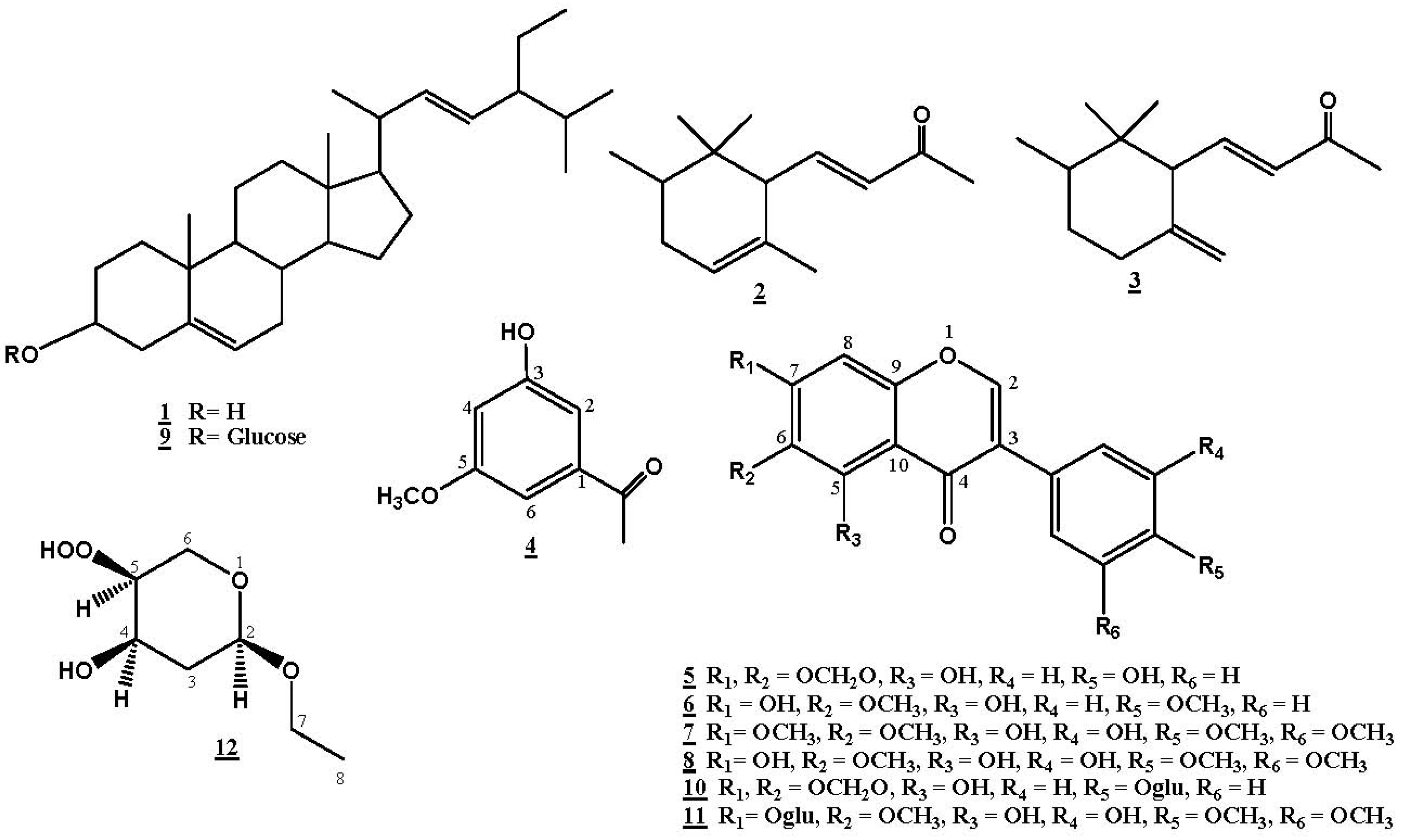
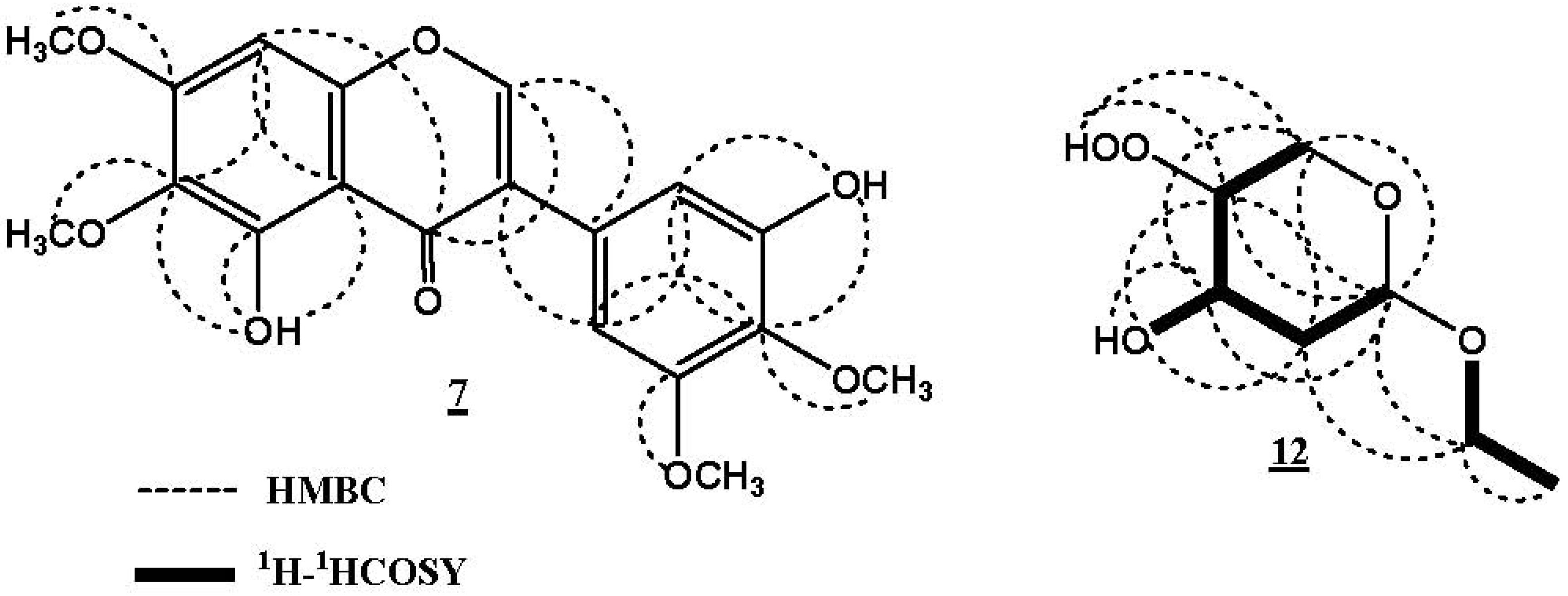
Irigenin S (7) was isolated as yellow needles (MeOH), m.p. 192–193 °C. The UV spectrum of 7 showed absorption bands at λmax 201, 265, and 323 nm, suggesting its isoflavone nature, further confirmed by the singlet signal at δH 8.42 for H-2 [29,35]. The IR spectrum showed absorption bands at 3437, 2956, 1651, and 1065 cm−1, indicating the presence of O-H, aromatic C-H, α,β-unsaturated carbonyl, and C-O functions in the molecule. Compound 7 possessed the molecular formula C19H18O8 by HRESIMS, that showed a pesudomolecular ion peak at m/z 375.1002 [M+H]+. The 1H-NMR spectrum showed resonances for 18 protons; three aromatic protons signals at δH 8.42 (s, H-2), 6.61 (s, H-8), 6.75 (d, J = 1.5 Hz, H-2', 6'), four methoxy group signals at δH 3.70 (4'-OCH3), 3.76 (6-OCH3), 3.79 (5'-OCH3), and 3.88 (7-OCH3), 3' and 5 chelated hydroxy groups at δH 9.31 and 12.97 (Table 1).
Table 1. NMR data of compounds 7 and 12 (DMSO-d6, 500 and 125 MHz).
712
PositionδH (J in Hz)δC, mult.HMBCδH (J in Hz)δC, mult.1H-1H COSYHMBC
H→CH→C
1-------
28.42 s154.8, d1', 3, 4, 95.08 dd (2.0, 4.1)103.2, CH33, 4, 5, 6, 7
3-126.3, C-1.96 m41.2, CH22, 42, 4, 5
1.85 m
4-180.7, C-4.09 t (4.5)70.9, CH3, 5, 3-OH-
5-151.3, C-3.68 m86.9, CH4, 62, 4, 6
6-132.1, C-3.36 m63.7, CH252, 4, 5
7-154.5, C-3.61 m62.1, CH282, 3, 5
3.29 m
86.61 s93.1, CH4, 6, 7, 101.06 t (6.5)15.1, CH377
9-152.6, C-----
10-104.5, C ----
1'-121.5, C ---
2'6.75 d (1.5)110.8, CH1', 3', 4', 6'----
3'-151.2, C -- -
4'-136.1, C ----
5'-152.2, C ----
6'6.75 d (1.5)103.8, CH1', 2', 4', 5'----
4-OH---4.95 d (3.5)-43, 4, 5
5-OOH---4.58 s -5, 6
5-OH12.97 s-4, 5, 6, 10----
3'-OH9.31 s-2, 4----
4'-OCH33.70 s59.9, CH34'----
5'-OCH33.79 s55.8, CH35'----
6-OCH33.76 s59.9, CH36----
7-OCH33.88 s55.9, CH37----
The 13C-NMR showed nineteen carbon signals, their multiplicities were determined through DEPT, as well as, direct correlations of protons to their respective carbons in the HMQC spectrum. The placement of the methoxy groups at C-4', C-6, C-5', and C-7 was supported by the HMBC spectrum that displayed correlations between 4'-OCH3 and C-4'/δC136.1, 6-OCH3 and C-6/δC 132.1, 5'-OCH3 and C-5'/δC 152.2, as well as 7-OCH3 and C-7/δC 154.5 (Figure 2). On the basis of the above evidences, the structure of 7 was elucidated as 3',5-dihydroxy-4',5',6,7-tetramethoxyisoflavone and considered as a new natural product, for which we propose the name irigenin S.
Figure 2. Important 1H-1H COSY and HMBC correlations of 7 and 12.
Molecules 17 02587 g002
Iriside A (12) was isolated as yellow oil. The molecular formula C7H14O5 for 12 was confirmed from the HRFABMS molecular ion peak at m/z 179.0840 [M+H]+, which required one degree of unsaturation. Its IR spectrum displayed an absorption band at 3452 cm−1, indicating the presence of a hydroperoxy group [36]. The 13C-NMR and HMQC spectra of 12 showed signals for one methyl carbon (δC 15.1, C-8), three methylenes, two of them which are oxygenated [δC 63.7 (C-6) and 62.1 (C-7)], and three oxygen-bonded methane carbons at δC 103.2, 70.9, and 86.9, ascribed to C-2, C-4, and C-5, respectively. The COSY spectrum displayed the presence of two spin systems. The first spin system consists of the triplet methyl signal at δH 1.06 (J = 6.5 Hz, H-8) coupled with the methylene protons at δH 3.61 and 3.29 indicating the presence of an ethoxy group, which further confirmed by the fragment ion peak at m/z 150.1503 [M-C2H5+H]+. The second spin system was assigned to a substituted pyran moiety, and confirmed by the fragment ion peak at m/z 84.0354 [C5H8O]+. This moiety was also validated by the correlations observed in the HMBC spectrum (Figure 2). H-2 showed HMBC correlations with C-4 and C-6 and a four bond coupling to C-5. H-6 correlated with C-2 and C-4 and H-3 with C-5. In the 1H-NMR spectrum, the two signals at δH 4.58 and 4.95 indicated the presence of two hydroxy groups; one for the hydroperoxy group at C-5 and the other for C-4-OH, which were confirmed by the observed COSY and HMBC correlations. The downfield shift of C-5 (δC 86.9) suggested the presence of a hydroperoxy functional group at C-5 and was confirmed by the fragment ion peaks at m/z 133.0905 [M-C2H6O+H]+ and 117.0782 [M-C2H6O2+H]+ [37]. The connectivity of the ethoxy group at C-2 of the pyran ring was established through the HMBC correlations of H-2 with C-7, H-7 with C-2 and C-3. On the basis of these findings the structure of 12 was unambiguously elucidated as 2-ethoxytetrahydro-5-hydroperoxy-(2R,4R,5S)-2H-pyran-4-ol, for which we propose the name iriside A. The relative stereochemistry of 12 was determined by comparing the 1H-, 13C-NMR chemical shifts, and the coupling constants values with literature [38,39], and the absolute stereochemistry was confirmed using the Mosher procedure [40].
The methanolic extract of Iris germanica (MIG) and compounds 5–8, 10, 11 were tested for their anti-inflammatory effects using the induced paw edema test. All the tested compounds as well as MIG exhibited potent anti-inflammatory effects; compound 7 showed the highest activity, which was almost similar to that of dexamethasone (Table 2). These results are in accordance with previous studies that attributed the anti-inflammatory activity of flavonoids to the C-2,3 double bond [41] and the presence of a methoxyl group at C-5 and C-7, and the pyran ring [42]. The activity of MIG may be due to the presence of different classes of terpenes and flavonoids. The MIG also showed potent antimicrobial activity against different bacterial and fungal strains (Table 3) at concentrations 250 and 500 µg per disc. It exhibited highest activity at a concentration of 250 µg per disc against S. aureus, S. marcescens, E. coli, C. albicans, and A. flavus. This observed anti-microbial effects of the extract might be due to the presence of phenolic constituents in the plant.
Table 2. Effect of injection of dexamethasone, MIG, and compounds 5–8, 10, and 11 on formalin-induced rat paw edema.
Groups n = 6Dose mg/kgPaw Edema Thickness (mm)
1 h2 h4 h24 h
Inflamed control 3.10 ± 0.183.36 ± 0.023.50 ± 0.133.40 ± 0.11
Inflamed + dexamethasone101.95 ± 0.09 *1.83 ± 0.11 *1.79 ± 0.06 *1.70 ± 0.08 *
Inflamed + MIG502.80 ± 0.25 *2.65 ± 0.03 *2.40 ± 0.11 *2.20 ± 0.09 *
1002.60 ± 0.32 *2.30 ± 0.18 *2.15 ± 0.14 *2.08 ± 0.07 *
Inflamed + 5102.40 ± 0.11 *2.20 ± 0.09 *1.98 ± 0.08 *1.90 ± 0.08 *
Inflamed + 6102.10 ± 0.13 *2.01 ± 0.18 *1.95 ± 0.14 *1.86 ± 0.07 *
Inflamed + 7101.98 ± 0.12 *1.85 ± 0.14 *1.78 ± 0.06 *1.72 ± 0.04 *
Inflamed + 8102.04 ± 0.09 *2.01 ± 0.11 *1.90 ± 0.04 *1.78 ± 0.07 *
Inflamed + 10102.50 ± 0.12 *2.30 ± 0.11 *2.10 ± 0.09 *2.01 ± 0.09 *
Inflamed + 11102.06 ± 0.09 *2.01 ± 0.18 *1.93 ± 0.13 *1.86 ± 0.07 *
* Significant different from inflamed control group at p < 0.01.
Table 3. Antimicrobial activity of MIG.
Inhibition zone in mm
Extract Bacterial StrainsFungal Strain
Conc. µg/discB. cereus (AUMC No. B-52)S. aureus (AUMC No. B-54)E. coli (AUMC No. B-53)P. aeruginosa (AUMC No. B-73)S. marcescens (AUMC No. B-55)A. flavus (AUMC No. 1276)C. albicans (AUMC No. 226)
MIG25021181212241912
50039322219363722
Chloroamphenicol a2505543272043 -
Clotriamazole b250-----4325
AUMC: Assiut University Mycology Center. a Positive control for antibacterial activity; b Positive control for antifungal activity.

3. Experimental

3.1. General Procedures

Optical rotation was measured on a Perkin-Elmer Model 341 LC polarimeter. Melting points were determined using an Electrothermal 9100 Digital Melting Point apparatus (Electrothermal Engineering Ltd., Essex, England). HRESI and FABMS were recorded on a LTQ Orbitrap and an API 2000 (ThermoFinnigan, Bremen, Germany) mass spectrometers, respectively. Low resolution mass spectra were determined using a Finnigan MAT TSQ-7000 mass spectrometer. UV spectra were recorded in absolute MeOH on a Shimadzu 1601 UV/VIS spectrophotometer. The IR spectra were measured on a Shimadzu Infrared-400 spectrophotometer (Kyoto, Japan). 1D and 2D NMR spectra (chemical shifts in ppm, coupling constants in Hz) were recorded on Bruker BioSpin GmbH 500 MHz Ultrashield spectrometer using standard Bruker software and DMSO-d6, CDCl3, and C5D5N as solvents, with TMS as the internal reference. Solvents were distilled prior spectroscopic measurements. Column chromatographic separations were performed on silica gel 60 (0.04-0.063 mm), RP-18 (0.04–0.063 mm, Merck), and Sephadex LH-20 (0.25–0.1 mm, Merck). TLC was performed on precoated TLC plates with silica gel 60 F254 (layer thickness 0.2 mm, Merck). The solvent systems used for TLC analyses were CHCl3-MeOH (97:3, solvent system I), CHCl3-MeOH (90:10, solvent system II), and CHCl3-MeOH (85:15, solvent system III). The compounds were detected by UV absorption at λmax 255 and 366 nm followed by spraying with anisaldehyde/H2SO4 reagent and heating at 110 °C for 1–2 min.

3.2. Plant Material

The rhizomes of Iris germanica L. were collected in April 2007 from plants growing at the botanical garden of the Faculty of Agriculture, Assiut University. The plant material was kindly identified by A. Fayed, Professor of Plant Taxonomy, Faculty of Science, Assiut University. A voucher specimen was deposited in herbarium of the Department of Pharmacognosy, Faculty of Pharmacy, Al-Azhar University, Assiut under the registration number IG-1-2007.

3.3. Extraction and Isolation

The air-dried powdered rhizomes of Iris germanica L. (320 g) were extracted with 70% methanol (6 × 2 L) at room temperature to afford a dark brown residue (18.1 g). The total methanolic extract was subjected to normal phase vacuum liquid chromatography (VLC) using n-hexane, CHCl3, EtOAc, and MeOH to afford four fractions; n-hexane IG-1 (3.5 g), CHCl3 IG-2 (4.0 g), EtOAc IG-3 (3.2 g), and MeOH IG-4 (5.3 g) fractions. Fraction IG-2 (4.0 g) was subjected to VLC by gradient elution using n-hexane-EtOAc as solvents, and four subfractions were obtained; IG-2-A (0.85 g), IG-2-B (0.90 g), IG-2-C (0.65 g), and IG-2-D (0.75 g). Subfraction IG-2-A (0.85 g) was chromatographed over silica gel column (140 g × 50 × 3 cm) using a n-hexane-EtOAc gradient to afford compounds 1 (40 mg), 2 (15 mg), and 3 (10 mg). Subfraction IG-2-B (0.90 g) was subjected to silica gel column chromatography (150 g × 50 × 3 cm) using CHCl3-MeOH (95:5) as an eluent to give compounds 4 (20 mg) and 5 (35 mg). A Sephadex LH-20 column chromatorgraphy (150 g × 100 × 5 cm) of subfraction IG-2-C (0.65 g) using MeOH:CHCl3 (9:1) give impure compounds 6, 7, and 8 which were further purified by repeated chromatography on a silica gel column using a CHCl3-MeOH gradient to afford pure compounds 6 (25 mg), 7 (30 mg), and 8 (55 mg). Compound 9 (100 mg) was obtained as a white amorphous powder upon crystallization of fraction IG-2-D using MeOH. Fraction IG-3 (3.2 g) was subjected to Sephadex LH-20 chromatography using MeOH as eluent to afford four subfractions IG-3-A (0.45 g), IG-3-B (0.51 g), IG-3-C (0.75 g), and IG-3-D (0.68 g). Subfraction IG-3-A was chromatographed over a silica gel column (100 g × 50 × 3 cm) using a CHCl3-MeOH gradient to give compound 10 (35 mg). RP-18 column chromatography (100 g × 50 × 3 cm) of subfraction IG-3-B using a MeOH-H2O gradient give compounds 11 (25 mg) and 12 (40 mg).

3.4. Spectral Data

Stigmasterol (1). White needles, m.p. 169–170 °C, EIMS: m/z 412 [M]+. 1H-NMR (CDCl3, 500 MHz): δH 3.55 (m, H-3), 5.38 (brs, H-6), 0.69 (s, H-18),1.02 (s, H-19), 0.93 (d, J = 6.1 Hz, H-21), 5.18 (dd, J = 15.4, 8.0 Hz, H-22), 5.11 (dd, J = 15.4, 8.0 Hz, H-23), 0.84 (d, J = 7.3 Hz, H-26), 0.84 (d, J = 7.3 Hz, H-27), 0.86 (t, J = 7.1 Hz, H-29). 13C-NMR (CDCl3, 125 MHz): δC 37.2 (C-1), 31.8 (C-2), 71.9 (C-3), 39.4 (C-4), 140.8 (C-5), 121.8 (C-6), 29.1 (C-7), 31.9 (C-8), 50.1 (C-9), 35.9 (C-10), 20.6 (C-11), 41.0 (C-12), 42.2 (C-13), 56.1 (C-14), 23.3 (C-15), 24.6 (C-16), 56.8 (C-17), 11.9 (C-18), 18.9 (C-19), 39.8 (C-20), 20.9 (C-21), 138.4 (C-22), 129.3 (C-23), 51.2 (C-24), 31.9 (C-25), 19.4 (C-26), 19.8 (C-27), 26.7 (C-28), 12.0 (C-29).
α-Irone (2). Colorless oil. EIMS: m/z 206 [M]+. 1H-NMR (CDCl3, 500 MHz): δH 2.33 (s, H-1), 6.10 (d, J = 15.8 Hz, H-3), 6.66 (dd, J = 15.8, 11.0 Hz, H-4), 2.59 (m, H-5), 5.54 (brs, H-7), 1.98, 1.78 (2H each m, H-8), 1.52 (m, H-9), 0.88 (s, H-11), 1.63 (s, H-12), 0.73 (s, H-13), 0.88 (d, J = 6.5 Hz, H-14). 13C-NMR (CDCl3, 125 MHz): δC 27.0 (C-1), 198.3 (C-2), 133.6 (C-3), 149.6 (C-4), 56.0 (C-5), 132.0 (C-6), 123.1 (C-7), 31.8 (C-8), 37.9 (C-9), 35.8 (C-10), 15.4 (C-11), 23.0 (C-12), 15.0 (C-13), 26.6 (C-14).
γ-Irone (3). Colorless oil. EIMS: m/z 206 [M]+. 1H-NMR (CDCl3, 500 MHz): δH 2.37 (s, H-1), 6.15 (d, J = 16.0 Hz, H-3), 6.95 (dd, J = 16.0, 10.5 Hz, H-4), 2.85 (m, H-5), 2.36, 2.15 (2H each m, H-7), 1.56, 1.25 (2H each m, H-8), 1.45 (m, H-9), 0.88 (s, H-11), 4.82, 4.45 (2 H each brs, H-12), 0.74 (s, H-13), 0.88 (d, J = 6.5 Hz, H-14). 13C-NMR (CDCl3, 125 MHz): δC 27.3 (C-1), 198.3 (C-2), 134.3 (C-3), 147.3 (C-4), 57.8 (C-5), 148.8 (C-6), 36.3 (C-7), 31.9 (C-8), 41.9 (C-9), 38.8 (C-10), 15.9 (C-11), 108.7 (C-12), 14.1 (C-13), 27.7 (C-14).
3-Hydroxy-5-methoxyacetophenone (4). Colorless crystals, (MeOH), Rf = 0.78 (solvent system I), m.p. 188–189 °C. EIMS: m/z 166 [M]+. 1H-NMR (CDCl3, 500 MHz): δH 7.55 (d, J = 1.5 Hz, H-2), 6.97 (d, J = 1.5 Hz, H-4), 7.55 (d, J = 1.5 Hz, H-6), 2.58 (s, H-8), 6.22 (s, 3-OH), 3.97 (s, 5-OCH3). 13C-NMR (CDCl3, 125 MHz): δC 130.2 (C-1), 124.1 (C-2), 150.4 (C-3), 113.8 (C-4), 146.6 (C-5), 109.7 (C-6), 196.9 (C-7), 26.2 (C-8), 56.1 (5-OCH3).
Irilone (5). Yellow needles (MeOH), m.p. 233–234 °C. EIMS: 298 [M]+. λmax 205, 261, and 324 nm. 1H-NMR (DMSO-d6, 500 MHz): δH 8.44 (s, H-2), 6.90 (s, H-8), 7.40 (2 H, d, J = 8.0 Hz, H-2', 6'), 6.83 (2 H, d, J = 8.0 Hz, H-3', 5'), 6.19 (s, -O-CH2-O-), 9.65 (s, 4'-OH), 12.94 (s, 5-OH). 13C-NMR (DMSO-d6, 125 MHz): δC 154.7 (C-2), 122.1 (C-3), 180.9 (C-4), 141.3 (C-5), 129.5 (C-6), 153.9 (C-7), 89.5 (C-8), 152.9 (C-9), 107.4 (C-10), 120.9 (C-1'), 130.2 (C-2', 6'), 115.1 (C-3', 5'), 157.5 (C-4'), 102.8 (-O-CH2-O-).
Irisolidone (6). Yellow amorphous powder. EIMS: m/z 314 [M]+. UV (MeOH) λmax nm: 207, 264, and 328. 1H-NMR (DMSO-d6, 500 MHz): δH 7.88 (s, H-2), 6.59 (s, H-8), 7.47 (d, J = 8.5 Hz, H-2', 6'), 6.99 (d, J = 8.5 Hz, H-3', 5'), 3.86 (s, 4'-OCH3), 4.05 (s, 6-OCH3), 13.15 (s, 5-OH). 13C-NMR (DMSO-d6, 125 MHz): δC 153.5 (C-2), 123.1 (C-3), 181.4 (C-4), 152.8 (C-5), 130.4 (C-6), 152.7 (C-7), 93.2 (C-8), 155.3 (C-9), 106.4 (C-10), 122.9 (C-1'), 130.2 (C-2', 6'), 159.7 (C-4'), 114.1 (C-3', 5'), 55.4 (4'-OCH3), 60.9 (6-OCH3).
Irigenin S (7). Yellow needles (MeOH), Rf = 0.47 (solvent system II), m.p. 192–193 °C. UV (MeOH) λmax nm: 201, 265, and 323. IR νmax (cm−1): 3437, 2956, 1651, and 1065. HRESIMS: m/z 375.1002 (calcd for C19H19O8 375.1001). NMR spectral data, see Table 1.
Irigenin (8). Yellow needles (MeOH), m.p. 185–186 °C. UV (MeOH) λmax nm: 203, 263, and 323 nm. EIMS: m/z 360 [M]+. 1H-NMR (DMSO-d6, 500 MHz): δH 8.40 (s, H-2), 6.52 (s, H-8), 6.72 (d, J = 1.5 Hz, H-2'), 6.71 (d, J = 1.5 Hz, H-6'), 3.70 (s, 4'-OCH3), 3.79 (s, 5'-OCH3), 3.76 (s, 6-OCH3), 9.31 (s, 3'-OH), 13.01 (s, 5-OH), 10.90 (s, 7-OH). 13C-NMR (DMSO-d6, 125 MHz): δC 154.8 (C-2), 126.0 (C-3), 180.3 (C-4), 153.3 (C-5), 131.4 (C-6), 157.5 (C-7), 93.9 (C-8), 152.8 (C-9), 104.8 (C-10), 121.7 (C-1'), 110.3 (C-2'), 150.2 (C-3'), 136.3 (C-4'), 152.6 (C-5'), 104.4 (C-6'), 59.9 (4'-OCH3), 55.7 (5'-OCH3), 59.9 (6-OCH3).
Stigmasterol-3-O-β-glucoside (9). White amorphous powder. FABMS: m/z 575 [M+H]+. 1H-NMR (C5D5N, 500 MHz): δH 3.97 (m, H-3), 0.62 (s, H-18), 1.21 (s, H-19), 0.96–0.81 (methyl groups), 5.06 (d, J = 7.6 Hz, H-1'), 4.58–3.86 (m, sugar protons). 13C-NMR (C5D5N, 125 MHz): δC 38.0 (C-1), 31.1 (C-2), 78.9 (C-3), 40.4 (C-4), 141.4 (C-5), 122.4 (C-6), 32.7 (C-7), 32.5 (C-8), 50.8 (C-9), 37.4 (C-10), 21.8 (C-11), 39.5 (C-12), 43.0 (C-13), 57.3 (C-14), 25.0 (C-15), 23.7 (C-16), 56.7 (C-17), 11.6 (C-18), 19.9 (C-19), 39.8 (C-20), 19.5 (C-21), 133.6 (C-22), 129.8 (C-23), 46.5 (C-24), 29.9 (C-25), 19.7 (C-26), 20.5 (C-27), 24.5 (C-28), 12.5 (C-29), 103.0 (C-1'), 72.2 (C-2'), 75.8 (C-3'), 68.7 (C-4'), 78.7 (C-5'), 63.3 (C-6').
Irilone 4'-O-β-D-glucopyranoside (10). Yellow amorphous powder. UV (MeOH) λmax nm: 203, 271, and 337 nm. FABMS: m/z 461 [M+H]+, 299 [M-162 (hexose unit)+H]+. 1H-NMR (DMSO-d6, 500 MHz): δH 8.51 (s, H-2), 7.01 (s, H-8), 7.34 (2 H, d, J = 8.5 Hz, H-2', 6'), 6.81 (2 H, d, J = 8.5 Hz, H-3', 5'), 6.19 (s, -O-CH2-O-), 13.42 (s, 5-OH), 4.98 (d, J = 6.7 Hz, H-1”). 13C-NMR (DMSO-d6, 125 MHz): δC 155.4 (C-2), 123.0 (C-3), 181.0 (C-4), 139.8 (C-5), 129.4 (C-6), 153.5 (C-7), 93.6 (C-8), 151.7 (C-9), 106.9 (C-10), 120.9 (C-1'), 130.2 (C-2', 6'), 114.8 (C-3', 5'), 157.9 (C-4'), 102.6 (-O-CH2-O-), 100.9 (C-1”), 73.6 (C-2”), 77.4 (C-3”), 69.8 (C-4”), 76.7 (C-5”), 60.6 (C-6”).
Iridin (11). Yellow needles, m.p. 210–211 °C. UV (MeOH) λmax nm: 203, 263, and 335 nm. FABMS: m/z 523 [M+H]+, 361 [M-162 (hexose unit)+H]+. 1H-NMR (DMSO-d6, 500 MHz): δH 8.51 (s, H-2), 6.70 (s, H-8), 6.91 (brs, H-2'), 6.74 (brs, H-6'), 3.71 s (s, 4'-OCH3), 3.80 (s, 5'-OCH3), 3.78 (s, 6-OCH3), 9.30 (s, 3'-OH), 12.95 (s, 5-OH), 5.11 (d, J = 6.7 Hz, H-1”). 13C-NMR (DMSO-d6, 125 MHz): δC 155.4 (C-2), 125.9 (C-3), 180.5 (C-4), 152.9 (C-5), 132.5 (C-6), 156.6 (C-7), 94.0 (C-8), 152.4 (C-9), 104.5 (C-10), 122.0 (C-1'), 110.3 (C-2'), 150.3 (C-3'), 136.4 (C-4'), 152.9 (C-5'), 104.5 (C-6'), 100.1 (C-1'), 73.1 (C-2'), 77.3 (C-3”), 69.6 (C-4”), 76.7 (C-5”), 60.3 (C-6”).
Iriside A (12). Yellow oil, Rf = 0.42 (solvent system III), [α]D +72.6° (c 0.3, MeOH). IR νmax (cm−1): 3452, 2945, 1455, 1103, 983. HRFABMS: m/z 179.0840 (calc. for C7H15O5, 179.0841). NMR spectral data, see Table 1.

3.5. Mosher Procedure [40]

Four mg of compound 12 was dissolved in pyridine-d5 (0.7 mL) and transferred to NMR tubes. The 1H-NMR spectrum of 12 was measured prior to adding 5 μL of (R)-MTPA-Cl and (S)-MTPA-Cl reagent (Fluka, Germany), respectively. The tubes were shaken thoroughly and were allowed to stand at room temperature for 72 h. The reaction was monitored by 1H-NMR spectroscopy after every 24 h.

3.6. Biological Studies

3.6.1. Anti-Inflammatory Activity

Hind paw edema (skin edema) was induced by 4% formalin solution injected into the subplantar region of the left hind paw [42]. Adult male albino rats 100–120 g (purchased from the Animal House, Pharmacology Department, Faculty of Medicine, Assiut University). The inflamed animals were divided randomly into ten groups (six in each group), inflamed control group, inflamed treated with dexamethasone (at a dose of 10 mg/kg subcutaneously), six groups of inflamed animals were treated with the tested compounds individually (at a dose of 10 mg/kg subcutaneously), and two groups were treated with MIG at doses of 50 and 100 mg/kg subcutaneously (the plant extract was dissolved in sterile distilled water). The change in paw thickness in all tested animals was measured at 1, 2, 4, and 24 h after formalin solution injection. The data were expressed as mean ± S.D. using the Student t test (Table 2).

3.6.2. Antimicrobial Assay

The procedure was carried out as previously described [43]. The antibacterial and antifungal activities were evaluated using the agar plate diffusion assay. Susceptibility discs (5.5 mm) were impregnated with solution of each of extract at concentrations 250 and 500 µg per disc. The discs were dried and placed on agar plates inoculated with the test bacterial strains: Bacillus cereus (AUMC No. B-52), Staphylococcus aureus (AUMC No. B-54), Escherichia coli (AUMC No. B-53), Pseudomonas aeruginosa (AUMC No. B-73), and Serratia marcescens (AUMC No. B-55), and the fungal strains: Candida albicans (AUMC No. 418), Geotrichum candidum (AUMC No. 226), Trichophyton rubrum (AUMC No. 1804), Fusarium oxysporum (AUMC No. 209), Scopulariopsis brevicaulis (AUMC No. 729), and Aspergillus flavus (AUMC No. 1276). Each plate was inoculated with a single organism and the test was run in duplicates. The plates were incubated at 37 °C and checked for inhibition zones after 24 h for bacteria and after 48 h for fungi. Chloroamphenicol and clotrimazole at concentration 250 µg per disc were used as positive reference standards for antibacterial and antifungal activities, respectively.

4. Conclusions

Two new compounds; irigenin S (7) and iriside A (12), were isolated from the rhizomes of I. germanica L. growing in Egypt along with ten known compounds. The methanolic extract (MIG) and the isolated flavonoids exhibited potent anti-inflammatory effects. Compound 7 showed activity similar to that of dexamethasone. The MIG showed highest anti-microbial effect against S. aureus, S. marcescens, E. coli, C. albicans, and A. flavus.

Acknowledgements

The authors are grateful to the research center for female scientific and medical colleges in King Saud University for the financial support and to Volker Brecht (Nuclear Magnetics Resonance, Institut fuer Pharmazeutische Wissenschaften, Albert-Ludwigs-Universität Freiburg, Germany) for MS spectral measurements and Mosher analysis and discussion.
  • Sample Availability: Samples of the compounds 1, 4, 5, 6, 8, 9, and 11 are available from the authors.

References

  1. Ali, S.I.; Mathew, B. Flora of Pakistan Department of Botany; Ali, S.I., Qaiser, M., Eds.; University of Karachi: Pakistan, Islamabad, Karachi, 1993; pp. 4–29. [Google Scholar]
  2. Hanawa, F.; Tahara, S.; Mizutani, J. Isoflavonoids produced by Iris pseudacorus leaves treated with cupric chloride. Phytochemistry 1991, 30, 157–163. [Google Scholar]
  3. Rigano, D.; Grassia, A.; Formisano, C.; Basile, A.; Sorbo, S.; Senatore, F. Antibacterial and allelopathic activity of methanolic extract from Iris pseudopumila rhizomes. Fitoterapia 2006, 77, 460–462. [Google Scholar] [CrossRef]
  4. Hideyuki, I.; Yoko, M.; Takashi, Y. New piscicidal triterpenes from Iris germanica. Chem. Pharm. Bull. 1995, 43, 1260–1262. [Google Scholar] [CrossRef]
  5. Miyake, Y.; Ito, H.; Yoshida, T. Identification of iridals as piscicidal components of iridaceous plants and their conformations associated with CD spectra. Can. J. Chem. 1997, 75, 734–741. [Google Scholar] [CrossRef]
  6. Bonfils, J.P.; Pinguet, F.; Culine, S.; Sauvaire, Y. Cytotoxicity of iridals, triterpenoids from Iris, on human tumor cell lines A2780 and K562. Planta Med. 2001, 67, 79–81. [Google Scholar] [CrossRef]
  7. Nadaroğlu, H.; Demir, Y.; Demir, N. Antioxidant and radical scavenging properties of Iris germanica. Pharm. Chem. J. 2007, 41, 409–415. [Google Scholar]
  8. Benoit-Vical, F.; Imbert, C.; Bonfils, J.P.; Sauvaire, Y. Anti-plasmodial and antifungal activities of iridal, a plant triterpenoid. Phytochemistry 2003, 62, 747–751. [Google Scholar] [CrossRef]
  9. Singab, A.B.; Ahmed, A.H.; Sinkkonen, J.; Ovcharenk, V.; Pihlaja, K. Molluscicidal activity and new flavonoids from Egyptian Iris germanica L. (var. alba). Z. Naturforsch. 2006, 61, 57–63. [Google Scholar]
  10. Takahashi, K.; Suzuki, S.; Hano, Y.; Nomura, T. Protein kinase C activation by iridal type triterpenoids. Biol. Pharm. Bull. 2002, 25, 432–436. [Google Scholar] [CrossRef]
  11. Rahman, A.U.; Nasim, S.; Baig, I.; Orhan, I.; Sener, B.; Ayanoglu, F.; Choudhary, I.M. Isoflavonoid glycosides from the rhizomes of Iris germanica. Helv. Chim. Acta 2003, 86, 3354–3362. [Google Scholar] [CrossRef]
  12. Rahman, A.U.; Nasim, S.; Biag, I.; Jalil, S.; Orhan, I.; Sener, B.; Choudhary, M.I. Anti-inflammatory isoflavonoids from the rhizomes of Iris germanica. J. Ethnopharmacol. 2003, 86, 177–180. [Google Scholar] [CrossRef]
  13. Choudhary, M.I.; Naheed, S.; Jalil, S.; Alam, J.M.; Rahman, A.U. Effect of ethanolic extract of Iris germanica on lipid profile of rats fed on a high-fat diet. J. Ethnopharmacol. 2005, 98, 217–220. [Google Scholar] [CrossRef]
  14. Rahman, A.U.; Nasim, S.; Baig, I.; Jahan, I.A.; Sener, B.; Orhan, I.; Choudhary, M.I. Isoflavonoid glycosides from the rhizomes of Iris germanica. Chem. Pharm. Bull. 2002, 50, 1100–1102. [Google Scholar] [CrossRef]
  15. Asghar, S.F.; Aziz, S.; Rehman, H.U.; Ahamed, I.; Hussein, H.; Rahman, A.U.; Choudhary, M.I. Secondary metabolites isolated from Iris germanica. Rec. Nat. Prod. 2009, 3, 139–152. [Google Scholar]
  16. Akashi, T.; Ishizaki, M.; Aoki, T.; Ayabe, S. Isoflavonoid production by adventitious-root culture of Iris germanica (Iridaceace). Plant Biotechnol. 2005, 22, 207–215. [Google Scholar] [CrossRef]
  17. Eckhard, W.; Frederik, S.J.; Karin, K.; Jutta, K.; Norbert, F.; Clarissa, G. Cancer chemoprotective in vitro activities of isoflavones isolated from Iris germanica. Planta Med. 2003, 69, 15–20. [Google Scholar] [CrossRef]
  18. Ali, A.A.; El-Emary, N.A.; Darwish, F.M.; Frahm, A.W. Three isoflavonoids from Iris germanica. Phytochemistry 1983, 22, 2061–2063. [Google Scholar] [CrossRef]
  19. Asghar, S.F.; Rehman, H.U.; Rahman, A.U. Phytochemical investigation of Iris germanica. Nat. Prod. Res. 2010, 24, 131–139. [Google Scholar] [CrossRef]
  20. Orhan, I.; Nasim, S.; Tener, B.; Ayanoglu, F.; Özgüven, M.; Choudhary, I.M.; Rahman, A.U. Two isoflavones and bioactivity spectrum of the crude extract if Iris germanica rhizomes. Phytother. Res. 2003, 17, 575–577. [Google Scholar] [CrossRef]
  21. Bonfils, J.; Sauvaire, Y. Localization of iridals in Iris germanica rhizomes. Phytochemistry 1996, 41, 1281–1285. [Google Scholar] [CrossRef]
  22. Ito, H.; Miyake, Y.; Yoshida, T. New piscicidal triterpenes from Iris germanica. Chem. Pharm. Bull. 1995, 43, 1260–1262. [Google Scholar] [CrossRef]
  23. Marner, F.J.; Krick, W.; Gellrich, B.; Jaenicke, L.; Winter, W. Irigermanal and Iridogermanal: Two new triterpenoids from rhizomes of Iris germanica L. J. Org. Chem. 1982, 47, 2531–2536. [Google Scholar]
  24. Crawford, M.M.R.; Lindsay, D.A.; Walton, J.C.; Wollenweber-Ratzer, B. Towards the characterization of radicals formed in rhizomes of Iris germanica. Phytochemistry 1994, 37, 979–985. [Google Scholar] [CrossRef]
  25. Jaenicke, L.; Marner, F.J. The irones and their origin. Pure Appl. Chem. 1990, 62, 1365–1368. [Google Scholar] [CrossRef]
  26. Orhan, I.; Sener, B.; Hashimoto, T.; Asakawa, Y.; Ozgüven, M.; Ayanoğlu, F. Iristectorone K, a novel monocyclic triterpene ester from Iris germanica rhizomes growing in Turkey. Fitoterapia 2002, 73, 316–319. [Google Scholar] [CrossRef]
  27. Mohamed, G.A.; Ibrahim, S.R.M. Eucalyptone G, a new phloroglucinol derivative and other constituents from Eucalyptus globulus Labill. ARKIVOC 2007, 2007, 281–291. [Google Scholar]
  28. Vysokogorskii, V.E.; Nozdrunova, A.A.; Plaksin, G.V.; Krivonos, O.I.; Mkrtchan, O.Z.; Petrosyan, L.Y. Antioxidant activity of liquid products of heat-treated sapropels. Pharm. Chem. J. 2009, 43, 191–194. [Google Scholar] [CrossRef]
  29. Harborne, B.; Mabry, T.J.; Mabry, H. The Flavonoids; Academic Press: New York, NY, USA, 1975; Volumes 1 and 2, pp. 50–55, 743–761. [Google Scholar]
  30. Al-Khalil, S.; Al-Eisaw, D. New isoflavones from Iris nigricans. J. Nat. Prod. 1994, 57, 201–205. [Google Scholar] [CrossRef]
  31. Kang, K.A.; Zhang, R.; Piao, M.J.; Ko, D.O.; Wang, Z.H.; Kim, B.J.; Park, J.W.; Kim, H.S.; Kime, D.H.; Hyun, J.W. Protective effect of irisolidone, a metabolite of kakkalide, against hydrogen peroxide induced cell damage via antioxidant effect. Bioorg. Med. Chem. 2008, 16, 1133–1141. [Google Scholar]
  32. Morita, N.; Arisawa, M.; Kondo, Y.; Takemoto, T. Studies on constituents of Iris genus plants. III. The constituents of Iris florentina L. (1). Chem. Pharm. Bull. 1973, 21, 600–603. [Google Scholar] [CrossRef]
  33. El-Askary, H.I. Terpenoids from Cleome droserifolia (Forssk.) Del. Molecules 2005, 10, 971–977. [Google Scholar] [CrossRef]
  34. Arisawa, M.; Morita, N. Studies on constituents of genus Iris. VII. The constituents of Iris unguicularis Poir. (1). Chem. Pharm. Bull. 1976, 24, 815–817. [Google Scholar] [CrossRef]
  35. Mabry, T.J.; Markham, K.R.; Thomas, T.B. The Systematic Identification of Flavonoids; Springer-Verlag: Berlin, Germany, 1970; pp. 35–61. [Google Scholar]
  36. Jung, C.M.; Kwon, H.C.; Seo, J.J.; Ohizumi, Y.; Matsunaga, K.; Saito, S.; Lee, K.R. Two new monoterpene peroxide glycosides from Aster scaber. Chem. Pharm. Bull. 2001, 49, 912–914. [Google Scholar] [CrossRef]
  37. El-Askary, H.I.; Meselhy, R.M.; Galal, A.M. Sesquiterpenes from Cymbopogon proximus. Molecules 2003, 8, 670–677. [Google Scholar] [CrossRef]
  38. Agrawal, P.K. NMR spectroscopy in the structural elucidation of oligosaccharides and glycosides. Phytochemistry 1992, 31, 3307–3330. [Google Scholar] [CrossRef]
  39. Bubb, W.A. NMR spectroscopy in the study of carbohydrates: Characterizing the structural complexity. Concepts Magn. Reson. 2003, 19A, 1–19. [Google Scholar] [CrossRef]
  40. Ibrahim, S.R.M.; Ebel, R.; Wray, V.; Müller, W.E.G.; Edrada-Ebel, R.; Proksch, P. Diacarperoxides, norterpene cyclic Peroxides from the sponge Diacarnus megaspinorhabdosa. J. Nat. Prod. 2008, 71, 1358–1364. [Google Scholar] [CrossRef]
  41. Della, L.R.; Sosa, S.; Tubaro, A.; Morazzoni, P.; Bombardelli, E.; Griffini, A. Oxo-anti-inflammatory activity of Ginkgo biloba constituents and of their phospholipid complexes. Fitoterapia 1996, 67, 257–264. [Google Scholar]
  42. Panthong, A.; Kanjanapotni, D.; Tuntiwachwuttikul, P.; Pancharoen, O.; Reutrakul, V. Antiinflammatory activity of flavonoids. Phytomedicine 1994, 1, 141–144. [Google Scholar] [CrossRef]
  43. Bonev, B.; Hooper, J.; Parisot, J. Principles of assessing bacterial susceptibility to antibiotics using the agar diffusion method. J. Antimicrob. Chemother. 2008, 61, 1295–1301. [Google Scholar] [CrossRef]

