Article

Superconducting Fullerene Nanowhiskers

by
Hiroyuki Takeya
1,2,*,
Kun’ichi Miyazawa
1,
Ryoei Kato
1,
Takatsugu Wakahara
1,
Toshinori Ozaki
1,2,
Hiroyuki Okazaki
1,2,
Takahide Yamaguchi
1,2 and
Yoshihiko Takano
1,2
1
National Institute for Materials Science, Tsukuba, Ibaraki 305-0047, Japan
2
Transformative Research Project on Iron Pnictides (TRIP), Japan Science and Technology Agency, Chiyoda-ku, Tokyo 102-0075, Japan
*
Author to whom correspondence should be addressed.
Molecules 2012, 17(5), 4851-4859; https://doi.org/10.3390/molecules17054851
Submission received: 31 March 2012 / Revised: 12 April 2012 / Accepted: 18 April 2012 / Published: 26 April 2012
(This article belongs to the Special Issue Fullerene Chemistry)

Abstract

:
We synthesized superconducting fullerene nanowhiskers (C60NWs) by potassium (K) intercalation. They showed large superconducting volume fractions, as high as 80%. The superconducting transition temperature at 17 K was independent of the K content (x) in the range between 1.6 and 6.0 in K-doped C60 nanowhiskers (KxC60NWs), while the superconducting volume fractions changed with x. The highest shielding fraction of a full shielding volume was observed in the material of K3.3C60NW by heating at 200 °C. On the other hand, that of a K-doped fullerene (K-C60) crystal was less than 1%. We report the superconducting behaviors of our newly synthesized KxC60NWs in comparison to those of KxC60 crystals, which show superconductivity at 19 K in K3C60. The lattice structures are also discussed, based on the x-ray diffraction (XRD) analyses.

1. Introduction

Fullerenes were discovered in 1985 [1], and the superconductivity of a potassium metal-doped fullerene was reported in 1991 [2]. Potassium metal (K)-doped fullerides KxC60 [0 ≤ x ≤ 6] are particularly interesting because their structures and electronic properties are strongly related to the doping concentration. They exhibit an fcc structure at x = 0 and 3, a bct structure at x = 4, and a bcc structure at x = 6 [3]. The compound, K3C60, shows superconductivity below 19 K, while the others exhibit insulating, semiconducting or metallic properties. In general, superconducting K3C60 bulk samples have been synthesized mainly by three methods, i.e., a solid-solid reaction, vapor evaporation, and a reaction using liquids. Much effort has been expended to produce the K3C60 superconductor, but a large volume fraction was difficult to obtain by a simple heating method. In addition, the obtained bulk superconductors of K3C60 by the above methods were usually in powder form. The above two were the problems inherent in the bulk application of K3C60 superconductors.
A recent study of fullerene-based supramolecular nanoarchitectures [4,5,6,7,8,9] is opening new possibilities for the application of fullerene materials; such applications include sensors, transistors, catalysts, and fuel cell electrodes. Fullerene nanowhiskers were obtained from the interface between the fullerene-saturated solution and fullerene-insoluble solvent. They vary in length from microns to centimeters. Most of the cuprate superconductors form powders or bulk polycrystals, which were encapsulated in metal tubes for making superconducting wires. This makes the process complicated and the wire heavy. In this study, we propose fullerene nanowhiskers for use as flexible and lightweight superconducting wire because of the advantages in the nanowhisker form.

2. Results and Discussion

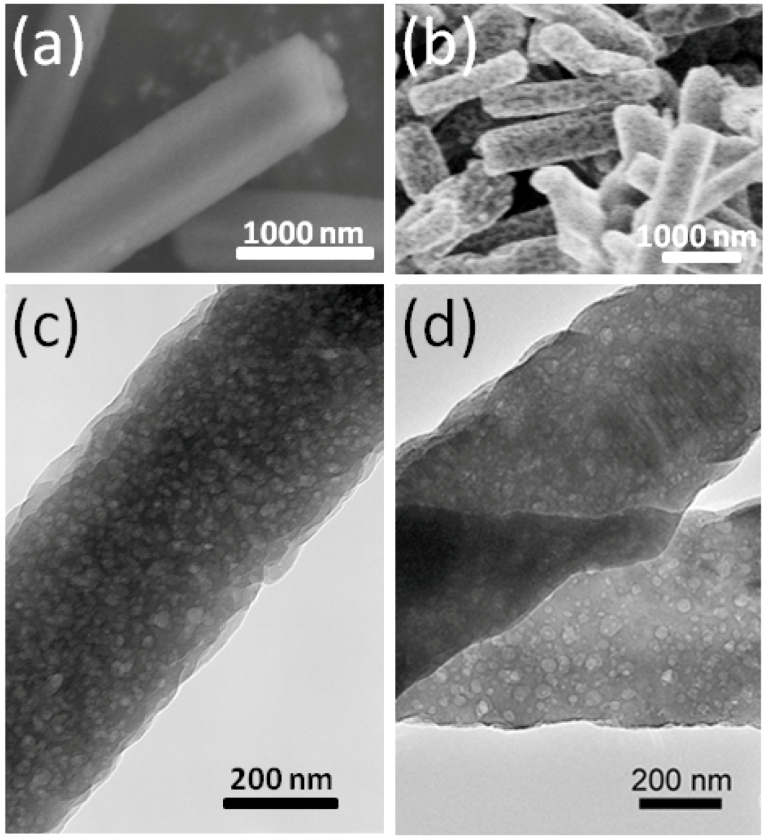
2.1. Observation of Morphologies

The morphology of C60 nanowihskers (C60NWs) used in this study are shown in Figure 1(a) observed using a scanning electron microscope (SEM). The shapes of C60NWs are mostly hexagonal prisms with the growth axis along the [001] direction [10].
Figure 1. SEM micrographs of (a) C60NWs and (b) K-doped C60NWs. (c) and (d) are the micrographs of (a) and (b) observed using a transmission electron microscope (TEM), respectively.
Molecules 17 04851 g001
C60NWs grow in a hexagonal crystal structure by the liquid-liquid interfacial method (LLIP method) [4,5], and the hexagonal structure then turns to the fcc structure while the solvent is drying. The C60NWs are composed of C60 units bound by van der Waals force. Figure 1(c) shows the micrograph of C60NWs observed using a transmission microscope (TEM), indicating disordered nanopores formed at the stage of the drying process in the LLIP method. The 10 mg C60NWs were taken in a quartz tube with an appropriate amount of K (molar ratio K/C60 = 0–6.0). The quartz tube was then sealed under an evacuated condition at about 3 × 10⁻³ Pa and heated at 200 °C for 24–36 h for K intercalation into C60NWs. The SEM micrograph and TEM image of the K-intercalated samples of K3.3C60NWs are shown in Figure 1(b) and 1(d), respectively. Basically, the morphology of the K-doped samples looks the same as the pristine samples showing the nanopore structure. The energy dispersive x-ray analysis (EDAX) revealed that K was taken in the C60NWs. In this report, all of the compositions denote nominal ones. We also prepared K-doped C60 crystals to compare the K-doped C60NWs. Several micro-cracks were observed in K3.3C60 crystals, while no crack was observed in K3.3C60NWs. K-doping into the C60 crystal expands the lattice of the C60 crystals; therefore, cracks seemed to be induced because of the boundary strain from the lattice mismatch between K-doped and undoped lattices. On the other hand, in the case of K-doped C60NWs, the disordered nanopores play a possible role in reducing the lattice strain.

2.2. Superconducting Properties and X-ray Diffraction Patterns

Figure 2 shows the superconducting transitions of K3.3C60NWs and K3.3C60 crystals heated at 200 °C for 24 h, which were measured upon warming under 20 Oe after cooling in a zero-field. The nominal K composition indicates the ratio vs. the C60 unit.
Figure 2. The temperature dependencies of the normalized magnetic moments m for K3.3C60NWs and K3.3C60 samples.
Molecules 17 04851 g002
The left and right ordinates of the graph set separately for K3.3C60NWs and K3.3C60 crystals are the magnetic moment normalized by the applied magnetic field and the weight of the samples. In K-doped C60 materials, the only fcc phase of stoichiometric K3C60 is a superconductor. We added a 10% excess amount of K, more than the crystallographically limiting composition K3C60NW and K3C60, because the K adsorption on the surface of crystallites or in the nanopores was considered to ensure complete reaction. As shown in the figure, there is a large difference of the superconducting signals between the two samples. The signal of the nanowhisker sample was 200 times larger than that of the crystal sample. The Tc at 17 K in K3.3C60NWs is lower than the reported Tc at 19 K in K3.3C60 crystals, as shown in Figure 2. We believe the Tc reduction in K3.3C60 NWs can be caused from the disordered nanopores as seen in Figure 1(d).
The powder x-ray diffraction (XRD) patterns of C60, K3.3C60 crystals, C60NWs, and K3.3C60NWs are shown in Figure 3. The XRD patterns of non-doped C60 crystals and C60NWs showed an fcc structure, as previously reported [3]. In addition, those of K3.3C60 crystals and K3.3C60NWs were also identified to be the fcc structure phase as the peaks were indexed in the figure. The lattice parameters of four samples, C60, K3.3C60 crystals, C60NWs, and K3.3C60NWs, were calculated to be 1.4180(5), 1.4180(2), 14.188(4), and 1.4200(5) nm, respectively. Therefore, K was hardly intercalated into the sites in the K3.3C60 crystals because its lattice parameter was almost identical to that of C60 crystals. This result is consistent with the difference of the superconducting shielding volume fractions between K3.3C60NWs and K3.3C60 crystals by the nominal compositions. According to reported processes for K-doping to C60, so far, a period longer than several days or several weeks is required to form the superconducting K3C60 phase [11,12]. We investigated the formation rate of the superconducting phase by measuring the superconducting volume fractions at 5 K as shown in Figure 4. In the case of K3.3C60NWs, it was saturated at around 24 h with heating at 200 °C. On the other hand, the shielding fraction of K3.3C60 crystals hardly increased up to 36 h. Such a small volume fraction of the superconducting KxC60 has already been reported, for example, by Hebard et al. [2] and Murphy et al. [3] Hebard et al. reported approximately 1% by heating at 200 °C for 36 h. We believe that the nanopores of K3.3C60NWs, shown in Figure 1(d), assist in the reaction of K-doping and the migration to form the K3C60NW superconducting phase, as explained in Figure 2. The nanopores in K3.3C60NWs play an important role for the formation of superconducting phase.
Figure 3. XRD patterns of C60 crystals, K3.3C60 crystals, C60NWs, and K3.3C60NWs (nominal composition), with fcc structure index.
Molecules 17 04851 g003
Figure 4. Formation of superconducting phase (%) in K3.3C60NWs estimated by shielding volume fractions vs. heating time (h) at 200 °C.
Molecules 17 04851 g004

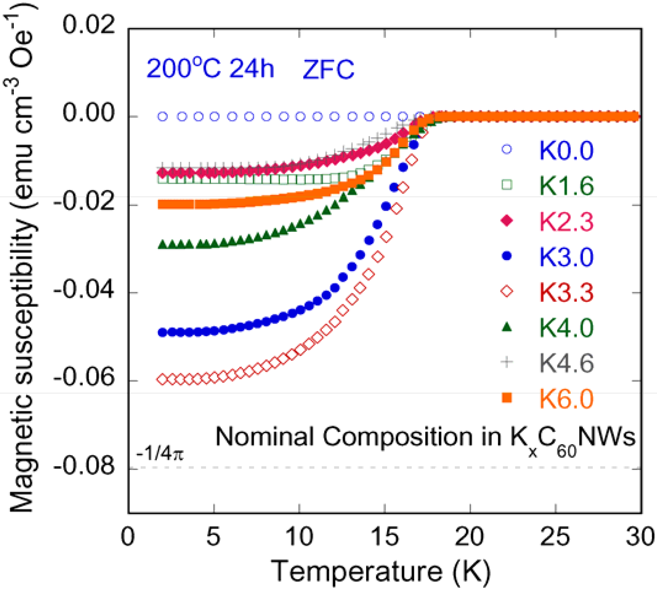
2.3. K-Compositional Dependence of Shielding Volume Fraction in KxC60NWs
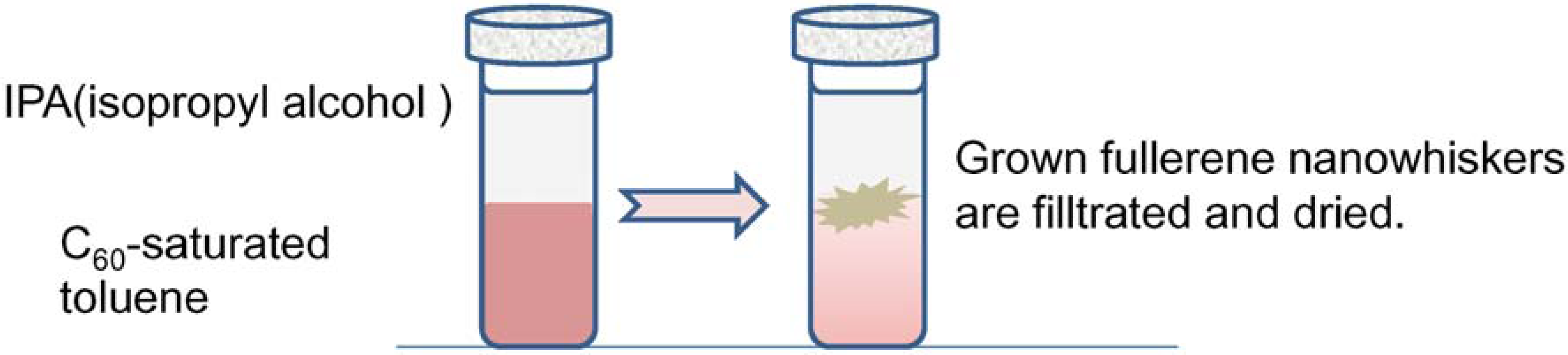
Figure 5 illustrates the bulk superconducting transitions of K-doped C60NWs for the K composition range of x = 0.0–6.0, which were measured on warming under 20 Oe after cooling in a zero-field.
Figure 5. The temperature dependencies of magnetic susceptibility for K3.3C60NWs (x = 0.0–6.0).
Molecules 17 04851 g005
The K composition indicates the nominal ratio vs. C60 unit in the C60NWs. The ordinate of the graph is the magnetic moment normalized by the applied magnetic field and the volume of the samples. None of the samples before heating showed any anomalies within a temperature range between 2 K and 30 K. The onset Tc’s of the materials after heat treatment at 200 °C were almost the same (17 K) independently of the K composition, while the superconducting shielding fractions depended on the K-ratio.
Figure 6 shows the compositional dependence of the superconducting volume fractions in KxC60NWs (nominal composition) heated at 200 °C for 24 h in comparison with those in KxC60 reported by Holczer et al. [11].
Figure 6. K content dependence of shielding volume fractions in KxC60NWs (at 5 K) compared with the result by Hoczer et al. [11] for KxC60 (at 4.2 K).
Molecules 17 04851 g006
Their heating procedure consists of three stages which are: (1) the first mixing stage heated at 200 °C for 20–24 h, (2) the second diffusion stage heated to 200 °C for 22 h and (3) the final relaxation stage heated at 250 °C for 6 h or more hours. Basically, their result coincides with our result in KxC60NWs. The shielding volume fractions are normalized by the volume of the perfect diamagnetism (−1/4π). The maximum fraction was observed at around 3.0–3.3 by the nominal K compositions. This value coincides with the carrier concentration at the Tc maximum in alkali-doped C60 superconductors [12]. In the KxC60 system, the superconducting phase is a line compound of K3C60 with the full occupancy of two tetrahedral and one octahedral sites by K in the fcc structure. The three electrons transferred from K to C60 occupy the triply degenerated t1u orbital, which becomes half-filled, and the high density of states at the Fermi level. We believe that this logic of K3C60 is analogical for K3C60NW. On the other hand, there is a difference in the case of x = 6.0 in KxC60NW, showing some superconducting volume fraction. Murphy et al. explained that the non-superconducting bcc phase (K6C60) was formed as a kinetically facile phase at the first step of the reaction [13]. Regarding our XRD measurement for the nominal K6C60NW in the heating condition at 200 °C by 36 h, no bcc phase was detected in contrast to the Murphy’s explanation. Since the lattice parameter of K6.0C60NW is 1.4210(9) nm, which is almost the same as 1.4200(5) nm of K3.3C60NW, an excessive amount of K over 3.0 might stay at the surface or in the nanopores of C60NW.

3. Experimental

The typical dimensions of the fullerene nanowhiskers (C60NWs) used in this experiment were 0.54 ± 0.16 μm in average diameter and 4.43 ± 2.63 μm in average length. The C60NWs were prepared by using the liquid-liquid interfacial method (LLIP method) [4,5]. The schematic diagram is illustrated in Figure 7. A C60-saturated toluene solution was taken in a glass bottle, and isopropyl alcohol was slowly added. The C60NWs form at the interface of the two solutions, then the nanowhiskers were filtered and dried in vacuum at 100 °C for 2 h [14]. According to the previous report [15], the residual toluene solvent in C60NWs is estimated to be about 0.2 mass %.
Figure 7. A schematic diagram of the liquid-liquid interfacial method (LLIP method).
Molecules 17 04851 g007
Ten mg C60NWs and an appropriate amount of potassium (K) were placed together into a thin quartz tube. The nominal K compositions were set at 0.0, 1.6, 2.3, 3.0, 3.3, 4.0, 4.6 and 6.0 mole ratio vs. C60 in KxC60NWs. We also prepared pristine and K-doped C60 crystals using the same procedures for a comparison with KxC60NWs. This process was conducted in a glove box to prevent potassium from oxidizing. The quartz tube was sealed under a vacuum condition at 3 × 10⁻³ Pa, followed by heating at 200 °C for 1–36 h in an electric oven. After the heat treatment, superconducting properties and structure analyses were performed as follows. Superconducting transitions were measured using a superconducting quantum interference device (SQUID) magnetometer (MPMS-5S, Quantum Design, San Diego, CA, USA) as the samples were kept in the quartz tube. The shapes and microstructures of those samples were observed with a scanning electron microscope (SEM 25kV, Hitachi SU-70, Tokyo, Japan) and a transmission electron microscope (TEM 400kV, JEOL JEM-4010, Tokyo, Japan). Their qualitative micro-analysis was achieved with an energy dispersive X-ray analyzer (EDAX, AMETEK, Mahwah, USA). The information of the crystal structure was analyzed by powder X-ray diffraction (XRD RINT-TTR3, RIGAKU, Akishima, Japan). To prevent the XRD samples from being oxidized, we used a tiny amount of paraffin oil to cover them on the holder plate. In the XRD patterns, the diffraction of oil was subtracted from the raw data.

4. Conclusions

We proved that K intercalation to C60NWs, rather than the process for K3C60, forms superconducting K3C60NWs with a short heating process. No bcc phase of K6C60NW was observed. The superconducting shielding volume fraction by heating at 200 °C for 24 h gave high values up to 80%, in contrast to the value of K-doped C60, K3.3C60, which was lower than 1%. This contrasting difference of the superconducting shielding fraction might be associated with the nanopores in KxC60NWs. These nanopores play an important role in the properties of KxC60NWs.

Acknowledgments

The authors are grateful for the partial financial support received from the Japan Society for the Promotion of Science and the Advanced Low Carbon Technology R&D Program (ALCA) of the Japan Science and Technology Agency.
  • Sample Availability: Samples of the compounds, K-doped fullerene nanowhiskers, are not available from the authors.

References and Notes

  1. Kroto, H.W.; Hearth, L.D.; O’Brien, S.C.; Curl, R.F.; Smalley, R.E. C60: Buckminsterfullerene. Nature 1985, 318, 162–163. [Google Scholar] [CrossRef]
  2. Hebard, A.F.; Rosseinsky, M.J.; Haddon, R.C.; Murphy, D.W.; Glarum, S.H.; Palstra, T.T.M.; Ramirez, A.P.; Kortan, A.R. Superconductivity at 18 K in potassium-doped C60. Nature 1991, 350, 600–601. [Google Scholar] [CrossRef]
  3. Murphy, D.W.; Rosseinsky, M.J.; Haddon, R.C.; Ramirez, A.P.; Hebard, A.F.; Tycko, R.; Fleming, R.M.; Dabbagh, G. Superconductivity in alkali metal fullerides. Physica C 1991, 185–189, 403–408. [Google Scholar]
  4. Miyazawa, K.; Kuwasaki, Y.; Obayashi, A.; Kuwabara, M. C60 nanowhiskers formed by the liquid-liquid interfacial precipitation method. J. Mater. Res. 2002, 17, 83–88. [Google Scholar] [CrossRef]
  5. Miyazawa, K.; Hamamoto, K.; Nagata, S.; Suga, T.J. Structural investigation of the C60/C70 whiskers fabricated by forming liquid-liquid interfaces of toluene with dissolved C60/C70 and isopropyl alcohol. Mater. Res. 2003, 18, 1096–1103. [Google Scholar] [CrossRef]
  6. Sathish, M.; Miyazawa, K. Size-tunable hexagonal fullerene (C60) nanosheets at the liquid-liquid interface. J. Am. Chem. Soc. 2007, 129, 13816–13817. [Google Scholar] [CrossRef]
  7. Geng, J.; Zhou, W.; Skelton, P.; Yue, W.; Kinloch, I.A.; Wimdle, A.H.; Johnson, B.F.G. Crystal structure and growth mechanism of unusually long fullerene (C60) nanowires. J. Am. Chem. Soc. 2008, 130, 2527–2534. [Google Scholar]
  8. Malik, S.; Fujita, N.; Muhopadhyay, P.; Goto, Y.; Kaneko, K.; Ikeda, T.; Shinkai, S. Creation of 1D [60]fullerene superstructures and its polymerization by γ-ray irradiation. J. Mater. Chem. 2007, 17, 2454–2458. [Google Scholar] [CrossRef]
  9. Miyazawa, K.; Ringor, C. Platinum chloride deposition into C60 nanotubes. Mater. Lett. 2008, 62, 410–413. [Google Scholar] [CrossRef]
  10. Kato, R.; Miyazawa, K. Cross-sectional structural analysis of C60 nanowhiskers by transmission electron microscopy. Diam. Relat. Mater. 2011, 20, 299–303. [Google Scholar] [CrossRef]
  11. Holczer, K.; Klein, O.; Griiner, G.; Huang, S.-M.; Kaner, R.B.; Fu, K.-J.; Whetten, R.L.; Diederich, F. Alkali-fulleride superconductors: Synthesis, composition, and diamagnetic shielding. Science 1991, 252, 1154–1157. [Google Scholar]
  12. Yildirim, T.; Barbedette, L.; Fischer, J.E.; Lin, C.L.; Robert, J.; Petit, P.; Palstra, T.T.M. Tc vs carrier concentration in cubic fulleride superconductors. Phys. Rev. Lett. 1996, 77, 167–170. [Google Scholar] [CrossRef]
  13. Murphy, D.W.; Rosseinsky, M.J.; Fleming, R.M.; Tycko, R.; Ramirez, A.P.; Haddon, R.C.; Siegrist, T.; Dabbagh, G.; Tully, J.C.; Walstedt, R.E. Synthesis and characterization of alkali metal fullerides: AxC60. J. Phys. Chem. Solids 1992, 53, 1321–1332. [Google Scholar] [CrossRef]
  14. Kato, R.; Miyazawa, K. Raman laser polymerization of C60 nanowhiskers. J. Nanotechnol. 2012, 2012. [Google Scholar] [CrossRef]
  15. Watanabe, M.; Hotta, K.; Miyazawa, K.; Tachibana, M. GC-MS analysis of the solvents contained in C60 nanowhiskers. J. Phys.: Conf. Ser. 2009, 159. [Google Scholar] [CrossRef]

