2. Results and Discussion
The structures of swertisin (
1) and embinoidin (
2) were confirmed and spectra assigned by a combination of
1H-
1H COSY [
11], HSQC [
12] and HMBC [
13] spectra. T-ROESY spectra [
14] were obtained to aid in interpretation of the origins of the spectral doubling. Complete spectral assignments for the flavone moieties of swertisin (
1) and embinoidin (
2) are given in
Table 1, while the spectral data for the sugar moieties are given in
Table 2.
The structure of swertisin (
1) is shown in
Figure 1, with carbons and the 5-OH proton giving rise to doubled signals marked with asterisks. The
1H-NMR spectrum of swertisin (
1) in DMSO-
d6 at 289 K showed that the relative proportion of the major and minor rotamers was 1.00:0.82. The duplication of the signals of the sugar unit and of carbons close to the
C-glycosidic bond implied that there was an energy barrier about this bond that prevented fast exchange between the two rotamers. VT
1H-NMR studies were conducted on swertisin (
1) to confirm this supposition. The VT
1H-NMR studies on swertisin (
1) were not performed using DMSO-
d6 as the solvent because the 5-OH signal markers were not detected individually just above the freezing temperature of DMSO-
d6. On lowering the temperature below 292 K, the sample froze. Therefore, swertisin (
1) was dissolved in (CD
3)
2CO-
d6/DMSO-
d6 (1:1) which had a freezing temperature of 258 K, thereby allowing the 5-OH signal markers to be individually detected. At 262 K and using the 5-OH signals as markers, the two rotamers of swertisin (1), with a relative proportion of 1.00:0.82, were independently detected since they were in slow exchange. At 305 K, the signals for the two rotamers moved even closer but were still detected. Eventually, the two signals coalesced to a single peak at ca. 13.54 ppm at a Coalescence Temperature,
Tc, of 321 K (
Figure 2). The coalescence of signals was observed for the entire
1H-NMR spectrum, although the other pairs of signals coalesced at lower temperatures, reflecting their smaller chemical shift differences.
Table 1.
1H-NMR (600 MHz) and 13C-NMR (150 MHz) spectral data of the flavone nucleus of swertisin (1) and embinoidin (2) in DMSO-d6 (δ in ppm, J in Hz).
Table 1.
1H-NMR (600 MHz) and 13C-NMR (150 MHz) spectral data of the flavone nucleus of swertisin (1) and embinoidin (2) in DMSO-d6 (δ in ppm, J in Hz).
| Position | 1 | 2 |
|---|
| δC | δH | δC | δH |
|---|
| C-2 | 164.01 | ― | 163.39 | ― |
| 163.87 | ― | 163.25 | ― |
| C-3 | 102.79 | 6.85 (
s ) | 103.78 | 6.95 (
s ) |
| 6.83 (
s
) | 103.70 | 6.93 (
s
) |
| C-4 | 182.21 | ― | 182.02 | ― |
| 181.88 | ― | 182.34 | ― |
| C-5 | 159.57 | ― | 159.66 | ― |
| 160.31 | ― | 160.53 | ― |
| C-6 | 109.65 | ― | 108.62 | ― |
| | 108.67 | ― |
| C-7 | 164.92 | ― | 165.14 | ― |
| 163.71 | ― | 163.86 | ― |
| C-8 | 91.00 | 6.82 (
s ) | 90.83 | 6.81 (
s ) |
| 90.16 | 6.83 (
s
) | 90.37 | 6.84 (
s
) |
| C-9 | 156.80 | ― | 157.12 | ― |
| 156.70 | ― | 157.01 | ― |
| C-10 | 104.07 | ― | 104.24 | ― |
| 104.58 | ― | 104.49 | ― |
| C-1′ | 120.37 | ― | 122.69 | ― |
| C-2′, C-6′ | 128.52 | 7.96 (
d, 8.9) | 128.37 | 8.08 (
d, 8.5) |
| C-3′, C-5′ | 116.16 | 6.92 (
d, 8.9) | 114.61 | 7.13 (
d, 8.5) |
| C-4′ | 162.12 | ― | 162.41 | ― |
| 7-OMe | 56.24 | 3.87 (
s ) | 56.13 | 3.92 (
s ) |
| 56.48 | 3.96 (
s
) | 56.55 | 3.91 (
s
) |
| 4′-OMe | ― | ― | 55.60 | 3.87 (
s) |
| 5-OH | ― | 13.52 (
s ) | ― | 13.46 (
s ) |
| ― | 13.54 (
s
) | ― | 13.58 (
s
) |
Table 2.
1H-NMR (600 MHz) and 13C-NMR (150 MHz) spectral data of the sugar moieties of swertisin (1) and embinoidin (2) in DMSO-d6 (δ in ppm, J in Hz).
Table 2.
1H-NMR (600 MHz) and 13C-NMR (150 MHz) spectral data of the sugar moieties of swertisin (1) and embinoidin (2) in DMSO-d6 (δ in ppm, J in Hz).
| Position | 1 | 2 |
|---|
| δC | δH | δC | δH |
|---|
| 6-C-Glc | | | | |
| C-1″ | 72.82 a | 4.58 (
d , 10) | 71.02 a | 4.68 (
d , 8.8) |
| 72.58 b | 4.60 (
d
, 10) | 70.70 b | 4.70 (
d
, 8.8) |
| C-2″ | 69.62 a | 4.19 (
dt , 10, 2.6) | 80.73 a | 4.47 (
t , 8.8) |
| 70.27 b | 4.00 (
dt
, 10, 2.6) | 81.21 b | 4.30 (
t
, 8.8) |
| C-3″ | 79.08 a | 3.18 (
m ) | 78.29 a | 3.42 (
m ) |
| 78.38 b | 3.18 (
m
) | 78.69 b | 3.44 (
m
) |
| C-4″ | 70.92 a | 3.09 (
m ) | 70.45 a,b | 3.15 (
m) |
| 70.83 b | 3.09 (
m
) | | |
| C-5″ | 81.68 a | 3.16 (
m ) | 81.60 a | 3.16 (
m ) |
| 81.86 b | 3.16 (
m
) | 81.93 b | 3.18 (
m
) |
| C-6″ | 61.75 a,b | 3.70 (
dd , 12.9, 4.3), | 61.45 a,b | 3.69 (
dd , 13, 4.4), |
| 3.37 (
m
) | | 3.38 (
m
) |
| 2″-O-Glc | | | | |
| C-1‴ | ― | ― | 105.42 a | 4.16 (
d , 8.8) |
| 105.25 b | 4.18 (
d
, 8.8) |
| C-2‴ | ― | ― | 74.54 a | 2.85 (
dt , 8.8, 2.6) |
| 74.70 b | 2.85 (
dt
, 8.8, 2.6) |
| C-3‴ | ― | ― | 76.32 a | 3.05 (
m ) |
| 76.37 b | 3.06 (
m
) |
| C-4‴ | ― | ― | 69.16 a | 3.02 (
m ) |
| 69.46 b | 2.96 (
m
) |
| C-5‴ | ― | ― | 76.42 a | 2.57 (
dt , 8.8, 2.6) |
| 76.68 b | 2.77 (
dt
, 8.8, 2.6) |
| C-6‴ | ― | ― | 60.07 a | 3.16 (
m), 2.96 (m ) |
| 60.60 b | 3.20 (
m
) |
Figure 1.
Diagram of swertisin showing atoms with duplicated signals indicated by asterisks; the arrow indicates rotation of the C-glycosidic bond.
Figure 1.
Diagram of swertisin showing atoms with duplicated signals indicated by asterisks; the arrow indicates rotation of the C-glycosidic bond.
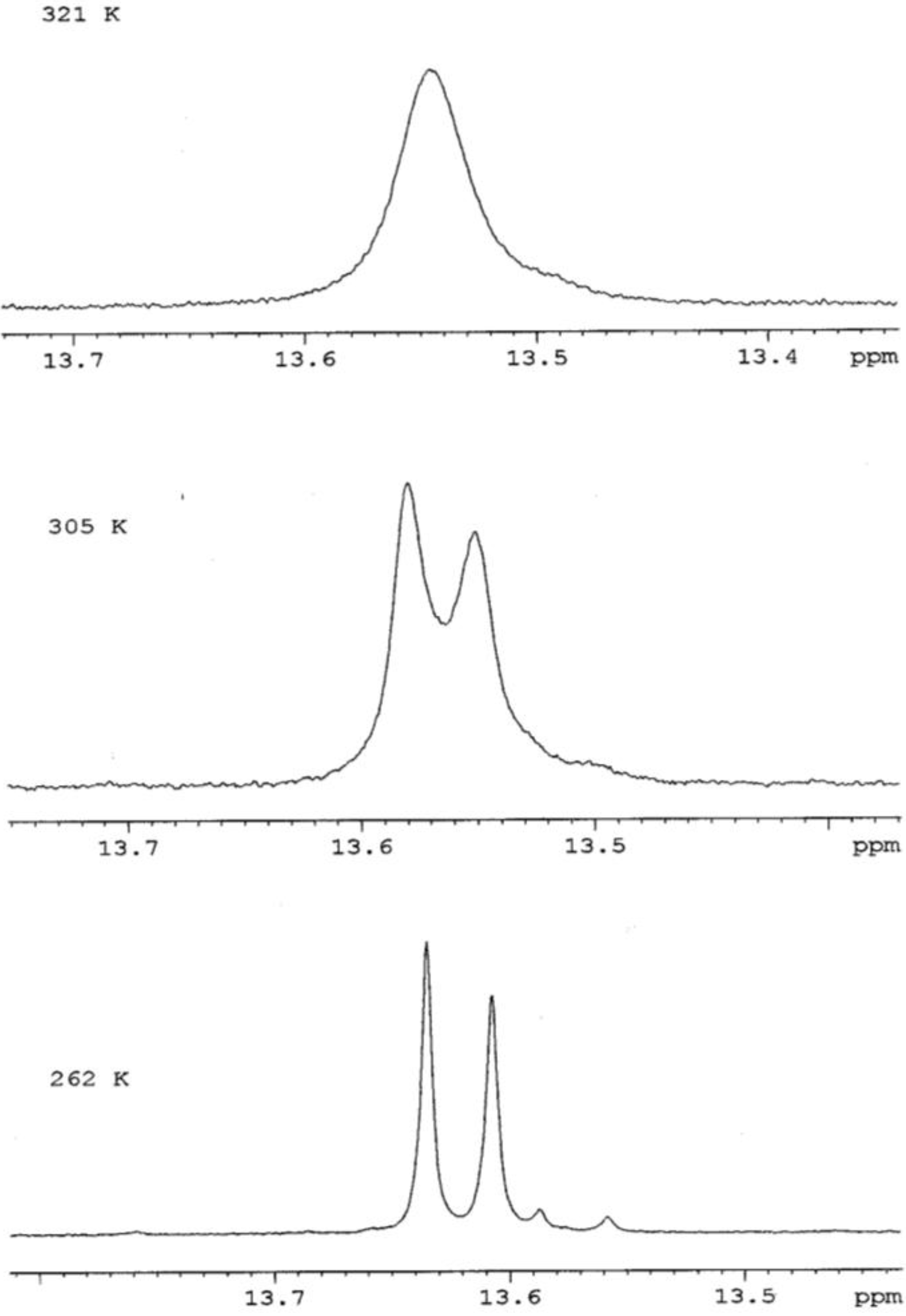
Figure 2.
Effect of Temperature on the 5-OH signal markers in the 1H-NMR spectrum of swertisin (1).
Figure 2.
Effect of Temperature on the 5-OH signal markers in the 1H-NMR spectrum of swertisin (1).
The free energy of activation for the interconversion between the two unequally populated rotamers of swertisin (
1) can be calculated using Eyring’s equations (
a and
b) as modified by Shanan-Atidi and Bar-Eli [
15]:
where X = 2πτ∆v and ∆P = PA − PB.
PA and PB represent the population of the conformers A and B (PA > PB, PA + PB = 1), respectively, and τ is the mean lifetime. Tc and ∆v are the coalescence temperature and the chemical shift difference between conformers A and B, respectively. X is obtained using equation (c):
From the 1H-NMR spectrum at 262 K, the frequency difference, Δν, between the 5-OH signals was 11.13 Hz (11.13 s−1). The Coalescence Temperature, Tc, was 321 K.
The VT 1H-NMR studies confirmed the hypothesis that the doubling of signals in the 1H- and 13C-NMR spectra at 262 K was due to the presence of two rotamers of swertisin (1) separated by a relatively high energy barrier.
The data derived from the T-ROESY spectrum suggested that the two rotamers slowly rotated about the C-glycosidic (C-1″—C-6) bond. Weak cross-peaks were observed between the respective methoxyl protons (7-OMe), and the H-1″a and H-2″b protons. This observation indicated that β-D-glucose rotates about the C-1″—C-6 bond. Therefore, in one rotamer, the H-1″ proton is oriented syn to the 7-OMe group whereas, in the other rotamer, the H-2″ proton is oriented syn to the 7-OMe group.
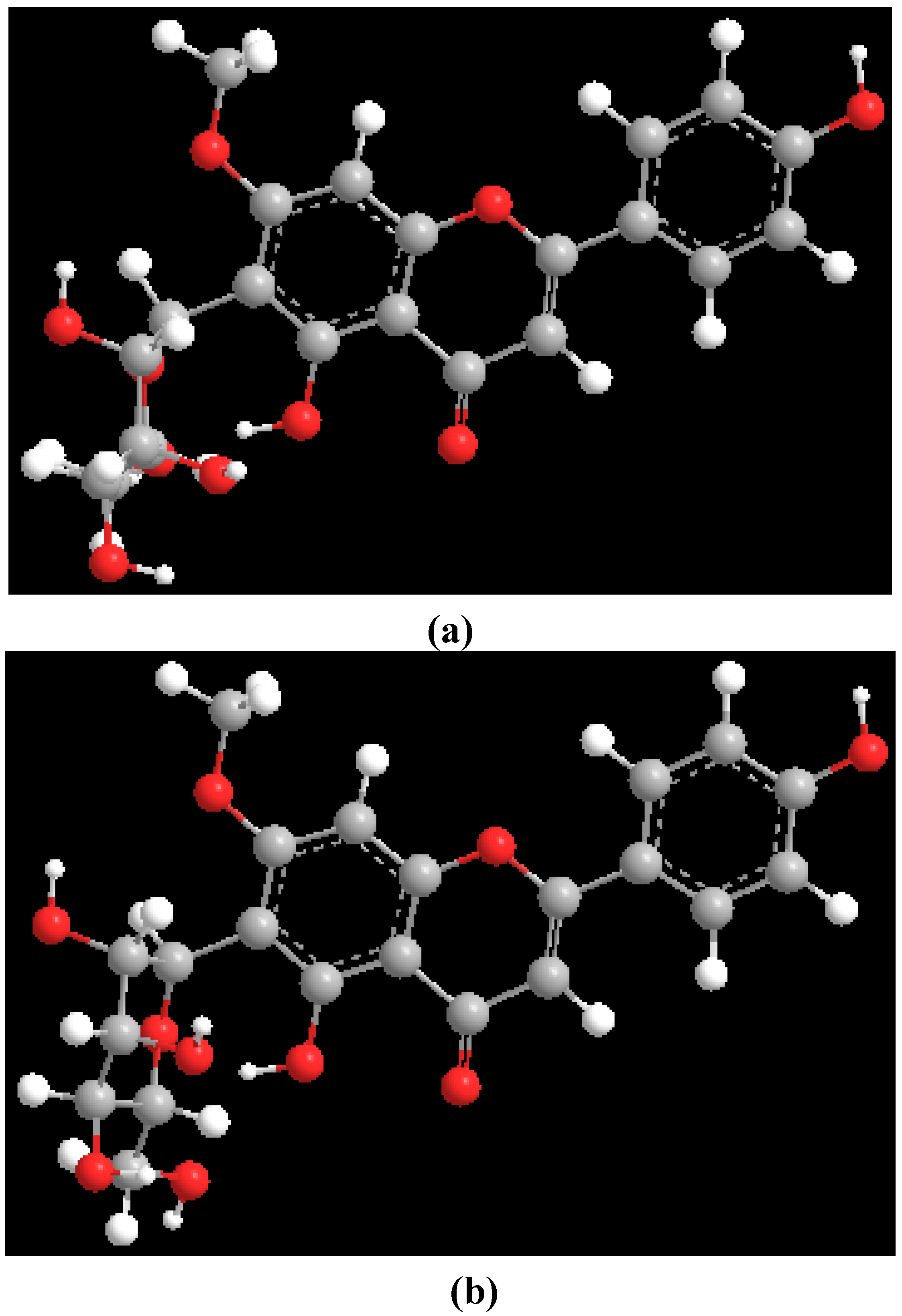
Theoretical (MM2) calculations of swertisin (
1) were performed utilizing the ChemDraw 3D Pro software, in which the dihedral angle was defined by highlighting four contiguous atoms [H-1", C-1", C-6 and C-7 (
Figure 1)]. Two minimum energy conformations (
Figure 3) were separated by a rotational energy barrier (Δ
G#rot) of 70 kJ mol
−1. The experimental Δ
G#rot (~71 kJ mol
−1) and theoretical Δ
G#rot (70 kJ mol
−1) of swertisin (
1) were essentially equal. Therefore, the theoretical (MM2) calculations in conjunction with the VT
1H-NMR studies and T-ROESY data of swertisin (
1) supported the proposal that the two rotamers of swertisin (
1) interchange by rotation about the
C-glycosidic (C-1″—C-6) bond.
Figure 3.
Molecular models of the rotamers of swertisin.
Figure 3.
Molecular models of the rotamers of swertisin.
The structure of embinoidin (
2) is shown in
Figure 4, with carbons and the 5-OH proton giving rise to doubled signals marked with asterisks. At 305 K, the
1H-NMR spectrum of embinoidin (
2) in DMSO-
d6 indicated a doubling of the signals which was attributed to the presence of two rotamers, with a relative proportion of 1.00:0.97, in solution. The duplicated
1H-NMR signals suggested that, at 305 K, there were two rotamers separated by an energy barrier about the
C-glycosidic bond which hindered rotation between the rotamers. VT
1H-NMR studies were performed on embinoidin (
2) in order to confirm this hypothesis.
Figure 4.
Diagram of embinoidin showing atoms with duplicated signals indicated by asterisks; the arrow indicates rotation of the C-glycosidic bond.
Figure 4.
Diagram of embinoidin showing atoms with duplicated signals indicated by asterisks; the arrow indicates rotation of the C-glycosidic bond.
At 305 K and using the 5-OH signals as markers, the two rotamers of embinoidin (
2) in DMSO-
d6 were in slow exchange so that each rotamer was detected independently. At 348 K, the signals for the two rotamers were still individually detected, although, the two 5-OH signals had broadened and moved closer together. Eventually, the two signals coalesced to a single peak at ca. 13.4 ppm at a Coalescence Temperature,
Tc, of 356 K (
Figure 5). The coalescence of signals was observed for the entire
1H-NMR spectrum, although the other pairs of signals coalesced at lower temperatures, reflecting their smaller chemical shift differences. The free energy of activation for the interconversion between the two rotamers of embinoidin (
2) was also calculated using Eyring’s equations (
a and
b) as modified by Shanan-Atidi and Bar-Eli [
15].
Figure 5.
Effect of Temperature on the 5-OH signal markers in the 1H-NMR spectrum of embinoidin (2).
Figure 5.
Effect of Temperature on the 5-OH signal markers in the 1H-NMR spectrum of embinoidin (2).
From the 1H-NMR spectrum at 305 K, the frequency difference, Δν, between the 5-OH signals was 46.6 Hz (46.6 s−1). The Coalescence Temperature, Tc, was 356 K.
Since the relative proportion of the two rotamers was approximately 1:1 at 305 K, the free energy of activation for rotation, Δ
G‡
rot, was also calculated using Eyring equation for equally populated rotamers [
16]. H. S. Gutowsky showed that the rate of rotation,
kc, at the temperature of coalescence,
Tc, 356 K, can be calculated using the following equation [
16]:
The free energy of activation for rotation, Δ
G‡
rot, at 356 K was calculated using the Eyring equation [
16]:
where k is the rate constant, kB is Boltzmann’s constant, h is Planck’s constant, K is the transmission coefficient, T is the temperature in K, R is the universal gas constant and ΔG‡ is the free energy of activation.
Assuming the transmission coefficient,
K, to be unity, converting natural log (ln) to log
10, and substituting
kc and
Tc into the Eyring equation, this equation becomes [
16]:
The VT 1H-NMR studies confirmed the hypothesis that the doubling of signals in the 1H- and 13C-NMR spectra at 305 K was due to the presence of two rotamers of embinoidin (2) separated by a relatively high energy barrier.
The data derived from the T-ROESY spectrum again suggested that the two rotamers slowly rotated about the C-glycosidic bond. Weak cross-peaks were observed between the respective methoxyl protons (7-OMe), and both signals of H-1″a and H-2″b. This observation indicated that β-D-glucose, which is directly attached to the flavone nucleus, rotates about the C-glycosidic bond. Therefore, in one rotamer, the H-1″ proton is oriented syn to the 7-OMe group whereas, in the other rotamer, the H-2″ proton is oriented syn to the 7-OMe group. Stronger cross-peaks were observed between the H-2″a and H-1‴a signals, and between the H-2″b and H-1‴b proton signals. Thus, in each rotamer, the rotation about the C-2″―O―C-1‴ bond brings the H-1‴ proton in close proximity to the H-2″ proton.
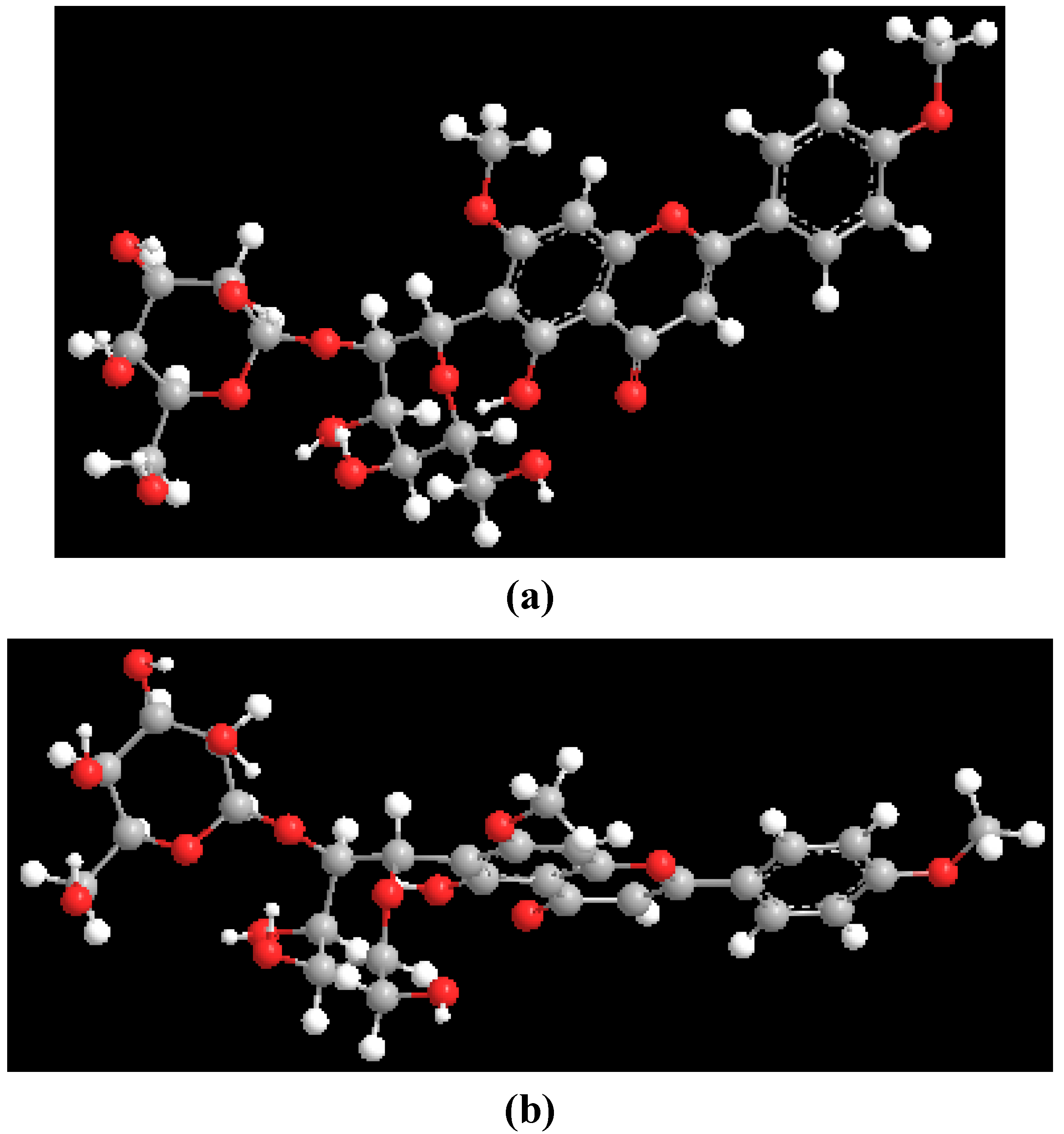
Theoretical (MM2) calculations of embinoidin (
2) were conducted utilizing the ChemDraw 3D Pro software, where the four contiguous atoms of H-1", C-1", C-6 and C-7 defined the dihedral angle. Two minimum energy conformations (
Figure 6) were separated by Δ
G#rot of 69 kJ mol
−1. The experimental Δ
G#rot (~74 kJ mol
−1) and theoretical Δ
G#rot (69 kJ mol
−1) of embinoidin (
2) were close in value. Therefore, the theoretical (MM2) calculations in conjunction with the VT
1H-NMR studies and T-ROESY data of embinoidin (
2) supported the proposal that the two rotamers of embinoidin (
2) interchange by rotation about the
C-glycosidic bond.
Figure 6.
Molecular models of the rotamers of embinoidin.
Figure 6.
Molecular models of the rotamers of embinoidin.


