Synthesis and Protective Effect of Scutellarein on Focal Cerebral Ischemia/Reperfusion in Rats

Abstract
:1. Introduction
2. Results and Discussion
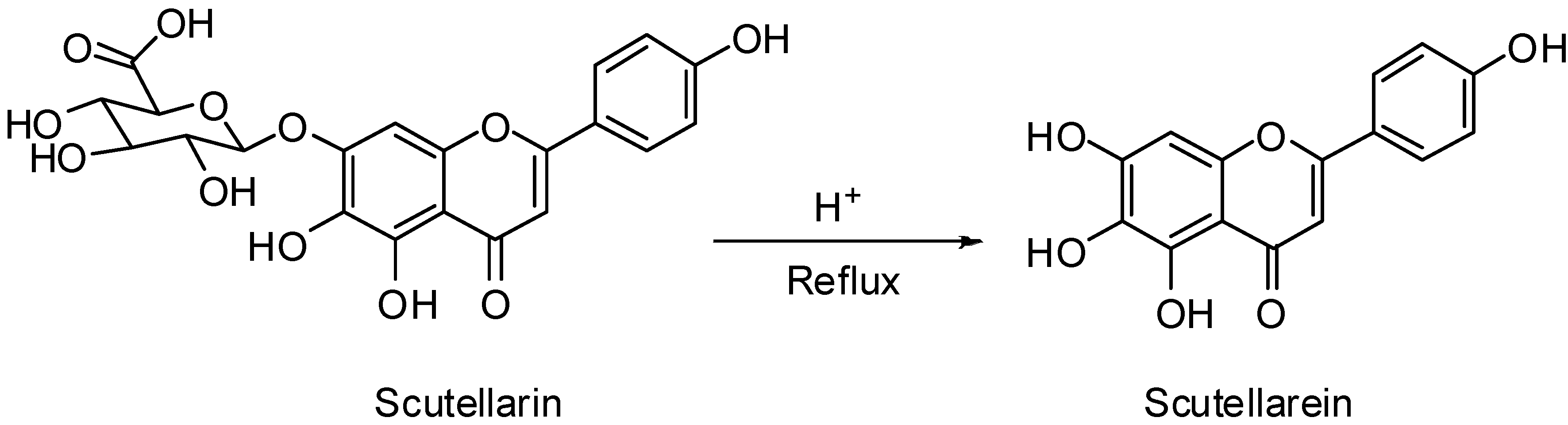
2.1. Optimization of Reaction Conditions for the Synthesis of Scutellarein

| Run | Reaction conditions | Yield (%) |
|---|---|---|
| 1 | 1.0 mol/L H2SO4 in water, 90 °C, 6~24 h | No product |
| 2 | 2.0 mol/L H2SO4 in water, 90 °C, 6~24 h | No product |
| 3 | 3.0 mol/L H2SO4 in water, 90 °C, 6~24 h | No product |
| 4 | 0.5 mol/L H2SO4 in 70% ethanol, 90 °C, 6~24 h | No product |
| 5 | 0.5 mol/L H2SO4 in 80% ethanol, 90 °C, 6~24 h | No product |
| 6 | 0.5 mol/L H2SO4 in 90% ethanol, 90 °C, 24 h | 2.1 |
| 7 | 1.0 mol/L H2SO4 in 90% ethanol, 90 °C, 24 h | 5.3 |
| 8 | 2.0 mol/L H2SO4 in 90% ethanol, 90 °C, 24 h | 8.5 |
| 9 | 3.0 mol/L H2SO4 in 90% ethanol, 90 °C, 24 h | 10.0 |
| 10 | 3.0 mol/L H2SO4 in 90% ethanol, 90 °C, 48 h | 12.1 |
| 11 | 3.0 mol/L H2SO4 in 90% ethanol, 100 °C, 48 h | 15.2 |
| 12 | 3.0 mol/L H2SO4 in 90% ethanol, 120 °C, 48 h | 17.3 |
2.2. Neurological Deficit Score
 ± s, n = 10).
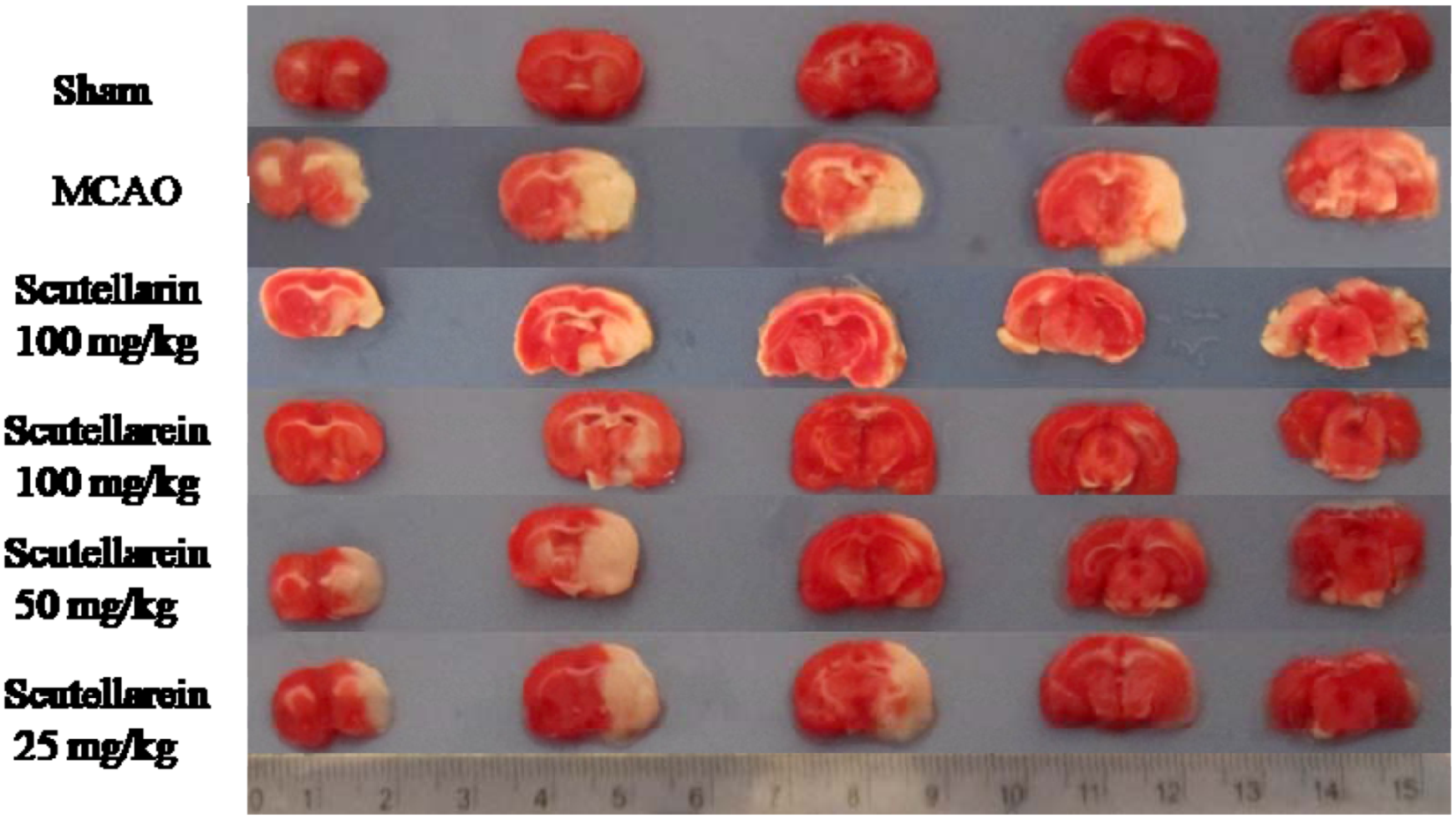
| Group | Dose (mg/kg) | Behavioral Scores | Cerebral Infarction Volume (%) |
|---|---|---|---|
| Sham | − | 0.00 ± 0.00 | 0.00 ± 0.00 |
| MCAO | − | 3.00 ± 0.53 ## | 22.24 ± 3.91 ## |
| Scutellarin | 100 | 2.16 ± 0.41 ** | 15.81 ± 1.48 ** |
| Scutellarein | 100 | 1.33 ± 0.69 **◆ | 9.66 ± 4.29 **◆ |
| 50 | 2.25 ± 0.46 ** | 15.91 ± 6.04 * | |
| 25 | 2.50 ± 0.55 | 19.04 ± 7.21 |
2.3. Cerebral Infarction Volume

3. Experimental
3.1. Materials and Subjects
3.2. General Procedure for the Synthesis of Scutellarein

3.3. Animal Model of MCAO
3.4. Neurological Deficit Score
3.5. Measurement of Cerebral Infarction Volume
4. Conclusions
Acknowledgments
- Sample Availability: Samples of scutellarein and scutellarin are available from the authors.
References
- Donnan, G.A.; Fisher, M.; Macleod, M.; Davis, S.M. Stroke. Lancet 2008, 371, 1612–1623. [Google Scholar]
- Cuzzocrea, S.; Riley, D.P.; Caputi, A.P.; Salvemini, D. Antioxidant therapy: A new pharmacological approach in shock, inflammation, and ischemia/reperfusion injur. Pharmacol. Rev. 2001, 53, 135–159. [Google Scholar]
- Zhang, H.Y.; Ping, Q.N.; Guo, J.X.; Cao, F. Pharmacokinetics of breviscapine and its β-cyclodextrin complex in rats. Acta Pharmcol. Sin. 2005, 40, 563–567. [Google Scholar]
- Cao, F.; Guo, J.X.; Ping, Q.N.; Shao, Y.; Liang, J. Ester prodrug of scutellarin: Synthesis, physicochemical property and degradation. Acta Pharmcol. Sin. 2006, 41, 595–602. [Google Scholar]
- Ge, Q.H.; Zhou, Z.; Zhi, X.J.; Ma, L.L.; Chen, X.H. Pharmacokinetics and absolute bioavailability of breviscapine in beagle dogs. Chin. J. Pharm. 2003, 34, 618–632. [Google Scholar]
- Zhang, J.L.; Che, Q.M.; Li, S.Z.; Zhou, T.H. Study on metabolism of scutellarin in rats by HPLC-MS and HPLC-NMR. J. Asian Nat. Prod. Res. 2003, 5, 249–256. [Google Scholar] [CrossRef]
- Che, Q.M.; Pan, L.Y.; Chen, Y.; He, H. Study on pharmacokinetics of scutellarein in rats. Chin. Pharm. J. 2007, 42, 1418–1421. [Google Scholar]
- Ju, W.Z.; Zhang, J.; Tan, H.S.; Jiang, M.; Chen, M.; Xiong, N.N. Determination of scutellarin in human plasma by LC-MS method and its clinical pharmacokinetics in Chinese healthy volunteers. Chin. J. Clin. Pharmacol. Ther. 2005, 10, 298–301. [Google Scholar]
- Qian, L.H.; Li, N.G.; Tang, Y.P.; Zhang, L.; Tang, H.; Wang, Z.J.; Liu, L.; Song, S.L.; Guo, J.M.; Ding, A.W. Synthesis and bio-Activity evaluation of scutellarein as a potent agent for the therapy of ischemic cerebrovascular disease. Int. J. Mol. Sci. 2011, 12, 8208–8216. [Google Scholar] [CrossRef]
- Song, Y.; Zhang, H.M.; Ma, J.J.; Che, Q.M.; Li, C.L. Protection of scutellarein on cerebral ischemia in rats. Chin. J. New Drug 2009, 18, 2061–2064. [Google Scholar]
- Farkas, L.; Mezey-Vándor, G.; Nórdj, M. Die sythese des scutellarins, plantaginins, scutellarein-7-β-rutinosids und die erste herstellung des isoscutellareins. Chem. Ber. 1974, 107, 3878–3882. [Google Scholar] [CrossRef]
- Cui, J.M.; Fang, G.; Duan, Y.B. Total synthesis of scutellarin-7-O-glucuronide. J. Asian Nat. Prod. Res. 2005, 7, 655–660. [Google Scholar] [CrossRef]
- Wang, X.S.; Ruan, X.Z.; Liu, M.L. Study of erigeron on brain protection after cerebral ischemia and reperfusion in rats. Chin. Tradit. Pat. Med. 2002, 24, 947–950. [Google Scholar]
- Pu, X.P.; Li, C.L. The revelation of cerebral ischemia cells promise on the development of new drugs. Chin. Pharm. J. 1999, 34, 579–581. [Google Scholar]
- Chen, X.; Cui, L.; Duan, X.; Ma, B.; Zhong, D. Pharmacokinetics and metabolism of the flavonoid scutellarin in humans after a single oral administration. Drug Metab. Dispos. 2006, 34, 1345–1352. [Google Scholar] [CrossRef]
- Longa, E.Z.; Weinstein, P.R.; Carlson, S.; Cummins, R. Reversible middle cerebral artery occlusion without craniectomy in rats. Stroke 1989, 20, 84–91. [Google Scholar] [CrossRef]
- 17. Zhang, Y.; Zhang, F.G.; Meng, C.; Tian, S.Y.; Wang, Y.X.; Zhao, W.; Chen, J; Zhang, X.S.; Liang, Y.; Zhang, S.D.; et al. Inhibition of sevoflurane postconditioning against cerebral ischemia reperfusion-induced oxidative injury in rats. Molecules 2012, 17, 341–354. [Google Scholar]
- Bederson, J.B.; Pitts, L.H.; Tsuji, M.; Nishimura, M.C.; Davis, R.L.; Bartkowski, H. Rat middle cerebral artery occlusion evaluation of the model and development of a neurologic examination. Stroke 1986, 17, 472–476. [Google Scholar] [CrossRef]
- Pei, H.T.; Su, X.; Zhao, L.; Li, H.Y.; Guo, Y.L.; Zhang, M.Z.; Xin, H. Primary study for the therapeutic dose and time window of picroside II in treating cerebral ischemic injury in rats. Int. J. Mol. Sci. 2012, 13, 2551–2562. [Google Scholar] [CrossRef]
- Li, Z.; Wang, B.S.; Kong, D.H.; Wang, L.C. The neuroprotective effect of pre-resveratrol on focal cerebral ischemia/reperfusion in rats. Chin. Pharmacol. Bull. 2010, 26, 802–806. [Google Scholar]
- Zhou, J.; Liu, J. Protective effect of astragaloside on local cerebral ischemia in rats and its mechanism involved. J. Clin. Res. 2008, 25, 814–816. [Google Scholar]
© 2012 by the authors; licensee MDPI, Basel, Switzerland. This article is an open-access article distributed under the terms and conditions of the Creative Commons Attribution license (http://creativecommons.org/licenses/by/3.0/).
Share and Cite
Qian, L.; Shen, M.; Tang, H.; Tang, Y.; Zhang, L.; Fu, Y.; Shi, Q.; Li, N.-G. Synthesis and Protective Effect of Scutellarein on Focal Cerebral Ischemia/Reperfusion in Rats. Molecules 2012, 17, 10667-10674. https://doi.org/10.3390/molecules170910667
Qian L, Shen M, Tang H, Tang Y, Zhang L, Fu Y, Shi Q, Li N-G. Synthesis and Protective Effect of Scutellarein on Focal Cerebral Ischemia/Reperfusion in Rats. Molecules. 2012; 17(9):10667-10674. https://doi.org/10.3390/molecules170910667
Chicago/Turabian StyleQian, Lihua, Minzhe Shen, Hao Tang, Yuping Tang, Li Zhang, Yifan Fu, Qianping Shi, and Nian-Guang Li. 2012. "Synthesis and Protective Effect of Scutellarein on Focal Cerebral Ischemia/Reperfusion in Rats" Molecules 17, no. 9: 10667-10674. https://doi.org/10.3390/molecules170910667
APA StyleQian, L., Shen, M., Tang, H., Tang, Y., Zhang, L., Fu, Y., Shi, Q., & Li, N. -G. (2012). Synthesis and Protective Effect of Scutellarein on Focal Cerebral Ischemia/Reperfusion in Rats. Molecules, 17(9), 10667-10674. https://doi.org/10.3390/molecules170910667