Anti-inflammatory Lignans from the Fruits of Acanthopanax sessiliflorus


Abstract
:1. Introduction
2. Results and Discussion
3. Experimental
3.1. General
3.2. Plant Material
3.3. Extraction and Isolation
3.4. Spectroscopic Data
3.5. Measurement of NO Production and Cell Viability
4. Conclusions
Supplementary Materials
Acknowledgments
References
- Song, L.; Wu, Y.; Hu, L.; Zhang, G.; Xu, G.; Xiao, P.; Ling, Y.; Ding, X.; Cao, C.; Li, Y. Zhong Hua Ben Cao; Shanghai Scientific Technologic Publisher: Shanghai, China, 1999; Volume 5, p. 765. [Google Scholar]
- Jung, B.S.; Shin, M.K. Hyang Yak Dae Sa Jeon, 3rd ed.; Young Lim Sa Publisher: Seoul, Korea, 2003; p. 432. [Google Scholar]
- Cai, X.F.; Lee, I.S.; Dat, N.T.; Shen, G.; Kang, J.S.; Kim, D.H.; Kim, Y.H. Inhibitory lignans against NFAT transcription factor from Acanthopanax koreanum. Arch. Pharm. Res. 2004, 27, 738–741. [Google Scholar] [CrossRef] [PubMed]
- Hong, S.S.; Hwang, J.S.; Lee, S.A.; Hwang, B.Y.; Ha, K.W.; Ze, K.R.; Seung, R.S.; Ro, J.S.; Lee, K.S. Isolation and quantitative analysis of acanthoside D from Acanthopanacis cortex. Saengyak Hakhoechi 2001, 32, 316–321. [Google Scholar]
- Yook, C.S.; Rho, Y.S.; Seo, S.H.; Leem, J.Y.; Han, D.R. Chemical components of Acanthopanax divaricatus and anticancer effect in leaves. Yakhak Hoechi 1996, 40, 251–261. [Google Scholar]
- Kim, H.M.; Kim, J.S.; Lee, S.H.; Lee, S.J.; Lee, G.P.; Kang, S.S.; Cho, S.H.; Cheoi, D.S. Quantitative analysis of lignans in the fruits of Acanthopanax species by HPLC. Food Sci. Biotechnol. 2006, 15, 778–780. [Google Scholar]
- Lee, S.H.; Son, D.W.; Ryu, J.Y.; Lee, Y.S.; Jung, S.H.; Kang, J.G.; Lee, S.Y.; Kim, H.S.; Shin, K.H. Anti-oxidant activities of Acanthopanax senticosus stems and their lignan components. Arch. Pharm. Res. 2004, 27, 106–110. [Google Scholar] [CrossRef] [PubMed]
- Lyu, S.Y.; Park, W.B. Modulation of IL-12 and IFN-γ secretions by eleutheroside E, tortoside A, and syringaresinol from Acanthopanax koreanum Nakai. Biomol. Ther. 2010, 18, 211–218. [Google Scholar] [CrossRef]
- Li, S.C. Bencao Gangmu (Compendium of Materia Medica); Ko Mun Sa Publisher: Seoul, Korea, 1993; Volume 36, p. 26. [Google Scholar]
- Hong, J.J.; Shin, K.H.; Lim, S.S.; Kwak, J.H.; Zee, O.P.; Ishihara, K.; Hirasawa, N.; Seyama, T.; Ohuchi, K. Lead compounds for anti-inflammatory drugs isolated from the plants of the traditional oriental medicine in Korea. Curr. Drug Targets Inflamm. Allergy 2008, 7, 195–202. [Google Scholar] [CrossRef]
- Jung, H.J.; Nam, J.H.; Choi, J.W.; Lee, K.T.; Park, H.J. Anti-inflammatory effects of chiisanoside and chiisanogenin obtained from the leaves of Acanthopanax chiisanensis in the carrageenan- and Freund’s complete adjuvant-induced rats. J. Ethnopharmacol. 2005, 97, 359–367. [Google Scholar] [CrossRef] [PubMed]
- Jung, H.J.; Park, H.J.; Kim, R.G.; Shin, K.M.; Ha, J.H.; Choi, J.W.; Kim, H.J.; Lee, Y.S.; Lee, K.T. In vivo anti-inflammatory and antinociceptive effects of liriodendrin isolated from the stem bark of Acanthopanax senticosus. Planta Med. 2003, 69, 610–616. [Google Scholar] [PubMed]
- Hanuman, J.B.; Mishra, A.K.; Sabata, B. A natural phenolic lignan from Tinospora cordifolia Miers. J. Chem. Soc. Perkin Trans. 1 1986, 7, 1181–1185. [Google Scholar] [CrossRef]
- Yamauchi, S.; Hayashi, Y.; Nakashima, Y.; Kirikihira, T.; Yamada, K.; Masuda, T. Effect of benzylic oxygen on the antioxidant activity of phenolic lignans. J. Nat. Prod. 2005, 68, 1459–1470. [Google Scholar] [CrossRef] [PubMed]
- Ma, J.; Dey, M.; Yang, H.; Poulev, A.; Pouleva, R.; Dorn, R.; Lipsky, P.E.; Kennelly, E.J.; Raskin, I. Anti-inflammatory and immunosuppressive compounds form Tripterygium wilfordii. Phytochemistry 2007, 68, 1172–1178. [Google Scholar] [CrossRef] [PubMed]
- Ryu, J.Y.; Son, D.W.; Kang, J.G.; Kim, H.S.; Kim, B.K.; Lee, S.H. A benzenoid from the stem of Acanthopanax senticosus. Arch. Pharm. Res. 2004, 27, 912–914. [Google Scholar] [CrossRef] [PubMed]
- Lee, D.Y.; Song, M.C.; Yoo, K.H.; Bang, M.H.; Chung, I.S.; Kim, S.H.; Kim, D.K.; Kwon, B.M.; Jeong, T.S.; Park, M.H. Lignans from the fruits of Cornus kousa Burg and their cytotoxic effects on human cancer cell lines. Arch. Pharm. Res. 2007, 30, 402–407. [Google Scholar] [CrossRef] [PubMed]
- Takaku, N.; Choi, D.H.; Mikame, K.; Okunishi, T.; Suzuki, S.; Ohashi, H.; Umezawa, T.; Shimada, M. Lignans of Chamaecyparis obtuse. J. Wood Sci. 2001, 47, 476–482. [Google Scholar] [CrossRef]
- Fang, J.M.; Lee, C.K.; Cheng, Y.S. Lignans from leaves of Juniperus chinensis. Phytochemistry 1992, 31, 3659–3661. [Google Scholar]
- Nishibe, S.; Kinoshita, H.; Takeda, H.; Okano, G. Phenolic compounds from stem bark of Acanthopanax senticosus and their pharmacological effect in chronic swimming stressed rats. Chem. Pharm. Bull. 1990, 38, 1763–1765. [Google Scholar] [CrossRef] [PubMed]
- Ghosal, S.; Banerjee, S.; Jaiswal, D.K. Chemical constituents of Justica. Part 2. New furofurano lignans from Justicia simplex. Phytochemistry 1980, 19, 332–334. [Google Scholar] [CrossRef]
- Calixto, J.B.; Campos, M.M.; Otuki, M.F.; Santos, A.R.S. Anti-inflammatory compounds of plant origin. Part II. Modulation of pro-inflammatory cytokines, chemokines and adhesion molecules. Planta Med. 2004, 70, 93–103. [Google Scholar] [PubMed]
- Jung, L.H.; Lee, D.Y.; Cho, J.G.; Lee, S.M.; Kang, H.C.; Seo, W.D.; Kang, H.W.; Kim, J.Y.; Baek, N.I. A new flavonolignan from the aerial parts of Oryza sativa L. inhibits nitric oxide production in RAW 264.7 macrophage cells. J. Korean Soc. Appl. Chem. 2011, 54, 865–870. [Google Scholar] [CrossRef]
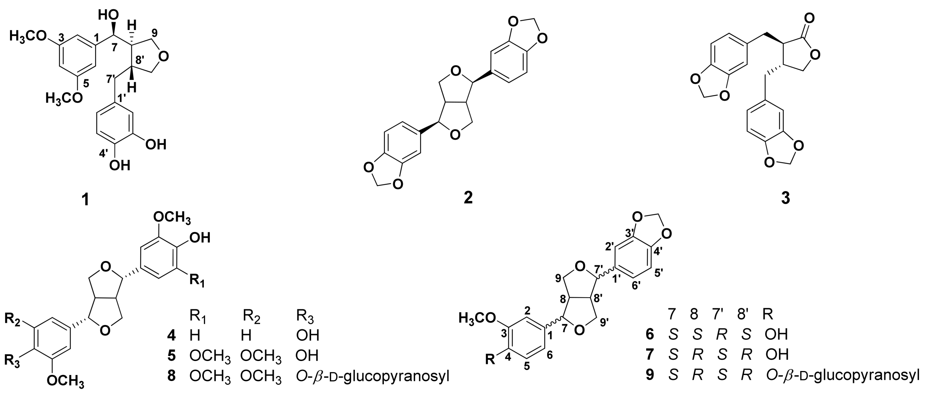
Sample Availability: Samples of the compounds 1−9 are available from the authors. |


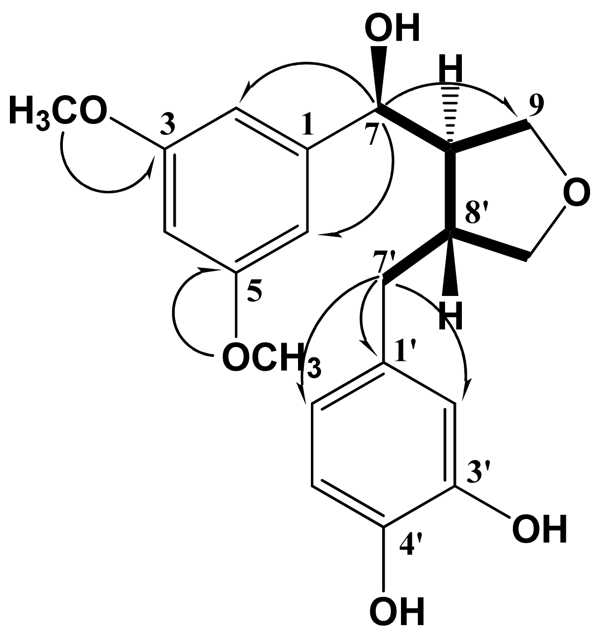
| No. | δH | δC | No. | δH | δC | |
|---|---|---|---|---|---|---|
| 1 | 135.7 | 1' | 133.5 | |||
| 2 | 6.76 (1H, overlapped) | 116.1 | 2' | 6.78 (1H, d, J = 2.0 Hz) | 113.3 | |
| 3 | 149.0 | 3' | 145.7 | |||
| 4 | 6.90 (1H, d, J = 3.2 Hz) | 110.6 | 4' | 147.0 | ||
| 5 | 149.0 | 5' | 6.70 (1H, d, J = 8.0 Hz) | 119.8 | ||
| 6 | 6.75 (1H, overlapped) | 115.9 | 6' | 6.63 (1H, dd, J = 8.0, 2.0 Hz) | 122.1 | |
| 7 | 4.74 (1H, d, J = 6.8) | 84.0 | 7' | 2.92 (1H, dd, J = 13.2, 4.8 Hz, H-7'a)2.48 (1H, dd, J = 13.2, 11.6 Hz, H-7'b) | 33.6 | |
| 8 | 2.34 (1H, m) | 54.0 | 8' | 2.72 (1H, m) | 43.8 | |
| 9 | 3.97 (1H, dd, J = 8.4, 6.8, H-9a)3.71 (1H, dd, J = 8.4, 6.8, H-9b) | 60.4 | 9' | 3.82 (1H, overlapped, H-9'a)3.62 (1H, dd, 10.8, 6.4 Hz, H-9'b) | 73.4 | |
| 3-OCH3 | 3.83 (3H, s) | 56.3 | ||||
| 5-OCH3 | 3.82 (3H, s) | 56.3 |
| Compound | IC50 (μM) a | Cell viability (%) b |
|---|---|---|
| 1 | 49.94 ± 6.56 | 84.81 ± 2.71 |
| 2 | 38.92 ± 2.86 | 95.52 ± 2.01 |
| 3 | 21.56 ± 1.19 | 50.21 ± 1.55 |
| 4 | 17.75 ± 1.15 | 80.21 ± 1.11 |
| 5 | 10.34 ± 2.37 | 81.50 ± 3.32 |
| 6 | 22.30 ± 1.10 | 84.11 ± 2.46 |
| 7 | 21.57 ± 1.28 | 88.43 ± 3.71 |
| 8 | 65.07 ± 8.02 | 82.42 ± 1.27 |
| 9 | 53.00 ± 2.75 | 54.52 ± 2.21 |
| Aminoguanidine c | 6.51 ± 1.15 | 84.61 ± 2.50 |
© 2013 by the authors. This article is an open access article distributed under the terms and conditions of the Creative Commons Attribution license (http://creativecommons.org/licenses/by/3.0/).
Share and Cite
Lee, D.-Y.; Seo, K.-H.; Jeong, R.-H.; Lee, S.-M.; Kim, G.-S.; Noh, H.-J.; Kim, S.-Y.; Kim, G.-W.; Kim, J.-Y.; Baek, N.-I. Anti-inflammatory Lignans from the Fruits of Acanthopanax sessiliflorus. Molecules 2013, 18, 41-49. https://doi.org/10.3390/molecules18010041
Lee D-Y, Seo K-H, Jeong R-H, Lee S-M, Kim G-S, Noh H-J, Kim S-Y, Kim G-W, Kim J-Y, Baek N-I. Anti-inflammatory Lignans from the Fruits of Acanthopanax sessiliflorus. Molecules. 2013; 18(1):41-49. https://doi.org/10.3390/molecules18010041
Chicago/Turabian StyleLee, Dae-Young, Kyeong-Hwa Seo, Rak-Hun Jeong, Sang-Min Lee, Geum-Soog Kim, Hyung-Jun Noh, Seung-Yu Kim, Gye-Won Kim, Ji-Young Kim, and Nam-In Baek. 2013. "Anti-inflammatory Lignans from the Fruits of Acanthopanax sessiliflorus" Molecules 18, no. 1: 41-49. https://doi.org/10.3390/molecules18010041