Four New Citrinin Derivatives from a Marine-Derived Penicillium sp. Fungal Strain

Abstract
:1. Introduction
2. Results and Discussion
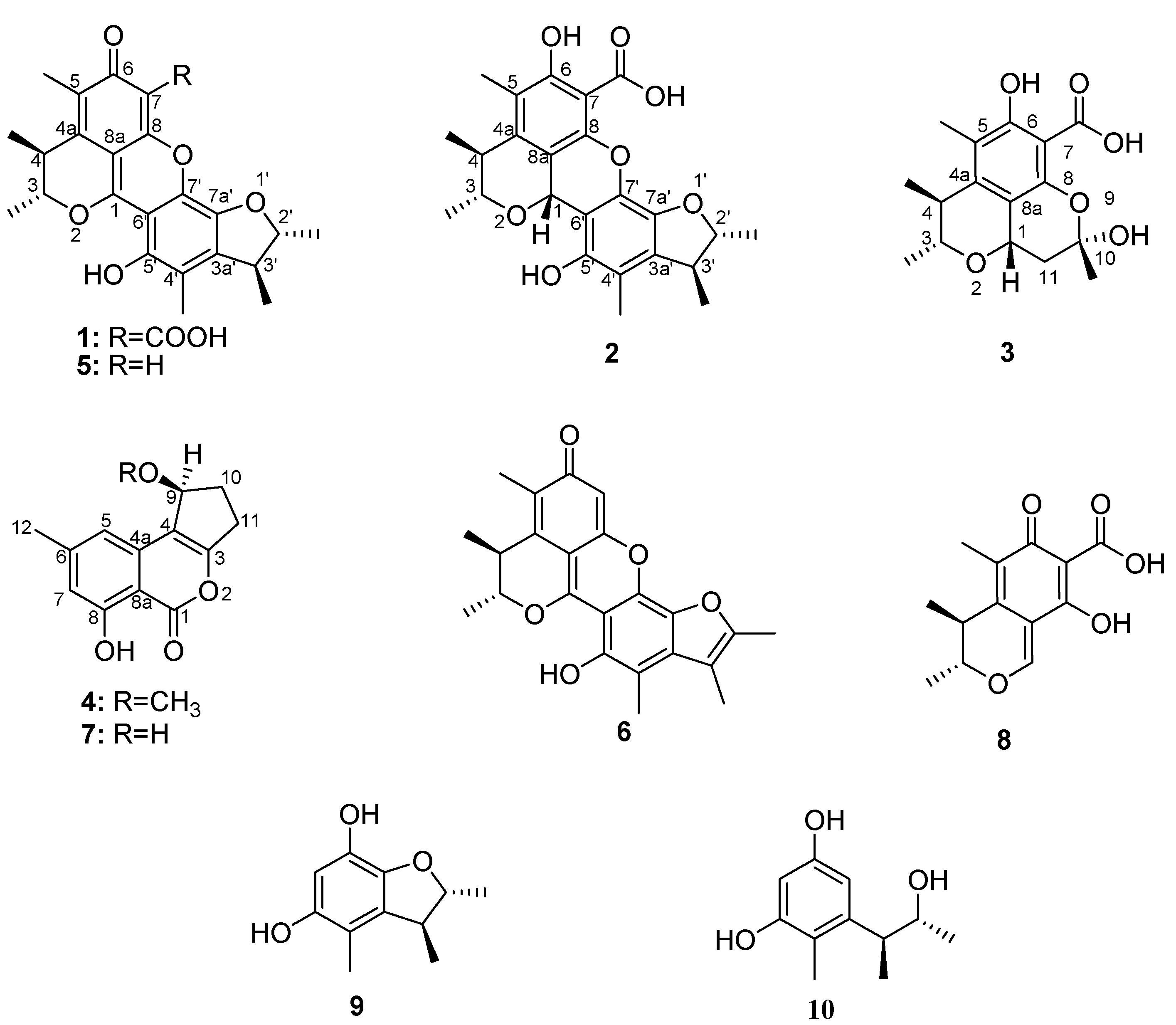
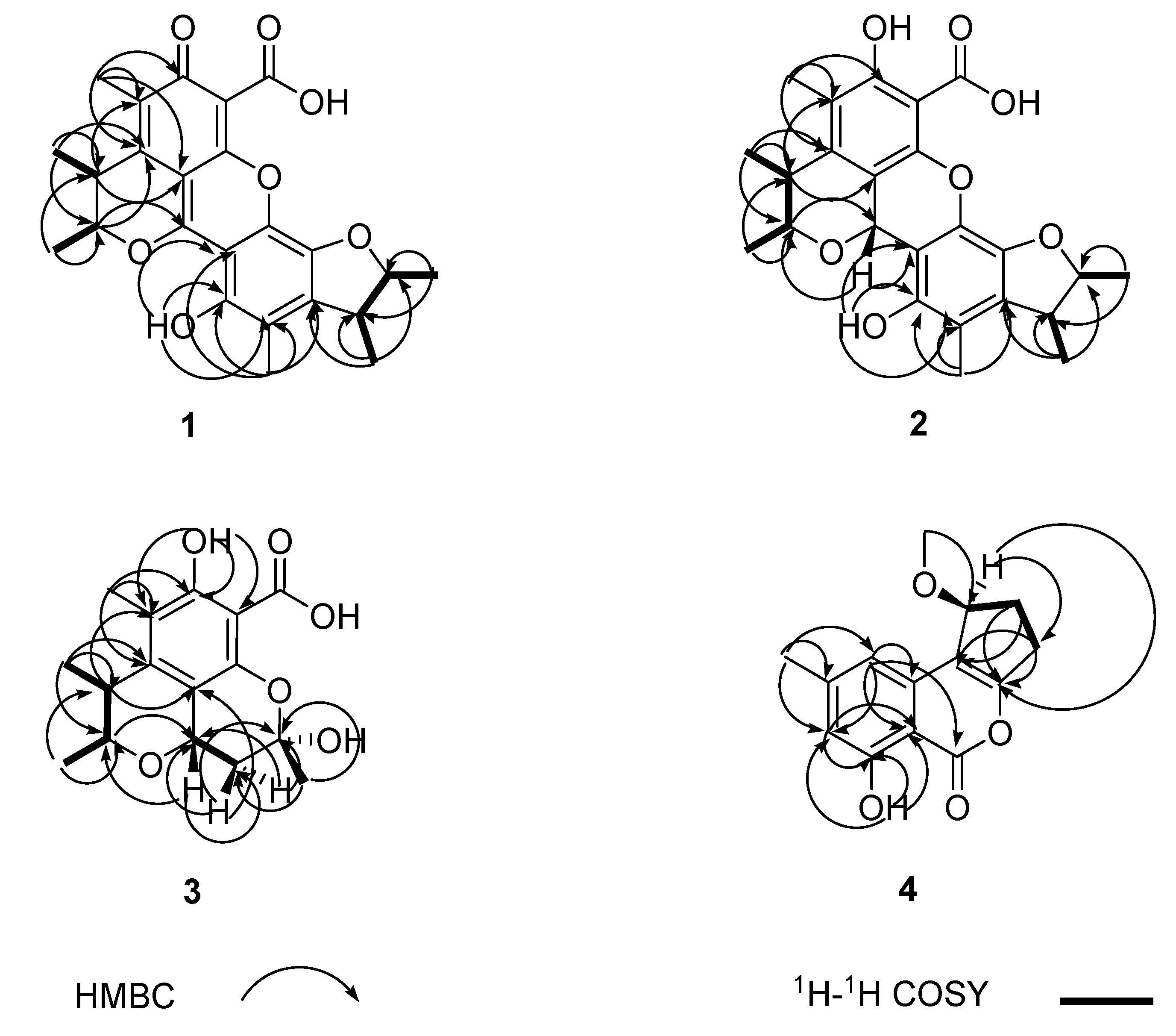
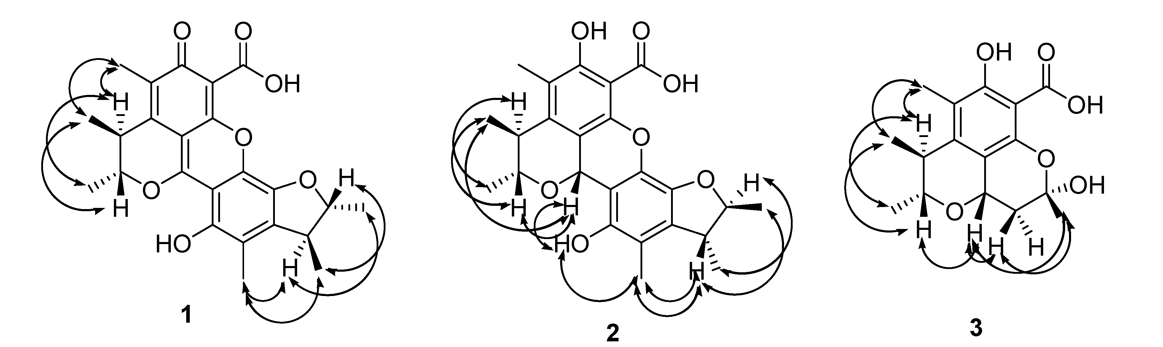
2.1. Structural Elucidation of Compounds

| No. | 1 | 2 | ||
|---|---|---|---|---|
| δH (mult., J [Hz], int.) | δc | δH (mult., J [Hz], int.) | δc | |
| 1 | - | 161.1s | 5.71 (s) | 66.3d |
| 3 | 5.17 (q, 6.7) | 83.2d | 4.10 (dq, 6.1, 6.8) | 79.0d |
| 3-CH3 | 1.49 (d, 6.7, 3H) | 19.04q | 1.47 (d, 6.8, 3H) | 21.9q |
| 4 | 3.29 (q, 7.2) | 34.8d | 3.03 (dq, 6.1, 7.0) | 37.7d |
| 4-CH3 | 1.38 (d, 7.2, 3H) | 18.97q | 1.34 (d, 7.0, 3H) | 19.6q |
| 4a | - | 132.7s | - | 144.7s |
| 5 | - | 130.8s | - | 120.6s |
| 5-CH3 | 2.22 (s, 3H) | 10.9q | 2.21 (s, 3H) | 11.2q |
| 6 | - | 183.8s | - | 161.9s |
| 6-OH | - | - | 12.52 (s) | - |
| 7 | - | 103.1s | - | 98.0s |
| 7-COOH | - | 165.4s | - | 170.6s |
| 8 | - | 160.9s | - | 145.3s |
| 8a | - | 99.5s | - | 108.9s |
| 2′ | 4.77 (dq, 4.3, 6.4) | 88.4d | 4.56 (m) | 88.3d |
| 2′-CH3 | 1.47 (d, 6.4, 3H) | 21.0q | 1.36 (d, 6.5, 3H) | 20.9q |
| 3′ | 3.25 (dq, 4.3, 7.1) | 45.0d | 3.09 (m) | 44.3d |
| 3′-CH3 | 1.36 (d, 7.1, 3H) | 18.82q | 1.35 (d, 7.2, 3H) | 19.3q |
| 3a′ | - | 142.4s | - | 133.0s |
| 4′ | - | 118.2s | - | 117.8s |
| 4′-CH3 | 2.28 (s, 3H) | 11.6q | 2.18 (s, 3H) | 11.6q |
| 5′ | - | 147.2s | - | 147.5s |
| 5′-OH | 8.25 (s) | - | 7.92 (s) | - |
| 6′ | - | 102.2s | - | 105.3s |
| 7′ | - | 136.2s | - | 130.7s |
| 7a′ | - | 139.2s | - | 138.1s |


| No. | δH (mult., J [Hz], int.) | δc |
|---|---|---|
| 1 | 4.70 (dd, 6.1, 11.5) | 66.2d |
| 3 | 3.74 (dq, 6.3, 6.2) | 78.8d |
| 3-CH3 | 1.39 (d, 6.2, 3H) | 21.6q |
| 4 | 2.88 (dq, 6.3, 6.9) | 38.1d |
| 4-CH3 | 1.23 (d, 6.9, 3H) | 19.3q |
| 4a | - | 145.8s |
| 5 | - | 118.2s |
| 5-CH3 | 2.14 (s, 3H) | 11.1q |
| 6 | - | 161.3s |
| 6OH | 12.16 (s) | - |
| 7 | - | 97.6s |
| 7-COOH | - | 171.6s |
| 8 | - | 146.9s |
| 8a | - | 111.7s |
| 10 | - | 101.4s |
| 10-CH3 | 1.87 (s, 3H) | 29.3q |
| 11a | 2.53 (dd, 6.1, 12.8) | 37.3t |
| 11b | 1.84 (dd, 11.5, 12.8) |
| No. | δH (mult., J [Hz], int.) | δc |
|---|---|---|
| 1 | - | 181.1s |
| 3 | - | 173.9s |
| 4 | - | 119.9s |
| 4a | - | 157.3s |
| 5 | 6.71 (s) | 107.6d |
| 6 | - | 146.7s |
| 7 | 6.63 (s) | 112.6d |
| 8 | - | 161.0s |
| 8-OH | 12.56 (s) | - |
| 8a | - | 109.0s |
| 9 | 4.95 (d, 6.8) | 79.4d |
| 10a | 2.35 (m) | 27.6t |
| 10b | 2.17 (m) | |
| 11a | 3.22 (m) | 30.2t |
| 11b | 2.81 (m) | |
| 12 | 2.40 (s, 3H) | 22.3q |
| 9-OCH3 | 3.50 (s, 3H) | 57.3q |
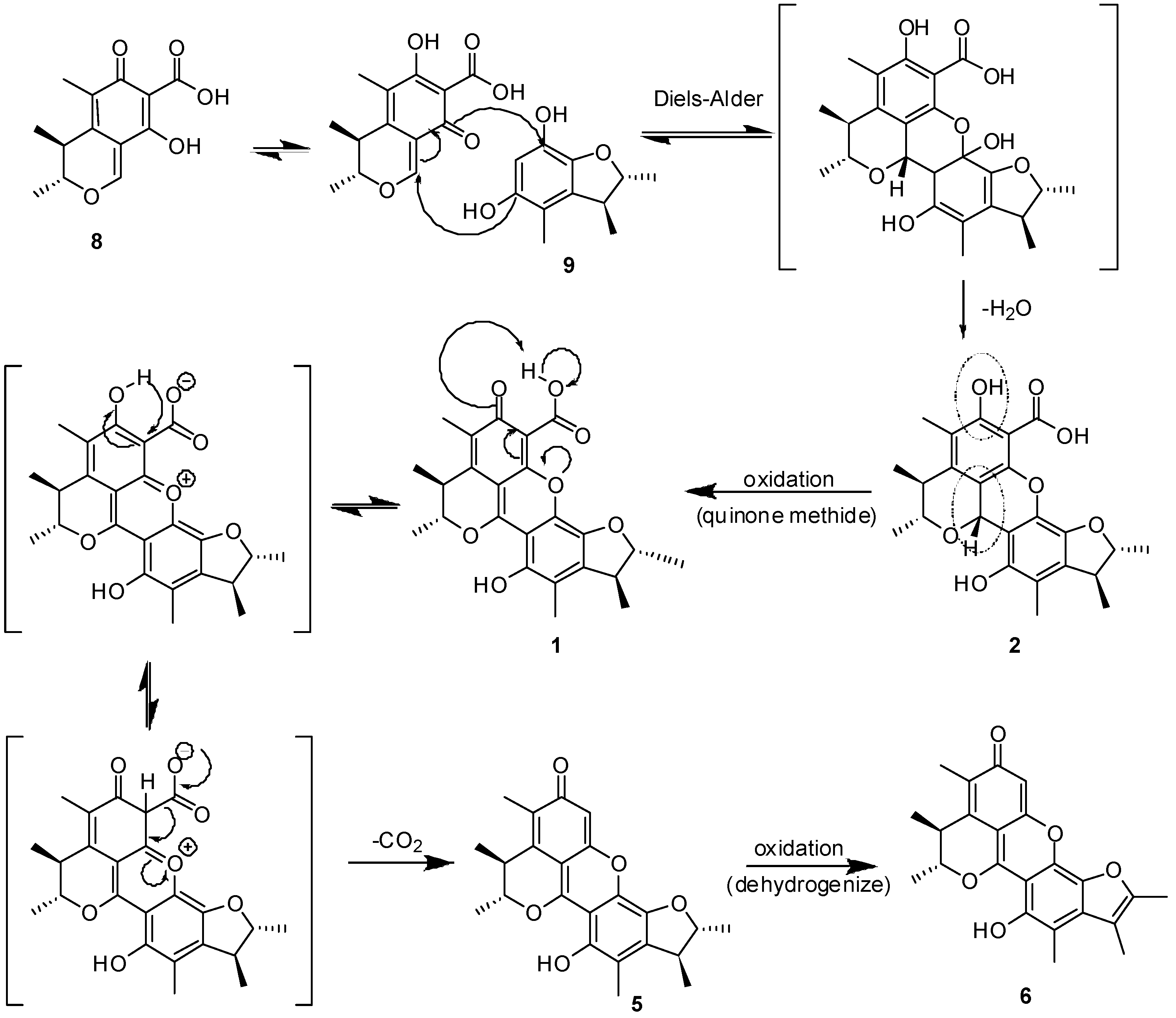
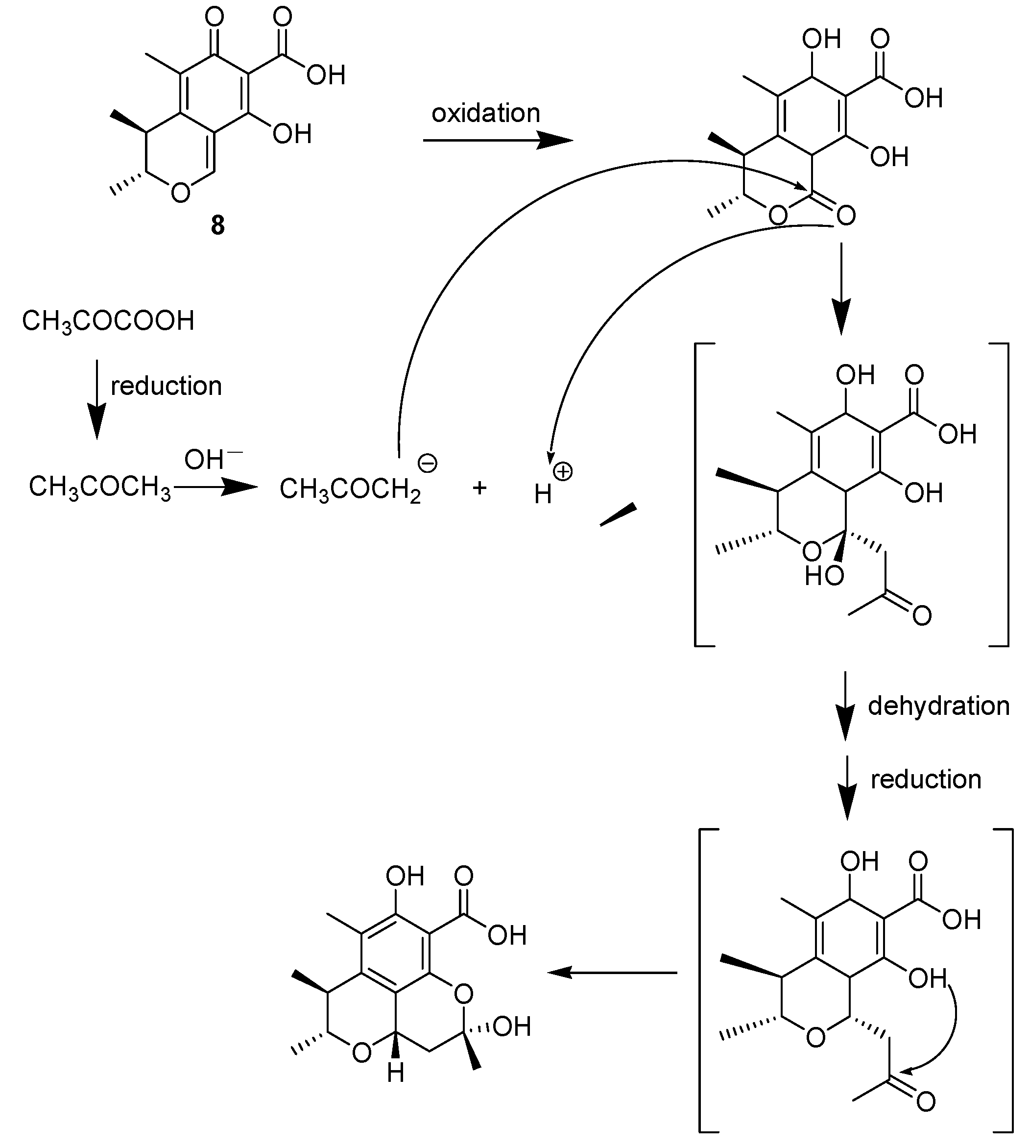
2.2. Biosynthesis


2.3. Cytotoxic and Antimicrobial Activity
| Compound | Inhibitory ratio (%) | |
| HeLa | HepG-2 | |
| 1 | - | 6.3 |
| 2 | - | 25.1 |
| 3 | - | 9.2 |
| 4 | 4.0 | 16.1 |
3. Experimental
3.1. General Procedures
3.2. Fungal Material
3.3. Fermentation and Extraction
3.4. Purification
3.5. Biological Assays
3.6. Spectral Data
4. Conclusions
Supplementary Materials
Acknowledgments
Conflicts of Interest
References
- Tamburini, E.; Mastromei, G. Do bacterial cryptic genes really exist? Res. Microbiol. 2000, 151, 179–182. [Google Scholar]
- Liberra, K.; Lindequist, U. Marine fungi—A prolific resource of biologically active natural products? Pharmazie 1995, 50, 583–588. [Google Scholar]
- Bugni, T.S.; Ireland, C.M. Marine-derived fungi: a chemically and biologically diverse group of microorganisms. Nat. Prod. Rep. 2004, 21, 143–163. [Google Scholar]
- Wakana, D.; Hosoe, T.; Itabashi, T.; Okada, K.; Takaki, G.M.D.; Yaguchi, T.; Fukushima, K.; Kawai, K. New citrinin derivatives isolated from Penicillium citrinum. J. Nat. Med. 2006, 60, 279–284. [Google Scholar] [CrossRef]
- Kuramata, M.; Fujioka, S.; Shimada, A.; Kawano, T.; Kimura, Y. Citrinolactones A, B and C, and Sclerotinin C, plant growth regulators from Penicillium citrinum. Biosci. Biotechnol. Biochem. 2007, 71, 499–503. [Google Scholar] [CrossRef]
- Hetherington, A.C.; Raistrick, H. Studies in the biochemistry of microorganisms. XI. On the production and chemical constitution of a new yellow coloring matter, citrinin, produced from glucose by Penicillium citrinum THOM. Philos. Trans. R. Soc. London Ser. B 1931, 220, 226–297. [Google Scholar]
- Chen, C.H.; Shaw, C.Y.; Chen, C.C.; Tsai, Y.C. 2,3,4-Trimethyl-5,7-dihydroxy-2,3-dihydrobenzofuran, a novel antioxidant, from Penicillium citrinum F5. J. Nat. Prod. 2002, 65, 740–741. [Google Scholar]
- Rodel, T.; Gerlach, H. Enantioselective Synthesis of the Polyketide Antibiotic (3R,4S)-(-)-Citrinin. Liebigs Ann. 1995, 1995, 885–888. [Google Scholar] [CrossRef]
- Lu, Z.Y.; Lin, Z.J.; Wang, W.L.; Du, L.; Zhu, T.J.; Fang, Y.C.; Gu, Q.Q.; Zhu, W.M. Citrinin dimers from the halotolerant fungus Penicillium citrinum B-57. J. Nat. Prod. 2008, 71, 543–546. [Google Scholar]
- Clark, B.R.; Capon, R.J.; Lacey, E.; Tennant, S.; Gill, J.H. Citrinin revisited: from monomers to dimers and beyond. Org. Biomol. Chem. 2006, 4, 1520–1528. [Google Scholar] [CrossRef]
- Chien, M.M.; Schiff, P.L., Jr.; Slatkin, D.J.; Knapp, J.E. Metabolites of aspergilli. III. The isolation of citrinin, dihydrocitrinone and sclerin from aspergilus carneus. Lloydia 1977, 40, 301–302. [Google Scholar]
- Chen, L.; Liu, W.; Hu, X.; Huang, K.; Wu, J.L.; Zhang, Q.Q. Citrinin derivatives from the marine-derived fungus Penicillium citrinum. Chem. Pharm. Bull. (Tokyo) 2011, 59, 515–517. [Google Scholar] [CrossRef]
- Xin, Z.H.; Wang, W.L.; Zhang, Y.P.; Xie, H.; Gu, Q.Q.; Zhu, W.M. Pennicitrinone D, a new citrinin dimer from the halotolerant fungus Penicillium notatum B-52. J. Antibiot. (Tokyo) 2009, 62, 225–227. [Google Scholar] [CrossRef]
- Chen, L.; Liu, W.; Huang, K.; Hu, X.; Fang, Z.X.; Wu, J.L.; Zhang, Q.Q. Penicitrinols F-I, New Citrinin Derivatives from the Marine-Derived Fungus Penicillium citrinum. Heterocycles 2011, 83, 1853–1858. [Google Scholar] [CrossRef]
- E, Z. Tissue Culture And Molecular Biology Technology; Beijing Publishing House: Beijing, China, 1995; pp. 1–141. [Google Scholar]
- Sample Availability: Samples of the compounds 1–10 are available from the authors.
© 2013 by the authors; licensee MDPI, Basel, Switzerland. This article is an open access article distributed under the terms and conditions of the Creative Commons Attribution license (http://creativecommons.org/licenses/by/3.0/).
Share and Cite
Wang, M.L.; Lu, C.H.; Xu, Q.Y.; Song, S.Y.; Hu, Z.Y.; Zheng, Z.H. Four New Citrinin Derivatives from a Marine-Derived Penicillium sp. Fungal Strain. Molecules 2013, 18, 5723-5735. https://doi.org/10.3390/molecules18055723
Wang ML, Lu CH, Xu QY, Song SY, Hu ZY, Zheng ZH. Four New Citrinin Derivatives from a Marine-Derived Penicillium sp. Fungal Strain. Molecules. 2013; 18(5):5723-5735. https://doi.org/10.3390/molecules18055723
Chicago/Turabian StyleWang, Mei Ling, Chun Hua Lu, Qing Yan Xu, Si Yang Song, Zhi Yu Hu, and Zhong Hui Zheng. 2013. "Four New Citrinin Derivatives from a Marine-Derived Penicillium sp. Fungal Strain" Molecules 18, no. 5: 5723-5735. https://doi.org/10.3390/molecules18055723