1. Introduction
Agaves have great economic and cultural importance for several native and cross-breed communities in Mexico, and for centuries people have used these plants as sources of food, fuel, shelter, and fiber, as fertilizer and ornamentals, but also in traditional medicine. These plant species belong to the family
Agavaceae, of which there are some 300 species in the World, over 250 of which are found in Mexico [
1].
The use of agave as food and fermented beverages source has persisted in Mexico for over 7,000 years. Tequila and mezcal, both distilled from agave, have become true Mexican symbols due to the fact that both have been certified with a “Denominación de origen” (protected designation of origin) according to the Norma Oficial Mexicana (NOM, Official Mexican Regulations) regulations NOM-006-SCFI-2005 (tequila) and NOM-070-SCFI-1994 (mezcal).
While these species are used as sources of fermented beverages, their use in traditional medicine has also been widely reported by Mexican herbalists [
2].
A. americana L. is one of the 500 more widely used medicinal plants in several countries [
3]. In Mexico it is used as diuretic and laxative, and also in wound, syphilis, scurvy and cancer treatments, but it’s also used to treat the lack of movement in extremities and postpartum belly inflammation [
4,
5,
6]. Sap from
A. angustifolia Haw is used as a treatment for digestive troubles and as a remedy for sprains and broken bones not only in people, but also in animals [
7]. The main use for
Agave tequilana (blue agave or blue weber) is the production of tequila.
The leaves of these species represent an important source of secondary metabolites like fructans, and flavonoids, but mainly terpenoids and steroidal saponins [
5,
8]. There are different saponins which have been isolated and identified in several species of the agave genus, such as smilagenin (steroidal saponin which precedes another eight saponins) and gitogenin [
9] isolated from
A. lechugilla. Manogenin and kamogenin, from
A. amanuensis callus culture [
10]. Hecogenin, tigogenin, agavasaponin E and H from
Agave americana [
11,
12,
13,
14,
15]. Steroidal saponins from
A. attenuata [
16,
17] and
A. shrevei [
18]. Data in the literature indicate that agave genus species have an anti-inflammatory effect, for example, the aqueous extract of
A. intermixta reduces carrageenan-induced plantar edema [
19]. Steroidal saponins isolated from
A. attenuata Salm-Dyck and
A. shevrei Gentry, exhibit anti-inflammatory activity in a membrane permeability induced by acetic acid model [
16,
18]. Hecogenin, and tiogenin isolated from
A. americana L., induce greater anti-inflammatory activity than the aqueous extract they come from, and even the anti-inflammatory steroidal drug, dexamethasone, in the essay of carrageenan-induced sub-plantar edema [
11]. Furthermore, Mana
et al, found that this species’ leaves have compounds with antitumor activity [
20].
A. americana extract orally administered to sheep, with different doses of saponins (120, 240 and 360 mg/kg) has antiprotozoal activity, as well as the capacity to lower the serum concentration of cholesterol and glucose, which helps the growth of these animals’ offspring [
21].
The goal of this investigation was to evaluate the anti-inflammatory effect of three species of the agave genus‒ A. americana, A. tequilana and A. angustifolia—due to the fact that there aren’t many pharmacological studies of these plants, which are widely used by several communities in Mexico. This was done using the auricular edema induced by 12-O-tetradecanoylphorbol 13-acetate (4β,9α,12β,13α,20-pentahydroxytiglia-1,6-dien-3-one 12-tetradecanoate 13-acetate, TPA) model, in addition to a chemical analysis of the species with better biological activity, which led to the structural elucidation of an anti-inflammatory compound.
2. Results and Discussion
Topical administration of TPA caused an approximately 12 mg edema in this test (
Figure 1, negative control group, Ctrl). The concentration employed in the present study was selected after performing some screening assays of TPA-induced auricular edema to analyze the anti-inflammatory activity of
A. americana y
A. tequilana and
A. angustifolia acetone extracts. We decided to evaluate them at 1.0, 2.0, 4.0, 6.0, y 10.0 mg/ear, and significant biological activity was seen starting from 2.0 mg/ear. There were no statistical differences between groups (data not shown), but the 6.0 mg/ear effect was higher, so we chose that concentration to perform our assays. It was observed that the groups that received
A. tequilana (At),
A. americana (Aam) and
A. angustifolia (Aan) showed a significant decrease (
p< 0.05) of edema caused by TPA, having inflammation levels of only 3.7 mg, 2.2 mg and 5.7 mg, corresponding to inhibition percentages of 68%, 81% and 51%, respectively. Mice that received indomethacin locally (indo to 1.0 mg/ear, positive control) showed an average level of 4.3 mg (67.2%) of edema inhibition, which was significantly different from control group (
p < 0.05,
Figure 1. The aqueous extract of
Agave intermixta was previously tested using the TPA model and showed an edema inhibition percentage of 54.27 and 56.55% with doses of 3 mg/ear and 5 mg/ear, respectively [
19].
Figure 1.
Effect of the topical administration of the acetone extract of A. tequilana (AT), A. americana (Aam), A. angusitfolia (Aan) at 6 mg/ear, on TPA induced ear edema. Indo = Indomethacin. ANOVA, post-hoc Bonferroni * p < 0.05 (n = 7, mean + SD, when it is compared with the negative control).
Figure 1.
Effect of the topical administration of the acetone extract of A. tequilana (AT), A. americana (Aam), A. angusitfolia (Aan) at 6 mg/ear, on TPA induced ear edema. Indo = Indomethacin. ANOVA, post-hoc Bonferroni * p < 0.05 (n = 7, mean + SD, when it is compared with the negative control).
So far, in the literature there was only one report in 1997 of any such activity from
A. americana. In that paper the authors showed that an extract of this species (at doses of 200 and 300 mg/kg, ip) and the corresponding mixture of genins (hecogenin and tiogenin) induced a decrease in carrageenan-induced plantar edema in Wistar rats [
11].
There are no reports of such activity in the species
A. tequilana and
A. angustifolia. As seen in
Figure 1, the
A. americana acetone extract showed higher activity than the other two species at the same dose, and even higher than the reference drug indomethacin. This nonsteroidal anti-inflammatory drug is widely used to treat edema due to its ability to block the inflammatory cascade associated with arachidonic acid by inhibiting the enzymes cyclooxygenase 1 (COX1) and 2 (COX2). There are pharmacological reports which indicate the anti-inflammatory effect of different species of the agave genus, which is mainly attributed to the presence of steroidal saponins [
16,
18] and terpenes [
22]. Thus, the anti-inflammatory activity of several species of the agave genus has already been established by several tests, including the one induced with TPA for
A. intermixta Trel [
19], and also in an induced vascular permeability test with acetic acid to
A. attenuata Salm-Dyck [
16] and
A. shevrei Gentry [
18].
Apparently,
A. americana has the ability to decrease the inflammatory process caused by different substances, and their effect may be due to local or systemic application. Both carrageenan [
23] and TPA [
24], activate the cyclo-oxygenase pathway, in addition to being sensitive to drugs that act as antagonists of the prostaglandins and glucocorticoids synthesis pathways.
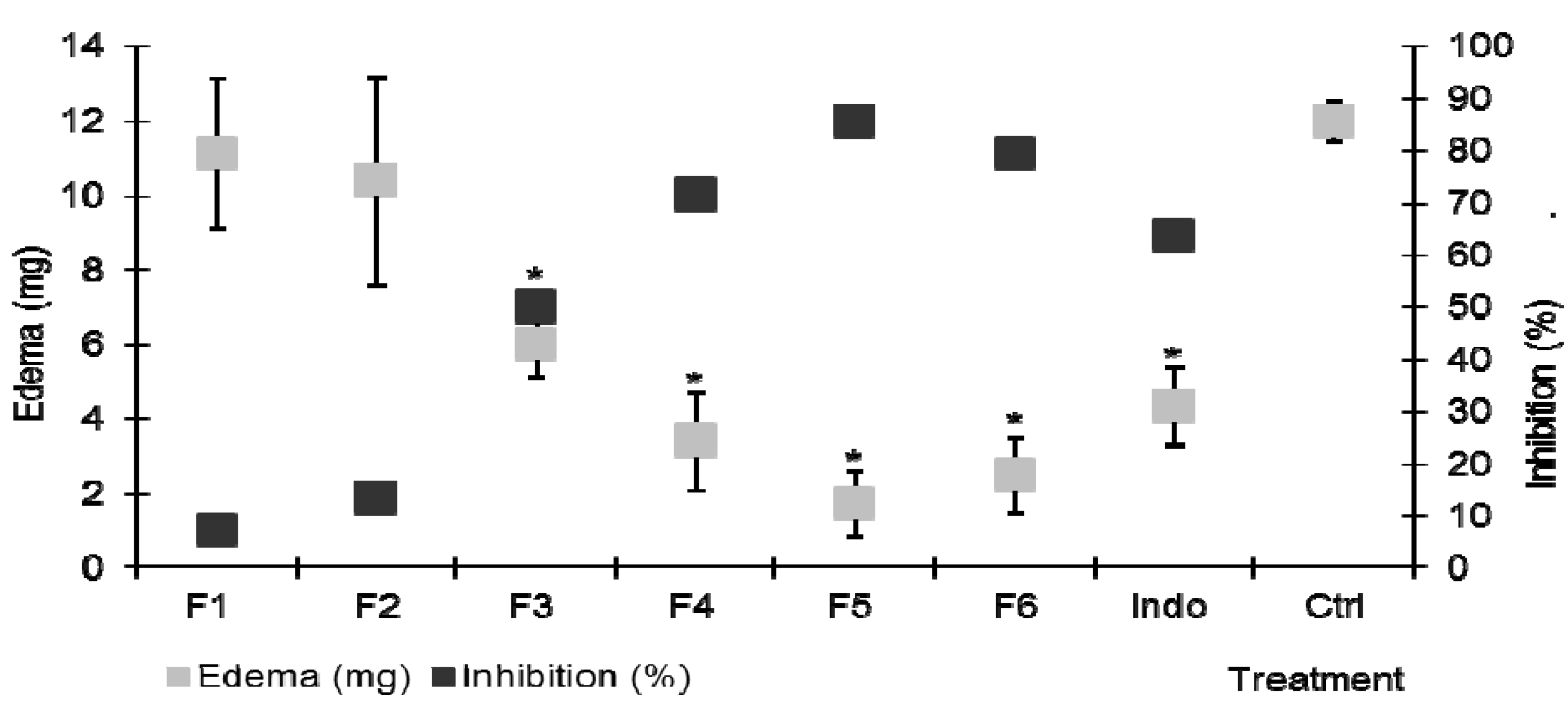
Due to its higher activity, a bio-guided chemical fractionation of
A. americana was carried out. Six fractions with different chromatographic profile (F1 to F6, with ascending polarity) could be separated by open column chromatography (CC). Each one of these in turn was evaluated in the TPA-induced ear edema at a dose of 2 mg/ear. Lower polarity fractions F1 and F2 did not cause any significant changes (
p > 0.05,
Figure 2) in ear edema level (11.14 mg and 10.4 mg, respectively) compared to the inflammation control group (12 mg).
Figure 2.
Effect of the topical administration (2 mg/ear) of the fractions with different polarity from acetone extract of A. americana on TPA induced ear edema. Indo = indomethacin (1 mg/ear). ANOVA, post-hoc Bonferroni * p < 0.05 (n = 7, mean ± SD, when it is compared with the negative control).
Figure 2.
Effect of the topical administration (2 mg/ear) of the fractions with different polarity from acetone extract of A. americana on TPA induced ear edema. Indo = indomethacin (1 mg/ear). ANOVA, post-hoc Bonferroni * p < 0.05 (n = 7, mean ± SD, when it is compared with the negative control).
Ear edema, measured as a weight difference between the ears, was 6.02 mg in the group that received fraction F3, which represents a 49% inhibition (
Figure 2), indicating the presence of some anti-inflammatory compounds. As the polarity of the extracts F4, F5 and F6 increased, the biological effect also increased, showing an edema level of 3.4, 1.7 and 2.4 mg, which represents a 71.7%, 85.6% and 79% edema inhibition, respectively, being significantly different from control group (
p < 0.05,
Figure 2). The data thus suggests that this plant contains more than one compound with the capacity to inhibit the local effects of TPA.
Fraction F5 was separated chemically because it caused a higher edema decrease. From the fractionation of F5, a precipitate (F5b) was isolated, which showed anti-inflammatory activity in the TPA model, so it was decided to evaluate it at three doses (1.0, 1.5 and 2.0 mg/ear). The tests indicated that the three doses employed induced inflammation decreases of 3.3, 1.4 and 1.2 mg, corresponding to inhibition percentages of 72.5%, 87.6% and 90%, respectively. Although there were no statistical differences between the analyzed groups, a dose-dependent behavior was observed, and F5b had a higher anti-inflammatory effect than indomethacin at a dose of 1.0 mg/ear (
Figure 3).
Figure 3.
Effect of the topical administration of different doses (1.0. 1.5 and 2.0 mg/ear) of precipitate F5b isolated of A. americana, on TPA induced ear edema. Indo = Indomethacin (1 mg/ear). ANOVA, post-hoc Bonferroni, * p < 0.05 (n = 7, mean ± SD, when it is compared with the negative control).
The precipitate F5b was exhaustively acetylated to give a white solid whose
13C-NMR spectroscopic analysis showed 57 signals, 29 of which could be assigned to an acetylated spirostanol skeleton type due to the characteristic ketal carbon signal at δ 108.6 (C-22). The other 28 carbon signals corresponded to the presence of two sugar moieties. The anomeric signals were observed at δ 4.62 (d, 8 Hz)/98.1 for H-1′ and C-1′ and δ 4.46 (d, 8 Hz)/101.7 for H-1′′ and C-1′′, respectively. The interglycosidic connectivities were determined by the long-range heteronuclear coupling correlations (
3JCH) observed in the HMBC experiment of C-1′ (δ
C 98.1) of glucose with H-3 (δ
H 3.49) and C-1′′ (δ
C 101.7) of glucose with H-6 (δ
H 3.26) (
Table 1). Taken together this indicated to us that the parent compound corresponded to (25
R)-5α-spirostan-3β,6α,23α-triol-3,6-di-
O-β-D-glucopyranoside (
Table 1). This saponin has been previously described from
A. cantala as cantalasaponin-1 [
25], later it was also identified in
A. americana [
13,
26].
Table 1.
1H (400 MHz) and 13C-NMR (100 MHz) data of the peracetylated derivative of cantalasaponin-1.
Table 1.
1H (400 MHz) and 13C-NMR (100 MHz) data of the peracetylated derivative of cantalasaponin-1.
| Position | δC | δH | Position | δC | δH |
|---|
| | | 3-
O-glc | | |
| 1 | 39.6 | | 1′ | 98.7 | 4.62 (d, 8) |
| 2 | 27.8 | | 2′ | 71.4 | 4.92 (dd, 8, 9.6) |
| 3 | 78.6 | 3.499 (m) | 3′ | 72.8 | 5.14 (dd, 9.2, 10) |
| 4 | 28.9 | | 4′ | 68.5 | 5.04 (dd, 9.6, 9.6) |
| 5 | 49.3 | | 5′ | 71.8 | 3.62 (dddd, 2, 4.8, 7.6, 12) |
| 6 | 81.12 | 3.26 (m) | 6′a | 62.2 | 4.05 (dd, 2, 12) |
| b | 4.22 (dd, 4.8, 12) |
| 7 | 37.1 | | 6-
O-glc | | |
| 8 | 33.8 | | 1′′ | 101.7 | 4.46 (d, 8) |
| 9 | 53.5 | | 2′′ | 71.6 | 4.92 (dd, 8, 9.6) |
| 10 | 36.6 | | 3′′ | 73.2 | 5.14 (dd, 9.2, 10) |
| 11 | 20.77 | | 4′′ | 68.6 | 5.04 (dd, 9.6, 9.6) |
| 12 | 39.69 | | 5′′ | 71.8 | 3.62 (dddd, 2, 4.8, 7.6, 12) |
| 13 | 41.1 | | 6′′ a | 62.3 | 4.05 (dd, 2, 12) |
| b | 4.22 (dd, 4.8, 12) |
| 14 | 56.0 | | | | |
| 15 | 31.8 | | | | |
| 16 | 81.17 | 4.41 (m) | | | |
| 17 | 61.7 | 0.75 (s) | | | |
| 18 | 13.5 | 0.77 (s) | | | |
| 19 | 16.2 | | | | |
| 20 | 36.1 | | | | |
| 21 | 14.2 | 0.90 (d, 6) | | | |
| 22 | 108.6 | | | | |
| 23 | 68.5 | 4.80 (dd, 11.6) | | | |
| 24 | 34.1 | 1.46 (m) | | | |
| 25 | 30.8 | | | | |
| 26 a | 65 | 3.37 (m) | | | |
| b | 3.29 (m) |
| 27 | 16.5 | 0.78 (d, 6) | | | |
This saponin was found in some members of the Agavacea family and could act as a marker, as reported in 2004, from its isolation in the species
Furcraea selloa which also belongs to that family [
27]. Importantly, there is some evidence that this compound has biological activity. For example, its cytotoxic effects against JTC-26 cells which induce human cervical carcinoma has been reported [
25]; the same activity was analyzed against human cells inducing HL-60 promyelocytic leukemia, but it was not active in those experiments [
28]. Recently, this saponin was isolated from
A. sisalana and it was evaluated again in a cytotoxicity test but on human MCF-7 breast cancer and NCI-H460 non-small cell lung cancer lines, and it was not active either [
29]. The present report is the first to demonstrate the dose-dependent anti-inflammatory activity of cantalasaponin-1.
It has been shown that secondary metabolites from
A. americana such as the steroidal saponins and triterpenes found in different parts of the plant, but mainly in its leaves, induced several activities. Examples of these componds are tigogenin, which has shown anticancer [
30] and anti-inflammatory activity [
11], besides promoting the union of nuclear factor Kappa B to DNA, which promotes the anti-proliferative effect associated to cancer [
31]. Chlorogenin, another steroidal saponin, induces toxicity against HL-60 human promyelocytic leukemia cells [
32], and also has anti-inflammatory activity [
33]. Considering that cantalasaponin-1 is structurally linked with these active compounds (it contains one C-23 hydroxyl more than clorogenin, and two more than tigogenin) we can propose a direct relation to the biological activity observed in this study.
3. Experimental
3.1. General
12-O-Tetradecanoylphorbol 13-acetate [4β,9α,12β,13α,20-pentahydroxytiglia-1,6-dien-3-one 12-tetradecanoate 13-acetate, TPA, ≥99% purity (by TLC)] and indomethacin [Indo, ≥99% purity (by TLC)] were purchased from Sigma Chemical Co. (St. Louis, MO, USA). All NMR spectra were recorded in CDC13 on a Varian INOVA-400 MHz instrument (operated at 400 MHz for 1H-NMR, NOESY, 1H-1H COSY, HSQC, and HMBC, and at 100 MHz for 13C-NMR). Chemical shifts are reported in parts per million (ppm) relative to tetramethylsilane (TMS).
3.2. Plant Material
The different agave leaves used were identified in the Biology Institute of the National University of Mexico (UNAM) by Dr. Abisai Josue García Mendoza as Agave tequilana F. A.C. Weber, Agave angustifolia Haw and Agave americana L. Marginata Hort. A. americana L. Marginata Hort. material was collected in the Toluca de Lerdo Municipality in the state of Mexico (19°17′29″N, 99°39′38″W); A. tequilana F.A.C. Weber and A. angustifolia Haw. were obtained from controlled cultivations in Tlaquiltenango (18°37′48′′N, 99°10′00′′W) in the state of Morelos.
3.3. Chemical Fractionation
The leaves were cut into pieces of approximately 10 cm in size, weighed and placed in trays for lyophilization (Heto Drywinner lyophilizer, Model DW3, Heto Holten A/S, Allerød, Denmark). Once the vegetable material was dry, it was weighed and ground (Pulvex plastic mill, D.F., Mexico), and then macerated in acetone for 72 h. The product thus obtained was filtered and the extract was concentrated under reduced pressure on a rotary evaporator (Heildolph Laborota Model 4000, Schwabach, Germany). Once the solvent was removed, the resulting A. tequilana (At), A. americana (Aam) and A. angustifolia (Aan) extracts were lyophilized and refrigerated at 4 °C until further use.
The most biologically active extract, in this case the
A. americana one, was subjected to partition with an immiscible 1:1 mixture of water/ethyl acetate (1 L, three times). The organic fraction (F-EtOAc 10 g) was concentrated by low-pressure distillation and separated on an open chromatographic column (50 × 500 mm, silica gel 70–230, 100 g, Merck, Darmstadt, Germany). The mobile phase consisted of a gradient of
n-hexane/ethyl acetate/MeOH mixtures (250 mL each sample). Concentrated fractions (characterized by eluent composition and amount isolated) were grouped according to their chemical similarities:
F1 (100:0:0, 0.32 g),
F2 (80:20:0, 0.45 g),
F3 (50:50:0, 0.6 g),
F4 (45:45:10, 2.1 g),
F5 (40:40:20, 3.8 g) and
F6 (0:0:100, 2.3 g). Fraction
F5 afforded in acetone a white precipitate (0.5 g) which was separated by Whatman paper filtration, and denominated F5b. In order to facilitate the structure elucidation, this precipitate (100 mg) was subjected to an acetylation process (1:2 pyridine-Ac
2O, 3 h), as previously described [
34]. The crude product of this reaction was partitioned with an ethyl acetate/water mixture. The organic fraction was concentrated to dryness. Crystallization from
n-hexane-ethyl acetate afforded the peracetate derivative (25 mg).
1H- and
13C-NMR analysis (
Table 1) and two dimensional experiments like COSY, HSQC and HMBC allowed us to confirm the chemical structures shown in
Figure 4.
Figure 4.
Chemical structure of cantalasaponin-1 and its peracetylated derivative.
Figure 4.
Chemical structure of cantalasaponin-1 and its peracetylated derivative.
Cantalasaponin-1 (R = H); peractylated derivative (R = COCH3)
3.4. Animals
Male ICR mice (35 gr weight) were used. All animals were purchased from Harlan Mexico (D.F, Mexico), and kept in an animal house for three weeks with a cycle of 12 h of light and 12 h of darkness and free access to water and food (pellets, Harlan). Three days before testing began the animals were conditioned to the laboratory environment and to the researcher. All experiments were conducted in accordance with the Federal Regulations for Animal Experimentation and Care (Ministry of Agriculture, NOM-062-ZOO-1999, Mexico). The experimental protocols were approved by the Research Committee of Mexican Institute of Social Security with the recording number R-2010-1701-21. The minimum number of animals and duration of observation required to obtain consistent data were employed.
3.5. Experimental Design
Groups of seven mice were formed, each corresponding to one of the following treatments: (a) negative control (TPA 2.5 μg dissolved in 20 µL of acetone), (b) positive control (indomethacin, 1 mg/ear) and (c) the acetone extracts of leaves of A. tequilana, A. americana and A. angustifolia at 6.0 mg/ear dose. The ear treatment administration procedure was initiated in the surgical anesthesia stage (pentobarbital sodium 50 mg/Kg via, ip). The left ear of each mouse served as control reference for all treatments because only 10 µL of 70% ethanol were administered on both sides of the ear. For all treatments with agave species, these were administrated 10 µL on both sides of the right ear. For positive control, the same volume of a solution of indomethacin (a non-steroidal anti-inflammatory drug) was applied and for the negative control group, only ethanol to 70%. Fifteen minutes after treatment administration, 10 µL of TPA (pro-inflammatory) solution was applied on both sides of the right ear.
Thereafter, 4 h later, the animals were sacrificed by cervical dislocation, and then 6 mm sections were taken from the ears of each mouse from all groups, the sections were weighed and the weight differential was determined [
35].
In the case of the extract that showed significant anti-inflammatory effect, was performed fractionation and some selected fractions were tested (2 mg/ear). Finally, we constructed a dose-response curve for the isolated compound at the doses of 1.0, 1.5 and 2.0 mg/ear. The results were used to determine the TPA induced edema inhibition percentage was obtained using expression below:
where Δw = wt − wnt; wt is the weight of the section of the treated ear; wnt is the weight of the section of the non-treated ear.

 ,
, 


