Article

First Chemical Constituents from Cordia exaltata Lam and Antimicrobial Activity of Two Neolignans

by
Tiago Bezerra de Sá de Sousa Nogueira
1,
Raquel Bezerra de Sá de Sousa Nogueira
1,
Davi Antas e Silva
2,
Josean Fechine Tavares
1,
Edeltrudes De Oliveira Lima
1,
Fillipe De Oliveira Pereira
1,
Milen Maria Magalhães De Souza Fernandes
1,
Fernando Antônio De Medeiros
3,
Rosangela Do Socorro Ferreira Rodrigues Do Socorro Ferreira Rodrigues Sarquis
3,
Raimundo Braz Filho
4,
Jéssica Karina Da Silva Maciel
5 and
Maria De Fátima Vanderlei de Souza
1,*
1
Postgraduate Program in Bioactive Natural and Synthetic Products, Health Sciences Center, Federal University of Paraíba, João Pessoa, PB 58051-970, Brazil
2
Department of Physiology and Pathology, Health Sciences Center, Federal University of Paraíba, João Pessoa, PB 58051-970, Brazil
3
Institute for Scientific and Technological Research of Amapá, Macapá, AP 68901-025, Brazil
4
Laboratório de Ciências Químicas, Universidade Estadual do Norte Fluminense Darcy Ribeiro-UFRRJ, Campos dos Goytacazes, RJ 28013-602, Brazil
5
Postgraduate Program in Development and Technological Innovation in Drug-Center for Health Sciences, Federal University of Paraíba, João Pessoa, PB 58051-970, Brazil
*
Author to whom correspondence should be addressed.
Molecules 2013, 18(9), 11086-11099; https://doi.org/10.3390/molecules180911086
Submission received: 21 June 2013 / Revised: 29 August 2013 / Accepted: 29 August 2013 / Published: 10 September 2013
(This article belongs to the Section Metabolites)

Abstract

:
The phytochemical study of Cordia exaltata Lam. (Boraginaceae) led to the isolation, through chromatographic techniques, of nineteen secondary metabolites: 8,8'dimethyl-3,4,3',4'-dimethylenedioxy-7-oxo-2,7'cyclolignan (1), 8,8'-dimethyl-4,5-dimethoxy-3',4'-methylenodioxy-7-oxo-2,7'cyclolignan (2), sitosterol (3a), stigmasterol (3b), sitosterol-3-O-β-d-glucopyranoside (4a), stigmasterol-3-O-β-d-glucopyranoside (4b), phaeophytin A (5), 132-hydroxyphaeophytin A (6), 173-ethoxypheophorbide A (7), 132-hydroxy-173-ethoxypheophorbide A (8), m-methoxy-p-hydroxybenzaldehyde (9), (E)-7-(3,4-dihydroxyphenyl)-7-propenoic acid (10), 1-benzopyran-2-one (11), 7-hydroxy-1-benzopyran-2-one (12), 2,5-bis-(3',4'-methylenedioxiphenyl)-3,4-dimethyltetrahydrofuran (13), 3,4,5,3',5'-pentamethoxy-1'-allyl-8.O.4'-neolignan (14), 3,5,7,3',4'-pentahydroxyflavonol (15),5,7-dihydroxy-4'-methoxyflavone (16), 5,8-dihydroxy-7,4’-dimethoxyflavone (17), kaempherol 3-O-β-d-glucosyl-6''-α-L-ramnopyranoside (18) and kaempherol 3,7-di-O-α-l-ramnopyranoside (19). Their structures were identified by 1H and 13C-NMR using one and two-dimensional techniques. In addition, the antimicrobial activity of compounds 1, 2, 13 and 14 against bacteria and fungi are reported here for the first time.

1. Introduction

The Boraginaceae family has 148 genera and 2,740 species distributed in the temperate and tropical zones of Europe, Asia, Australia and America [1]. The genus Cordia, one of the most representative in the Boraginaceae family [2], consists of approximately 320 species, which are presented as trees, shrubs or herbs, with the main habitat in South America [3]. Its species are used in the folk medicine as diuretics, weight loss agents, healants and emollients [4]. The leaves of the species Cordia leucocephala, popularly known as “Maria Preta”, are used as an infusion for the treatment of dysmenorrhea [5]. Biological studies related to the extracts and isolated substances from Cordia report antiviral effects against herpes type I, cytotoxic effects on tumor cells CA-9KB [6], anti-inflammatory [7,8] spasmolytic and vasorelaxant [9] properties. The compound 1-(3'-methoxypropanoyl)-2,4,5-trimethoxybenzene, reported as the active principle of C. alliadora and commonly found in other species of this genus, was identified as a larvicide for Aedes aegypti [10]. Some substances of C. goetzei and C. corymbosa, are mentioned in the literature for their fungicidal effects against phytopatogens [11,12]. The present study aimed to isolate and identify other classes of secondary metabolites from C. exaltata in order to associate them to the observed pharmacological properties. We now report the isolation and identification of four neolignans 1, 2, 13 and 14, two mixtures of steroids 3a, 3b and 4a, 4b, four porphyrins 5-8, two phenolic acids 9 and 10, two coumarins 11 and 12 and five flavonoids 15-19. Besides we report for the first time the antimicrobial activity of compounds 1, 2, 13 and 14 against bacteria and fungi.

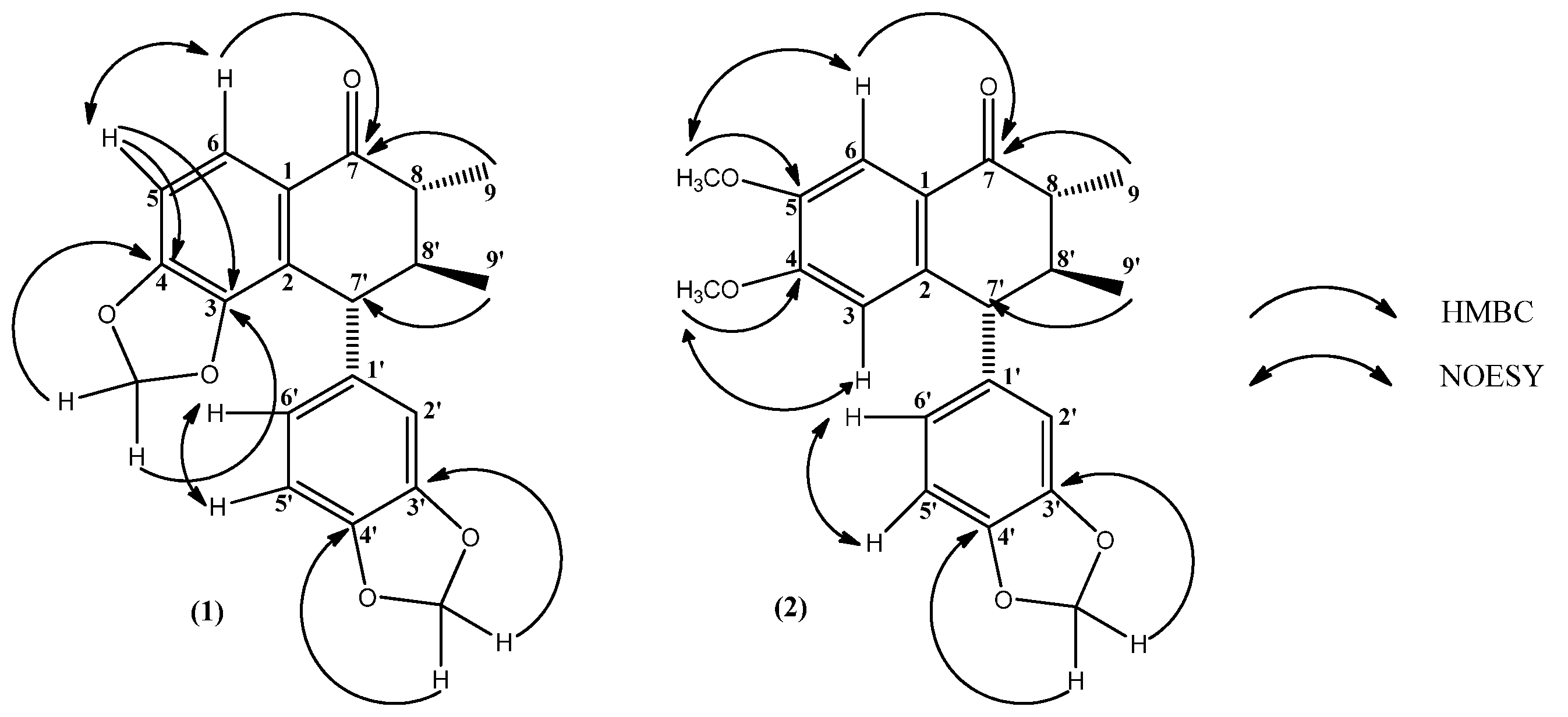
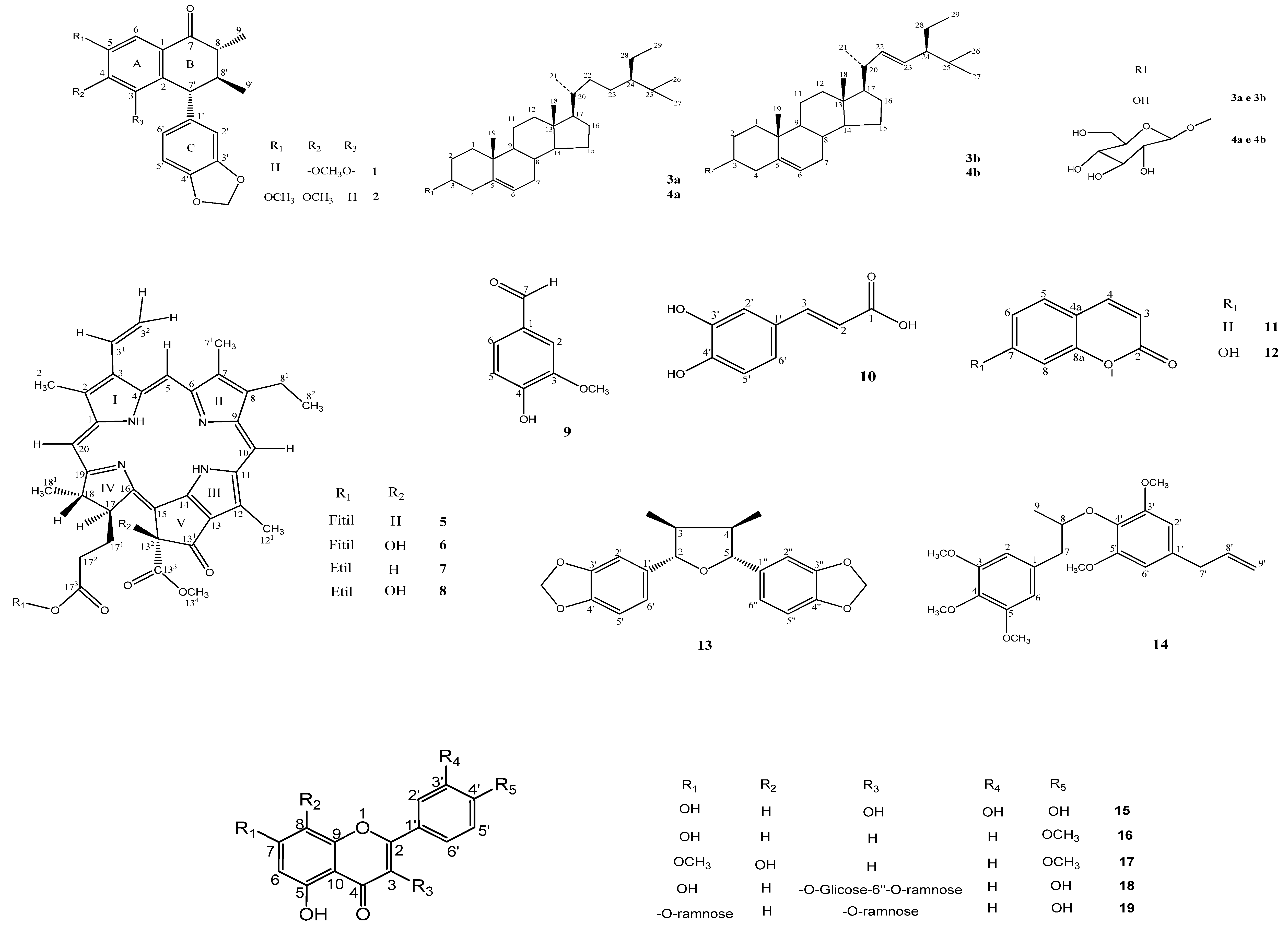
2. Results and Discussion

Compound 1 was obtained as colorless crystals. Its IR spectrum exhibited the characteristic absortion of a carbonyl group at 1,678 cm−1 and an aromatic nucleus at 1,475 cm−1 [13]. The 1H- and 13C-NMR spectra exhibited the general absortions of a neolignan, with two methyl and two methylenedioxy groups on aromatic rings [14]. The 1H-NMR spectrum of 1 exhibited a singlet at δH 3.80, a multiplet at δH 2.41 and another multiplet at δH 1.93, which were attributed to H-7', H-8 and H-8', respectively, while signals at δH 1.09 and 0.90 were assigned to two methyl groups (H-9 and H-9'). In the aromatic region, an ABX-type system was observed at δH 6.80 (1H, d, J = 8.0 Hz, H-5'), 6.62 (1H, dd, J = 8.0 Hz and 1.8 Hz, H-6') and 6.60 (1H, d, J = 1.8 Hz, H-2'), attributed to hydrogens of a 1',3',4'-trisubstituted aryl group. Two additional aromatic hydrogens resonate at δH 7.55 (1H, d, J = 8.2 Hz, H-6) and δH 6.95 (1H, d, J = 8.2 Hz, H-5). The 13C-NMR spectrum showed 20 carbon resonances, which were assigned by the DPT spectrum as two methyl carbons, three methine carbons, five phenyl methine carbons, two alcoholic methylene carbons, seven phenyl quaternary carbons and a carbonyl carbon. The long-range 1H-13C-correlations of the HMBC spectrum showing 3J between C-4 (δC 151.41), C-3 (δC 144.90) with δH 5.93 (OCH2O-A, 2H, s), C-4' (δC 145.50), C-3' (δC 147.00) with δH 5.83 (1H, s) and δH 5.78 (1H, s) (OCH2O-C), allowed the location of the two methylenedioxy groups to be established and also the assignments of the two methyl positions by way of correlations between C-8 (δC 42.60) and C-8' (δC 46.48) with δH 2.41 (1H, m, H-8) and δH 1.09 (3H, d, J = 6.7 Hz, H-9), respectively, 2J and 3J. Additionally, a carbonyl group was located at C-7 by the correlation between the resonances at δC 197.48 (C-7) and δH 2.41 (1H, m, 2J, H-8) and δH 7.55 (H-6, 1H, d, J = 8.7 Hz, 3J). Thus, compound 1 was identified as 8,8'-dimethyl-3,4,3',4''-dimethylenedioxy-7-oxo-2,7'-cyclolignan, isolated before from another source [15], but for the first time in genus Cordia.
Compound 2 formed white needle-like crystals. Spectral analysis revealed similarities with compound 1, thus suggesting a neolignan structure for 2 as well. The main difference consisted of two methoxyl groups instead of a methylenedioxy on ring A. This could be inferred by the IR spectrum, which exhibited a C-H absortion from a methoxyl group at 2,850 cm−1. The HMBC spectrum showed 3J correlations between resonances at δC 149.10 (C-5) and δH 3.83 (3H, d, J = 1.0 Hz, OCH3-5), and also δC 155.06 (C-4) and δH 3.58 (3H, d, J = 1.0, OCH3-4), allowing the assignments of the methoxyl groups’ positions. Thus, through a complete spectral analysis, it was possible to identify compound 2as 8,8'-dimethyl-4,5-dimethoxy-3',4'-methylenodioxy-7-oxo-2,7'-cyclolignan [16], a compound isolated for the first time in the Boraginaceae family. The main correlations of HMBC and NOESY spectra, which allowed a complete assignment for both compounds 1 and 2 are shown in Figure 1.
Figure 1. Main HMBC and NOESY interactions for compounds 1 and 2.
Molecules 18 11086 g001
Compound 13 showed in its 1H-NMR spectrum absorptions in the aromatic region, which corresponded to two ABX-systems: at δH 6.86 (dd, J = 8.0 and 1.7 Hz, 2H), at δH 6.78 (d, J = 8.0 Hz, 2H) and another doublet at δH 6.94 (J = 1.7Hz, 2H), suggesting a symmetrical structure. This information was supported by a strong singlet at δH 5.94, attributed to four hydrogens of two methylenedioxy groups, which is consistent with a neolignan structure. Also, a tetrahydrofuran ring could be inferred fromy two multiplets at δH 2.24 (2H) and δH 4.40 (2H), related to H-3/4 and H-2/5, respectively. Two symmetric methyl groups were revealed at δH 0.99 (J = 6.8 Hz, 6H). The 13C-NMR spectrum of 13 showed 10 strong peaks, which allowed the identification of two methyl carbons, four methine carbons, two alcoholic methylene carbons, six phenyl methine carbons and six phenyl quaternary carbons, corresponding to 20 chemically equivalent carbons. The HMBC spectrum showed long-range 1H-13C-correlations between H-2/H-5 (δH 4.40) and the aromatic carbons at C-6'/C-6' (δC 120.10) through 3J and also C-1'/C-1 (δC 136.16) through 2J, allowing the location of the two aromatic rings at C-2/C-5. This spectrum could also establish the locations of the two methyl groups, showing 2J interactions between H-3/H-4 (δH2.24, 2H) and CH3-3/CH3-4 (δC 12.97). The 1H-1H-NOESY spectrum reinforced these positions by showing spatial interactions between H-2/H-5 (δH 4.40), H-3/H-4 (δH 2.24) and CH3-3/CH3-4 (δH 0.99). Therefore, compound 13 was identified as 2,5-bis-(3',4'-methylenedioxiphenyl)-3,4-dimethyltetrahydrofuran [17], described for the first time in the Boraginaceae family.
Compound 14 appeared as yellow oil. Its 1H-NMR spectrum showed resonances for five methoxyl groups, being two peaks at δH 3.81 (s, 6H) and δH 3.77 (s, 6H), which correspond to two pair of symmetric aromatic methoxyls and another group at δH 3.80 (s, 3H). An allyl group was shown at δH 5.95 (ddt, J = 17.0, 10.5 and 6.5 Hz, 1H) (H-8'), δH 5.09 (ddt, J = 10.5, 2.0 and 1.6 Hz, 1H, H-9'a), δH 5.05 (ddt, J = 17.0, 2.0, e 1.6 Hz, 1H, H-9'b) and δH 3.31 (d, J = 6.5 Hz, 2H, H-7'). Additionally, two singlets were observed at δH 6.44 (2H) and δH 6.38 (2H), referring to two pairs of symmetric phenyl hydrogens, H-2/H-6 and H-2'/H-6', respectively. From these absorptions it was possible to suggest an aryloxyarylpropane structure for compound 14, consistent with a neolignan skeleton. Through HMBC analysis, the methoxyl groups positions could be assigned by 3J 1H-13C correlations showed between resonances at δH 3.81 (H3CO-3/5) with δC 153.64 (C-3/5) and δH 3.77 (H3CO-3'/5') with δC 152.85 (C-3'/5'). Also, hydrogen H-7' (δH 3.31, J = 6.5 Hz, 2H) showed 3J and 2J correlations with C-2'/6' (δC 105.94) and C-9' (δC 115.94), respectively, leading to the location of the allyl group at C-1'. Therefore, compound 14 was identified as 3,4,5,3',5'-pentamethoxy-1'-allyl-8.O.4'-neolignan [17], described here for the first time in the Boraginaceae family.
The spectral analysis using IR and 1H/13C-NMR data by means of one and two-dimensional techniques was also performed for the other compounds, which made possible their identification as sitosterol (3a), stigmasterol (3b), sitosterol-3-O-β-d-glucopyranoside [18] (4a), stigmasterol-3-O-β-d-glucopyranoside (4b) [18], phaeophytin A (5) [19], 132-hydroxyphaeophytin A (6) [20], 173-ethoxypheophorbide A (7) [21], 132-hydroxy-173-ethoxypheophorbide A (8) [22], m-methoxy-p-hydroxy-benzaldehyde (9) [23], (E)-7-(3, 4-dihydroxyphenyl)-7-propenoic acid (10) [24], 1-benzopyran-2-one (11) [25], 7-hydroxy-1-benzopyran-2-one (12) [26], 3,5,7,3',4'–pentahydroxyflavone (15) [27], 5,7-dihydroxy-4'-methoxyflavone (16) [27], 5,8-dihydroxy-7,4'-dimethoxyflavone (17) [28], kaempferol 3-O-β-d-glucosyl-6''-α-L-ramnopyranoside (18) [29], and kaempferol 3,7-di-O-α-l-ramnopyranoside (19) [30] (Figure 2).
The MIC values of compound 1 are shown in Table 1. As can be seen, 90% of the strains were susceptible to the compound up to the concentration of 300 µg/mL, and the percentage of inhibition was higher than that observed with the drug controls—chloramphenicol for bacteria and ketoconazole for yeasts—where 60% of strains where inhibited by the respective antibiotics. The MIC of the compound 1 was 150 µg/mL to almost all strains tested, where about 75% of the strains had their growth inhibited by this concentration. It was observed that compound 1 showed the greatest inhibiting effect on strains of C. guilliermondii LM-2101 and C. guilliermondii LM-011, with showed the lowest MIC values (75 µg/mL).
Figure 2. Constituents isolated from Cordia exaltata.
Molecules 18 11086 g002
The MIC data of the compound 2 are also summarized in Table 1. It was observed that the MIC was 300 for 35% of the strains. The percentage of strains resistant to the compound 2 to the highest concentration was 40%, therefore higher than that found with the compound 1 (10%). As for the antibiotics tested, they showed inhibitory effect on 60% of the strains, similar to compound 2.
Thus, one can conclude that both the tested compounds have detectable antimicrobial activity against a variety of pathogens, including bacteria and yeasts, in infectious processes relevant from a clinical point of view. It also may be noted the compound 1 had the strongest antimicrobial effect, which could be explained by the lower MIC values against microorganisms [31,32,33,34]. Compounds 13 and 14 were also tested against the same strains and following the same protocol described for 1 and 2, but did not show any significant activity.
Table 1. MIC values of compounds 1 and 2 against bacteria and yeasts.
Microorganisms(1) (µg/mL)(2) (µg/mL)Controls
Tween 80MicrorganismsAntimicrobyal
S. aureus ATCC-6538150>300++-
S. aureus ATCC-25923150>300+++
S. epidermidis ATCC-12228150>300++-
B. subtilis ATCC-6633150>300++-
P. aeruginosa ATCC-25853150300+++
P. aeruginosa ATCC-9027150300+++
E. coli (clássica C)150300++-
E. coli ATCC-18739150300+++
E. coli ATCC-8733150>300+++
S. flexineri LM-412150150++-
C. albicans ATCC-90028150150++-
C. albicans ATCC-76615150150++-
C. albicans LM-142 V15075++-
C. albicans ICB-12 150300++-
C. tropicalis ATCC-13803>300300+++
C. tropicalis LM-028>300>300++-
C. krusei ATCC-625815075+++
C. krusei LM-12300300++-
C. guilliermondii LM-210175>300++-
C. guilliermondii LM-01175>300+++
+: Presence of microbial growth; −: Absence de microbial growth.

3. Experimental

3.1. General

Silica gel 60 (Merck) 7734 (0.063–0.2 mm particle, 70–230 mesh), flash silica (0.04–0.063 mm particles, 230–400 mesh) and Sephadex LH-20 were used for the fractionation and isolation of the secondary metabolites from C. exaltata. TLC was used to analyse and compare the fractions obtained from chromatographic column procedures. Solvents used in TLC were basically mixtures of the same solvents of the chromatographic experiments. For pure compounds analysed by TLC, three different systems of solvents were tested, to make sure that these compounds were free of impurities. The melting point of the constituents was recorded on a MQAPF-302 apparatus. IR spectra were recorded on a FT-IR-1750 Perkin-Elmer spectrometer. 1H and 13C-NMR spectra were recorded on a Varian Oxford 200 NMR spectrometer (200/50 MHz) and on a Varian 500 NMR spectrometer (500/125 MHz).

3.2. Collection, Extraction and Isolation

The leaves, stems, fruits and stem bark of Cordia exaltata Lam, were collected near the city of Porto Grande–AP in July 2006 and were identified by Prof. Dr. Rosangela do Socorro Ferreira Rodrigues Sarquis-IEPA (Instituto de Pesquisa Científica e Tecnológica do Estado do Amapá/BR-Institute for Scientific and Technological Research of Amapá). A voucher specimen is deposited in the Herbarium Amapaense/AP/BR (HAMAP), under the code 2528.
Leaves, stems, fruits and stem bark of Cordia exaltata Lam were dehydrated in an oven at 40 °C for 72 h and ground separately in a mechanical mill to yield 847.5 g, 1000.5 g, 281.0 g and 2.310.0 g of a powder, respectively. Each powdered part of the plant was submitted to maceration with methanol (2.5 L, 3.0 L, 1.0 L and 4 L, respectively) for three consecutive days at room temperature and this process was repeated until the maximum amount of chemical constituents had been extracted. The obtained methanol extract solutions were concentrated in a rotatory evaporator, yielding 150.0 g, 150.0 g, 11.0 g and 211.0 g of the respective crude extracts.
An amount of the crude methanolic extract from fruits (12.0 g) was subjected to filtration under reduced pressure using silica gel 60 as stationary phase and eluted with hexane (hex.), ethyl acetate (EtOAc) and methanol (MeOH) alone or in binary mixtures following an increasing gradient polarity. After that, the extracts were concentrated under pressure, leading to the respective phases. The hexane phase (1695.0 mg) yielded 847.8 mg of a precipitate, which was chromatographed on a silica gel column and eluted with hex., EtOAc and MeOH. This procedure led to 28 fractions, analysed by TLC (hexane, EtOAc and MeOH at least in three different polarities), from which the combined fractions 5/14 (200.0 mg) and 17/20 (170.0 mg) gave compounds 1 and 2, respectively.
The methanolic crude extract from the leaves of C. exaltata Lam. (100.0 g) was suspended in methanol-H2O (9:1) and successively partitioned with hex., chloroform (CHCl3), EtOAc and n-butanol, increasing the gradient polarity. The hexane phase (10.0 g) was chromatographed under the same conditions as described previously for the crude extract of fruits, giving 80 fractions, which were combined by TLC (hexane, EtOAc and MeOH mixtures of at least three different polarities). Fractions 1/19, after purification, led to 100.0 mg of a mixture of compounds 3a and 3b. Fraction 20/27 was chromatographed on a gel silica column, from which 84 fractions were obtained and analysed by TLC. From this chromatography, fractions 01/08, 09/18 and 28/36 were purified and this led to the isolation of compounds 4 (40 mg), 5 (100.0 mg) and 6 (100.0 mg), respectively. The CHCl3 extract (8.18 g) was also chromatographed following the described methodology, which allowed the isolation and purification of 30.0 mg of compound 11, 35.0 mg of compound 12 and 17.0 mg of 16. The EtOAc (3.7 g) and n-butanol (1.4 g) phases were submitted to chromatography on Sephadex LH-20, affording compound 15 (40.0 mg) and 19 (15.0 mg), respectively.
The methanolic crude extract from the stem bark (100 g) was solubilized in methanol-H2O (7:3) and successively partitioned under vacuum using silica gel with hex., CHCl3, EtOAc and n-butanol as described in the previous procedures. The hexane phase (5.0 g) was submitted to column chromatography with silica gel using hexane (hex.), ethyl acetate (EtOAc) and methanol (MeOH) alone or in binary mixtures with increasing polarity, leading to 93 fractions, which were further combined according to TLC (hex., AcOEt and MeOH, in three different mixtures at least). Using this technique fractions 16/24, 37/47 and 57/70 were pure and gave 25.0 mg of compound 8, 18.0 mg of compound 9 and 40.0 mg of compound 10. Through column chromatography on Sephadex LH-20, the EtOAc (2.9 g) and n-butanol (2.6) phases yielded compound 17 (15.0 mg) and compound 18 (30.0 mg), respectively.
The bark extract of C. exaltata Lam (7.5 g) was chromatographed on silica gel under reduced pressure with the following solvents: hex., hex-EtOAc (9:1), hex.-EtOAc (6:4), hex.-EtOAc (1:1), Hex.-EtOAc (4:6), AcOEt, EtOAc-MeOH (9:1) and EtOAc-MeOH (1:1). Fraction Hex.:AcOEt (6:4) (175.0 mg) was fractionated by column using gel silica and an increasing polarity system of solvents, such as Hex., EtOAc and MeOH, which result in 30 fractions. These fractions were analysed by TLC, using Hex.-EtOAc in three different mixtures of polarity. Fractions 02/04 and 08/11 were purified and led to the isolation of compound 13 (10.0 mg) and compound 14 (12.0 mg), respectively. The fraction eluted with EtOAc-MeOH (9:1) yield a supernatant and a precipitate. The latter was separated from the former, purified and analysed by TLC using EtOH-MeOH (9:1) as eluting solvent, which resulted in a mixture of compounds 4a and 4b (30 mg).

3.3. Spectral Data

8.8'-Dimethyl-3,4,3', 4'-dimethylenedioxy-7-oxo-2,7'-cyclolignan (1). Colorless crystals; mp 176 °C; IR (KBr) vmáx (cm−1): 3081, 2963, 1678 (C=O), 1475; 1H-NMR (D­MSO-d6, 500 MHz): δ 7.55 (1H, d, J = 8.2 Hz, H-6), 6.95 (1H, d, J = 8.2 Hz, H-5), 6.80 (1H, d, J = 7.9 Hz, H-5'), 6.62 (1H, dd, J = 8.0 Hz e 2.0 Hz, H-6'), 6.60 (1H, d, J = 1.7 Hz, H-2'), 5.93 (2H, s, OCH2O-A), 5.83 (2H, s, OCH2O-C), 3,80 (1H, d, J = 9.8 Hz, H-7'), 2.41 (m, H-8), 1.09 (1H, d, J = 6.7 Hz, H-9), 1.93 (m, H-8'), 0.90 (1H, d, J = 6.7 Hz, H-9'); 13C- NMR (D­MSO-d6, 125 MHz): δ 197.48 (C-7), 151.41(C-4), 147.00 (C-3'), 145.50 (C-4), 144.90 (C-3), 137.50 (C-1'), 126.84 (C-2), 122.05 (C-6), 121.83 (C-6'), 108.67 (C-2'), 107.82 (C-5'), 107.46 (C-5), 100.72 (OCH2O-A), 101.54 (OCH2O-C), 47.97 (C-7'), 46.48 (C-8'), 42.60 (C-8), 17.11 (C-9), 12.45 (C-9').
8,8'-Dimethyl-4,5-dimethoxy-3',4'-methylenedioxy-7-oxo-2,7'cyclelignan (2). Fine white crystals; mp 131 °C; IR (KBr) vmáx (cm−1): 3081, 2963, 2850 (OCH3), 1678 (C=O), 1475; 1H-NMR (CD3OD, 500 MHz): δH 7.47 (1H, d, J = 1.0 Hz, H-6), 6.24 (1H, sl, H-3), 6.81 (1H, dd, J = 8.0 Hz and 2.0 Hz, H-6'), 6.70 (1H, d, J = 8.0 Hz, H-5'), 6.59 (1H, sl, H-2'), 5.94 (2H, s, OCH2O-C), 3.83 (3H, d, J = 1.0, OCH3-5), 3.74 (1H, d, J = 11.50 Hz, H-7'), 3.58 (3H, d, J = 1.0, OCH3-4), 2.39 (1H, dq, J = 13.0 Hz and 6.5, H-8), 2.04 (1H, ddq, J = 12.5, 11.5 and 6.0 Hz, H-8'), 1.26 (1H, dd, J = 6.5 and 0.5 Hz, H-9), 0.93 (1H, dd, J = 6.5 and 1.0 Hz, H-9'); 13C-NMR (CD3OD, 125 MHz): δC 200.79 (C-7), 155.06 (C-4), 149.49 (C-3'), 149.10 (C-5), 143.51 (C-2), 138.89 (C-1'), 126.68 (C-1), 124.20 (C-5'), 112.57 (C-3), 110.14 (C-2'), 109.42 (C-6), 109.10 (C-6'), 102.40 (OCH2O-C), 56.17 (OCH3-4), 54.28 (C-7'), 49.52 (C-8), 44.98 (C-8'), 18.17 (C-9'), 12.87 (C-9).
Sitosterol (3a) and stigmasterol (3b). 1H-NMR and 13C-NMR data were consistent with the literature values [17].
Sitosterol-3-O-β-d-glucopyranoside (4a) and stigmasterol-3-O-β-d-glucopyranoside (4b). 1H-NMR and 13C-NMR data were in agreement with the literature [17].
Phaeophytin A (5). IR, 1H-NMR and 13C-NMR data were in agreement with the literature [21].
132-Hydroxyphaeophytin A (6) (without phytyl ester). Green powder. IR (KBr) vmax: 3494, 2926, 2854, 1739, 1706, 1620, 1377 cm−1. 1H-NMR and 13C-NMR data were in agreement with the literature [35].
173-Ethoxyphaeophorbide A (7). 1H-NMR and 13C-NMR data were in agreement with the literature [21].
132-Hydroxy-173-ethoxyphaeophorbide A (8). Bluish green powder with a metallic luster. 1H-NMR and 13C-NMR data were in agreement with the literature [22].
m-Methoxy-p-hydroxybenzaldehyde (9). White crystals; mp 81 °C–82 °C. 1H-NMR and 13C-NMR data were in agreement with the literature [23].
Kaempferol 3,7-di-O-α-L-ramnopyranoside (19). Yellow crystals; mp 201 °C–203 °C. 1H-NMR and 13C-NMR data were in agreement with the literature [36].
(E)-7-(3,4-Dihydroxyphenyl)-7-propenoic acid (10). White solid; mp 220–221 °C. 1H-NMR (CD3OD, 200 MHz): δH7.40 (1H, d, J = 15.9 Hz, H-7), 6.08 (1H, d, J = 15.9 Hz, H-8), 6.91 (1H, d, J = 1.9 Hz, H-2), 6.79 (1H, dd, J = 8.2, 1.9, Hz, H-6), 6.64 (1H, d, J = 8.2 Hz, H-5); 13C-NMR (CD3OD, 50 MHz): δC127.71 (C-1), 115.43 (C-2), 116.45 (C-5), 122.88 (C-6), 149.35(C-4), 146.66 (C-3), 171.12 (C-9), 114.98 (C-8), 147.04 (C-7).
1-Benzopyran-2-one (11). White solid, mp 69–70 °C. 1H-NMR (CDCl3, 200 MHz): δH 7.52 (1H, d, J = 9.6 Hz, H-3), 6.22 (1H, d, J = 9.6 Hz, H-4), 7.12 (1H, dd, J = 7.0 and 1,0 Hz, H-8), 7.06 (1H, dd, J = 7.86, 6.4 and 1.0 Hz, H-6), 7.35 (1H, dd, J = 7.86, 6.4 and 1.25 Hz, H-7) 7.30 (1H, dd, J = 6.4 and 1.25 Hz, H-5); 13C-NMR (CDCl3, 50 MHz): δC 160.89 (C-2), 116.96 (C-3), 116.76 (C-8), 118.90 (C-4a), 154.10 (C-8a), 143.57 (C-4), 131.93 (C-7), 127,97 (C-5), 124.53 (C-6).
7-Hydroxy-1-benzopiran-2-one (12). White solid; mp 230–231 °C.1H-NMR (CD3OD, 500 MHz): δH 7.87 (1H, d, J = 9.4 Hz, H-4), 6.22 (1H, d, J = 9.4 Hz, H-3), 7.48 (1H, d, J = 8.5 Hz, H-5), 6.83 (1H, dd, J = 8.5 and 1.9 Hz, H-6), 6.75 (1H, s, H-8); 13C-NMR (CD3OD, 125 MHz): δC 163.66 (C-2), 112.34 (C-3), 103.41 (C-8), 113.49 (C-4a), 157.21 (C-8a), 145.99 (C-4), 163.09 (C-7), 130,62 (C-5), 114.49 (C-6).
2,5-bis-(3',4'-Methylenedioxyphyenyl)-3,4-dimethyltetrahydrofurane (13). 1H-NMR (CD­Cl3, 500 MHz): δH 6.94 (2H, d, J = 1.7 Hz, H-2' and 2''), 6.86 (2H, d, J = 8.0 and 1.7 Hz, H-6' and 6''), 6.78 (2H, d, J = 8.0 Hz, H-5' and 5''), 5.94 (4H, s, H-OCH2O-), 4.40 (2H, d, J = 6.8 Hz, H-2 and 5), 2.24 (2H, m, H-3 and H-6) and 0.99 (6H, d, J = 6.8 Hz, H-CH3-3 and CH3-4), 13C-NMR (CD­Cl3, 125 MHz): δC 147.92 (C-3' and 3''), 147.14 (C-4' and 4''), 136. 16 (C-1' and 1''), 120.10 (C-6' and C-6''), 108.15 (C-5' and 5''), 106.91 (C-2' and 2''), 101.09 (-OCH2O-), 87.59 (C-2 and 5), 44.80 (C-3 and C-4), 12.97 (C-CH3-3 and CH3-4).
3,4,5,3',5'-Pentamethoxy-1'-allyl-8.O.4'-neolignan (14). 1H-NMR (CDCl3, 500 MHz): δH 6.44 (2H, s, H-2 and 6), 6.38 (2H, s, H-2' and 6'), 5.95 (1H, ddt, J = 17.0 Hz, 10.0 Hz and 6.5 Hz, H-8), 5'.09 (1H, ddt, J=10.5 Hz, 2.0 Hz and 1.6 Hz, H-9'a) 5,05 (1H, ddt, J = 17.0 Hz, 2.0 Hz and 1.6 Hz, H-9'b), 4.33 (1H, dt, J=7.6 Hz and 5.3 Hz, H-8), 3.81 (6H, s, H- H3CO-3 and 5), 3.80 (3H, s, H3CO-4), 3.77 (6H, s, H3CO-3' and 5') 3.31 (2H, d, J = 6.5Hz, H-7'), 3.10 (1H, dd, J = 13.6 Hz, and 5.3 Hz , H-7a), 2.72 (1H, dd, J = 13.6 Hz and 7.6 Hz, H-7b), 1.20 (3H, d, J = 6.2 Hz, H-CH3-9), 0.93 (1H, dd, J = 6.5 and 1.0 Hz, H-9'); 13C-NMR (CDCl3, 125 MHz): δC 153.64 (C-3 and 5), 152.85 (C-1'), 137.24 (C-8'), 136.33 (C-4), 135.49 (C-4'), 134.89 (C-1), 134.35 (C-1'), 115.94 (C-9'), 106.58 (C-2 and 6), 105.94 (C-2' and 6'), 79.66 (C-8), 60.82 (C- H3CO-4), 56.08 (OCH2O-C), 56.17 (C- H3CO-3 and 5), 56.01 (C- H3CO-3' and 5'), 43.66 (C-7), 40.51 (C-7') and 19,76 (C- CH3-9).
3,5,7,3',4'-Pentahydroxyflavone (15). Yellow crystals, mp 313–314 °C. 1H-NMR (CD3OD, 500 MHz) δH: 6.38 (1H, d, J = 2.0 Hz, H-8), 6.17 (1H, d, J = 2.0 Hz, H-6), 7.62 (1H, dd, J = 8.5 and 2.5 Hz, H-6'), 6.88 (1H, d, J = 8.0 Hz, H-5'), 7.72 (1H, d, J = 2.5 Hz, H-2'). 13C-NMR (CD3OD, 125 MHz) δC: 148.01 (C-2), 137.23 (C-3), 177.36 (C-4), 162.50 (C-5), 99.27 (C-6), 165.62 (C-7), 94.43 (C-8), 158.24 (C-9), 104.51 (C-10), 124.16 (C-1'), 116.23 (C-2'), 146.21 (C-3'), 148.76 (C-4), 116.01 (C-5'), 121.68 (C-6').
5,7-Dihydroxy-4’-methoxyflavone (16). Yellow crystals, m.p. 261–262 °C. 1H-NMR (D­MSO-d6, 200 MHz) δH: 6.86 (s,1H, H-3), 6.20 (1H, d, J = 2.1 Hz, H-6), 6.50 (1H, d, J = 2.1 Hz, H-8), 8.03 (2H, d, J = 9.0 Hz, H-2'/H-6'), 7.10 (2H, d, J = 9.0 Hz, H-3'/H-5'), 3.85 (s, 3H, OCH3, H-4'). 13C-NMR (DMSO-d6, 50 MHz) δC: 164.34 (C-2), 103.38 (C-3), 181.86 (C-4), 161.52 (C-5), 98.99 (C-6), 163.38 (C-7), 94.13 (C-8), 157.42 (C-9), 103.81 (C-10), 122.87 (C-1'), 128.40 (C-2'/C-6'), 114.67 (C-3'/C-5'), 162.38 (C-4), 55.64 (OCH3 C-4').
5,8-Dihydroxy-7,4'-dimethoxyflavone (17). Yellow powder. mp 265–266 °C. 1H-NMR (DMSO-d6, 500 MHz) δH: 6.84 (s,1H, H-3), 6.53 (s, 1H, H-6), 8.09 (2H, dd, J = 7.0 and 2,0 Hz, H-2'/H-6'), 7.12 (2H, dd, J = 7.0 and 2.0 Hz, H-3'/H-5'), 3.88 (s, 3H, OCH3 H-7), 3.85 (s, 3H, OCH3 H-4') and 12.41 (s, 1H, OH-5). 13C-NMR (DMSO-d6, 125 MHz) δC: 163.56 (C-2), 103.03 (C-3), 182.41 (C-4), 153.08 (C-5), 95.73 (C-6), 154.39 (C-7), 126.26 (C-8), 144.49 (C-9), 103.90 (C-10), 123.00 (C-1'), 128.51 (C-2'/C-6'), 114.59 (C-3'/C-5'), 162.41 (C-4'), 56.37 (OCH3 C-7) and 55.58 (OCH3 C-4').
Kaempferol 3-O-β-d-glucosyl-6''-α-L-ramnopyranoside (18). Yellow powder; mp 220–221 °C. 1H-NMR (CD3OD, 500 MHz): δH 8.08 (2H, d, J = 8.6 Hz, H-2', 6'), 6.92 (2H, d, J = 8.6 Hz, H-3', 5'), 6.42 (1H, s, H-8), 6.23 (1H, s, H-6), 5.15 (1H, d, J = 7.5 Hz, H-’’), 3.30 (1H, d, J = 4.9 Hz, H-2'', 4.55 (1H, s, H-1'''), 3.84 (2H, d, J = 9.0 Hz, H-6'') 3.47 (3H, m, H-3'',4'',5''), 3.68 (1H, d, J = 7.1 Hz, H-2'''), 3.55 (1H, d, J = 3.3 Hz, H-3'''), 3.36 (1H, d, J = 5.4 Hz, H-4'''), 3.29 (1H, d, J = 5.4 Hz, H-4'''), 1.16 (3H, d, J = 6.2 Hz, H-6'''). 13C-NMR (CD3OD, 125 MHz): δC 159.36 (C-2), 135.51 (C-3), 179.35 (C-4), 162.92 (C-5), 100.02 (C-6), 166.15 (C-7), 94.96 (C-8), 158.24 (C-9), 105.59 (C-10), 122.73 (C-1'), 132.35 (C-2', C-6'), 116.12 (C-3', C-5'), 161.44 (C-4'), 104.64 (C-1''), 71.43 (C-2'', 78.14 (C-3''), 75,76 (C-4''), 69.71 (C-5'', 68.57 (C-6'', 102.40 (C-1'''), 72.07 (C-2'''), 72.30 (C-3'''), 77.18 (C-4'''), 73.90 (C-5'''), 17.90 (C-6''').

3.4. Antimicrobial Activity Experiments

The microorganisms used througout the tests were: S. aureus (ATCC 6538), S. aureus (ATCC 25923), S. epidermidis (ATCC 12228), Bacillus subtilis (ATCC 6633), P. aeruginosa (ATCC 25853), P. aeruginosa (ATCC 9027), E. coli (Classical C), E. coli (ATCC 18739), E. coli (ATCC 8733), Shigella flexineri (LM 412), C. albicans (ATCC 90028), C. albicans (ATCC 76,615), C. albicans (LM 142V), C. albicans (JCB 12), C. tropicalis (ATCC 13,803), C. tropicalis (LM 028), C. krusei (ATCC 6258), C. krusei (12 LM), C guilliermondii (LM 2101) and C. guilliermondii (LM 011). The strains were acquired from the collection of the Mycology Laboratory (LM), Institute of Biomedical Sciences, University of São Paulo (ICB-USP/SP) and the Oswaldo Cruz Foundation-FIOCRUZ (Rio de Janeiro). The stock bacterial strains were maintained in Muller Hinton agar (AMH) and the yeast on Sabouraud dextrose agar (SDA) under refrigeration (8 °C). The inoculum of microorganisms was prepared and standardized in sterilized saline (0.85%), containing Tween 80 (1%). Turbidity was visually compared and adjusted to the range of 0.5 tube McFarland, which corresponds to an inoculum of approximately 106 CFU/mL (CFU/mL) [37,38,39]. The MIC determination was made by the microdilution method using 96 well plates (INLAB/Brazilian Industry) as protocol [39,40]. In each well of the plate was added 100 µL of liquid medium CSD or HCM doubly concentrated. Then was added 100 µL of each compound in the cavities of the first row of the plate, and by serial dilution concentrations were obtained 300 µg/mL to 9 µg/mL. Then was added 10 µL of the inoculum of microorganisms into the wells. Controls were: Tween 80 (10% in distilled water), chloramphenicol microbial growth control (30 µg/mL, Sigma-Aldrich®) for the bacteria and ketoconazole (50 µg/mL, Sigma-Aldrich®) for yeasts.
The assay was performed in duplicate and incubated at 35 °C/24 h. After the incubation time was added 20 µL of resazurin sodium 0.01% (w/v) (Sigma-Aldrich®) recognized as an indicator of oxidation-reduction colorimetric for bacteria. And in parallel, 20 μL of triphenyltetrazolium chloride (TTC) 1% (Sigma-Aldrich®), a colorimetric indicator for the redox yeast [41,42,43]. The assay was incubated at 35 °C. The reading of the test was performed by viewing the color change from blue to pink cavities tests of bactéria, and colorless to pink tests of yeast, indicating growth of the microorganism. MIC for each product was defined as the lowest concentration able to inhibit fungal growth visually, checked by the permanence of the color of the growth indicator.
Biological properties of the compounds were considered either active or non-active according to the criteria reported [31,32,33,34], in which natural compounds showing MIC between 50 and 500 μg/mL are classified as possessing strong antimicrobial activity, those with MIC from 500 to 1,500 μg/mL possess moderate activity and those with MIC larger than 1,500 μg/mL are considered with weak antimicrobial activity.

4. Conclusions

The chemical study of the crude extracts from C. exaltata, resulted in nineteen compounds: two mixtures of steroids 3a, 3b, 4a and 4b, four neolignans 1, 2, 13 and 14, four porphyrins 5-8, two phenolic acids 9 and 10, two coumarins 11 and 12 and five flavonoids 1519. For the first time compounds 4a/4b, 7, 11, 12, 13, 14, 16, 19 were isolated in Boraginaceae family, compounds 1, 2, 5, 6, 8, 9 and17 in genus Cordia and compounds 10, 15 and 18 in species C. exaltata. Compounds 1 and 2 showed significant antimicrobial activity against bacteria and yeasts, whereas compounds 13 and 14 did not show a relevant effect.

Acknowledgments

The authors are grateful to CNPq and CAPES for financial support, to UFPB for technical assistance, to the Central Analytical Laboratory Multiuser (LMCA-UFPB) for obtaining the spectra.

Conflicts of Interest

The authors declare no conflict of interest.

References

  1. Costa, J.F.O.; David, J.P.L.; David, J.M.; Giulietti, A.M.; Queiroz, L.P.; Santos, R.R.; Soares, M.B. Immunomodulatory activity of extracts from Cordia. superba Cham. and Cordia. rufescens A. DC. (Boraginaceae), Plant species native from Brazilian Semi-arid. Rev. Bras. Farmacogn. 2008, 18, 11–15. [Google Scholar]
  2. Arrebola, M.R.B.; Peterlin, M.F.; Bastos, D.H.M.; Rodrigues, R.F. De O.; Carvalho, P. De O. Estudo dos componentes lipídicos das sementes de três espécies do gênero Cordia. L. (Boraginaceae). Rev. Bras. Farmacogn. 2004, 14, 57–65. [Google Scholar] [CrossRef]
  3. Stevens, F. Angiosperm Phylogeny Website. Available online: http://www.mobot.mobot.org (accessed on 13 April 2013).
  4. Saito, M.L.; De Oliveira, F. Physical and chemical characteristics of a fuid extract of Cordia. ecalyculata Vell–Boraginaceae. Rev. Bras. Farmacogn. 1986, 1, 3–11. [Google Scholar] [CrossRef]
  5. Agra, M.F. Plantas. da medicina popular dos Cariris. Velhos, Paraíba-Brasil, 1st ed.; Editora União: João Pessoa, Brasil, 1996. [Google Scholar]
  6. Hayashi, K.; Hayashi, T.; Morita, N.; Niwayama, S. Antiviral activity of an extract of Cordia. salicifolia on Herpes simplex virus type I. Planta Med. 1990, 56, 439–443. [Google Scholar] [CrossRef]
  7. Rapisarda, A.; Barbeira, R.; De Pasquale, A.; Ficarra, P.; Ficarra, R.; Tommasini, S.; Calabrò, M.L.; Ragusa, S. Cordia francisci, C. martinicencis, C. myxa, C. serratifolia, and C. ulmifolia leaves as new sources of rutin: Analgesic and anti-inflammatory activity. Planta Med. S1. 1992, 58, 643–644. [Google Scholar]
  8. Ficarra, R.; Ficarra, P.; Tommasini, S.; Calabro, M.L.; Ragusa, S.; Barbera, R.; Rapisarda, A. Leaf extracts of some Cordia species: analgesic and anti-inflamatory activities as well as their chromatographic analysis. Farmaco 1995, 50, 245–256. [Google Scholar]
  9. Feng, P.C.; Haynes, L.J.; Magnus, K.E.; Plimmer, J.R.; Sherrat, H.S.A. Pharmacological screening of some west Indian medicinal plants. J. Pharmacol. 1962, 14, 556–561. [Google Scholar] [CrossRef]
  10. Ioset, J.R.; Martson, A.; Grupta, M.P.; Hostettmann, K. Antifungal and larvicidal compounds from the root bark of Cordia. alliadora. J. Nat. Prod. 2000, 63, 424–426. [Google Scholar]
  11. Marton, A.; Zagorski, M.G.; Hostettmann, K. Antifungal polyphenols from Cordia. goetzeigurke. Helv. Chim. Acta 1988, 71, 1210–1219. [Google Scholar] [CrossRef]
  12. Da Silva-Filho, A.A.; Lima, A.R.C.; Nascimento, S.C.; Silva, E.C.; Andrade, M.S; Lins, S.; Bieber, L.W. Biological activity of cordia quinones A and B, Isolated from Cordia. corymbosa. Fitoterapia 1993, 64, 78–80. [Google Scholar]
  13. Silverstein, R.M.; Webster, F.X. Identificação. Espectrométrica de Compostos Orgânicos, 7ª Edição ed; LTC: Rio de Janeiro, Brazil, 2000. [Google Scholar]
  14. Lee, T.H.; Yeh, M.H.; Chang, C.I.; Lee, C.K.; Shao, Y.Y.; Kuo, Y.H. New lignans from the Heartwood of Cunnighamialanceolata. Biosci. Biotechnol. Biochem. 2008, 71, 2075–2078. [Google Scholar]
  15. Kuo, Y.H.; Chen, C.H.; Chiang, Y.M. Three novel and one new lignan, Chamaecypanones A, B, obtulignolide and isoobanone from the heartwood of Chamaecyparisobtusa. var. formosana. Tetrahedron Lett. 2001, 42, 6731–6735. [Google Scholar] [CrossRef]
  16. Lopes, N.P.; Blumenthal, E.E.A.; Cavalheiro, A.J.; Kato, M.J.; Yoshida, M. Lignans, γ-Lactones and propiophenones of Virolasurinamensis. Phvtochemistry 1996, 43, 1089–1092. [Google Scholar]
  17. França, V.C. Estudo Fitoquímico de duas espécies de Aristolochiaceae: A. birostris Duchtr. e A. Papillaris. Master’s Thesis, Universidade Federal da Paraíba, João Pessoa, Brazil, 2003. [Google Scholar]
  18. Kojima, H.; Sato, N.; Hatano, A.; Ogura, H. Sterol glucosides from Prunellavulgaris. Phytochemistry 1990, 29, 2351–2355. [Google Scholar] [CrossRef]
  19. Matsuo, A.; Ono, K.; Hamasaki, K.; Nozaki, H. Phaeophytins from a cell suspension culture of the liverwort Plagiochilaovalifolia. Phytochemistry 1996, 42, 427–430. [Google Scholar]
  20. Jerz, G. Structural Chracterization of 132-hydroxy-(132-S)-phoephytin-A from leaves and stems of Amaranthus tricolor isolated by high-speed countercurrent chromatography. Innov. Food Sci. Emerg. Technol. 2007, 8, 413–418. [Google Scholar] [CrossRef]
  21. Chaves, O.S.; Gomes, R.A.; Tomaz, A.C.A.; Fernandes, M.G.; Junior, L.G.M.; Agra, M.F.; Braga, V.A.; Souza, M.F.V. Secondary metabolites from Sida. rhombifolia L. (Malvaceae) and the vasorelaxant activity of cryptolepinone. Molecules 2013, 18, 2769–2777. [Google Scholar] [CrossRef]
  22. Silva, D.A.; Silva, T.M.S.; Lins, A.C.S.; Costa, D.A.; Cavalcante, J.M.S.; Matias, W.N.; Souza, M.F.V. Constituintes químicos e atividade antioxidante de Sida. galheirensis Ulbr. (Malvaceae). Quim. Nova 2006, 29, 1250–1254. [Google Scholar] [CrossRef]
  23. Cavalcante, J.M.S.; Nogueira, T.B.S.S.; Tomaz, A.C.A.; Silva, D.A.; Agra, M.F.E.; Souza, M.F.V.; Carvalho, P.R.C.; Ramos, S.R.; Nascimento, S.C.; Gonçalves-Silva, T. Steroidal and Phenolic Compounds from Sidastrum. paniculatum(L.) Fryxell and Evaluation of Cytotoxic and Anti-inflammatory Activities. Quim. Nova 2010, 33, 846–849. [Google Scholar] [CrossRef]
  24. Queiroga, M.A. Investigação fitoquímica de Tillandsia recurvata L (Bromeliaceae). Master’s Thesis, Programa de Pós-Graduação em Produtos Naturais e Sintéticos Bioativos Universidade Federal da Paraíba, João Pessoa, Brasil, 2001. [Google Scholar]
  25. Chang, C.; Floss, H.G.; Steck, W. Carbon-13 Magnetic Resonance Spectroscopy of Coumarins. Carbon- 13-Proton Long-Range Couplings. J. Org. Chem. 1977, 42, 1337–1340. [Google Scholar] [CrossRef]
  26. Gottlieb, H.E.; Lima, R.A.; Monache, F.D. 13C nuclear magnetic resonance spectroscopy of 6- and 7-substituted coumarins. Correlation with Hammett constants. J. Chem. Soc., Perkin Trans. 2 1979, 4, 435–437. [Google Scholar] [CrossRef]
  27. Gomes, R.A.; Ramirez, R.R.A.; Maciel, J.K.S.; Agra, M.F.; Souza, M.F.V. Phenolic compounds from Sidastrum. micranthum (a. st.-hil.) fryxell and evaluation of acacetin and 7,4'-di-o-methylisoscutellarein as motulator of bacterial drug resistence. Quim. Nova 2011, 34, 1385–1388. [Google Scholar] [CrossRef]
  28. Horie, T.; Ohtsuru, Y.; Shibata, K.; Yamashiata, K.; Tsukayama, M.; Kawamura, Y. 13C-NMR Spectral Assignment of the A-ring of Polyoxygenated Flavones. Phytochemistry 1998, 47, 865–874. [Google Scholar] [CrossRef]
  29. Musayeib, N.A.; Perveen, S., Fátima; Nasir, M.; Hussain, A. Antioxidant, Anti-Glycation and Anti-inflammatory activities of phenolic constituents from Cordiasinensis. Molecules 2011, 16, 10214–10226. [Google Scholar] [CrossRef]
  30. Silva, D.A.; Costa, D.A.; Silva, D.F.; Agra, M.F.; Medeiros, I.A.; Braz Filho, R.; Souza, M.F.V. Flavonoides glicosilados de Herissantia. tiubae (K. Schum) Brizicky (Malvaceae) e testes farmacológicos preliminares do canferol 3,7-di-O-a-l-ramnopiranosídeo. Rev. Bras. Farmacogn. 2005, 15, 23–29. [Google Scholar] [CrossRef]
  31. Mitscher, L.A.; Lev, R.P.; Wu, N.; Beal, J.L.; Whitw, R. Antimicrobial agents from higher plants In: Introduction, Rational and methodology. Lloydia 1972, 35, 157–166. [Google Scholar]
  32. Alligianais, N.; Kalpoutzakis, E.; Mitaku, S.; Chinou, I.B. Composition and antimicrobial activity of the essential oil of two Origanum species. J. Agric. Food. Chem. 2001, 49, 4168–4179. [Google Scholar]
  33. Sartoratto, A.; Machado, A.L.M.; Delarmelina, C.; Figueira, G.M.; Duarte, M.C.T.; Rehder, V.L.G. Composition and antimicrobial activity of essential oils from aromatic plants used in Brazil. Braz. J. Microbiol. 2004, 35, 275–280. [Google Scholar]
  34. Houghton, P.; Fang, R.; Techatanawat, I.; Steveton, G.; Hilands, P.J.; Lee, C.C. The sulphordamine (SRB) assay and other approaches to testing plant extracts and derived compounds for activies related to reputed anticancer activity. Methods 2007, 42, 377–387. [Google Scholar]
  35. Nunes, P.X.; Mesquita, R.F.; Silva, D.A.; Lira, P.L.; Costa, V.C.O.; Silva, V.B.; Xavier, A.L.; Diniz, M.F.F.M; Agra, M.F. Chemical constituents, Evaluation of the cytotoxic and antioxidant activities of Mimosa paraibana Barneby (Mimosaceae). Rev. Bras. Farmacogn. 2008, 18, 718–723. [Google Scholar]
  36. Costa, D.A.; Matias, W.N.; Lima, I.O.; Xavier, A.L.; Costa, V.B.M.; Diniz, M.F.F.M.; Agra, M.F.; Batista, L.M.; Souza, M.F.V. First secondary metabolites from Herissantia. crispa L (Brizicky) and the toxicity activity against Artemia. salina Leach. Quim. Nova 2009, 32, 48–50. [Google Scholar] [CrossRef]
  37. Bauer, A.W.M.M.; Kirby, J.C.; Turck, M. Antibiotic susceptibility testing by a standardized single disk method. Amer. J. Clin. Pathol. 1966, 45, 493–496. [Google Scholar]
  38. Cleeland, R.; Squires, E. Evaluation of new antimicrobials “in vitro” and experimental animal infection. In Antibiotics in Laboratory Medicine; Willians. & Wikins: Baltimore, MD, USA, 1991. [Google Scholar]
  39. Hadacek, F.; Greger, H. Testing of antifungal natural products: Methodologies, Comparatibility of results and assay choice. Phytochem. Anal. 2000, 11, 137–147. [Google Scholar] [CrossRef]
  40. Koneman, E.W.; Allen, S.D.; Janda, W.M.; Schreckenberger, P.C.; winn Jr, W.C. Diagnóstco Microbiológico: Texto e Atlas Colorido, 5ª Edição ed; MEDSI-Editora Médica Científica Ltda: São Paulo, Brasil, 2001. [Google Scholar]
  41. Grabe, D.F. Manual de Teste. de Tetrazólio; Agiplan: Brasília, Brazil, 1976. [Google Scholar]
  42. Deswal, D.P.; Chand, U. Standardization of the tetrazolium test for viability estimation in ricebean (Vignaumbellata (Thumb) Ohwi&Ohashi) seeds. Seed Sci. Technol. 1997, 25, 409–417. [Google Scholar]
  43. Duarte, M.C.T.; Figueira, G.M.; Sartoratto, A.; Rehder, V.L.G.; Delarmelina, C. Anti-Candida activity of Brazilian medicinal plants. J. Ethnopharmacol. 2005, 97, 305–311. [Google Scholar] [CrossRef]
  • Sample Availability: Samples of the compounds 114 are available from the authors.

