Discrimination of Dendrobium officinale and Its Common Adulterants by Combination of Normal Light and Fluorescence Microscopy

Abstract
:1. Introduction
2. Results and Discussion
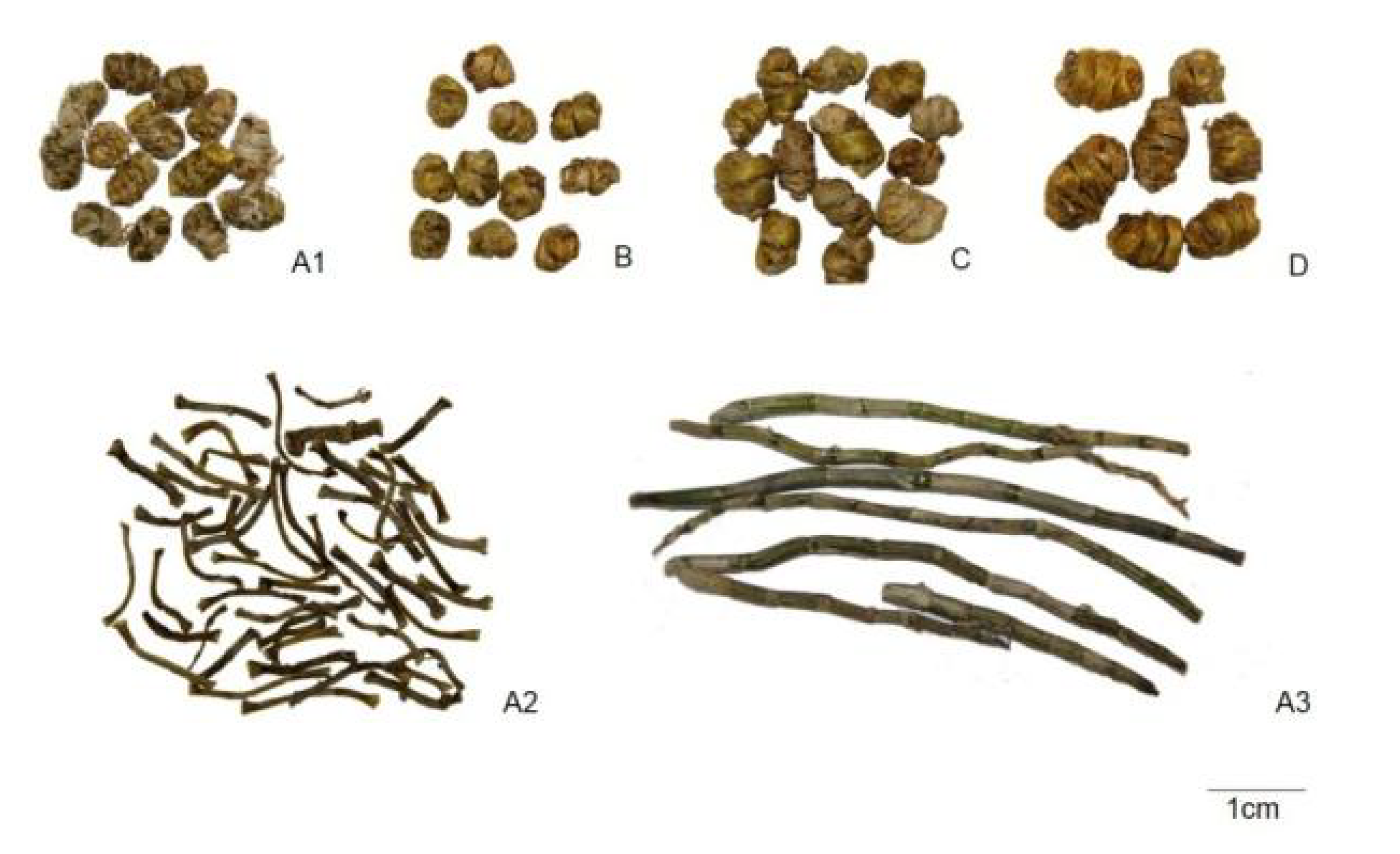
2.1. Macroscopic Characters
2.1.1. Stems of D. officinale

2.1.2. Stems of D. devonianum
2.1.3. Stems of D. aphyllum
2.1.4. Stems of D. gratiosissimum
2.2. Microscopic Characters
2.2.1. Transverse Section of Stems (Observed Under Normal Light Microscope)
2.2.1.1. Stems of D. officinale–Outline is nearly circular (Table 1 and Table 2)
- (1).
- Epidermis: a row of cells, thin and flat, 17–45 μm in diameter, lateral walls were slightly lignified, covered with yellow to orange cuticles. A layer of pericladium consisting of parenchymatous cells and vascular bundles can be observed outside the epidermis sometimes.
- (2).
- Parenchyma: Parenchymatous cells similar in size, usually smaller near the vascular bundles. Parenchymatous cells containing raphides, starch granules or silica masses can be observed.
- (3).
- Vascular bundles: Closed collateral vascular bundles, 78 (52)–134, arranged in 4–5 whorls in parenchyma, with similar size.
- (4).
- Fiber groups: outside vascular bundles, two hat-shaped, consisting of 9–58 fiber cells, 5–37 μm in diameter. Occasionally, a hat-shaped or a ring-shaped fiber group can be observed.
- (5).
- Raphides: occurring in parenchymatous cells near the epidermis, (10) 52–194 μm in length.
- (6).
- Silica masses: occurring in parenchymatous cells outside the vascular bundles.
2.2.1.2. Stems of D. devonianum—Outline is nearly circular (Table 1 and Table 3)
- (1).
- Epidermis: a row of cells, thin and flat, 17–85 μm in diameter, lateral walls were slightly lignified, covered with yellow to orange cuticles. A layer of pericladium consisting of parenchymatous cells and vascular bundles can sometimes be observed outside the epidermis.
- (2).
- Parenchyma: Parenchymatous cells various in size, usually small near the vascular bundles. Parenchymatous cells containing raphides, starch granules or silica masses can be observed.
- (3).
- Vascular bundles: closed collateral vascular bundles, 57–91, various in size.
- (4).
- Fiber groups: outside vascular bundles, hat-shaped, consisting of 7–43 of fiber cells, 7–45 μm in diameter.
- (5).
- Raphides: occurring in parenchymatous cells near the epidermis and vascular bundles, 51–132 μm in length.
- (6).
- Silica masses: occurring in parenchymatous cells outside the vascular bundles.
2.2.1.3. Stems of D. aphyllum—Outline is nearly circular (Table 1 and Table 4)
- (1).
- Epidermis: a row of cells, thin and flat, 13–38 μm in diameter, lateral walls were slightly lignified, covered with yellow to orange cuticles. A layer of pericladium consisting of parenchymatous cells and vascular bundles can be observed outside the epidermis sometimes.
- (2).
- Parenchyma: Parenchymatous cells similar in size, usually smaller near the vascular bundles. Parenchymatous cells containing raphides, starch granules or silica masses can be observed.
- (3).
- Vascular bundles: closed collateral vascular bundles, 53–85, with similar size.
- (4).
- Fiber groups: outside vascular bundles, hat-shaped, consisting of 8–47 fiber cells, 5–33 μm in diameter.
- (5).
- Raphides: scattered or in bundles, non-specific raphide distribution, 65–202 μm in length.
- (6).
- Silica masses: occurring in parenchymatous cells outside the vascular bundles.
2.2.1.4. Stems of D. gratiosissimum—Outline is nearly circular (Table 1 and Table 5)
- (1).
- Epidermis: a row of cells, thin and flat, 11–35 μm in diameter, lateral walls were slightly lignified, covered with yellow to orange cuticles.
- (2).
- Parenchyma: Parenchymatous cells various in size, usually small near the vascular bundles. Parenchymatous cells containing raphides, starch granules or silica masses can be observed.
- (3).
- Vascular bundles: closed collateral vascular bundles, 57–84 (124), various in size.
- (4).
- Fiber groups: outside vascular bundles, hat-shaped, consisting of 19–66 fiber cells, 6–31 μm in diameter.
- (5).
- Raphides: occurring in parenchymatous cells near vascular bundles, 39–124 μm in length.
- (6).
- Silica masses: occurring in parenchymatous cells outside the vascular bundles.
2.2.2. Transverse Section of Stems (Observed Under Fluorescence Microscope)

| 40× * | 100× * | 200× * | |
|---|---|---|---|
| D. officinale |  | ||
| D. devonianu | |||
| D. aphyllum | |||
| D. gratiosissimum | |||
| 500 µm | 250 µm | 100 µm | |
| Sample No. | Cuticle | Epidermis | Fiber | Vascular bundles | Raphide | |||
|---|---|---|---|---|---|---|---|---|
| Thickness | Diameter | Number | Diameter | Number | Diameter | Length | ||
| Tangential | radial | |||||||
| TP-1 | 9–12 | 14–34 | 17–29 | 8–19 | 78–90–102 | 73–125 | 85–229 | 65–68 |
| TP-2 | 10–15 | 16–45 | 15–26 | 12–27 | 52–61–77 | 55–83 | 92–173 | 100–112 |
| TP-3 | 8–13 | 12–29 | 16–37 | 10–28 | 84–92–108 | 61–118 | 80–187 | 75–79 |
| TP-4 | 8–10 | 17–33 | 21–26 | 6–23 | 87–98–111 | 59–100 | 72–183 | 65–71 |
| TP-5 | 8–10 | 13–26 | 20–58 | 8–24 | 84–88–108 | 67–104 | 90–236 | 100–103 |
| TP-6 | 10–11 | 14–39 | 12–25 | 9–25 | 83–92–101 | 46–101 | 60–158 | 76–82 |
| TP-7 | 9–11 | 12–37 | 14–22 | 11–29 | 54–69–86 | 42–140 | 51–147 | 59–96 |
| TP-8 | 10–13 | 23–37 | 10–35 | 8–19 | 84–96–118 | 45–102 | 67–168 | 68–72 |
| TP-9 | 9–10 | 17–27 | 20–30 | 5–18 | 85–92–103 | 45–89 | 50–135 | 53–92 |
| TP-10 | 10–12 | 15–30 | 18–46 | 6–25 | 89–93–108 | 67–103 | 98–159 | 10–15 |
| TP-11 | 10–18 | 13–38 | 18–33 | 6–20 | 79–93–99 | 50–102 | 63–169 | 146–194 |
| TP-12 | 8–12 | 12–28 | 12–26 | 7–24 | 85–97–103 | 48–83 | 62–129 | 62–93 |
| TP-13 | 10––12 | 12–36 | 11–46 | 7–31 | 61–65–97 | 56–119 | 73–196 | 62–156 |
| TP-14 | 8–15 | 13–30 | 12–34 | 6–34 | 89–94–110 | 49–127 | 72–197 | 48–81 |
| TP-15 | 9–21 | 11–39 | 9–31 | 8–37 | 93–109–134 | 54–145 | 71–237 | 52–190 |
| Total | 8–21 | 11–45 | 9–58 | 5–37 | 78(52) –134 | 42–145 | 50–237 | 10–194 |
| Sample No. | Cuticle | Epidermis | Fiber | Vascular bundles | Raphide | |||
|---|---|---|---|---|---|---|---|---|
| Thickness | Diameter | Number | Diameter | Number | Diameter | Length | ||
| Tangential | Radial | |||||||
| ZP-1 | 10–11 | 17–26 | 14–29 | 5–19 | 58–72–84 | 74–134 | 135–208 | 74–90 |
| ZP-2 | 11–12 | 32–85 | 31–37 | 11–45 | 72–76–91 | 172–283 | 296–487 | 51–132 |
| ZP-3 | 13–20 | 50–72 | 7–43 | 7–25 | 57–64–80 | 62–171 | 82–265 | 52–63 |
| Total | 11–20 | 17–85 | 7–43 | 7–45 | 57–91 | 62–283 | 82–487 | 51–132 |
| Sample No. | Cuticle | Epidermis | Fiber | Vascular bundles | Raphide | |||
|---|---|---|---|---|---|---|---|---|
| Thickness | Diameter | Number | Diameter | Number | Diameter | Length | ||
| Tangential | Radial | |||||||
| SC-1 | 9–14 | 17–37 | 11–36 | 8–25 | 56–67–84 | 84–174 | 82–251 | 93–115 |
| SC-2 | 14–16 | 13–30 | 20–47 | 5–33 | 64–72–85 | 88–127 | 99–281 | 65–182 |
| SC-3 | 10–12 | 19–38 | 8–22 | 7–20 | 53–66–79 | 73–166 | 81–207 | 143–202 |
| Total | 9–16 | 13–38 | 8–47 | 5–33 | 53–85 | 73–174 | 81–281 | 65–202 |
| Sample No. | Cuticle | Epidermis | Fiber | Vascular bundles | Raphide | |||
|---|---|---|---|---|---|---|---|---|
| Thickness | Diameter | Number | Diameter | Number | Diameter | Length | ||
| Tangential | Radial | |||||||
| GJ-1 | 8–10 | 11–32 | 20–66 | 6–31 | 57–65–124 | 104–346 | 89–218 | 53–124 |
| GJ-2 | 17–18 | 15–33 | 23–52 | 6–29 | 59–72–81 | 108–293 | 92–173 | 39–73 |
| GJ-3 | 16–17 | 17–35 | 19–31 | 7–24 | 68–71–84 | 127–278 | 102–225 | 51–71 |
| Total | 8–18 | 11–35 | 19–66 | 6–31 | 57–84(124) | 104–346 | 89–225 | 39–124 |
| 40× * | 200× * | |||
|---|---|---|---|---|
| B-1 | G-1 | B-1 | G-1 | |
| D. officinale |  | |||
| D. devonianum | ||||
| D. aphyllum | ||||
| D. gratiosissimum | ||||
| 500 µm | 100 µm | |||
2.3. Discussion
- (1)
- Cuticle: color and thickness of cuticle of four studied Dendrobium species are similar.
- (2)
- Epidermis: size of epidermal cells of D. officinale, D. aphyllum and D. gratiosissimum are similar, epidermal cells of D. devonianum is bigger than the other three species, up to 85 μm in diameter.
- (3)
- Vascular bundle: Vascular bundles of D. officinale, usually more than 90, are more abundant than in the three adulterants. The size of vascular bundles in D. officinale is similar apart from those near the epidermis which are slightly smaller. Vascular bundles of D. devonianum are about 70 in number, various in size, and much bigger in the centre than at the margins of the stem. Vascular bundles of D. aphyllum are about 65, with similar size, apart from those near to theepidermis that are slightly smaller. Vascular bundles of D. gratiosissimum are about 70, althoughmore than 100 can be observed occasionally. Various in size with no apparent distribution rules.
- (4)
- Fiber groups: D. officinale with “two hat-shaped” fiber groups, while the other three species only have “one hat-shaped” fiber groups.
- (5)
- Raphides: D. officinale scattered in parenchmatous cells near epidermis. D. devonianum scatteredin parenchmatous cells near epidermis and vascular bundles. D. aphyllum scattered in parenchmatous cells throughout. D. gratiosissimum scattered in parenchmatous cells near vascular bundles.
| (1). “Two hat-shaped” fiber groups emitted green fluorescence can be easily observed, Raphides distribute near the epidermis | D. officinale | |
| (1). “One hat-shaped” fiber groups emitting green fluorescence can be easily observed | ||
| (2). Vascular bundles similar in size, non-specific raphide distribution | D. aphyllum | |
| (2). Vascular bundles various in size | ||
| (3). Vascular bundles are much bigger in the centre than in the margin of the stem. Raphides distribute near the epidermis and vascular bundles | D. devonianum | |
| (3). Vascular bundles of different size distribute with no obvious rule. Raphides distribute near the vascular bundle | D. gratiosissimum | |
3. Experimental Section
3.1. Materials
3.1.1. Samples
| Sample No. | Origin | Collection area/market | Collection date | Trade name |
|---|---|---|---|---|
| TP-1 | D. officinale | GAP bases, Pu’er, Yunnan Province | September 2011 | Tie-pi-shi-hu |
| TP-2 | D. officinale | GAP bases, Tiantai, Zhejiang Province | October 2011 | Tie-pi-shi-hu |
| TP-3 | D. officinale | GAP bases, Tiantai, Zhejiang Province | September 2011 | Tie-pi-shi-hu |
| TP-4 | D. officinale | GAP bases, Tiantai, Zhejiang Province | August 2011 | Tie-pi-shi-hu |
| TP-5 | D. officinale | GAP bases, Tiantai, Zhejiang Province | April 2013 | Tie-pi-shi-hu |
| TP-6 | D. officinale | GAP bases, Tiantai, Zhejiang Province | May 2013 | Tie-pi-shi-hu |
| TP-7 | D. officinale | GAP bases, Tiantai, Zhejiang Province | Marrch 2013 | Tie-pi-shi-hu |
| TP-8 | D. officinale | Pan’an market, Zhejiang Province | September 2013 | Tie-pi-shi-hu |
| TP-9 | D. officinale | Pan’an market, Zhejiang Province (fresh) | September 2013 | Tie-pi-shi-hu |
| TP-10 | D. officinale | Pan’an market, Zhejiang Province (fresh) | September 2013 | Tie-pi-shi-hu |
| TP-11 | D. officinale | Wuyi, Zhejiang Province (fresh) | July 2013 | Tie-pi-shi-hu |
| TP-12 | D. officinale | Wuyi, Zhejiang Province (fresh) | July 2013 | Tie-pi-shi-hu |
| TP-13 | D. officinale | Pu’er, Yunnan Province (fresh) | July 2013 | Tie-pi-shi-hu |
| TP-14 | D. officinale | Pu’er, Yunnan Province (fresh) | July 2013 | Tie-pi-shi-hu |
| TP-15 | D. officinale | Pu’er, Yunnan Province (fresh) | July 2013 | Tie-pi-shi-hu |
| ZP-1 | D. devonianum | Myanmar (fresh) | January 2013 | Zi-pi-shi-hu |
| ZP-2 | D. devonianum | Pan’an market, Zhejiang Province | September 2013 | Zi-pi-shi-hu |
| ZP-3 | D. devonianum | Pan’an market, Zhejiang Province | September 2013 | Zi-pi-shi-hu |
| SC-1 | D. aphyllum | Myanmar (fresh) | January 2013 | Shui-cao-shi-hu |
| SC-2 | D. aphyllum | Pan’an market, Zhejiang Province | September 2013 | Shui-cao-shi-hu |
| SC-3 | D. aphyllum | Pan’an market, Zhejiang Province | September 2013 | Shui-cao-shi-hu |
| GJ-1 | D. gratiosissimum | Myanmar (fresh) | January 2013 | Guang-jie-shi-hu |
| GJ-2 | D. gratiosissimum | Pan’an market, Zhejiang Province | September 2013 | Guang-jie-shi-hu |
| GJ-3 | D. gratiosissimum | Pan’an market, Zhejiang Province | September 2013 | Guang-jie-shi-hu |
3.1.2. Apparatus
- (a)
- Optika digital camera DS-Fi1
- (b)
- Optika Microscope equipped with CCD from Photometrics Coolsnap to capture photos.
- (c)
- Optika Fluorescence Microscope B-600TiFL
- (d)
- Canon digital camera 550D
3.1.3. Software
3.2. Method
3.2.1. Morphological Characteristics of four Dendrobium Stems
3.2.2. Transverse Section of Four Dendrobium Stems

4. Conclusions
Acknowledgments
Author Contributions
Conflictts of Interest
References
- Editorial Board of Flora of China. Angiospermae, Monocotyledoneae. Flora Reipublicae Popularis Sinica; (in Chinese). Science Press: Beijing, China, 1999; Volume 19, pp. 67–146. [Google Scholar]
- China Pharmacopoeia Committee. Pharmacopoeia of China; Medical Science Press: Beijing, China, 2010; Volume I, pp. 265–266. [Google Scholar]
- Lin, X.; Shaw, P.C.; Sze, S.C.; Tong, Y.; Zhang, Y. Dendrobium officinale polysaccharides ameliorate the abnormality of aquaporin 5, pro-inflammatory cytokines and inhibit apoptosis in the experimental Sjogren’s syndrome mice. Int. Immunopharmacol. 2011, 11, 2025–2032. [Google Scholar] [CrossRef]
- Xiang, L.; Stephen Sze, C.W.; Ng, T.B.; Tong, Y.; Shaw, P.C.; Sydney Tang, C.W.; Kalin Zhang, Y.B. Polysaccharides of Dendrobium officinale inhibit TNF-alpha-induced apoptosis in A-253 cell line. Inflamm. Res. 2013, 62, 313–324. [Google Scholar]
- Xiao, L.; Ng, T.B.; Feng, Y.B.; Yao, T.; Wong, J.H.; Yao, R.M.; Li, L.; Mo, F.Z.; Xiao, Y.; Shaw, P.C.; et al. Dendrobium candidum extract increases the expression of aquaporin-5 in labial glands from patients with Sjogren’s syndrome. Phytomedicine 2011, 18, 194–198. [Google Scholar]
- Kowitdamrong, A.; Chanvorachote, P.; Sritularak, B.; Pongrakhananon, V. Moscatilin inhibits lung cancer cell motility and invasion via suppression of endogenous reactive oxygen species. Biomed. Res. Int. 2013, 2013, 765894. [Google Scholar]
- Xiao, Y.Y.; Wen, J.Y; Fan, J.S. Pharmacognostic identification of Dendrobium officinale and Dendrobium devonianum. Strait Pharm. J. 2011, 23, 52–53. [Google Scholar]
- Li, J.; Li, S.X.; Huang, D. Advances in the resources, constituents and pharmacological effects of Dendrobium officinale. Sci. Technol. Rev. 2011, 29, 74–79. [Google Scholar]
- Chen, X.; Wang, F.; Wang, Y.; Li, X.; Wang, A.; Wang, C.; Guo, S. Discrimination of the rare medicinal plant Dendrobium officinale based on naringenin, bibenzyl, and polysaccharides. Science China. Life Sci. 2012, 55, 1092–1099. [Google Scholar]
- Yan, M.Q.; Chen, S.H.; Lv, G.Y.; Zhou, G.F.; Liu, X. HPLC specific chromatogram of Dendrobium officinale. China J. Chin. Mater. Med. 2013, 38, 516–519. [Google Scholar]
- Lv, X.K.; Cheng, C.G.; Yang, G.P.; Jin, Y.; Ye, H.; Xu, D.W. Appl ication of FTIR spectroscopy to the analysis of eleven kinds of Dendrobium. China J. Chin. Mater. Med. 2005, 30, 738–740. [Google Scholar]
- Zhang, W.; Ding, X.; Xie, M.; Feng, Z.; Lu, S.; Li, X.; Zhang, F.; Ding, G. Authentication of three valuable Dendrobium species by adapter ligation-mediated allele-specific amplification. Eur. Food Res. Technol. 2009, 229, 1–7. [Google Scholar] [CrossRef]
- Xu, H.; Ying, Y.; Wang, Z.T.; Cheng, K.T. Identification of Dendrobium species by dot blot hybridization assay. Biol. Pharm. Bull. 2010, 33, 665–668. [Google Scholar] [CrossRef]
- Xu, L.S.; Xu, G.J.; Sha, W.L.; Luo, J.Y. Studies on the microscopic identification of Chinese drug Shi-Hu. J. China Pharm. Univ. 1980, 2, 1–7. [Google Scholar]
- Guan, Y.H.; Li, H.T.; Wang, Y.Q.; Li, X.L. Comparison of Microscopical characters between Dendrobium officinale and Dendrobium devonianum. J. Chin. Med. Mater. 2010, 33, 1869–1871. [Google Scholar]
- Liu, X.P.; Tang, M.H.; Dai, Y.; Liu, H.J.; Xu, G.J.; Xu, L.S. Microscopic identification of the powder of Chinese drug Shihu. J. China Pharm. Univ. 1992, 22, 148–151. [Google Scholar]
- Liang, Z.T.; Jiang, Z.H.; Leung, K.S.; Peng, Y.; Zhao, Z.Z. Distinguishing the medicinal herb Oldenlandia diffusa from similar species of the same genus using fluorescence microscopy. Microsc. Res. Tech. 2006, 69, 277–282. [Google Scholar] [CrossRef]
- Wang, Y.Q.; Liang, Z.T.; Li, Q.; Yang, H.; Chen, H.B.; Zhao, Z.Z.; Li, P. Identification of powdered Chinese herbal medicines by fluorescence microscopy, Part 1: Fluorescentcharacteristics of mechanical tissues, conducting tissues, and ergastic substances. Microsc. Res. Tech. 2011, 74, 269–280. [Google Scholar] [CrossRef]
- Wan, X.J.; Liang, Z.T.; Chen, H.B.; Zhao, Z.; Li, P. Identification of Daqingye and Banlangen including crude drugs and decoction dregs from three plant species by normal light and fluorescence microscopy. Microsc. Res. Tech. 2013, 76, 774–782. [Google Scholar] [CrossRef]
- Tang, Y.N.; He, X.C.; Chen, Q.L.; Fan, L.L.; Zhang, J.Y.; Zhao, Z.Z.; Dong, L.S.; Liang, Z.T.; Yi, T.; Chen, H.B. A mixed microscopic method for differentiating seven species of “Bixie”-related Chinese Materia Medica. Microsc. Res. Tech. 2014, 77, 57–70. [Google Scholar] [CrossRef]
- Sample Availability: Not available.
© 2014 by the authors. Licensee MDPI, Basel, Switzerland. This article is an open access article distributed under the terms and conditions of the Creative Commons Attribution license ( http://creativecommons.org/licenses/by/3.0/).
Share and Cite
Chu, C.; Yin, H.; Xia, L.; Cheng, D.; Yan, J.; Zhu, L. Discrimination of Dendrobium officinale and Its Common Adulterants by Combination of Normal Light and Fluorescence Microscopy. Molecules 2014, 19, 3718-3730. https://doi.org/10.3390/molecules19033718
Chu C, Yin H, Xia L, Cheng D, Yan J, Zhu L. Discrimination of Dendrobium officinale and Its Common Adulterants by Combination of Normal Light and Fluorescence Microscopy. Molecules. 2014; 19(3):3718-3730. https://doi.org/10.3390/molecules19033718
Chicago/Turabian StyleChu, Chu, Huimin Yin, Li Xia, Dongping Cheng, Jizhong Yan, and Lin Zhu. 2014. "Discrimination of Dendrobium officinale and Its Common Adulterants by Combination of Normal Light and Fluorescence Microscopy" Molecules 19, no. 3: 3718-3730. https://doi.org/10.3390/molecules19033718