Article

Evaluation of the Nano-TiO2 as a Novel Deswelling Material

1
Department of Immunology, School of Basic Medical Sciences, Peking University, Beijing 100191, China
2
Key Laboratory of Medical Immunology, Ministry of Health, Beijing 100191, China
3
Pharmacy Department, Liao Ning University of Traditional Chinese Medicine, Shenyang 116600, Liao Ning, China
*
Authors to whom correspondence should be addressed.
Molecules 2016, 21(1), 57; https://doi.org/10.3390/molecules21010057
Submission received: 26 September 2015 / Revised: 18 December 2015 / Accepted: 28 December 2015 / Published: 4 January 2016
(This article belongs to the Collection Nanomedicine)

Abstract

:
Nano-TiO2 is widely applied in the automobile exhaust hose reels as a catalyst to reduce oxynitride emissions, including nitric oxide (NO). In the biomedicine field, NO plays an important role in vasodilation and edema formation in human bodies. However, the deswelling activity of nano-TiO2 has not been reported. Here, we demonstrated that nano-TiO2 can significantly degrade the production of NO in LPS-induced RAW264.7 mouse macrophages. Further study indicated that nano-TiO2 exhibited an effect on vascular permeability inhibition, and prevented carrageenan-induced footpad edema. Therefore, we prepared a nano-TiO2 ointment and observed similar deswelling effects. In conclusion, nano-TiO2 might act as a novel deswelling agent related with its degradation of NO, which will aid in our ability to design effective interventions for edema involved diseases.

1. Introduction

Nitric oxide is a by-product of combustion of the substances in the air, and it is abundant in automobile engines. Because of the large vehicle population, significant amounts of NOx are emitted to the atmosphere. Attention is given to the catalyst system which simultaneously promotes the reduction of NO. Most reports show that NO can be broke into N2 and O2 by nano-TiO2 as a catalyst with the help of UV light and sensible light [1,2]. In mammals including humans, NO is also an important cellular signaling molecule involved in many physiological and pathological processes. NO, known as an endothelium-derived relaxing factor (EDRF), is a powerful vasodilator with a short half-life of a few seconds in the blood. The endothelium of blood vessels uses NO to signal the surrounding smooth muscle to relax, thus resulting in vasodilation [3,4,5]. Recent studies have shown that NO is also essential for host innate immune responses to pathogens such as viruses, bacteria, fungi, and parasites, which is mainly generated by macrophages [6,7,8,9]. However, excessive production of NO is a common feature of most diseases associated with infection and acute or chronic inflammation, which contributes to edema formation and pain sensitization [10,11].
In the present study, we examined whether nano-TiO2 might act as a deswelling material. To gain insights into the molecular mechanism, we further investigated the effects of nano-TiO2 on degrading NO and inhibiting vascular permeability. We believe that further study on nano-TiO2 will aid in our ability to design effective interventions and treatments for edema involved diseases.

2. Results and Discussion

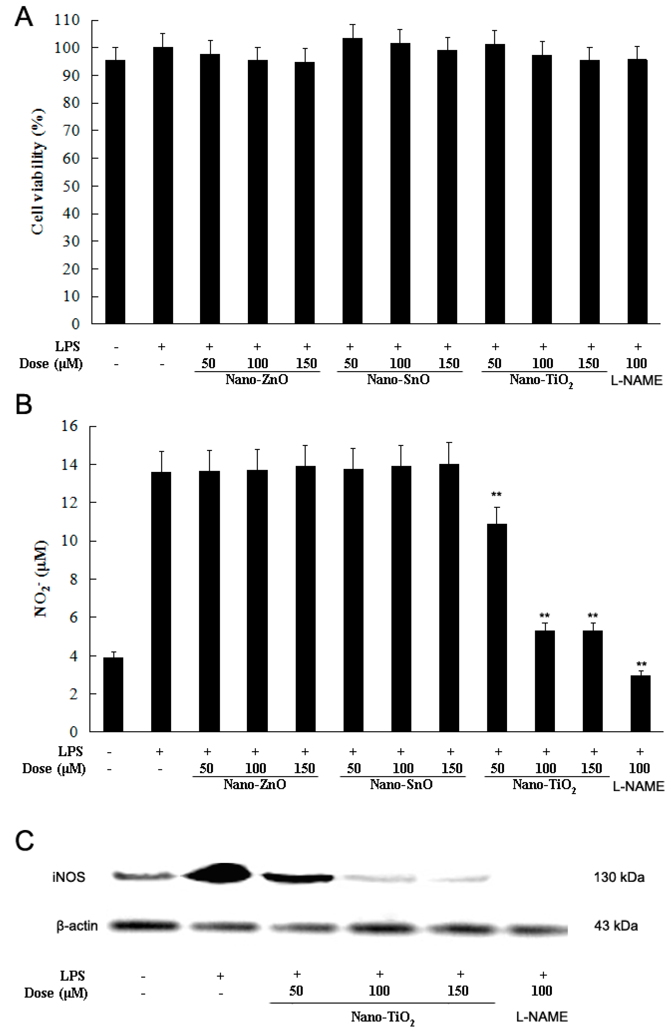
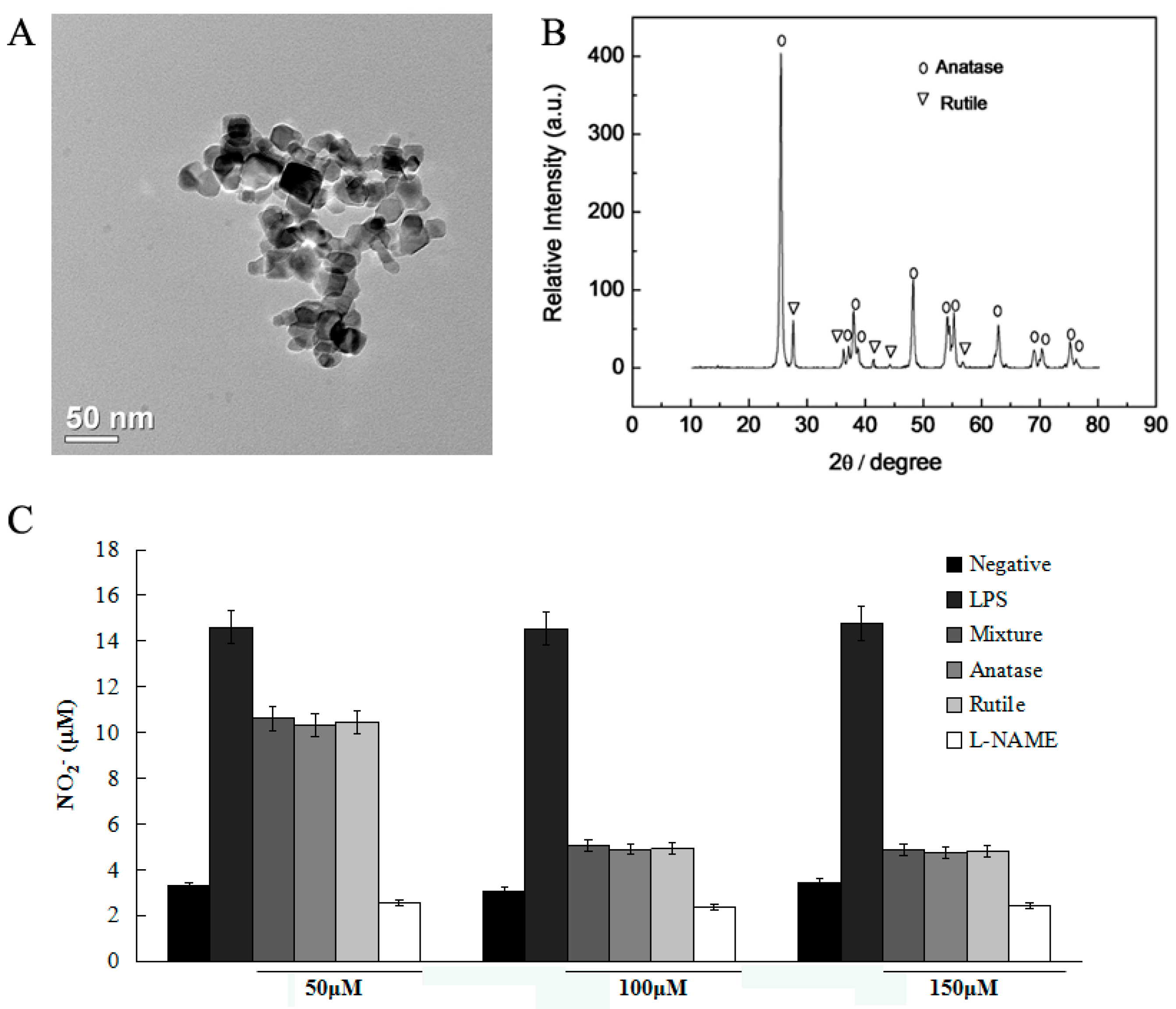
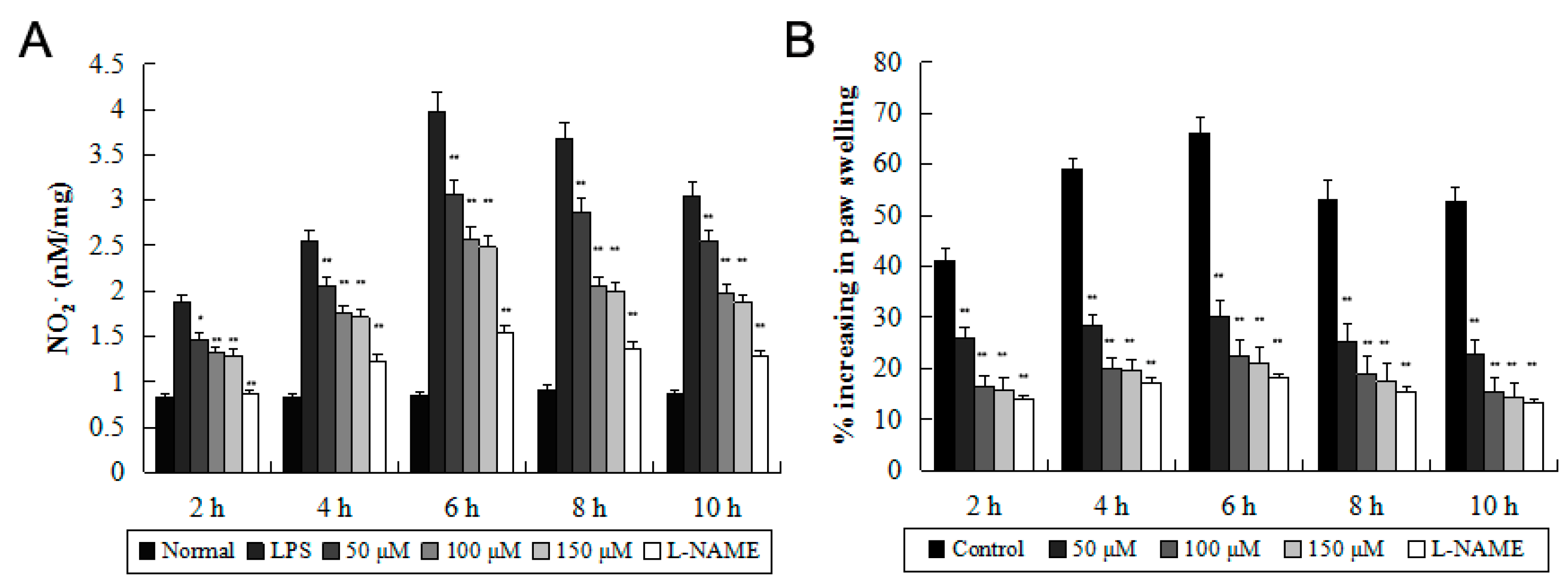
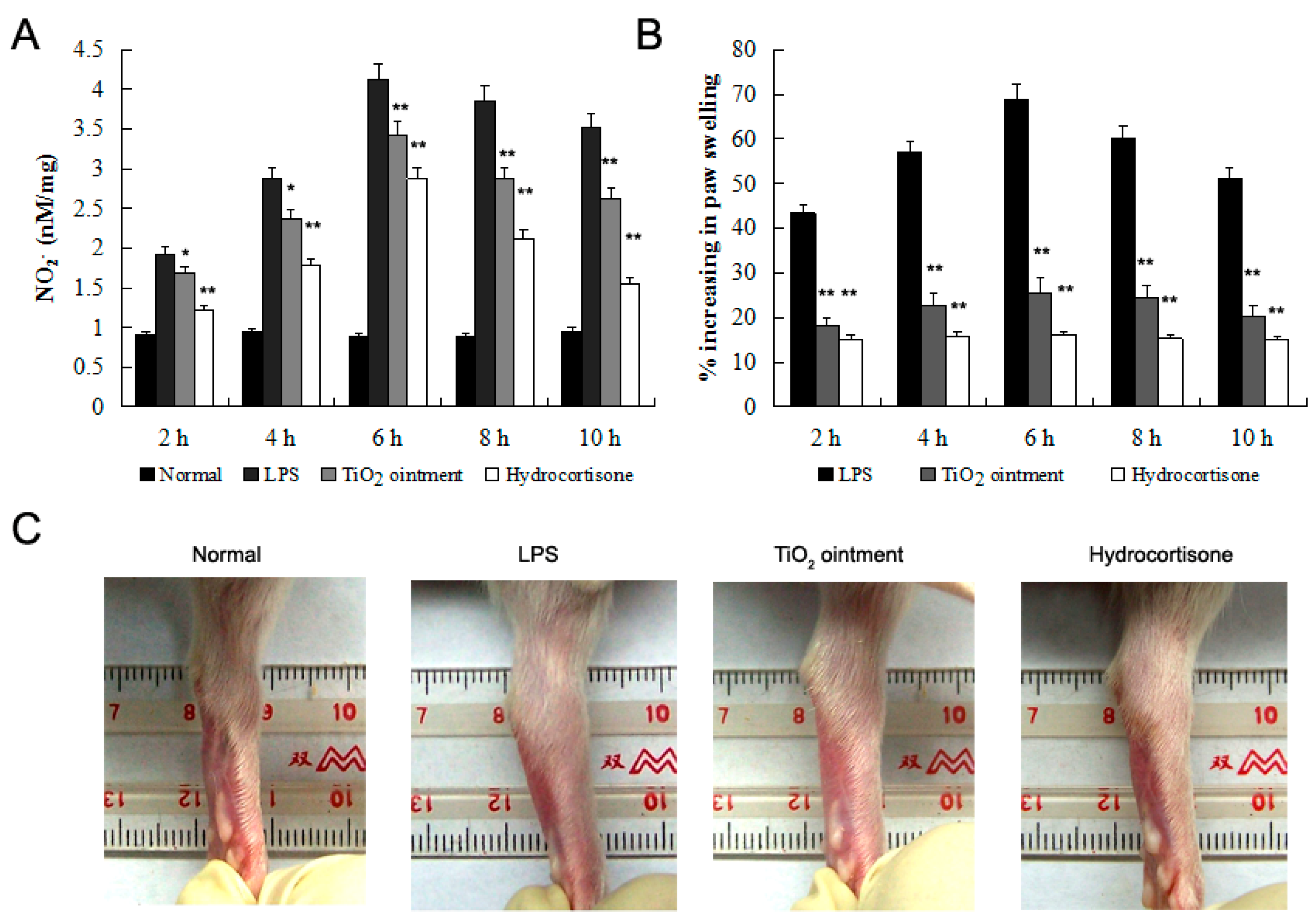
2.1. Effects of Nano-TiO2 on LPS-Induced NO Production

Nano-TiO2 is demonstrated to be active on NO absorption in the air. In mammals including humans, NO is mainly generated by phagocytes (monocytes, macrophages, and neutrophils). Here, we used RAW264.7 mouse macrophages to investigate the effects of nano-TiO2 on LPS-induced NO production in vitro.
We first measured the cytotoxicity of nano-materials in RAW264.7 cells by using the MTT assay. RAW264.7 cells were cultured with LPS (100 ng/mL) in the presence or absence of nano-materials. As shown in Figure 1A, nano-ZnO, nano-SnO and nano-TiO2 at the concentrations of 50, 100 and 150 μM had no cytotoxic effect.
Figure 1. Effects of nano-TiO2 on cell viability and nitric oxide (NO) production in LPS-induced RAW 264.7 mouse macrophages. After 24 h treatment, cell viability (A) was evaluated by MTT assay and NO production (B) was measured by the Griess reaction. Normal group was treated with media only. Control group was treated with LPS (100 ng/mL) alone. Data were shown as means ± standard deviation (SD) of three independent experiments. ** p < 0.01 against control group; (C) The expression of iNOS was dectected by Western blot.
Molecules 21 00057 g001
NO is a relatively unstable molecule, which is produced at low concentrations and rapidly converted into nitrate within 10 s of its formation [12]. Thus, the concentration of nitrate is commonly used to reflect the levels of NO production. Following 24 h of LPS stimulation, a higher level of NO production were measured in the culture media of RAW264.7 cells treated with LPS alone when compared to the untreated cells. However, the significant induction of NO production was reduced by nano-TiO2 in a dose-dependent manner, whereas other nano-materials did not reduce the nitrate levels (Figure 1B). We further demonstrated that the expression of iNOS in macrophages decreased after treated with nano-TiO2 at the concentration of 50, 100 and 150 μM. Thus, nano-TiO2 might be a candidate material for relieving vasodilation concerned with excessive NO production.
Moreover, we analyzed the nano-size of the nano-TiO2 used in this study, and separated it into anatase and rutile type (Figure 2). Then, we detected the effect of nano-TiO2 in different types on NO degradation. Both anatase and rutile nano-TiO2 presented NO degradation action with no significant differences (Figure 2C). Thus, we chose the anatase nano-TiO2 for the further study.
Figure 2. Analysis of nano-TiO2. (A) Nano-TiO2 was measured by electron microscope to a diameter of 21 nm; (B) Nano-TiO2 was analyzed by XRD method, and separated into anatase and rutile type; (C) Anatase and rutile nano-TiO2 presented NO degradation action with no significant differences. Data were shown as means ± SD of three independent experiments.
Molecules 21 00057 g002

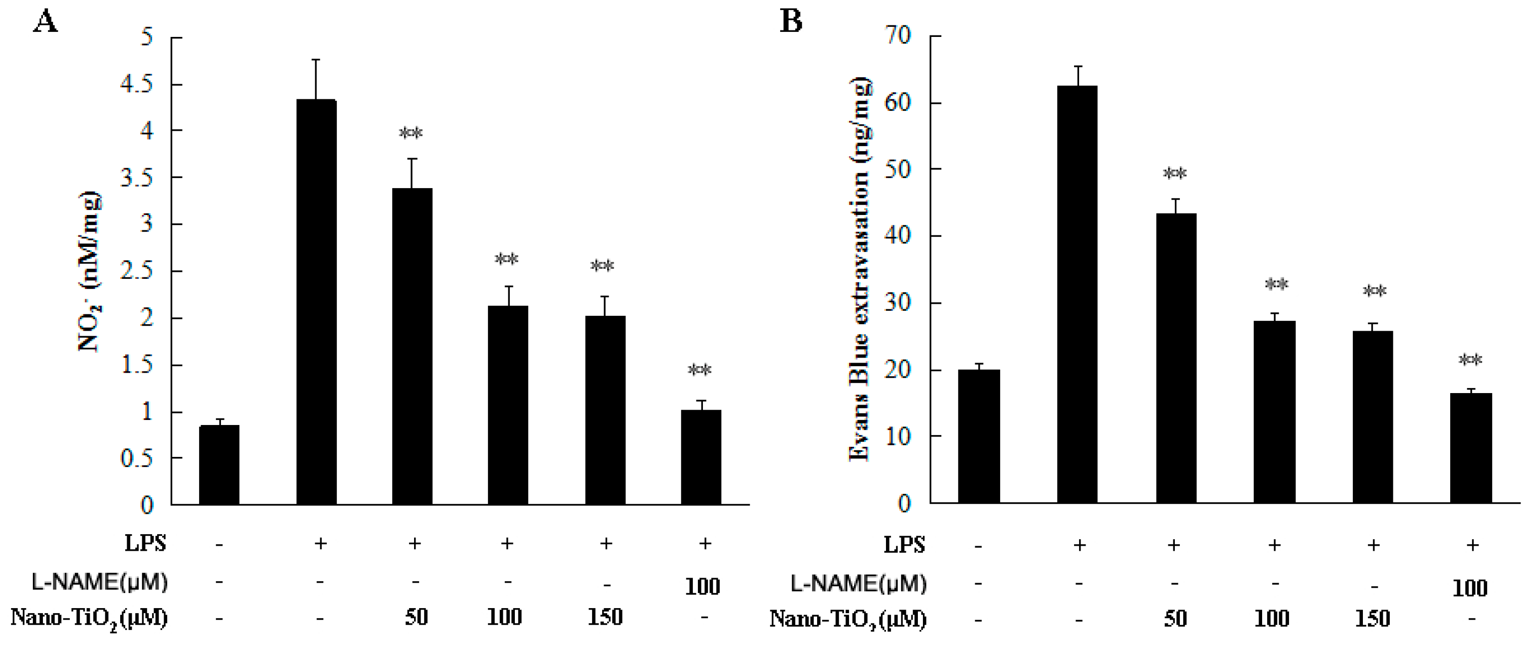
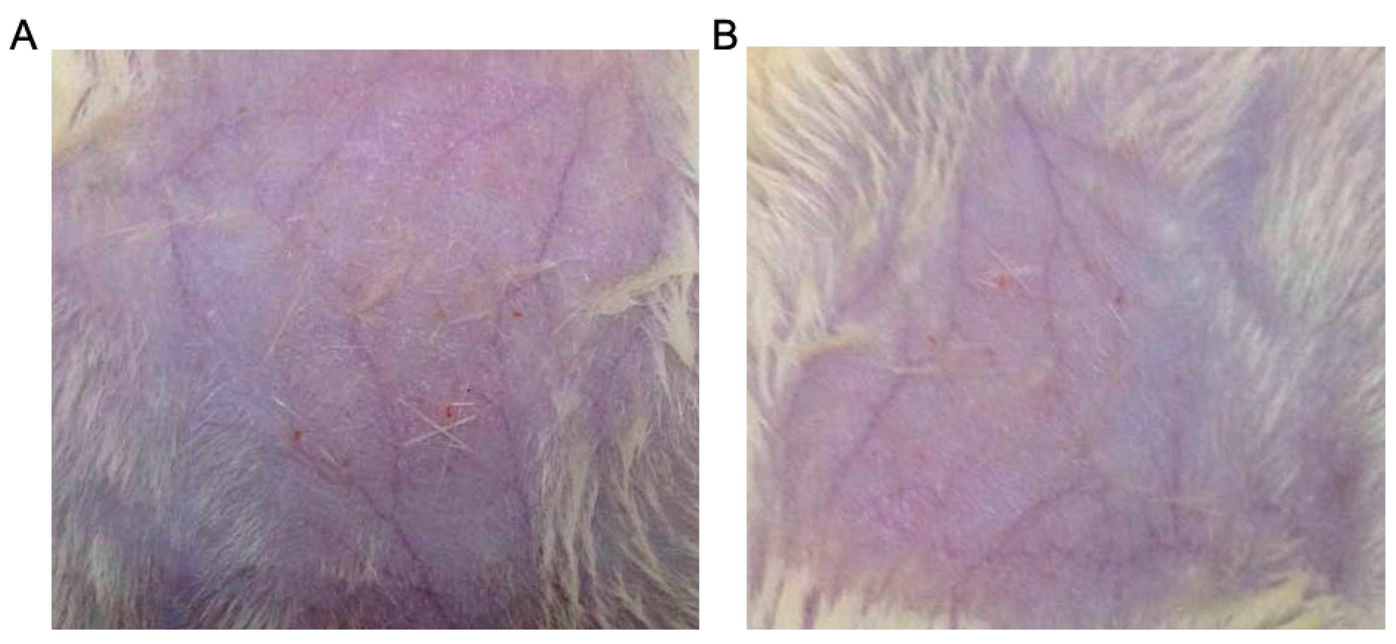
2.2. Effects of Nano-TiO2 on Inhibiting Vascular Permeability

In 1980, Furchgott and Zawadsko reported the crucial role endothelium in the relaxation of arterial smooth muscle by acetylcholine. The report was based on the integrity of endothelial cells and suggested that the endothelial cells might generate a special transfer molecule causing vascular smooth muscle cell (VSMC) relaxation. Thus, they named this molecule as endothelium-derived relaxing factor (EDRF) [3,4]. Later in 1986, Furchgott and Ignarro further proved that NO is the specific transfer molecule that played the role of EDRF by using spectral analysis of hemoglobin [5]. It is now known that NO can induce the synthesis of cyclic guanosine monophosophate (cGMP) through guanylyl cyclase (GC) leading to relaxation of myosin [13,14,15,16]. Thus, we wondered if nano-TiO2 might present an inhibitory effect on vascular permeability via degradation of NO in vivo.
We first investigated the inhibitory effect of nano-TiO2 on vascular permeability in female SD rats. The rats were subcutaneous injected with 50 μM, 100 μM and 150 μM nano-TiO2 for 1 h prior to the addition of LPS stimulation. NO production decreased significantly in the nano-TiO2 treated group (Figure 3A). Evans blue extravasation was employed to evaluate the vascular permeability [17]. As shown in Figure 3B, Evans blue extravasation was significantly decreased in the nano-TiO2 treated groups in a dose dependent manner when compared to the rats stimulated with LPS alone (p < 0.01).
Figure 3. Effects of nano-TiO2 on inhibiting vascular permeability. SD rats were injected with 50, 100, and 150 μM nano-TiO2 for 1 h prior to the addition of LPS stimulation, compared to 100 μM L-NAME. (A) NO2 in the skin was detected using the Griess method; (B) Evans blue extravasation was used to evaluate the vascular permeability. Data were shown as means ± SD of three independent experiments. ** p < 0.01 against control group.
Molecules 21 00057 g003
Nano-TiO2 is a white pigment widely used in foods, sunscreens, and cosmetic products [18,19,20,21]. Here, we prepared ointments which contained 5.0%, 10.0% and 15.0% of nano-TiO2 with vaseline and lanolin in the ratio of 1:2 as an accessory. Three points were marked on the median line of the depilated dorsal skin, where 2 mg of the nano-TiO2 ointment was rubbed carefully for 1 min on a circular area with a 2 cm diameter with each point. The nano-TiO2 ointment was applied 1 h before LPS subcutaneous injection. NO production decreased significantly in the nano-TiO2 treated group (Figure 4A). More importantly, the vascular permeability was significantly inhibited by nano-TiO2 ointment in a dose dependent manner when compared to the rats treated with accessories (Figure 4B).
Figure 4. Effects of nano-TiO2 ointment on inhibiting vascular permeability. Ointment contained 5.0%, 10.0%, and 15.0% of nano-TiO2 were rubbed on the depilated dorsal skin of rats. (A) NO2 in the skin was detected using the Griess method; (B) Evans blue dye extracted from the skin was measured. Data were shown as means ± SD of three independent experiments. ** p < 0.01 against control group.
Molecules 21 00057 g004

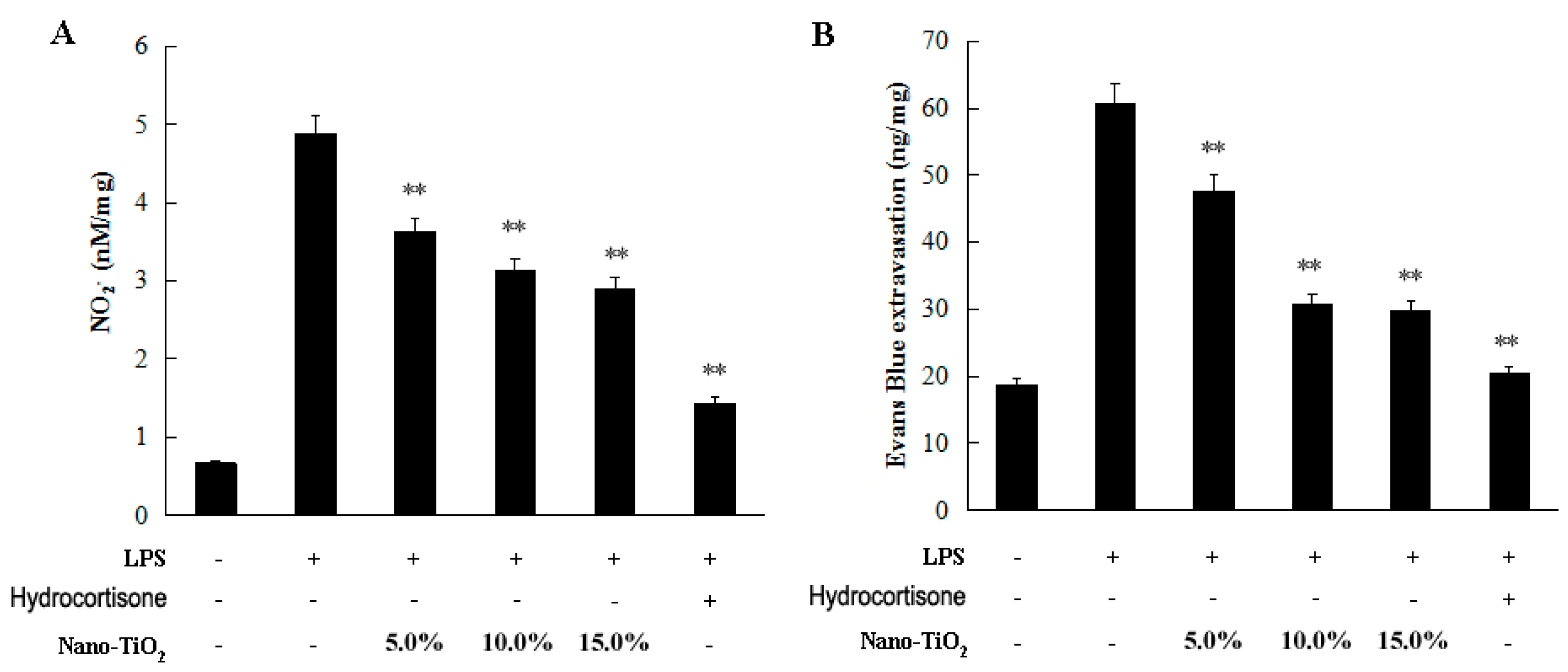
2.3. Effects of Nano-TiO2 on Carrageenan-Induced Paw Edema

An increase in vessel wall permeability contributes to the formation of edemas. Here, we used the carrageenan-induced paw edema model to evaluate the deswelling effect of nano-TiO2 [22,23,24].
In this study, SD rats were divided into five groups (eight animals in each group), and 0.1 mL of nano-TiO2 was administered 1 h before carrageenan stimulation. NO production decreased significantly in the footpad of nano-TiO2 treated rats (Figure 5A). In addition, subcutaneous injection of nano-TiO2 resulted in a significant reduction in rat paw edema. The deswelling effect of nano-TiO2 at doses of 50–150 μM was statistically significant for reducing paw edema of rats at 2, 4, 6, 8 and 10 h after induction of edema (Figure 5B).
Figure 5. Effect of nano-TiO2 on carrageenan-induced paw edema. 0.1 mL of nano-TiO2 (50–100 μM) was administered 1 h before carrageenan stimulation. (A) NO2 in the footpad was detected using the Griess method; (B) The degree of swelling in the footpads was measured using a plethysmometer. Data were shown as means ± SD of three independent experiments. * p < 0.05 against control group; ** p < 0.01 against control group.
Molecules 21 00057 g005
Then, we evaluate the inhibitory effect of nano-TiO2 ointment on carrageenan-induced paw edema. The volumes of the unilateral hind paws of these animals were measured, and on each paw, nano-TiO2 ointment was carefully rubbed 1 h before the carrageenan was given. This ointment-treated part was covered softly with a fiber cloth in order to prevent the rats from licking off the ointment. Then, carrageenan was injected subcutaneously into the paw, and the volume of the hind paw was measured at 2, 4, 6, 8, and 10 h. Ointment with 10% nano-TiO2 showed a significant reduction of NO release and rat paw edema (Figure 6). Carrageenan-induced edema develops through mediators in three phases. The early phase is caused by histamine release, the second phase is mediated by kinin, and the late phase is caused by prostaglandins. According to the NO degradation effects of nano-TiO2, we proposed that nano-TiO2 are effective for the first phase of edema formation.
Figure 6. Effect of nano-TiO2 ointment on carrageenan-induced paw edema. Ointment contained 10.0% of nano-TiO2 was smeared on the SD rats’ right footpads 1 h prior to the addition of carrageenan induction. (A) NO2 in the footpad was detected using the Griess method; (B) The degree of swelling in the footpads was measured using a plethysmometer; (C) The degree of swelling in the foot pads was photographed. Data were shown as means ± SD of three independent experiments. * p < 0.05 against control group; ** p < 0.01 against control group.
Molecules 21 00057 g006
Moreover, the application of the ointment with 15% nano-TiO2 to the skin of the rats for 1 month had no irritation on the animal skin (Figure 7). It is reported that nano TiO2 does not appear to significantly penetrate the intact skin [25]. However, the compromised skin allows nano-sized particle penetration through the skin [26]. Therefore, we believe that the nano-TiO2 ointment is available for the prevention and treatment of edema formation in the skin wounds.
Figure 7. Irritation of nano-TiO2 ointment on the skin of rats. Accessory (A) and ointment with 15% nano-TiO2 (B) was rubbed on the skin of rats for month.
Molecules 21 00057 g007

3. Experimental Section

3.1. Animals

All experiments was performed in compliance with relevant laws and institutional guidelines and approved by the Peking University Biomedical Ethics Committee. Female SD rats (4–6 weeks old) were provided by Beijing Laboratory Animal Research Center. The experimental animals were housed separately at room temperature (20 ± 2 °C), humidity 55%–60%.

3.2. Cell Culture

RAW264.7 mouse macrophages were purchased from American Type Culture Collection (ATCC, Manassas, VA, USA) and cultured in Dulbecco’s modified Eagle’s medium (DMEM, Gobico, NY, USA) supplemented with 100 µg/mL of penicillin/streptomycin and 10% heat-inactivated fetal bovine serum (FBS, Gibco, NY, USA), in a humidified atmosphere of 5% CO2 at 37 °C, until reaching 80% confluency [27]. The medium was changed every 3 days.

3.3. Nano Materials

Nano-TiO2, nano-ZnO and nano-SnO were purchased from Shanghai Huijing Sub-Nanoseale New Material Co., Ltd. (Shanghai, China). To prepare nano TiO2 ointment, we mixed vaseline and lanolin in the ratio of 1:2 as an accessory and then mixed with nano-TiO2 in different proportions.

3.4. Griess Method

We used a total Nitrate/Nitrite Parameter Assay Kit for determining NO according to the instructions (Catalog KGE001, R & D, Minneapolis, MN, USA) [28]. The completed reaction was read at 540 nm. The concentrations of NO2 in the cell culture supernatant were expressed as NO2 (µM). In addition, the concentration of NO2 in the skin or the footpad of rats were calculated as NO2 divided by the weight of tissue (nM/mg).

3.5. Western Blot Analysis

Total protein of RAW-264.7 cells were extracted and quantified respectively. After SDS-PAGE, the protein was transferred to polyvinylidene fluoride membrane. The membrane was incubated with antibodies against iNOS and β-actin (Sigma-Aldrich, Saint Louis, MO, USA) overnight at 4 °C. Then the membrane was incubated with the secondary antibodies and detected with enhanced chemiluminescence kit.

3.6. Vascular Permeability Assay

Evans blue dye at a concentration of 2% (40 mg/kg; Sigma-Aldrich) was injected into the great saphenous vein of 4- to 6-week-old mice. After 60 min, 2 cm-diameter free back skin graft was removed, blotted dry, and weighed. The Evans blue dye was extracted from the skin with 1 mL of formamide overnight at 55 °C and measured spectrophotometrically at 630 nm [17].

3.7. Carrageenan-Induced Paw Edema Method

The deswelling effect of nano-TiO2 were evaluated by the carrageenan-induced paw edema method [22]. Nano-TiO2 was administered 1 h before the rats received 0.1 mL of carrageenan (1%, w/v) into the subplantar area of the right hind paw. The control group was treated with the vehicle only. The paw volume of rats was measured before injecting carrageenan and at 2, 4, 6, 8, 10 h after carrageenan stimulation using a plethysmometer (MK-101P, Tokyo, Japan). The edema was expressed as the increase in paw volume, and the percentage of inhibition of edema was expressed as the reduction in volume with respect to the control group [24].

3.8. Statistical Analysis

Data are expressed as means ± SD of three replicate determinations and were analyzed by SPSS (SPSS Inc., Chicago, IL, USA) [29]. Statistical significance was determined by one way Analysis of Variance (ANOVA). Data were regarded as statistically significant when p < 0.01.

4. Conclusions

At the onset of an infection, macrophages undergo activation and release NO responsible for vasodilation. Increased permeability of the blood vessels results in an exudation of plasma proteins and fluid into the tissue, which manifests itself as swelling. During the edema formation process, nano-TiO2 plays an important role in reducing NO generated by phagocytes in vivo, so as to inhibit vascular permeability, and reduce swelling. In addition, we further prepared nano-TiO2 ointment, and proved it to be effective on deswelling. These results suggest that nano-TiO2 might act as an deswelling material through reducing NO, which will aid in our ability to design effective interventions and treatments for edema involved diseases.

Acknowledgments

This work was supported by Leading Academic Discipline Project of Beijing Education Bureau (BMU20110254), and the fund for Fostering Talents in Basic Science of the National Natural Science Foundation of China (J1030831/J0108).

Author Contributions

M.C., Z.-Y.C. and Y.-D.W. conceived the experiments. M.C., Y.-L.H. and L.X. conducted the experiments. M.-B.Z. analyzed the results. M.C. and Y.-L.H. prepared the figures and wrote the main manuscript text. All authors reviewed the manuscript.

Conflicts of Interest

The authors declare that there is no conflict of interests regarding the publication of this paper.

References

  1. Ângelo, J.; Andrade, L.; Madeira, L.M.; Mendes, A. An overview of photocatalysis phenomena applied to NOx abatement. J. Environ. Manag. 2013, 129, 522–539. [Google Scholar] [CrossRef] [PubMed]
  2. Ismail, A.A.; Bahnemann, D.W. Metal-free porphyrin-sensitized mesoporous titania films for visible-light indoor air oxidation. ChemSusChem 2010, 3, 1057–1062. [Google Scholar] [CrossRef] [PubMed]
  3. Furchgott, R.F.; Zawadzki, J.V. The obligatory role of the endothelial cell in the relaxation of arterial smooth muscle by acetylcholine. Nature 1980, 288, 373–376. [Google Scholar] [CrossRef] [PubMed]
  4. Furchgott, R.F. Role of endothelium in responses of vascular smooth muscle. Circ. Res. 1983, 53, 557–573. [Google Scholar] [CrossRef] [PubMed]
  5. Ignarro, L.J.; Buga, G.M.; Wood, K.S.; Byrns, R.E.; Chaudhuri, G. Endothelium-derived relaxing factor produced and released from artery and vein is nitric oxide. Proc. Natl. Acad. Sci. USA 1987, 84, 9265–9269. [Google Scholar] [CrossRef] [PubMed]
  6. Zhang, B.B.; He, B.Q.; Sun, J.B.; Zeng, B.; Shi, X.J.; Zhou, Y.; Niu, Y.; Nie, S.Q.; Feng, F.; Liang, Y.; et al. Diterpenoids from Saliva plebeian R. Br. and Their Antioxidant and Anti-Inflammatory Activities. Molecules 2015, 20, 14879–14888. [Google Scholar] [CrossRef] [PubMed]
  7. Kim, Y.A.; Kong, C.S.; Park, H.H.; Lee, E.; Jang, M.S.; Nam, K.H.; Seo, Y. Anti-inflammatory Activity of Heterocarpin from the Salt Marsh Plant Corydalis heterocarpa in LPS-Induced RAW 264.7 Macrophage Cells. Molecules 2015, 20, 14474–14486. [Google Scholar] [CrossRef] [PubMed]
  8. Lee, J.Y.; Park, W. Anti-inflammatory Effect of Wogonin on RAW 264.7 Mouse Macrophages Induced with Polyinosinic-Polycytidylic Acid. Molecules 2015, 20, 6888–6900. [Google Scholar] [CrossRef] [PubMed]
  9. Lu, C.L.; Zhu, Y.F.; Hu, M.M.; Wang, D.M.; Xu, X.J.; Lu, C.J.; Zhu, W. Optimization of astillbin extraction from the rhizome of Smilax glabra, and evalution of its anti-inflammatory effect and probable underlying mechanism in lipopolysaccharide-induced RAW264.7 macrophages. Molecules 2015, 20, 625–644. [Google Scholar] [CrossRef] [PubMed]
  10. Olson, N.; van der Vliet, A. Interactions between nitric oxide and hypoxia-inducible factor signaling pathways in inflammatory disease. Nitric Oxide 2011, 25, 125–137. [Google Scholar] [CrossRef] [PubMed]
  11. Chu, M.; Ding, R.; Chu, Z.Y.; Zhang, M.B.; Liu, X.Y.; Xie, S.H.; Zhai, Y.J.; Wang, Y.D. Role of berberine in anti-bacterial as a high-affinity LPS antagonist binding to TLR4/MD-2 receptor. BMC Complement. Altern. Med. 2014, 14, 89–97. [Google Scholar] [CrossRef] [PubMed]
  12. Hall, C.N.; Garthwaite, J. What is the real physiological NO concentration in vivo? Nitric Oxide 2009, 21, 92–103. [Google Scholar] [CrossRef] [PubMed]
  13. Stuehr, D.J.; Santolini, J.; Wang, Z.Q.; Wei, C.C.; Adak, S. Update on mechanism and catalytic regulation in the NO synthases. J. Biol. Chem. 2004, 279, 36167–36170. [Google Scholar] [CrossRef] [PubMed]
  14. Francis, S.H.; Busch, J.L.; Corbin, J.D. cGMP-dependent protein kinases and cGMP phosphodiesterases in nitric oxide and cGMP action. Pharmacol. Rev. 2010, 62, 525–563. [Google Scholar] [CrossRef] [PubMed]
  15. Gao, Y. The multiple actions of NO. Pflug. Arch. Eur. J. Phys. 2010, 459, 829–839. [Google Scholar] [CrossRef] [PubMed]
  16. De Lima, R.G.; Silva, B.R.; da Silva, R.S.; Bendhack, L.M. Ruthenium complexes as NO donors for vascular relaxation induction. Molecules 2014, 19, 9628–9654. [Google Scholar] [CrossRef] [PubMed]
  17. Yang, B.; Cai, B.; Deng, P.; Wu, X.; Guan, Y.; Zhang, B.; Cai, W.; Schaper, J.; Schaper, W. Nitric oxide increases arterial endotheial permeability through mediating VE-cadherin expression during arteriogenesis. PLoS ONE 2015, 10, e0127931. [Google Scholar] [CrossRef] [PubMed]
  18. Duan, Y.; Liu, J.; Ma, L. Toxicological characteristics of nanaparticulate anatase titanium dioxide in mice. Biomaterials 2010, 31, 894–899. [Google Scholar] [CrossRef] [PubMed]
  19. Furukawa, F.; Doi, Y.; Suguro, M. Lack of skin carcinogenicity of topically applied titanium dioxide nanoparticles in the mouse. Food Chem. Toxicol. 2010, 49, 744–749. [Google Scholar] [CrossRef] [PubMed]
  20. Newman, M.D.; Stotland, M.; Ellis, J.I. The safety of nanosized particles in titanium dioxide- and zinc oxide-based sunscreens. J. Am. Acad. Dermatol. 2009, 61, 685–692. [Google Scholar] [CrossRef] [PubMed]
  21. Auttachoat, W.; McLoughlin, C.E.; White, K.L., Jr.; Smith, M.J. Route-dependent systemic and local immune effects following exposure to solutions prepared from titanium dioxide nanoparticles. J. Immunotoxicol. 2014, 11, 273–282. [Google Scholar] [CrossRef] [PubMed]
  22. Winter, C.A.; Risley, E.A.; Nuss, G.W. Carregeenin-induced edema in hind paw of the rat as assay for anti-inflammatory drugs. Proc. Soc. Exp. Biol. Med. 1962, 11, 544–547. [Google Scholar] [CrossRef]
  23. EI-Haggar, R.; AI-Wabli, R.I. Anti-inflammatory screening and molecular modeling of some novel coumarin derivatives. Molecules 2015, 20, 5374–5391. [Google Scholar] [CrossRef] [PubMed]
  24. Im, K.H.; Nquyen, T.K.; Shin do, B.; Lee, K.R.; Lee, T.S. Appraisal of antioxidant and anti-inflammatory activities of various extracts from the fruiting bodies of Pleurotus florida. Molecules 2014, 19, 3310–3326. [Google Scholar] [CrossRef] [PubMed]
  25. Sadrieh, N.; Wokovich, A.M.; Gopee, N.V.; Zheng, J.; Haines, D.; Parmiter, D.; Siitonen, P.H.; Cozart, C.R.; Patri, A.K.; McNeil, S.E.; et al. Lack of significant dermal penetration of titanium dioxide from sunscreen formulations containing nano- and submicron-size TiO2 particles. Toxicol. Sci. 2010, 115, 156–166. [Google Scholar] [CrossRef] [PubMed]
  26. Gopee, N.V.; Roberts, D.W.; Webb, P.; Cozart, C.R.; Siitonen, P.H.; Latendresse, J.R.; Warbritton, A.R.; Yu, W.W.; Colvin, V.L.; Walker, N.J.; et al. Quantitative determination of skin penetration of PEG-coated CdSe quantum dots in dermabraded but not intact SKH-1 hairless mouse skin. Toxicol. Sci. 2009, 111, 38–48. [Google Scholar] [CrossRef] [PubMed]
  27. Chu, M.; Xu, L.; Zhang, M.B.; Chu, Z.Y.; Wang, Y.D. Role of Baicalin in anti-influenza virus A as a potent inducer of IFN-gamma. BioMed Res. Int. 2015, 2015. [Google Scholar] [CrossRef]
  28. Hajishengallis, G.; Wang, M; Liang, S.; Triantafilou, M; Triantafilou, K. Pathogen induction of CXCR4/TLR2 cross-talk impairs host defense function. Proc. Natl. Acad. Sci. USA 2008, 105, 13532–13537. [Google Scholar] [CrossRef] [PubMed]
  29. Chu, M.; Kang, J.-R.; Wang, W.; Li, H.; Feng, J.-H.; Chu, Z.-Y.; Zhang, M.-B.; Xu, L.; Wang, Y.-D. Evaluation of human epidermal growth factor receptor 2 in breast cancer with a novel specific aptamer. Cell. Mol. Immunol. 2015. [Google Scholar] [CrossRef] [PubMed]
  • Sample Availability: Not Available.

