Article

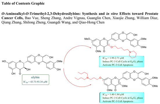
O-Aminoalkyl-O-Trimethyl-2,3-Dehydrosilybins: Synthesis and In Vitro Effects Towards Prostate Cancer Cells

1
Department of Chemistry, California State University, Fresno, 2555 E. San Ramon Avenues, M/S SB70, Fresno, CA 93740, USA
2
Department of Chemistry and RCMI Cancer Research Center, Xavier University of Louisiana, 1 Drexel Drive, New Orleans, LA 70125, USA
*
Author to whom correspondence should be addressed.
Molecules 2018, 23(12), 3142; https://doi.org/10.3390/molecules23123142
Submission received: 31 October 2018 / Revised: 26 November 2018 / Accepted: 29 November 2018 / Published: 29 November 2018
(This article belongs to the Special Issue Silymarin and Derivatives: From Biosynthesis to Health Benefits)

Abstract

:
As part of our ongoing silybin project, this study aims to introduce a basic nitrogen-containing group to 7-OH of 3,5,20-O-trimethyl-2,3-dehydrosilybin or 3-OH of 5,7,20-O-trimethyl-2,3-dehydrosilybin via an appropriate linker for in vitro evaluation as potential anti-prostate cancer agents. The synthetic approaches to 7-O-substituted-3,5,20-O-trimethyl-2,3-dehydrosilybins through a five-step procedure and to 3-O-substituted-5,7,20-O-trimethyl-2,3- dehydrosilybins via a four-step transformation have been developed. Thirty-two nitrogen-containing derivatives of silybin have been achieved through these synthetic methods for the evaluation of their antiproliferative activities towards both androgen-sensitive (LNCaP) and androgen-insensitive prostate cancer cell lines (PC-3 and DU145) using the WST-1 cell proliferation assay. These derivatives exhibited greater in vitro antiproliferative potency than silibinin. Among them, 11, 29, 31, 37, and 40 were identified as five optimal derivatives with IC50 values in the range of 1.40–3.06 µM, representing a 17- to 52-fold improvement in potency compared to silibinin. All these five optimal derivatives can arrest the PC-3 cell cycle in the G0/G1 phase and promote PC-3 cell apoptosis. Derivatives 11, 37, and 40 are more effective than 29 and 31 in activating PC-3 cell apoptosis.

Graphical Abstract

1. Introduction

Silybin ((1), Figure 1), also known as silibinin, exists in nature as an approximately equimolar mixture of two diastereomers of silybin A and silybin B with opposite configurations at C-10 and C-11 [1]. They are hard to separate by conventional purification methods, but can be separated by the HPLC method [2]. Silybin A and silybin B biogenetically originate from a taxifolin moiety (flavonoid) and a coniferyl alcohol unit (lignan), which was presumed to follow a non-stereoselective radical coupling reaction [3]. The mixture of diastereoisomeric silybin A and silybin B was originally thought to be a pure compound and named silybin [4]. Later, it was re-termed as silibinin to emphasize that it is a mixture [5]. In this article, silibinin was thus used to represent the mixture of silybin A and silybin B. Silibinin (1) is the first and well-studied member of flavonolignans [4] and the key chemical and medicinal component of milk thistle (Silybum marianum L. Gaertner, Asteraceae) [6]. The earliest record on the medicinal merit of Milk thistle for preventing and treating various hepatotoxicity is Hieronymus Bock’s book published in 1539 [7,8]. Milk thistle and silibinin are now attractive to scientists, not only for their well-known chemotherapeutic use for hepatotoxicity in Europe [9], but also for their potential in treating and preventing cancers [5,10,11,12]. Silymarin (crude extract of milk thistle), silibinin (diastereomeric mixture), silybin A (optically pure), and silybin B (optically pure) have been demonstrated by various in vitro cell-based and in vivo animal-based assays to possess therapeutic potential in treating prostate cancer [12,13,14]. The detailed mechanism of silibinin and structurally-related flavonolignans as anti-prostate cancer agents has previously been summarized by us in a review article [15]. Briefly, this group of natural products can arrest the prostate cancer cell cycle at G1 phase and induce cancer cell apoptosis. Also, they have been demonstrated to inhibit the secretion of prostate cancer specific antigen (PSA) and androgen receptor (AR). They have been revealed to possess anti-angiogenesis properties by decreasing the density of prostate tumor microvessels and VEGF immunoreactivity. Additionally, silibinin and structurally-related compounds can suppress prostate cancer cell migration and invasion through down-regulating the vimentin protein and MMP-2 mRNA, up-regulating E-cadherin expression, and reversing the epithelial-to-mesenchymal transition (EMT). Additionally, the non-toxic profiles of silibinin have been confirmed by its long-term use as a dietary supplement and a Phase I clinical trial of silybin-phytosome, a formula of silibinin, at a dose of 13 g/day [16]. However, the development of silibinin as an anti-prostate cancer drug is hindered, at least partly, by its moderate potency, with its IC50 values of 40–106 µM in prostate cancer cell models [12,13,15,17] and by its poor pharmacokinetic profiles [16]. Structural modification of silibinin can serve as a viable strategy to enhance its potency. Methylated silybins have been reported to be capable of increasing antiproliferative activities towards prostate cancer cells [18]. Additionally, 2,3-dehydrosilybin has been shown to be a significantly better anticancer agent than silibinin [19]. Structural manipulations on the phenolic and alcoholic hydroxyl groups of silibinin and 2,3-dehydrosilybin have been applied to overcome their pharmacokinetic limitations [20,21,22].
Our previous studies on the structure-activity relationships of silibinin revealed that the antiproliferative potency of 2,3-dehydrosilybin in three prostate cancer cell models could be further improved through introducing a suitable alkyl group on 7-OH and 3-OH, as exemplified by 7-O-ethyl-2,3-dehydrosilybin (3) and 3-O-propyl-2,3-dehydrosilybin (4) (Figure 1) [23,24]. This encouraged us to further investigate the effects of nitrogen-containing groups on 7-OH and 3-OH of 2,3-dehydrosilybin on the biological profiles in prostate cancer cell models. Consequently, this study started with the development of general synthetic approaches to 7-O-substituted-3,5,20-O-trimethyl-2,3-dehydrosilybins and 3-O-substituted-5,7,20-O-trimethyl-2,3- dehydrosilybins, followed by the synthesis of thirty-two new derivatives of silibinin, including six 7-O-aminoalkyl-3,5,20-O-trimethyl-2,3-dehydrosilybins and 26 3-O-aminoalkyl-5,7,20-O- trimethyl-2,3-dehydrosilybins. Additionally, the phenolic hydroxyl groups in all synthetic derivatives were converted to methoxyl groups. This conversion was expected to overcome, to some degree, pharmacokinetic limitations caused by the phenolic hydroxyl groups and to pave an avenue to the selective incorporation of a basic nitrogen-containing group into the phenolic hydroxyl group at either C-7 or C-3. The in vitro anticancer activities of these derivatives have been evaluated in three prostate cancer cell models. The design, synthesis, antiproliferative activity, and structure-activity relationships of these silibinin derivatives are presented in this paper. The cell apoptosis induction and cell cycle regulation by five representative derivatives are also reported.

2. Results and Discussion

2.1. Chemistry

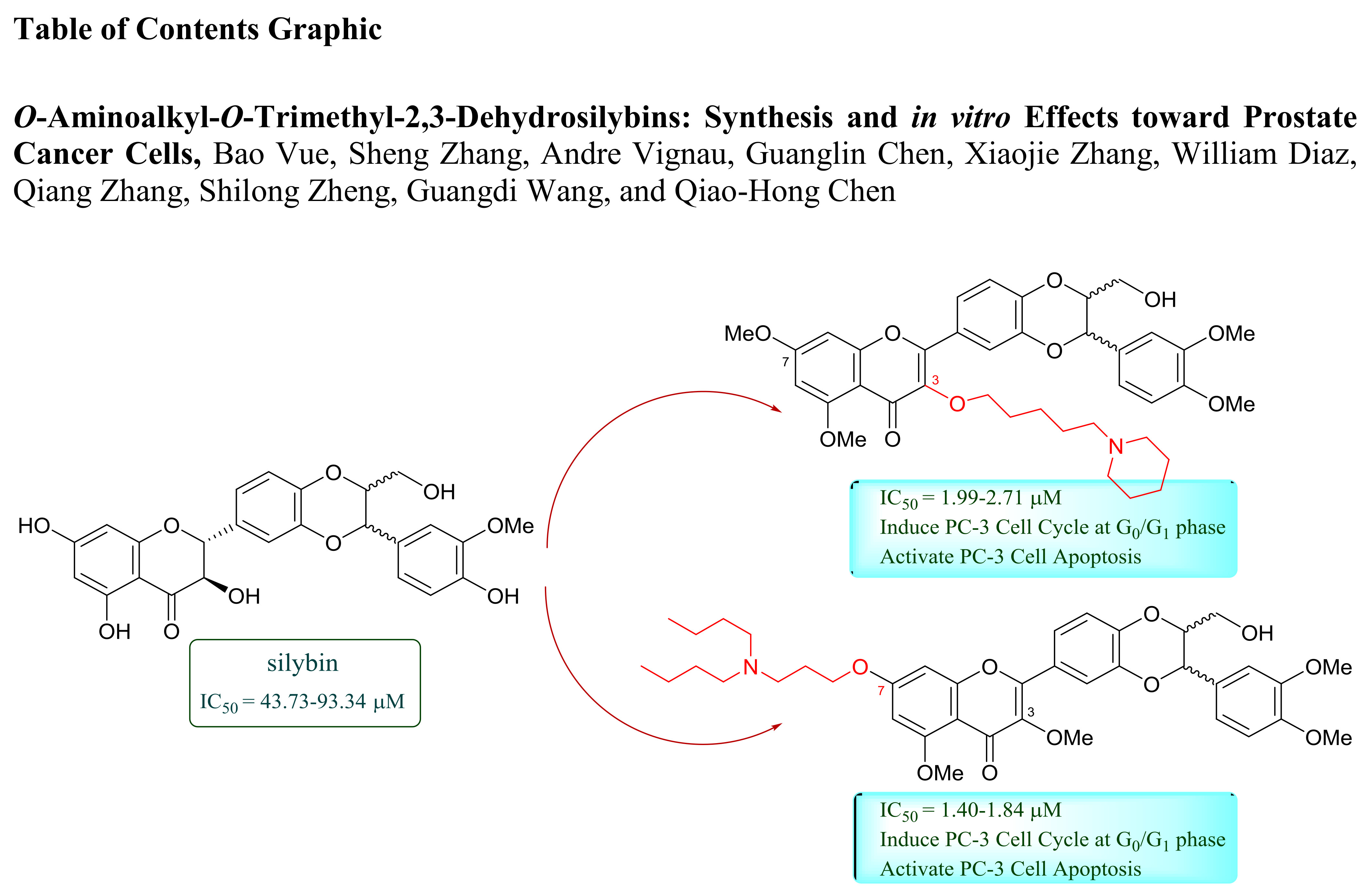
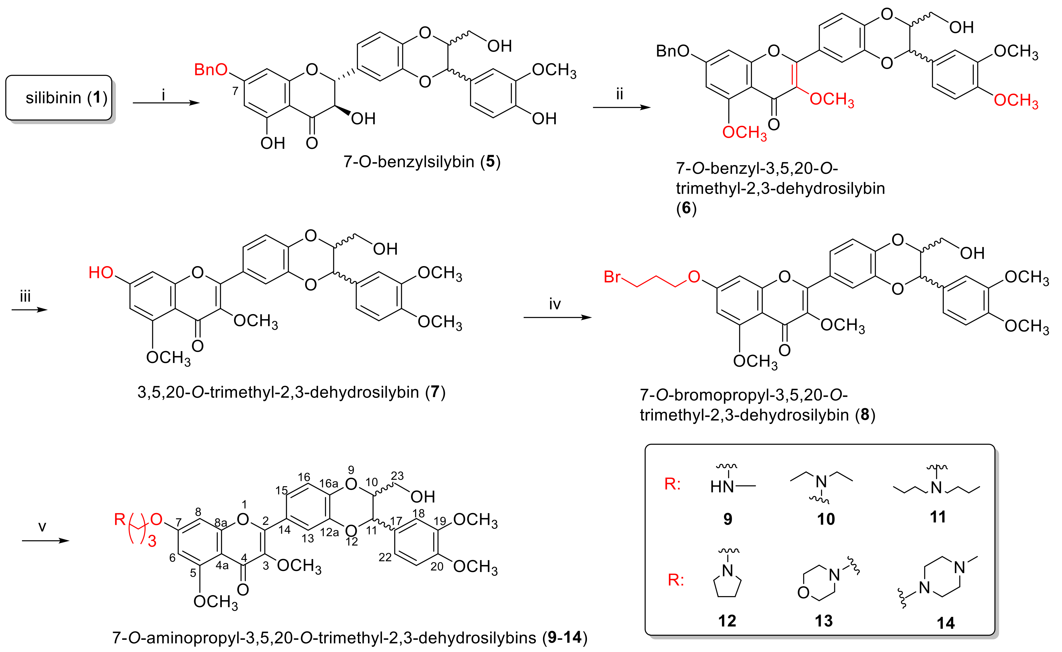
2.1.1. Synthesis of 7-O-Aminoalkyl-3,5,20-O-Trimethyl-2,3-Dehydrosilybins (914)

Some oxidative conversions of silibinin to 2,3-dehydrosilybin have been reported [25,26,27]. As illustrated in Scheme 1, the synthesis of 7-O-substituted-2,3-dehydrosilybins (914) started with selective benzylation (81%) of the C-7 phenolic hydroxyl group of silibinin, according to the procedure reported by Kren et al. and us [23,28]. It is worth noting that anaerobic conditions are essential to achieving high yields for the selective benzylation. This is because the simultaneous presence of a base and air led to the aerobic oxidation of silibinin to 2,3-dehydrosilybin [8,24] and the 3-OH in 2,3-dehydrosilybin is readily benzylated or alkylated [8,24,28], which has been rationalized by the electrochemistry measurements and bond dissociation energy calculations [29]. 7-O-Benzyl-3,5,20-O-trimethyl-2,3-dehydrosilybin (6) was then achieved by the one-pot reaction of the base-mediated oxidation of 7-O-benzylsilybin (5), followed by trimethylation of the corresponding 7-O-benzyl-2,3-dehydrosilybin. 3,5,20-O-Trimethyl-2,3-dehydrosiliybin (7) was obtained by debenzylation of aryl benzyl ether 6 using ammonium formate as the hydrogen source, catalyzed by palladium on carbon. 7-O-bromopropyl-3,5,20-O-trimethyl-2,3-dehydrosilybin (8) was prepared by O-alkylating 7 with 1,3-dibromopropane mediated by potassium carbonate. 7-O-Aminoalkyl-3,5,20-O-trimethyl-2,3-dehydrosilybins (914) were achieved by N-alkylation of the bromoalkyl side chain of 8 with the appropriate amine.

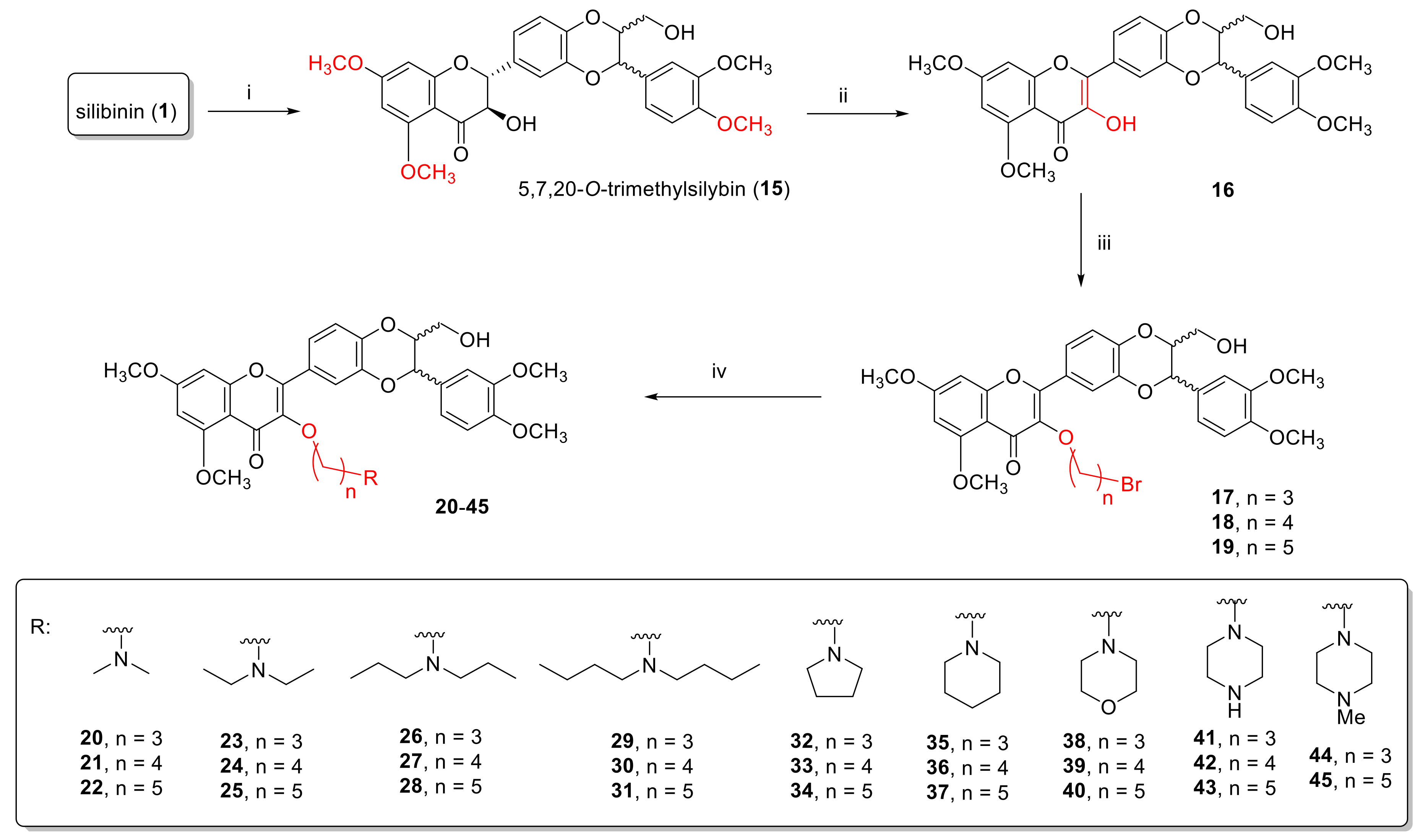
2.1.2. Synthesis of 3-O-Aminoalkyl-5,7,20-O-Trimethyl-2,3-Dehydrosilybins (2045)

3-O-Substituted-2,3-dehydrosilybins (2045) were synthesized following a four-step procedure, as shown in Scheme 2. Specifically, 5,7,20-O-trimethylsilybin (15) was achieved by treating silibinin (1) with dimethylsulfate in the presence of potassium carbonate under strictly anaerobic conditions. Note that a small amount of 3,5,7,20-O-tetramethyl-2,3-dehydrosilybin can be formed if anaerobic conditions are not well-controlled, which would complicate the purification process and decrease the yield. Even though 5,7,20-O-tribenzylsilybin was much easier to aerobically oxidize than that in silibinin [30], the oxidation of 5,7,20-O-trimethylsilybin (15) under the same conditions led to a mixture of products instead of the desired oxidation product. After several trials with different oxidation conditions, 5,7,20-O-trimethyl-2,3-dehydrosilybin (16) was eventually obtained by the oxidation of 15 with sodium hydroxide and hydrogen peroxide. A 10–14 h reaction time serves as a critical factor for the optimal yield (40–55%) of this oxidation reaction. We also found that this oxidation cannot be quenched with hydrochloric acid because it selectively demethylated the 5-OMe of the product. 5,7,20-O-Trimethyl-2,3-dehydrosilybin (16) was then converted to 3-O-bromoalkyl-5,7,20-O-trimethyl-2,3-dehydrosilybins (1719) via O-alkylation with the appropriate dibromoalkanes, using potassium carbonate as the base and DMF as the aprotic solvent. The subsequent N-alkylation of the 3-O-bromoalkyl-5,7,20-O-trimethyl-2,3-dehydrosilybins (1719) with the corresponding amine furnished the respective 3-O-aminoalkyl-5,7,20-O-trimethyl-2,3-dehydrosilybin (2045).

2.1.3. Structure Determination

The 1H-NMR and 13C-NMR data of 5 are consistent with those reported in the literature [28]. The structure of 6 was characterized by the presence of three single signals at 3.94, 3.924, and 3.916 in its 1H-NMR spectrum and at 60.1, 56.6, and 56.2 in its 13C-NMR spectrum for three additional methoxyl groups when compared with 5 and the absence of the 1H-NMR signal at 4.52 for the H-3 of 5. The structure of 7 was established based on the absence of the 1H-NMR signals at 7.48–7.34 (m, 5H) and 5.12 (s, 2H) for the benzyl group of 6. The signals in the 1H-NMR spectrum [4.17 (t, J = 5.7 Hz, 2H), 3.61 (t, J = 6.3 Hz, 2H), 2.36 (quin, J = 6.0 Hz, 2H)] of compound 8 confirmed the addition of a 3-bromopropyl group when compared with 7. The structures of the six 7-O-aminoalkyl-3,5,20-O-trimethyl-2,3-dehydrosilybins (914) were characterized by interpreting their NMR, HRMS, and FTIR data (for details see Experimental Section and Supporting Information). Specifically, the presence of the 1H-NMR and 13C-NMR signals for each alkylamine group suggests the incorporation of the corresponding alkylamine group, which was corroborated by the HRMS data for each compound. 1H-NMR and 13C-NMR data for 914 were assigned (Experimental Section and Supporting Information) by comparing them with the NMR data of 7-O-propyl-2,3-dehydrosilybin because derivatives 914 and 7-O-propyl-2,3-dehydrosilybin share an identical core structure and all NMR signals of 7-O-propyl-2,3-dehydrosilybin have been fully assigned by us, according to the interpretation of its COSY, HMQC, and HMBC data [23].
The structure of 15 was determined by the existence of three single signals for three additional methoxyl groups at 3.91, 3.90, and 3.90 in its 1H-NMR spectrum when compared with silibinin. The structure of 16 was confirmed by the absence of the 1H-NMR signal at 4.42 (1H) for H-3 of 15. The signals in the 1H-NMR spectra [4.14 (t, 2H), 3.60 (t, 2H), 2.31–2.26 (m, 2H) of 17; 4.14 (t, 2H), 3.46 (t, 2H), 2.09–2.00 (m, 2H), 1.90–1.81 (m, 2H) of 18; 4.02 (t, 2H), 3.34 (t, 2H), 1.85 (quin, 2H), 1.74 (quin, 2H), 1.59–1.49 (m, 2H) of 19] confirmed the addition of an appropriate bromoalkyl group to each of 1719 when compared with 16. The structures of the 26 3-O-aminoalkyl-5,7,20-O-trimethyl-2,3-dehydrosilybins (2045) were characterized by interpreting their NMR, HRMS, and FTIR data (for details, see Experimental Section and Supporting Information). Specifically, the presence of the 1H-NMR and 13C-NMR signals for each alkylamine group suggests the incorporation of the corresponding alkylamine group, which was further supported by the HRMS data for each compound. 1H-NMR and 13C-NMR data for 2045 were assigned (Experimental Section and Supporting Information) by comparing them with the NMR data of 3-O-propyl-2,3-dehydrosilybin (4), due to the fact that derivatives 2045 and compound 4 possess an identical core structure and all NMR signals of compound 4 have been fully assigned by us, according to the interpretation of its COSY, HMQC, and HMBC data [24].

2.2. Antiproliferative Activity towards Prostate Cancer Cell Lines and Structure-Activity Relationships

The in vitro antiproliferative activities of six 7-O-substituted and 26 3-O-substituted silybin derivatives were evaluated using the WST-1 cell proliferation assay, according to the procedure as described in the Experimental Section in both androgen-sensitive (LNCaP) and androgen-insensitive (PC-3 and DU145) human prostate cancer cell lines. Silibinin and docetaxel were used as a positive control for comparison in the parallel experiments and the IC50 values calculated from the dose-response curves are summarized in Table 1. Clearly, 7-O-aminoalkyl-3,5,20-O-trimethyl-2,3-dehydrosilybins and 3-O-aminoalkyl-5,7,20-O-trimethyl-2,3-dehydrosilybins are more potent in suppressing both androgen-sensitive and androgen-insensitive prostate cancer cell proliferation than silibinin. This conclusion is supported by the following data: i) the optimal 7-O-substituted derivative (11) is 52-, 51-, and 24-fold more potent than silibinin toward PC-3, DU145, and LNCaP prostate cancer cell lines, respectively; and ii) the optimal 3-O-substituted derivatives (29, 31, 37, and 40) are 26–27, 31–37 and 17–22 times more potent than silibinin against PC-3, DU145, and LNCaP prostate cancer cell lines, respectively. Furthermore, the most potent derivatives 11, 29, 31, 37, and 40 are more active than 2,3-dehydrosilybin (2), 7-O-ethyl-2,3-dehydrosilybin (3), and 3-O-propyl-2,3-dehydrosilybin (4) in suppressing DU145 prostate cancer cell proliferation. Additionally, the dibutylamino group in derivatives 11, 29 and 31; the morpholino moiety in 37; and the piperidino unit in 40, are the favorable nitrogen-containing groups for the greater potency. A three-carbon linker in 11 and 29, as well as a five-carbon linker in 31, 37 and 40, are beneficial to the potency.

2.3. Antiproliferative Activity towards MCF 10A and PWR-1E Non-Neoplastic Human Epithelial Cell Lines

Silibinin and five potent derivatives (11, 29, 31, 37, and 40) were selected for further evaluation against the MCF 10A non-neoplastic human mammary epithelial cell line and PWR-1E non-neoplastic human prostate epithelial cells. The five derivatives were chosen based on the following grounds: 11 is the most potent 7-O-aminoalkyl-3,5,20-O-trimethyl-2,3-dehydrosilybin considering its overall potency towards three prostate cancer cell models; 29, 31, 37, and 40 are the most promising 3-O-aminoalkyl-5,7,20-O-trimethyl-2,3-dehydrosilybins. As shown in Table 2, silibinin demonstrates a significantly higher capability of suppressing non-neoplastic cell (MCF 10A and PWR-1E) proliferation than prostate cancer cell proliferation. Four 3-O-aminoalkyl-5,7,20-O-trimethyl-2,3-dehydrosilybins (29, 31, 37, and 40) did not exhibit significantly different responses to prostate cancer cells and to non-neoplastic MCF 10A and PWR-1E cells. However, the 7-O-aminoalkyl-3,5,20-O-trimethyl-2,3-dehydrosilybin 11 does not demonstrate apparent antiproliferative activity towards PWR-1E and MCF 10A non-neoplastic epithelial cells up to a 50 µM concentration. Consequently, 7-O-aminoalkyl-3,5,20-O-trimethyl-2,3-dehydrosilybin 11 illustrates a good selectivity of inhibiting prostate cancer cell proliferation over non-neoplastic MCF 10A and PWR-1E human epithelial cell proliferation (Table 2), which suggests that modification at 7-OH of 3,5,20-O-trimethyl-2,3-dehydrosilybin only improves the antiproliferative potency towards prostate cancer cells, and not non-neoplastic epithelial cells.

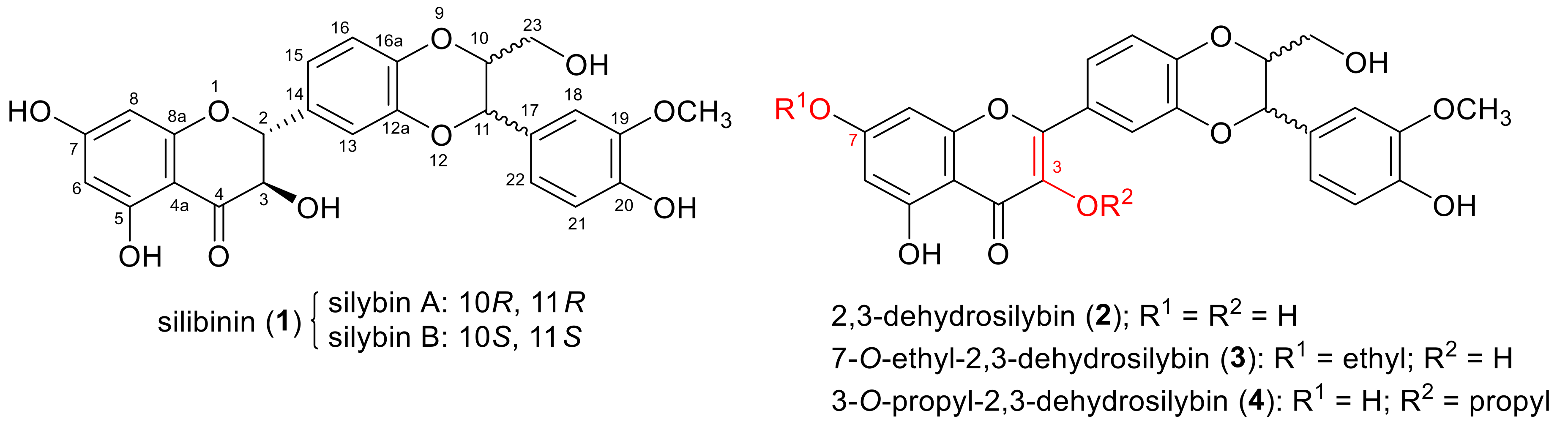
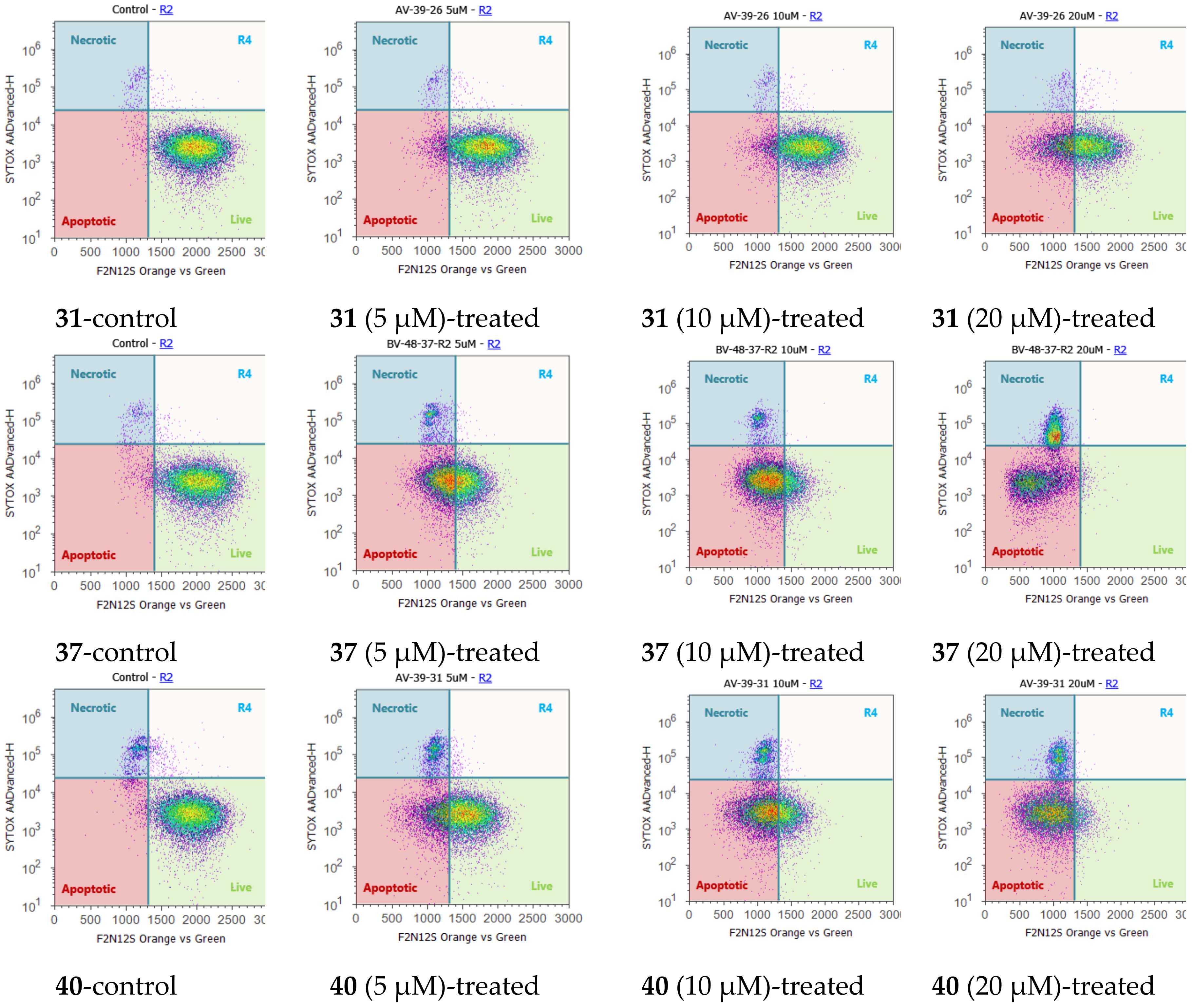
2.4. Cell Cycle Regulation and Cell Apoptosis

Silibinin can arrest the rat (H-7 and I-8) and human prostate cancer cell (LNCaP) cycle at the G1 phase [31,32], and cause G1 and G2-M PC-3 prostate cancer cell cycle arrest [33]. Five optimal derivatives, consisting of 11, 29, 31, 37, and 40, were selected for flow cytometry evaluation of their effect on PC-3 cell cycle regulation because they exhibited optimal cell proliferation inhibition on both androgen-dependent LNCaP and androgen-independent PC-3 prostate cancer cell models, with ≤3.0 µM IC50 values. At 20 µM, all these five derivatives can cause PC-3 cell accumulation in a G0/G1 phase by increasing the cell population in this phase at 16 h from 55.7% (control) to 66.3% (treated with 11), from 36.2% (control) to 50.3% (treated with 29), from 33.1% (control) to 35.9% (treated with 31), from 33.1% (control) to 34.6% (treated with 37), and from 33.1% (control) to 43.4% (treated with 40).
Silibinin was revealed by Agarwal and co-workers to activate cell apoptosis in PC-3 tumor xenografts [34]. An F2N12S and SYTOX AADvanced double staining flow cytometry-based assay was used to discriminate PC-3 cells dying from apoptosis from those dying from necrosis in response to various concentrations of derivatives 11, 29, 31, 37, and 40. PC-3 cells were incubated with the test compound for 16 h. As shown in Figure 2, derivatives 11, 37, and 40 induced appreciable levels of apoptotic cell death in the androgen-insensitive PC-3 prostate cancer cell line in a dose-responsive manner after 16 h of treatment. Specifically, 5 µM of derivatives 11, 37, and 40 could induce a substantial early phase of apoptosis (26–59%) in PC-3 cells compared with control cells: treatment with 10 µM of these three optimal derivatives led to 56–76% early apoptotic cells and 6–40% late apoptotic/necrotic cells; 20 µM of derivatives 11, 37, and 40 also activated notable apoptosis, with 54–75% early apoptotic cells and 16–44% late apoptotic/necrotic cells. The apoptotic cell population reached its maximum when PC-3 cancer cells were exposed to derivative 11, 37, and 40 at 5µM, 10 µM, and 30 µM, respectively. In contrast, derivatives 29 and 31 did not induce significant levels of apoptotic cell death (less than 10%) up to a 10 µM concentration. Only 50 µM of derivatives 29 and 31 resulted in the maximum apoptotic cell population (71% and 95%, respectively).

3. Materials and Methods

3.1. General Procedures

HRMS were obtained on an Orbitrap mass spectrometer with electrospray ionization (ESI) (Thermo Fisher Scientific, Waltham, MA, USA). NMR spectra were obtained on a Bruker Fourier 300 spectrometer (Billerica, MA, USA) in CDCl3, or DMSO-d6. The chemical shifts are given in ppm referenced to the respective solvent peak, and coupling constants are reported in Hz. Anhydrous THF and dichloromethane were purified by the PureSolv MD 7 Solvent Purification System from Innovative Technologies (MB-SPS-800) (Herndon, VA, USA). All other reagents and solvents were purchased from commercial sources and were used without further purification. Silica gel column chromatography was performed using silica gel (32–63 µm) (SiliCycle Inc, Quebect, QC, Canada). Preparative thin-layer chromatography (PTLC) separations were carried out on thin layer chromatography plates loaded with silica gel 60 GF254 (EMD Millipore Corporation, MA, USA). Silibinin (>98.0%) was purchased from Fisher Scientific (TCI America, Portland, OR, USA, Cat # 50-014-46874).

3.2. Synthesis of 7-O-Benzylsilybin (5)

Following the procedure described in the literature [23,28], 7-O-benzylsilybin (5) was prepared from silibinin in an 80% yield as a light yellow solid. m.p. 93–95 °C. IR (film) νmax: 3432, 2937, 1634, 1571, 1507 cm−1; 1H-NMR (300 MHz, CDCl3): δ 11.25 (11.24) (s, 1H), 7.43–7.34 (m, 5H), 7.19 (dd, J = 4.2, 1.5 Hz, 1H), 7.10–7.01 (m, 2H), 6.97–6.88 (overlapped, 3H), 6.21 (d, J = 1.8 Hz, 1H), 6.13 (6.12) (d, J = 2.4 Hz, 1H), 5.96 (br.s, 1H), 5.07 (s, 2H), 4.99 (d, J = 11.7 Hz, 1H), 4.93 (d, J = 8.4 Hz, 1H), 4.52 (dd, J = 11.7, 3.3 Hz, 1H), 4.09–3.99 (m, 1H), 3.89 (s, 3H), 3.80 (dd, J = 12.3, 2.1 Hz, 1H), 3.55 (dd, J = 12.3, 3.6 Hz, 1H); 13C-NMR (75 MHz, CDCl3): δ 196.2, 167.8, 163.6, 162.8, 147.0, 146.4, 144.1, 143.9, 135.6, 129.5, 128.8, 128.4, 127.9, 127.5, 121.2 (121.1), 120.8, 117.3 (117.2), 116.6, 114.9, 109.8, 101.1, 96.4, 95.4, 83.0, 78.4, 76.3, 72.4, 70.5, 61.6, 56.1; HRMS-ESI m/z [M + H]+ calcd for C32H29O10: 573.1761, found: 573.1769.

3.3. Synthesis of 7-O-Benzyl-3,5,20-O-Trimethyl-2,3-Dehydrosilybin (6)

Potassium carbonate (3 eq.) was added to a solution of benzylsilybin (1 eq.) in DMF (0.5 M) and the reaction mixture was exposed to air with stirring at room temperature (or 60 °C) for 3 h. When most of the 7-O-benzylsilybin was oxidized to 7-O-benzyl-2,3-dehydrobenzylsilybin, as monitored by TLC, the reaction mixture was cooled down to room temperature. Potassium carbonate (3 eq.), followed by methyl iodide (6 eq.), were added to the reaction mixture and the reaction was allowed to proceed at room temperature overnight prior to being quenched with HCl (1 M). The subsequent mixture was diluted with water and extracted with ethyl acetate. The combined organic extracts were rinsed with brine and dried over anhydrous sodium sulfate. After filtration, the volatile components were evaporated under vacuum to give the crude product, which was purified by column chromatography over silica gel or PTLC eluting with 5% methanol in dichloromethane to generate 7-O-benzyl-3,5,20-O-trimethyl-2,3-dehydrosilybin (6): 48% yield, yellow solid, m.p. 118–119 °C. IR (film) νmax: 3401, 2932, 1601, 1504, 1440 cm−1; 1H-NMR (300 MHz, CDCl3): δ 7.76 (d, J = 2.1 Hz, 1H), 7.73 (dd, J = 8.7, 2.1 Hz, 1H), 7.48–7.34 (m, 5H), 7.06 (d, J = 8.7 Hz,1H), 7.04 (dd, J = 8.4, 2.1Hz, 1H), 6.98 (d, J = 1.8 Hz, 1H), 6.93 (d, J = 8.1 Hz, 1H), 6.56 (d, J = 2.1 Hz, 1H), 6.41 (d, J = 2.4 Hz, 1H), 5.12 (s, 2H), 5.02 (d, J = 8.4 Hz,1H), 4.17–4.10 (m,1H), 3.94 (s, 3H), 3.924 (s, 3H), 3.916 (s, 3H), 3.89 (s, 3H), 3.85 (dd, J = 12.3, 2.1 Hz, 1H), 3.58 (dd, J = 12.6, 3.9 Hz, 1H); 13C-NMR (75 MHz, CDCl3): δ 174.2, 163.1, 161.2, 158.8, 152.1, 150.0, 149.6, 145.3, 143.8, 141.6, 135.8, 128.9, 128.6, 128.4, 127.7, 124.5, 122.4, 120.3, 117.3, 117.2, 111.5, 110.3, 109.8, 96.5, 93.4, 78.8, 76.5, 70.6, 61.8, 60.1, 56.6, 56.2, 56.1; HRMS-ESI m/z [M + H]+ calcd for C35H33O10: 613.2074, found: 613.2071.

3.4. Synthesis of 3,5,20-O-Trimethyl-2,3-Dehydrosilybin (7)

To the solution of 7-O-benzyl-3,5,20-O-trimethyl-2,3-dehydrosilybin (6, 1 eq.) in methanol (0.2 M), Pd-C (50% wet, 10% w/w) and ammonium formate (10 eq.) were sequentially added. The reaction mixture was refluxed overnight under argon. After being cooled to room temperature, the reaction mixture was filtered through silica gel pad eluting with THF. The filtrate was concentrated under vacuum to afford 3,5,20-O-trimethyl-2,3-dehydrosilybin (7) in 67% yield as a light yellow solid. M.p. 235–236 °C. IR (film) νmax: 3545, 2955, 2924, 2853, 2177, 2159, 2028, 1992, 1978, 1968, 1728, 1593, 1557, 1508 cm−1; 1H-NMR (300 MHz, DMSO-d6): δ 7.74–7.64 (m, 1H), 7.60–7.55 (m, 2H), 7.13–6.98 (m, 6H), 6.42 (s, 1H), 6.33 (s, 1H), 5.02 (d, J = 7.8 Hz, 1H), 4.36–4.28 (m, 1H), 3.78 (s, 12H), 3.76 (s, 3H), 3.57 (dd, J = 12.9, 2.1 Hz, 1H), 3.35 (dd, J = 12.9, 4.8 Hz, 1H). HRMS-ESI m/z [M + H]+ calcd for C28H27O10: 523.1604, found: 523.1598.

3.5. Synthesis of 7-O-(3′-Bromo)Propyl-3,5,20-O-Trimethyl-2,3-Dehydrosilybin (8)

To a solution of 3,5,20-O-trimethyl-2,3-dehydrosilybin in DMF (1 M), potassium carbonate (4 eq.) followed by 1,3-dibromopropane (4 eq.) were added. The reaction mixture was stirred at 60 °C overnight prior to being quenched by the addition of 1 M HCl. The subsequent reaction mixture was diluted with water and extracted with ethyl acetate. The combined organic extracts were rinsed with brine, dried over anhydrous sodium sulfate, and concentrated under vacuum to yield a crude product, which was subjected to column chromatography or PTLC purification over silica gel eluting with 5% methanol in dichloromethane to afford 7-O-(3′-bromo)propyl-3,5,20-O-trimethyl-2,3-dehydrosilybin (8) as a yellow solid. m.p. 115–116 °C. IR (film) νmax: 2932, 1604, 1506, 1463, 1441, 1345, 1264 cm−1; 1H-NMR (300 MHz, CDCl3): δ 7.76 (d, J = 2.1 Hz, 1H), 7.73 (dd, J = 8.7, 2.4 Hz, 1H), 7.07–6.97 (m, 3H), 6.92 (d, J = 8.4 Hz, 1H), 6.48 (d, J = 2.1 Hz, 1H), 6.32 (d, J = 2.1 Hz, 1H), 5.02 (d, J = 8.4 Hz, 1H), 4.20–4.11 (m, 1H), 4.17 (t, J = 5.7 Hz, 2H), 3.95 (s, 3H), 3.92 (s, 3H), 3.91 (s, 3H), 3.89 (s, 3H), 3.85 (dd, J = 12.6, 2.7 Hz, 1H), 3.66-3.56 (m, 1H), 3.61 (t, J = 6.3 Hz, 2H), 2.36 (quin, J = 6.0 Hz, 2H); 13C-NMR (75 MHz, CDCl3): δ 174.2, 163.0, 161.2, 158.8, 152.1, 149.9, 149.6, 145.3, 143.8, 141.5, 128.4, 124.4, 122.3, 120.3, 117.3, 117.2, 111.4, 110.2, 109.7, 96.1, 93.0, 78.8, 76.4, 66.0, 61.8, 60.1, 56.5, 56.2, 56.1, 32.1, 29.6; HRMS-ESI m/z [M + H]+ calcd for C31H32BrO10: 643.1179, 645.1158, found: 643.1173, 645.1151.

3.6. General Procedure for the Synthesis of 7-O-(N,N-Dialkylamino)Propyl-3,5,20-O-Trimethyl-2,3-Dehydrosilybins

Potassium carbonate (3 eq.) and the appropriate amine (3 eq.) were added to a solution of 7-O-(3′-bromo)propyl-3,5,20-O-trimethyl-2,3-dehydrosilybin (8) in dry acetone (0.1 M). The reaction mixture was refluxed overnight before the removal of acetone under vacuum. The residue was diluted with water and extracted with ethyl acetate. The combined organic extracts were rinsed with brine, dried over anhydrous sodium sulfate, and concentrated in vacuo to generate the crude products, which were subjected to PTLC purification eluting with 10% methanol in dichloromethane to yield the respective 7-O-(3′-amino)propyl-3,5,20-O-trimethyl-2,3-dehydrosilybin.
7-O-(N-Methylaminopropyl)-3,5,20-O-trimethyl-2,3-dehydrosilybin (9). 40% yield, light yellow solid, m.p. 125–127 °C. IR (film) νmax: 3401, 2928, 1625, 1606, 1507 cm−1; 1H-NMR (300 MHz, CDCl3): δ 7.59 (d, J = 2.1 Hz, 1H, H-13), 7.53 (dd, J = 8.7, 2.1 Hz, 1H, H-15), 7.04 (dd, J = 8.1, 1.2 Hz, 1H, H-16), 6.97 (d, J = 1.5 Hz, 1H, H-18), 6.93 (dd, J = 8.7, 2.7 Hz, 2H, H-21 & H-22), 6.22 (d, J = 1.8 Hz, 1H, H-8), 6.18 (d, J = 1.8 Hz, 1H, H-6), 4.99 (d, J = 8.4 Hz, 1H, H-11), 4.17–4.06 (overlapped, 3H, H-10 & 7-OCH2), 3.92 (s, 3H, OCH3), 3.91 (s, 3H, OCH3), 3.90 (s, 3H, OCH3), 3.80 (s, 3H, OCH3), 3.85–3.80 (overlapped, 1H, H-23), 3.57 (dd, J = 12.9, 3.9 Hz, 1H, H-23), 3.25 (t, J = 6.3 Hz, 2H, 7-O-CH2CH2CH2-), 2.81 (s, 3H, NHCH3), 2.34–2.30 (m, 2H, 7-O-CH2CH2CH2-), 2.01 (d, J = 1.2 Hz, 1H, 23-OH); 13C-NMR (75 MHz, CDCl3): δ 174.4 (C-4), 162.9 (C-7), 160.4 (C-5), 158.1 (C-8a), 152.6 (C-19), 150.0 (C-20), 149.6 (C-16a), 145.6 (C-2), 143.7 (C-12a), 140.9 (C-3), 128.3 (C-17), 123.5 (C-14), 122.0 (C-15), 120.3 (C-22), 117.2 (C-16), 117.1 (C-13), 111.5 (C-21), 110.4 (C-18), 108.7 (C-4a), 96.3 (C-6), 92.9 (C-8), 78.9 (C-10), 76.3 (C-11), 65.2 (7-OCH2), 61.6 (C-23), 59.9 (OCH3), 56.8 (OCH3), 56.2 (OCH3), 56.1 (OCH3), 47.5 (7-O-CH2CH2CH2-), 34.2 (NHCH3), 25.7 (7-O-CH2CH2CH2-); HRMS-ESI m/z [M + H]+ calcd for C32H36NO10: 594.2339, found: 594.2334. The yields and spectral data for compounds 1014 are included in Supporting Information.

3.7. Synthesis of 5,7,20-O-Trimethylsilybin (15)

A three-neck round bottom flask was charged with silibinin (2.01 g, 4.2 mmol) and potassium carbonate (3.43 g, 25.1 mmol), which was vacuumed three times under argon prior to the addition of acetone (30.0 mL). The reaction mixture was refluxed for 15 min before dimethylsulfate (3.13 mL, 33.1 mmol) was added through a needle. The reaction was continued with refluxing for an additional 4 h when the reaction was completed, as monitored by TLC. After cooling down to room temperature, saturated ammonium chloride was added to quench the reaction, and the subsequent mixture was extracted with ethyl acetatethree times. The organic layers were combined, washed with brine twice, and dried over anhydrous sodium sulfate. Purification of the crude product through column chromatography, eluting with ethyl acetate/hexane (50/50 to 70/30, v/v), gave the product (15) as a white crystal in 80% yield. 1H-NMR (300 MHz, CDCl3): δ 7.21 (dd, J = 9.9, 1.8 Hz, 1H), 7.08(dd, J = 8.4, 2.1 Hz, 1H), 7.04 (d, J = 3.3 Hz, 1H), 7.00 (dd, J = 8.7, 2.7 Hz, 1H), 6.96 (s, 1H), 6.90 (d, J = 8.4 Hz, 1H), 6.12 (d, J = 2.1 Hz, 1H), 6.11 (d, J = 1.5 Hz, 1H), 4.98 (d, J = 9.0 Hz, 1H), 4.94 (d, J = 12.3 Hz, 1H), 4.42 (dd, J = 12.3, 5.1 Hz, 1H), 4.06–4.03 (m, 1H), 3.91 (s, 3H), 3.90 (s, 3H), 3.90 (s, 3H), 3.81 (d, J = 1.8 Hz, 3H), 3.81 (dd, J = 12.0, 2.4 Hz, 1H), 3.55 (dd, J = 12.3, 3.9 Hz).

3.8. Synthesis of 5,7,20-O-Trimethyl-2,3-Dehydrosilybin (16)

A 10 mL round flask was charged with 5,7,20-O-trimethylsiybin (150.0 mg, 0.23 mmol) in methanol (2.0 mL) and tetrahydrofuran (2.0 mL). The solution was stirred for 10 min at room temperature prior to being added to hydrogen peroxide (0.85 mL, 30%) and sodium hydroxide aqueous solution (0.65 mL, 16%) at 0 °C. The reaction mixture was slowly warmed to room temperature and then stirred overnight, before being quenched with saturated ammonium chloride. The subsequent mixture was extracted with dichloromethane three times, and the combined extracts were dried over sodium sulfate and concentrated under vacuum. The crude product was obtained in 49% yield, which is pure enough for the next step of the reaction without purification. 1H-NMR (300 MHz, CDCl3): δ 7.89 (s, 1H), 7.86 (d, J = 8.4 Hz, 1H), 7.14 (d, J = 8.7 Hz, 1H), 7.04 (d, J = 8.4 Hz, 1H), 6.98 (s, 1H), 6.93 (d, J = 8.1 Hz, 1H), 6.53 (d, J = 1.8 Hz, 1H), 6.35 (d, J = 2.1 Hz, 1H), 5.03 (d, J = 8.1 Hz, 1H), 4.17–4.11 (m, 1H), 3.98 (s, 3H), 3.93 (s, 3H), 3.92 (s, 3H), 3.90 (s, 3H), 3.85 (d, J = 13.8 Hz, 1H), 3.59 (d, J = 10.5 Hz, 1H).

3.9. General Procedure for the Synthesis of 3-O-Bromoalkyl-5,7,20-O-Trimethyl-2,3-Dehydrosilybins (1719)

A round bottom flask (10 mL) was charged with 5,7,20-O-trimethyl-2,3-dehydrosilybin (16, 83.2 mg, 0.16 mmol), potassium carbonate (352.0 mg, 2.55 mmol), and DMF (5.0 mL). The mixture was stirred for 10 min prior to being added to 1,3-, 1,4-, or 1,5-dibromalkane (2.56 mmol, 16 equiv.). The reaction was continued with stirring at room temperature for 24–48 h, before the reaction was quenched with water. The subsequent mixture was extracted with ethyl acetate three times, and the combined extracts were dried over anhydrous sodium sulfate and concentrated in vacuo. The crude products were subjected to PTLC purification eluting with DCM/methanol (100/5, v/v) to yield the respective 3-O-bromoalkyl-5,7,10-O-trimethyl-2,3-dehydrosilybin. 3-O-(3′-Bromo)propyl-5,7,20-O-trimethyl-2,3-dehydrosilybin (17). 81% yield, 1H-NMR (300 MHz, CDCl3): δ 7.72 (s, 1H), 7.70 (d, J = 9.6 Hz, 1H), 7.07 (d, J = 7.8 Hz, 1H), 7.06 (dd, J = 8.1, 1.8 Hz, 1H), 7.00 (d, J = 1.8 Hz, 1H), 6.94 (d, J = 8.4 Hz, 1H), 6.49 (d, J = 2.1 Hz, 1H), 6.35 (d, J = 2.4 Hz, 1H), 5.05 (d, J = 8.4 Hz, 1H), 4.14 (t, J = 6.0 Hz, 2H), 4.16–4.12 (overlapped, 1H), 3.96 (s, 3H), 3.94 (s, 3H), 3.93 (s, 3H), 3.89 (s, 3H), 3.86 (dd, J = 12.6, 2.7 Hz, 1H), 3.60 (t, J = 6.9 Hz, 2H), 3.63–3.58 (overlapped, 1H), 2.31–2.26 (m, 2H). The yields and spectral data for compounds 1819 are included in Supporting Information.

3.10. General Procedure for the Synthesis of 3-O-(Alkylamino)Alkyl-5,7,20-O-Trimethyl-2,3-Dehydrosilybins

A round bottom reaction flask (10 mL) was charged with 3-O-bromoalkyl-5,7,10-O-trimethyl- 2,3-dehydrosilybin (1 eq.) and potassium carbonate (10 eq.) in acetone (2.0 mL, 0.029 M). The solution was stirred for 10 min prior to being added the appropriate amine (16 eq.). The reaction was allowed to proceed with stirring at room temperature for 24–48 h, before being quenched with water. The subsequent mixture was extracted with ethyl acetate three times, and the combined extracts were dried over anhydrous sodium sulfate and concentrated in vacuo. The crude product was subjected to PTLC purification eluting with DCM/methanol (100:5, v/v). Each desired nitrogen-containing compound was retrieved from PTLC silica gel by washing with dichloromethane/methanol/ammonium hydroxide (100:10:5, v/v/v).
3-O-(N,N-Dimethylamino)propyl-5,7,20-O-trimethyl-2,3-dehydrosilybin (20). 97% yield, white solid, white wax. IR (film) νmax: 3364, 2940, 2837, 1625, 1604, 1517, 1505, 1492, 1462 cm−1; 1H-NMR (300 MHz, CDCl3): δ 7.64 (d, J = 6.2 Hz, 1H, H-15), 7.63 (s, 1H, H-13), 7.10 (d, J = 10.1 Hz, 1H, H-16), 7.03 (d, J = 8.4 Hz, 1H, H-22), 6.98 (s, 1H, H-18), 6.91 (d, J = 8.1 Hz, 1H, H-21), 6.47 (s, 1H, H-8), 6.33 (s, 1H, H-6), 5.05 (d, J = 7.7 Hz, 1H, H-11), 4.19–4.16 (m, 1H, H-10), 4.00–3.97 (overlapped, 2H, 3-OCH2), 3.94 (s, 3H, OCH3), 3.91 (s, 3H, OCH3), 3.90 (s, 3H, OCH3), 3.86 (s, 3H, OCH3), 3.86–3.83 (overlapped, 1H, H-23), 3.57 (dd, J = 12.2, 2.9 Hz, 1H, H-23), 3.52–3.42 (m, 2H, NCH2), 2.88 (s, 6H, 2 × NCH3), 2.33–2.22 (m, 2H, 3-OCH2CH2); 13C-NMR (75 MHz, CDCl3): δ 174.4 (C-4), 164.5 (C-7), 160.8 (C-5), 158.9 (C-8a), 153.4 (C-16a), 149.8 (C-19), 149.4 (C-20), 145.8 (C-2), 143.9 (C-12a), 139.6 C-3), 128.2 (C-17), 123.3 (C-14), 122.2 (C-15), 120.2 (C-22), 117.5 (C-13), 117.0 (C-16), 111.4 (C-21), 110.3 (C-18), 108.9 (C-4a), 96.2 (C-6), 92.5 (C-8), 78.7 (C-10), 76.3 (C-11), 69.3 (3-OCH2), 61.4 (C-23), 56.5 (OCH3), 56.1 (OCH3), 56.0 (OCH3), 55.9 (OCH3), 43.3 (NCH2), 42.3 (NCH3), 25.4 (3-O-CH2CH2CH2-); HRMS-ESI m/z [M + H]+ calcd for C33H38NO10: 608.2496, found: 608.2490. The yields and spectral data for compounds 2145 are included in Supporting Information.

3.11. Cell Culture

All cell lines were initially purchased from American Type Culture Collection (ATCCTM) (Manassas, VA, USA). The PC-3 and LNCaP prostate cancer cell lines were routinely cultured in RPIM-1640 medium supplemented with 10% FBS and 1% penicillin/streptomycin. The DU145 prostate cancer cells were routinely cultured in Eagle’s Minimum Essential Medium (EMEM) supplemented with 10% FBS and 1% penicillin/streptomycin. Cultures were maintained in a high humidity environment supplemented with 5% carbon dioxide at a temperature of 37 °C.

3.12. WST-1 Cell Proliferation Assay

PC-3, DU145, or LNCaP cells were plated in 96-well plates at a density of 3,200 per well in 200 µL of culture medium. The cells were then separately treated with silibinin, or synthesized silibinin derivatives, at five different doses for three days, while equal treatment volumes of DMSO (0.25%) were used as the vehicle control. The cells were cultured in a CO2 incubator at 37 °C for three days. A total of 10 µL of the premixed WST-1 cell proliferation reagent (Clontech) was added to each well. After mixing gently for one min on an orbital shaker, the cells were incubated for an additional 3 h at 37 °C. To ensure a homogeneous distribution of color, it is important to mix gently on an orbital shaker for one min. The absorbance of each well was measured using a microplate reader (Synergy HT, BioTek, Winooski, VT, USA) at a wavelength of 430 nm. The IC50 value is the concentration of each compound that inhibits cell proliferation by 50% under the experimental conditions and is the average of at least triplicate determinations so is reproducible and statistically significant. For calculating the IC50 values, a linear proliferative inhibition was made based on at least five dosages for each compound.

3.13. Cell Cycle Analysis

PC-3 cells were plated in 24-well plates at a density of 200,000 per well in 400 µL of culture medium. After 3 h of cell attachment, the cells were then treated with compound 30 at 5 μM for 15 h, while equal treatment volumes of DMSO were used as the vehicle control. The cells were cultured in a CO2 incubator at 37 °C for 22 h and 31 h, respectively. Both attached and floating cells were collected in a centrifuge tube by centrifugation at an rcf value of 450 g for 5 min. After discarding the supernatant, the collected cells were re-suspended with 500 µL 80% cold ethanol to fix them for 30 min in 4 °C. The fixed cells could be stored at −20 °C for one week. After fixation, the ethanol was removed after centrifuging and the cells were washed with PBS. The cells were then re-suspend with 100 μL of 100 mg/mL ribonuclease and were cultured at 37 °C for 30 min to degrade all RNA. The cells were stained with 200 μL of 50 μg/mLpropidium iodide stock solution for 30 min at −20 °C, and then the fluorescence intensity of PI was detected in individual PC-3 cells using an Attune flow cytometer (Life Technologies, Carlsbad, CA, USA) within 0.5 to 1 h after staining.

3.14. F2N12S and SYTOX AADvanced Double Staining Assay

PC-3 cells were plated in 24-well plates at a density of 200,000 per well in 400 µL of culture medium. After 3 h of cell attachment, the cells were then treated with each test compound at different concentrations for 15 h, while equal treatment volumes of DMSO were used as the vehicle control. The cells were cultured in a CO2 incubator at 37 °C for 15 h. Both attached and floating cells were collected in a centrifuge tube by centrifugation at an rcf value of 450 g for 5 to 6 min. The collected cells were re-suspended with 500 µL HBSS to remove proteins which may affect the flow signal and were centrifuged again. After discarding the supernatant, the collected cells were re-suspended with 300 µL HBSS and stained with 0.3 µL of F2N12S for 3–5 min, followed by 0.3 µL SytoxAAdvanced for an additional 5 min. The fluorescence intensity of the two probes was further measured in individual PC-3 cells using an Attune flow cytometer (Life Technologies) 0.5 to 1 h after staining.

3.15. Statistical Analysis:

All data are represented as the mean ± standard deviation (SD) for the number of experiments indicated. Other differences between treated and control groups were analyzed using the Student’s t-test. A p-value < 0.05 was considered statistically significant.

4. Conclusions

In summary, six 7-O-aminoalkyl-3,5-20-O-trimethyl-2,3-dehydrosilybins and 26 3-O-aminoalkyl-5,7,20-O-trimethyl-2,3-dehydrosilybins have been successfully synthesized through a five-step and four-step sequence, respectively. The synthetic methods can be used for the general synthesis of 7-O-substituted-3,5,20-O-trimethyl-2,3-dehydrosilybins and 3-O-substituted-5,7,20-O- trimethyl-2,3-dehydrosilybins. The antiproliferative activities of the 32 derivatives against three prostate cancer cell lines have been evaluated using the WST-1 cell proliferation assay. All of them showed better prostate cancer cell proliferation inhibition than silybin. Derivatives 11, 29, 31, 37, and 40 were identified as the optimal derivatives, with IC50 values in the range of 1.40–3.06 µM toward these three prostate cancer cell lines, representing a 17- to 52-fold improvement in potency compared to silibinin. All these five optimal derivatives can cause PC-3 cell accumulation in a G0/G1 phase by increasing the cell population in this phase at 16 h. Derivatives 11, 37, and 40 show a stronger ability than derivatives 29 and 31 in activating PC-3 cell apoptosis by inducing appreciable levels of apoptotic cell death at a 5 µM concentration after 16 h of treatment. In contrast, derivatives 29 and 31 did not induce significant levels of apoptotic cell death (less than 10%) up to a 10 µM concentration.

Supplementary Materials

The following are available online at https://www.mdpi.com/1420-3049/23/12/3142/s1, The yields and spectral data for compounds 1014, 1819, and 2145; NMR-spectra (1H and 13C) of the new silybin derivatives.

Author Contributions

Conceptualization and design: Q.C., B.V., and G.W.; performed the experiment and analyzed data: B.V., S.Z., A.V., G.C., X.Z., W.D., Q.Z., S.Z., and Q.C.; drafted and proofread the paper: Q.C., B.V., S.Z., and G.W.; all authors finally reviewed and approved the manuscript.

Funding

This work was financially supported by California State University (CSU)-Fresno. HRMS were supported by the NIH RCMI program at Xavier University of Louisiana through Grant [2G12MD007595] (G. Wang). We are also grateful to (i) the ASI at CSU-Fresno for a Graduate Research Grant (to S. Zhang), (ii) the Graduate Net Initiative at CSU-Fresno for 2015–2016 Graduate Research Fellowships (to S. Zhang and X. Zhang), and (iii) the Undergraduate Research Grant program at CSU-Fresno for the funding (to A. Vignau and W. Diaz). We are grateful to the Henry Madden Library Open Access mini-grant program (CSU-Fresno) for an Open Access Mini-Grant.

Acknowledgments

We thank the Department of Chemistry and College of Science and Mathematics at CSU-Fresno for all administrative support. We also thank Mr. Douglas Kliewer (CSU-Fresno) and Mr. Alan Preston (CSU-Fresno) for technical help.

Conflicts of Interest

The authors declare no conflict of interest.

References

  1. Kim, N.-C.; Graf, T.N.; Sparacino, C.M.; Wani, M.C.; Wall, M.E. Complete isolation and characterization of silybins and isosilybins from milk thistle (Silybum marianum). Org. Biomol. Chem. 2003, 1, 1684–1689. [Google Scholar] [CrossRef] [PubMed]
  2. Fabio, G.D.; Romanucci, V.; Marino, C.D.; De Napoli, L.; Zarrelli, A. A rapid and simple chromatographic separation of diastereomers of silibinin and their oxidation to produce 2,3-dehydrosilybin enantiomers in an optically pure form. Planta Med. 2013, 79, 1077–1080. [Google Scholar] [CrossRef] [PubMed]
  3. Althagafy, H.S.; Meza-Avina, M.E.; Oberlies, N.H.; Croatt, M.P. Mechanistic study of the biomimetic synthesis of flavonolignan diastereoisomers in milk thistle. J. Org. Chem. 2013, 78, 7594–7600. [Google Scholar] [CrossRef] [PubMed]
  4. Pelter, A.; Haensel, R. The structure of silybin (Silybum substance E6), the first flavonolignan. Tetrahedron Lett. 1968, 2911–2916. [Google Scholar] [CrossRef]
  5. Kroll, D.J.; Shaw, H.S.; Oberlies, N.H. Milk thistle nomenclature: Why it matters in cancer research and pharmacokinetic studies. Integr. Cancer Ther. 2007, 6, 110–119. [Google Scholar] [CrossRef] [PubMed]
  6. Carrier, D.J.; Crowe, T.; Sokhansanj, S.; Wahab, J.; Barl, B. Milk thistle, Silybum marianum (L.) Gaertn., flower head development and associated marker compound profile. J. Herbs Spices Med. Plants 2002, 10, 65–74. [Google Scholar] [CrossRef]
  7. Abenavoli, L.; Capasso, R.; Milic, N.; Capasso, F. Milk thistle in liver diseases: Past, present, future. Phytother. Res. 2010, 24, 1423–1432. [Google Scholar] [CrossRef] [PubMed]
  8. Biedermann, D.; Vavrikova, E.; Cvak, L.; Kren, V. Chemistry of silybin. Nat. Prod. Rep. 2014, 31, 1138–1157. [Google Scholar] [CrossRef] [PubMed]
  9. Flora, K.; Hahn, M.; Rosen, H.; Benner, K. Milk thistle (Silybum marianum) for the therapy of liver disease. Am. J. Gastroenterol. 1998, 93, 139–143. [Google Scholar] [CrossRef] [PubMed]
  10. Agarwal, R.; Katiyar, S.K.; Lundgren, D.W.; Mukhtar, H. Inhibitory effect of silymarin, an anti-hepatotoxic flavonoid, on 12-O-tetradecanoylphorbol-13-acetate-induced epidermal ornithine decarboxylase activity and mRNA in SENCAR mice. Carcinogenesis 1994, 15, 1099–1103. [Google Scholar] [CrossRef] [PubMed]
  11. Lahiri-Chatterjee, M.; Katiyar, S.K.; Mohan, R.R.; Agarwal, R. A flavonoid antioxidant, silymarin, affords exceptionally high protection against tumor promotion in the SENCAR mouse skin tumorigenesis model. Cancer Res. 1999, 59, 622–632. [Google Scholar] [PubMed]
  12. Agarwal, R.; Agarwal, C.; Ichikawa, H.; Singh, R.P.; Aggarwal, B.B. Anticancer potential of silymarin: From bench to bed side. Anticancer Res. 2006, 26, 4457–4498. [Google Scholar] [PubMed]
  13. Vue, B.; Zhang, S.; Chen, Q.-H. Flavonoids with therapeutic potential in prostate cancer. Anticancer Agents Med. Chem. 2016, 16, 1205–1229. [Google Scholar] [CrossRef] [PubMed]
  14. Davis-Searles, P.R.; Nakanishi, Y.; Kim, N.-C.; Graf, T.N.; Oberlies, N.H.; Wani, M.C.; Wall, M.E.; Agarwal, R.; Kroll, D.J. Milk thistle and prostate cancer: Differential effects of pure flavonolignans from Silybum marianum on antiproliferative end points in human prostate carcinoma cells. Cancer Res. 2005, 65, 4448–4457. [Google Scholar] [CrossRef] [PubMed]
  15. Vue, B.; Chen, Q.-H. The potential of flavonolignans in prostate cancer management. Curr. Med. Chem. 2016, 23, 3925–3950. [Google Scholar] [CrossRef] [PubMed]
  16. Flaig, T.W.; Gustafson, D.L.; Su, L.J.; Zirrolli, J.A.; Crighton, F.; Harrison, G.S.; Pierson, A.S.; Agarwal, R.; Glode, L.M. A phase I and pharmacokinetic study of silybin-phytosome in prostate cancer patients. Invest. New Drugs 2007, 25, 139–146. [Google Scholar] [CrossRef] [PubMed]
  17. Zheng, D.; Wang, Y.; Zhang, D.; Liu, Z.; Duan, C.; Jia, L.; Wang, F.; Liu, Y.; Liu, G.; Hao, L.; et al. In vitro antitumor activity of silybin nanosuspension in PC-3 cells. Cancer Lett. 2011, 307, 158–164. [Google Scholar] [CrossRef] [PubMed]
  18. Sy-Cordero, A.A.; Graf, T.N.; Runyon, S.P.; Wani, M.C.; Kroll, D.J.; Agarwal, R.; Brantley, S.J.; Paine, M.F.; Polyak, S.J.; Oberlies, N.H. Enhanced bioactivity of silybin B methylation products. Bioorg. Med. Chem. 2013, 21, 742–747. [Google Scholar] [CrossRef] [PubMed] [Green Version]
  19. Agarwal, C.; Wadhwa, R.; Deep, G.; Biedermann, D.; Gazak, R.; Kren, V.; Agarwal, R. Anti-cancer efficacy of silybin derivatives – A structure-activity relationship. PLoS ONE 2013, 8, e60074. [Google Scholar] [CrossRef] [PubMed]
  20. Romanucci, V.; Agarwal, C.; Agarwal, R.; Pannecouque, C.; Iuliano, M.; De Tommaso, G.; Caruso, T.; Di Fabio, G.; Zarrelli, A. Silibinin phosphodiester glycol-conjugates: Synthesis, redox behavior and biological investigations. Bioorg. Chem. 2018, 77, 349–359. [Google Scholar] [CrossRef] [PubMed]
  21. Yang, L.X.; Huang, K.X.; Li, H.B.; Gong, J.X.; Wang, F.; Feng, Y.B.; Tao, Q.F.; Wu, Y.H.; Li, X.K.; Wu, X.M.; et al. Design, synthesis, and examination of neuron protective properties of alkenylated and amidated dehydro-silybin derivatives. J. Med. Chem. 2009, 52, 7732–7752. [Google Scholar] [CrossRef] [PubMed]
  22. Manivannan, E.; Amawi, H.; Hussein, N.; Karthikeyan, C.; Fetcenko, A.; Narayana Moorthy, N.S.H.; Trivedi, P.; Tiwari, A.K. Design and discovery of silybin analogues as antiproliferative compounds using a ring disjunctive – Based, natural product lead optimization approach. Eur. J. Med. Chem. 2017, 133, 365–378. [Google Scholar] [CrossRef] [PubMed]
  23. Vue, B.; Zhang, S.; Zhang, X.; Parisis, K.; Zhang, Q.; Zheng, S.; Wang, G.; Chen, Q.-H. Silibinin derivatives as anti-prostate cancer agents: Synthesis and cell-based evaluations. Eur. J. Med. Chem. 2016, 109, 36–46. [Google Scholar] [CrossRef] [PubMed] [Green Version]
  24. Zhang, S.; Vue, B.; Huang, M.; Zhang, X.; Lee, T.; Zhang, Q.; Zheng, S.; Wang, G.; Chen, Q.-H. 3-O-Alkyl-2,3-dehydrosilibinins: Two synthetic approaches and in vitro effects toward prostate cancer cells. Bioorg. Med. Chem. Lett. 2016, 26, 3226–3231. [Google Scholar] [CrossRef] [PubMed]
  25. Di Fabio, G.; Romanucci, V.; De Nisco, M.; Pedatella, S.; Di Marino, C.; Zarrelli, A. Microwave-assisted oxidation of silibinin: A simple and preparative method for the synthesis of improved radical scavengers. Tetrahedron Lett. 2013, 54, 6279–6282. [Google Scholar] [CrossRef]
  26. Gazak, R.; Svobodova, A.; Psotova, J.; Sedmera, P.; Prikrylova, V.; Walterova, D.; Kren, V. Oxidized derivatives of silybin and their antiradical and antioxidant activity. Bioorg. Med. Chem. 2004, 12, 5677–5687. [Google Scholar] [CrossRef] [PubMed]
  27. Zarrelli, A.; Sgambato, A.; Petito, V.; De Napoli, L.; Previtera, L.; Di Fabio, G. New C-23 modified of silybin and 2,3-dehydrosilybin: Synthesis and preliminary evaluation of antioxidant properties. Bioorg. Med. Chem. Lett. 2011, 21, 4389–4392. [Google Scholar] [CrossRef] [PubMed]
  28. Dzubak, P.; Hajduch, M.; Gazak, R.; Svobodova, A.; Psotova, J.; Walterova, D.; Sedmera, P.; Kren, V. New derivatives of silybin and 2,3-dehydrosilybin and their cytotoxic and P-glycoprotein modulatory activity. Bioorg. Med. Chem. 2006, 14, 3793–3810. [Google Scholar] [CrossRef] [PubMed]
  29. Psyzkova, M.; Biler, M.; Biedermann, D.; Valentova, K.; Kuzma, M.; Vrba, J.; Ulrichova, J.; Sokolova, R.; Mojovic, M.; Popovic-Bijelic, A.; et al. Flavonolignan 2,3-dehydroderivatives: Preparation, antiradical and cytoprotective activity. Free Radic. Biol. Med. 2016, 90, 114–125. [Google Scholar] [CrossRef] [PubMed]
  30. Vue, B.; Zhang, X.; Lee, T.; Nandini, N.; Zhang, S.; Chen, G.; Zhang, Q.; Zheng, S.; Wang, G.; Chen, Q.-H. 5- or/and 20-O-Alkyl-2,3-dehydrosilybins: Synthesis and biological profiles on prostate cancer cell models. Bioorg. Med. Chem. 2017, 25, 4845–4854. [Google Scholar] [CrossRef] [PubMed]
  31. Tyagi, A.; Bhatia, N.; Condon, M.S.; Bosland, M.C.; Agarwal, C.; Agarwal, R. Antiproliferative and apoptotic effects of silibinin in rat prostate cancer cells. Prostate 2002, 53, 211–217. [Google Scholar] [CrossRef] [PubMed]
  32. Zi, X.; Agarwal, R. Silibinin decreases prostate-specific antigen with cell growth inhibition via G1 arrest, leading to differentiation of prostate carcinoma cells: Implications for prostate cancer intervention. Proc. Natl. Acad. Sci. USA 1999, 96, 7490–7495. [Google Scholar] [CrossRef] [PubMed] [Green Version]
  33. Deep, G.; Singh, R.P.; Agarwal, C.; Kroll, D.J.; Agarwal, R. Silymarin and silibinin cause G1 and G2-M cell cycle arrest via distinct circuitries in human prostate cancer PC3 cells: A comparison of flavone silibinin with flavanolignan mixture silymarin. Oncogene 2006, 25, 1053–1069. [Google Scholar] [CrossRef] [PubMed]
  34. Singh, R.P.; Deep, G.; Blouin, M.-J.; Pollak, M.N.; Agarwal, R. Silibinin suppresses in vivo growth of human prostate carcinoma PC-3 tumor xenograft. Carcinogenesis 2007, 28, 2567–2574. [Google Scholar] [CrossRef] [PubMed] [Green Version]
Sample Availability: Samples of the compounds 914 and 2045 are available from the authors.
Figure 1. Structures of silibinin and derivatives.
Molecules 23 03142 g001
Scheme 1. Synthesis of 7-O-aminopropyl-3,5,20-O-trimethyl-2,3-dehydrosilybins (914). Reagents and conditions: i. K2CO3 (4 equiv.), BnBr (1 equiv.), acetone (0.1 M), reflux overnight, 80%; ii. (a) K2CO3 (3 equiv.), DMF (0.5 M), rt, 3 h; (b) MeI (6 equiv.), rt, overnight, 48%; iii. HCOONH4 (10 equiv.), Pd/C (10%, w/w), MeOH (0.2 M), reflux, overnight, 67%; iv. 1,3-dibromopropane (4 equiv.), K2CO3 (4 equiv.), DMF (1 M), 60 °C, overnight; v. amine (3 equiv.), K2CO3 (3 equiv.), acetone (0.1 M), reflux, overnight, 40%.
Molecules 23 03142 sch001
Scheme 2. Synthesis of 3-O-aminoalkyl-5,7,20-O-trimethyl-2,3-dehydrosilybins (2045). Reagents and conditions: i. dimethyl sulfate (8 equiv.), K2CO3 (8 equiv.), acetone, argon, reflux 4 h; ii. H2O2 (30%), NaOH (16%), methanol/THF, rt overnight; iii. Dibromoalkane (10 equiv.), K2CO3 (6 equiv.), DMF, rt 24–48 h; iv. Amine (16 equiv.), K2CO3 (6 equiv.), acetone, rt 48 h.
Molecules 23 03142 sch002
Figure 2. Apoptosis in PC-3 cells treated with 11, 29, 31, 37, and 40 at 5 µM, 10 µM, and 20 µM (by F2N12S and SYTOX AADvanced double staining).
Molecules 23 03142 g002aMolecules 23 03142 g002b
Table 1. Anti-proliferative activity of dialkylaminoalkyl-2,3-dehydrosilybins.
CompoundIC50 (µM) aIC50 (Silibinin)/IC50(Derivative)
PC-3 bDU145 cLNCaP dPC-3bDU145 cLNCaP d
Docetaxel0.0019 ± 0.00060.0012 ± 0.00030.0002 ± 0.0001---
Silibinin72.65 ± 3.1593.34 ± 13.6743.73 ± 10.90---
2 [23]9.45 ± 0.5611.48 ± 1.423.09 ± 1.308814
3 [24]3.25 ± 0.317.59 ± 0.662.58 ± 0.07221217
4 [24]1.71 ± 0.45 11.04 ± 0.682.07 ± 0.1842821
826.09 ± 3.5811.47 ± 2.385.71 ± 2.13388
942.64 ± 6.6139.64 ± 9.4912.08 ± 1.81224
109.92 ± 0.438.62 ± 0.327.49 ± 0.167116
111.40 ± 0.171.84 ± 0.141.82 ± 0.14525124
1225.05 ± 1.0019.59 ± 0.4711.00 ± 0.99354
1326.47 ± 1.0045.10 ± 11.5312.72 ± 6.28323
1425.65 ± 3.3919.42 ± 0.8816.35 ± 3.47353
195.76 ± 1.368.13 ± 0.425.19 ± 2.0813118
209.09 ± 1.4832.71 ± 5.3220.69 ± 5.34832
215.15 ± 2.139.97 ± 3.345.13 ± 0.891499
223.47 ± 2.236.73 ± 0.455.07 ± 0.5221149
235.16 ± 0.949.21 ± 0.373.09 ± 0.15141014
245.76 ± 1.368.18 ± 0.425.19 ± 2.0811813
256.41 ± 0.406.64 ± 0.546.33 ± 0.3411147
262.03 ± 0.698.39 ± 1.383.89 ± 0.54361111
273.77 ± 0.415.39 ± 0.534.02 ± 1.55191711
283.30 ± 0.865.78 ± 1.362.70 ± 0.47221616
292.73 ± 0.122.51 ± 0.042.21 ± 0.17273720
303.77 ± 0.413.07 ± 0.513.84 ± 0.51193011
312.84 ± 0.102.85 ± 0.232.51 ± 0.31263317
322.86 ± 0.7914.12 ± 2.795.89 ± 0.302577
337.49 ± 1.9819.84 ± 2.388.06 ± 1.441055
342.95 ± 0.765.90 ± 1.303.92 ± 1.67251611
352.37 ± 0.707.26 ± 1.122.28 ± 0.42311319
3624.09 ± 10.5566.96 ± 13.6527.30 ± 5.42312
372.71 ± 0.232.69 ± 0.031.99 ± 0.23273522
382.94 ± 0.1310.60 ± 0.631.74 ± 0.9725925
393.49 ± 0.246.36 ± 0.372.84 ± 0.22211515
402.72 ± 0.083.06 ± 0.132.23 ± 0.18273120
419.98 ± 4.94>5011.41 ± 5.917<24
425.30 ± 0.666.85 ± 0.774.58 ± 1.77141410
437.87 ± 1.4748.12 ± 16.2714.39 ± 7.49923
448.45 ± 2.8918.57 ± 7.6712.80 ± 7.80953
454.72 ± 0.888.59 ± 2.235.66 ± 0.4415118
a IC50 value is the compound concentration effective at inhibiting 50% of the cell viability measured by the WST-1 cell proliferation assay after three days of exposure. The data are presented as the mean ± SD from n = 3. b Human androgen-insensitive prostate cancer cell line derived from bone metastasis of a prostate tumor. c Human androgen-insensitive prostate cancer cell line derived from brain metastasis of a prostate tumor. d Human androgen-sensitive prostate cancer cell line derived from lymph node metastasis of a prostate tumor.
Table 2. Antiproliferative activity of selected derivatives against MCF 10A and PWR-1E cells.
CompoundIC50 (µM) a
PC-3DU145LNCaPMCF 10APWR-1E
Silibinin72.65 ± 3.1593.34 ± 13.6743.73 ± 10.9023.84 ± 0.9620.45 ± 4.09
111.40 ± 0.171.84 ± 0.141.82 ± 0.14>50>50
292.73 ± 0.122.51 ± 0.042.21 ± 0.17<5<5
312.84 ± 0.102.85 ± 0.232.51 ± 0.31<5<5
372.71 ± 0.232.69 ± 0.031.99 ± 0.23<5<5
402.72 ± 0.083.06 ± 0.132.23 ± 0.18<5<5
a IC50 is the drug concentration effective at inhibiting 50% of the cell viability measured by the WST-1 cell proliferation assay after three days of exposure. The data are presented as the mean ± SD from n = 3.

