Article

Antifeedant Activities of Lignans from Stem Bark of Zanthoxylum armatum DC. against Tribolium castaneum

1
Beijing Key Laboratory of Traditional Chinese Medicine Protection and Utilization, Faculty of Geographical Science, Beijing Normal University, No. 19 Xinjiekouwai Street, Beijing 100875, China
2
Analytical and Testing Center, Beijing Normal University, No. 19 Xinjiekouwai Street, Haidian District, Beijing 100875, China
*
Author to whom correspondence should be addressed.
Molecules 2018, 23(3), 617; https://doi.org/10.3390/molecules23030617
Submission received: 24 January 2018 / Revised: 18 February 2018 / Accepted: 7 March 2018 / Published: 9 March 2018
(This article belongs to the Collection Herbal Medicine Research)

Abstract

:
The speciation of a methanolic extract of Zanthoxylum armatum stem bark has enabled the isolation and characterization of 11 known lignans. Among them, five compounds (6, 811) are reported in this plant for the first time. All of the chemical structures were elucidated on the basis of NMR spectral analysis. Additionally, their antifeedant activities against Tribolium castaneum were evaluated scientifically. Among them, asarinin (1), with an EC50 of 25.64 ppm, exhibited a much stronger antifeedant activity than the positive control, toosendanin (EC50 = 71.69 ppm). Moreover, fargesin (2), horsfieldin (3), and magnolone (10), with EC50 values of 63.24, 68.39, and 78.37 ppm, showed almost the same antifeedant activity as the positive control. From the perspective of structure-effectiveness relationship, compounds with the chemical group of methylenedioxy exhibited higher antifeedant activities and have potential to be developed into novel antifeedants or potential lead compounds to protect food and crops in storage.

1. Introduction

Tribolium castaneum, the red flour beetle, is one of the most destructive storage pests and has a direct effect on the reduction of the quantity and quality of grain during postharvest storage [1]. According to reports, the damages to stored grains and their related grain products by storage pests could be as high as 20–30% in tropical countries [2]. As an invertebrate model organism, T. castaneum is used in various biological disciplines, including developmental biology, immunology, evolutionary studies, and ecology [3,4,5]. Nowadays, synthetic insecticides have been commonly and heavily used to protect stored grains and their related products against pests. However, massive and repeated usage of synthetic insecticides has resulted in a series of problems, including pesticide residue, environmental pollution, and insect resistance [6,7]. Therefore, increased attention has been given to develop alternative strategies which include using plant-derived compounds as effective natural anti-insect agents or leading compounds for the control of insects [6,8]. Plants containing bioactive compounds could provide an alternative source of insect control agents and many of them are selective and have little or no harmful effects on the environment and non-targeted organisms [9,10].
Zanthoxylum armatum DC., belonging to the family Rutaceae, is a traditional evergreen shrub that is distributed in Southeast Asia [11]. In China, it is widely cultivated in the South of China, especially in Sichuan province [12]. As a Chinese traditional folk medicinal herb, Z. armatum is applied for toothaches, stomachaches, and wet sores [13]. According to reports, the chemical composition category of Z. armatum is various, and including alkaloids, saponins, phenolic glycosides, coumarins, lignans, sterols, flavonoids, and terpenes [14,15,16,17,18,19]. Different chemical constituents result in different bioactivities, such as larvicidal [18], insecticidal [20], antioxidant [21,22], antibacterial [23], cytotoxic [24,25], antimicrobial [11], and anthelmintic [26] activities. In our present screening program, 11 lignans were isolated from Zanthoxylum armatum and some of them showed significant antifeedant activity. Moreover, their antifeedant activities were evaluated for the first time.

2. Results

In total, 11 known lignans, asarinin (1), fargesin (2), horsfieldin (3), eudesmin (4), planispine A (5), planispine B (6), yangambin (7), acuminatolide (8), salicifoliol (9), magnolone (10), and magnone A (11), were isolated from the methanol extract of Z. armatum through a variety of column chromatography and preparative HPLC processes. Their chemical structures are displayed in Figure 1. Among them, five compounds, planispine B (6), acuminatolide (8), salicifoliol (9), magnolone (10), and magnone A (11), were isolated from this plant for first time. The structural characteristics of the 11 lignans are obvious. As shown in Figure 1, compounds 17, compounds 89, and compounds 1011 each have the same basic mother nucleus. Compared with compounds 17, the basic mother nucleus of compounds 89 only has one benzene cycle on one side, while for compounds 89, one of the furan cycles on the mother nucleus is opened. Therefore, different chemical structures with different substituents may result in different bioactivities.
The antifeedant activities of the 11 lignans displayed in Figure 1 were measured, and the results are presented in Table 1. At the lowest concentration of 25 ppm, asarinin (1) exhibited the highest antifeedant activity against T. castaneum with an antifeedant index of 49.45%. Furthermore, fargesin (2), acuminatolide (8), magnolone (10), yangambin (7), eudesmin (4), and horsfieldin (3) also showed higher antifeedant activities against T. castaneum than the positive control, toosendanin, at the lowest concentration of 25 ppm, with antifeedant indices of 40.65%, 39.15%, 38.93%, 37.80%, 35.73%, and 34.99%, respectively. The EC50 of 10 lignans are displayed in Figure 2. As shown in Figure 2, eudesmin (4), planispine B (6), yangambin (7), acuminatolide (8), salicifoliol (9), and magnone A (11) exhibited significant differences compared to the positive control, toosendanin, at p < 0.05. Meanwhile, fargesin (2), horsfieldin (3), and magnolone (10) showed nearly the same antifeedant level against T. castaneum. Asarinin (1) was the only compound that exhibited a lower EC50 than the positive control with the significant difference level at p < 0.05.

3. Discussion

Compared with traditional isolation method, the application of preparative HPLC provided an effective way to achieve the isolation and purification of compounds. Moreover, this method provides a basis for the establishment of the method of compound separation and purification. After that, 11 compounds were identified as lignan compounds. As reported in the literature [27], lignans are a large group of natural products, and many of them exhibit potential bioactivities with antioxidant, antitumor, anti-inflammatory, and antiviral properties. Here, the antifeedant activities of lignans isolated from Z. armatum were investigated.
As shown in Table 1, there was a dose-dependent antifeedant activity, and the antifeedant activity increased with the increase of the concentration gradient from 25 ppm to 2000 ppm. Most of the lignans exhibited antifeedant activities against T. castaneum. At a concentration of 74 ppm, asarinin (1) and fargesin (2) showed high antifeedant activities with an antifeedant index above 50%. At a concentration of 222 ppm, asarinin (1), fargesin (2), horsfieldin (3), and magnolone (10) exhibited higher antifeedant activities than other lignans. Moreover, asarinin (1) and fargesin (2) showed nearly the same antifeedant index as the positive control, toosendanin, at a concentration of 2000 ppm. According to the antifeedant results at different concentrations, asarinin (1) is the most active compound, which has potential to be developed into novel antifeedant formulations for insect control in storage. In Figure 2, asarinin (1), with an EC50 of 25.64 ppm, exhibited a significant difference compared to the positive control, toosendanin (EC50 = 71.69 ppm) at p < 0.05. What is more, fargesin (2), horsfieldin (3), and magnolone (10) showed the same antifeedant activity as the positive control, with EC50 values of 63.24, 68.39, and 78.37 ppm, respectively. Meanwhile, eudesmin (4), planispine B (6), yangambin (7), and acuminatolide (8), with EC50 values of 245.72, 219.84, 132.50, and 102.12 ppm, respectively, showed less antifeedant activity compared to the positive control at p < 0.05. Salicifoliol (9) (EC50 = 1069.20 ppm) and magnone A (11) (EC50 = 1203.53 ppm) exhibited much less antifeedant activity, and planispine A (5) showed no antifeedant activity. From the perspective of structure-effectiveness relationship, asarinin (1), having two methylenedioxy groups, showed the highest antifeedant activity. Moreover, fargesin (2), horsfieldin (3), magnolone (10), and acuminatolide (8), each having one group of methylenedioxy, displayed less antifeedant activity than asarinin (1). Salicifoliol (9), magnone A (11), and planispine A (5), having no methylenedioxy groups, showed much less or no antifeedant activity. As it was reported, compounds with methylenedioxy groups have various bioactivities, such as improving skin condition, antioxidant activity [28], anti-inflammatory activity [29], radical scavenging activity [30], insecticidal activity [31], antineuroinflammatory effects [32], antiparasitic activity, and anti-osteoporotic activity [33]. Therefore, methylenedioxy, as an active chemical group, may play a key role in antifeedant activities. Moreover, compounds with one or more methylenedioxy groups might be developed into potential lead compounds or conventional antifeedant alternatives. Since the natural resources of Z. armatum are abundant, it is meaningful to take advantage of its non-medicinal parts to achieve a comprehensive utilization of resources.

4. Materials and Methods

4.1. General Information

NMR spectra were recorded on a Bruker Avance III 500 instrument (Bruker-Biospin, Billerica, MA, USA) at room temperature. Preparative HPLC was performed on a Waters Delta Prep 4000 system, which was equipped with a Waters 2487 dual λ absorbance detector. A Rainbow Kromasil-C18 (10 mm × 250 mm, 10 μm) column was selected and performed for preparative HPLC. Column chromatography was performed on silica gel (160–200 mesh) and Thin Layer Chromatography (TLC) analysis was carried out on silica gel G plates (Qingdao Marine Chemical Plant, Qingdao, China). Sephadex LH-20 was purchase from Amersham Pharmacia Biotech (Beijing, China). Mitsubishi Chemical Ion (MCI) gel CHP20P (75–150 μm) was supplied by Kaiteki Company (Tokyo, Japan). All of the analytical reagents were purchased from Beijing Chemical Factory (Beijing, China). The deuterated solvents (CDCl3, Deuterated ratio, 99.8%) with tetramethylsilane (TMS) were purchased from Cambridge Isotope Laboratories, Inc. (Andover, MA, USA).

4.2. Material

4.2.1. Plants

The fresh stem bark of Z. armatum was collected in Wen county, Gansu Province, China (32.95 N latitude, 104.70° E longitude) on 2 September 2015. The plant was identified by Dr. Liu, Q.R. (College of Life Sciences, Beijing Normal University, Beijing, China) and the voucher specimen (BNU-CMH-DuSS-2015-09-02-001) was deposited at the Herbarium (BNU) of the Faculty of Geographical Science, Beijing Normal University.

4.2.2. Insects

The red flour beetle, Tribolium castaneum, was identified by Liu, Z.L. (College of Plant Protection, China Agricultural University, Beijing, China). A laboratory culture of T. castaneum followed the same method mentioned in the recent literature [34]. The red flour beetle was maintained in the dark cabinet of an incubator at 29–30 °C and 70–80% relative humidity (RH). Glass containers (0.5 L) containing wheat flour at 12–13% moisture content mixed with yeast (10:1, w/w) were used for insect culture. The unsexed insect adults used in the experiments were about 7 days old.

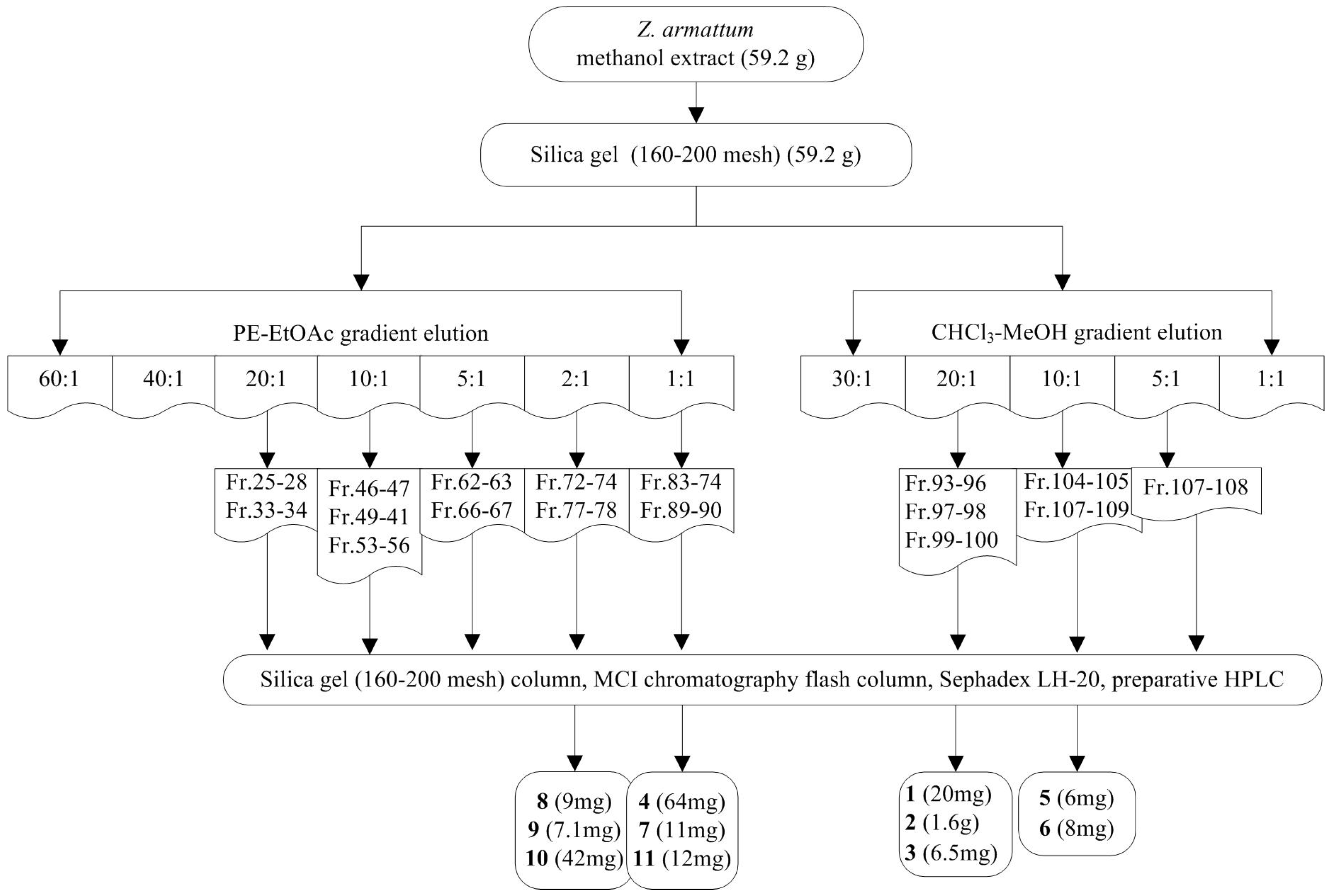
4.3. Extraction and Isolation

The fresh stem bark (1.6 kg) of Z. armatum was roughly crushed and extracted with methanol (20 L) under ultrasound three times. The filtrate was concentrated into crude extract (59.2 g) under vacuum. The extract was crudely fractionated by silica gel column chromatography (160–200 mesh, 10.0 × 63 cm, 1300 g), eluting with a stepwise gradient of PE (petroleum ether)/EtOAc (ethyl acetate) (60:1, 40:1, 20:1, 10:1, 5:1, 2:1 and 1:1), and then CHCl3/CH3OH (30:1, 20:1, 10:1, 5:1, 1:1 and MeOH) to receive 108 fractions. In total, 11 lignans were isolated from the stem bark of Z. armatum. The chemical isolation scheme of Z. armatum methanol extract is shown in Figure 3. All purified products were analyzed by 1H and 13C-NMR spectra and then compared with literature values. Accordingly, lignans 111 were characterized and identified as asarinin (1) [35], fargesin (2) [31], horsfieldin (3) [36], eudesmin (4) [37], planispine A (5) [36], planispine B (6) [38], yangambin (7) [39], acuminatolide (8) [40], salicifoliol (9) [41], magnolone (10) [42], and magnone A (11) [43].
Asarinin (1). White powder. 1H-NMR (500 MHz, CDCl3) δ ppm: 6.89 (2H, s, H-2,2′), 6.85~6.79 (4H, m, H-5,5′,6,6′), 5.98 (2H, s, -O-CH2-O-), 5.97 (2H, s, -O-CH2-O-), 4.85 (1H, s, H-7′), 4.42 (1H, s, 7.0 Hz, H-7), 4.12 (1H, s, H-9′α), 3.85 (2H, m, H-9α,9′β), 3.32 (2H, m, H-9β,8′), 2.88 (1H, m, H-8); 13C-NMR (500 MHz, CDCl3) δ ppm: 147.9 (C-4′), 147.6 (C-4), 147.2 (C-3), 146.6 (C-3′), 135.1 (C-1), 132.3 (C-1′), 119.6 (C-6), 118.7 (C-6′), 108.2 (C-5′), 106.6 (C-2′), 106.4 (C-2), 101.1 (-O-CH2-O-), 101.0 (-O-CH2-O-), 87.7 (C-7), 82.0 (C-7′), 70.9 (C-9′), 69.9 (C-9), 54.7 (C-8), 50.2 (C-8′).
Fargesin (2). Colorless cluster crystals. 1H-NMR (500 MHz, CDCl3) δ ppm: 6.95 (1H, s, H-2), 6.90 (1H, s, H-2′), 6.88 (2H, s, H-5, 6), 6.85 (1H, d, J = 8.0 Hz, H-6′), 6.80 (1H, d, J = 8.0 Hz, H-5′), 5.97 (2H, s, -OCH2O-), 4.89 (1H, d, J = 5.0 Hz, H-7), 4.44 (1H, d, J = 7.0 Hz, H-7′), 4.14 (1H, d, J = 9.5 Hz, H-9′α), 3.93 (3H, s, 3-OCH3), 3.90 (3H, s, 4-OCH3), 3.86 (2H, dd, J = 8.0 Hz, J = 7.0 Hz, H-9α, 9′β), 3.44 (2H, m, H-9β, 8), 2.90 (1H, dd, J = 8.0 Hz, H-8′); 13C-NMR (125 MHz,CDCl3) δ ppm: 148.8 (C-3), 148.0 (C-4), 148.0 (C-3′), 147.2 (C-4′), 135.2 (C-1′), 130.9 (C-1), 119.6 (C-6′), 117.7 (C-6), 111.0 (C-5), 109.0 (C-2), 108.2 (C-5′), 106.5 (C-2′), 101.1 (-OCH2O-), 87.7 (C-7′), 82.0 (C-7), 71.0 (C-9′), 69.8 (C-9), 55.9 (3-OCH3), 55.9 (4-OCH3), 54.6 (C-8′), 50.2 (C-8).
Horsfieldin (3). Colorless needle crystals. 1H-NMR (500 MHz, CDCl3) δ ppm: 6.97 (1H, s, H-2), 6.91 (1H, d, J = 8.0 Hz, H-2′), 6.90 (1H, s, H-5), 6.85 (1H, d, J = 8.0 Hz, H-5′), 6.80 (2H, d, J = 8.0 Hz, H-6,6′), 5.98 (2H, s, -OCH2O-), 5.60 (1H, s, 3-OH), 4.87 (1H, d, J = 5.0 Hz, H-7′), 4.44 (1H, d, J = 7.0 Hz, H-7), 4.14 (1H, d, J = 9.5 Hz, H-9′α), 3.94 (3H, s, 4-OCH3), 3.86 (2H, m, H-9β,9′β), 3.33 (2H, m, H-9α,8′), 2.89 (1H, m, H-8); 13C-NMR (125 MHz, CDCl3) δ ppm: 148.0 (C-3′), 147.2 (C-4), 146.4 (C-3), 144.6 (C-4′), 135.2 (C-1), 130.3 (C-1′), 119.6 (C-6), 118.4 (C-6′), 114.2 (C-2), 108.3 (C-5), 108.2 (C-5′), 106.6 (C-2′), 101.1 (-OCH2O-), 87.7 (C-7), 82.0 (C-7′), 71.0 (C-9′), 69.8 (C-9), 56.0 (4-OCH3), 54.6 (C-8), 50.2 (C-8′).
Eudesmin (4). Colorless cluster crystals. 1H-NMR (500 MHz, CDCl3) δ ppm: 6.93 (2H, s, H-2,2′), 6.90 (2H, d, J = 8.0 Hz, H-5,5′), 6.87 (2H, d, J = 8.0 Hz, H-6,6′), 4.78 (2H, t, J = 3.0 Hz, H-7,7′), 4.28 (2H, t, d, J = 8.0 Hz, H-9α,9′α), 3.92 (8H, s, 3,3′-OCH3,H-9β,9′β), 3.90 (6H, s, 4,4′-OCH3), 3.14 (2H, s, H-8,8′), 13C-NMR (125 MHz, CDCl3) δ ppm: 149.2 (C-3,3′), 148.6 (C-4,4′), 133.6 (C-1,1′), 118.3 (C-6,6′), 111.0 (C-2,2′), 109.2 (C-5,5′), 85.8 (C-7,7′), 71.7 (C-9,9′), 56.0 (C-3,3′), 55.9 (C-4,4′), 54.2 (C-8,8′).
Planispine A (5). Colorless oil. 1H-NMR (500 MHz, CDCl3) δ ppm: 6.98 (1H, s, H-6), 6.94 (1H, s, H-6′), 6.91 (1H, d, J = 8.5 Hz, H-5), 6.88 (2H, d, H-5′,2), 6.81 (1H, d, J = 8.0 Hz, H-2′), 5.61 (1H, s, 4′-OH), 5.54 (1H, t, J = 6.0 Hz, H-2″), 4.88 (1H, d, J = 5.0 Hz, H-7), 4.60 (2H, d, J = 6.5 Hz, H-1″), 4.46 (1H, d, J = 7.0 Hz, H-8), 4.15 (1H, d, J = 7.0 Hz, H-9α), 3.94 (3H, s, 3′-OCH3), 3.91 (3H, s, 3-OCH3), 3.87 (2H, t, J = 7.0 Hz, H-9β,9′β), 3.34 (2H, d, J = 5.0 Hz, H-8,9′α), 2.94 (1H, m, H-8′); 13C-NMR (125 MHz, CDCl3) δ ppm: 149.7 (C-3), 148.0 (C-4), 146.4 (C-3′), 144.6 (C-4′), 137.6 (C-1), 133.7 (C-1′), 130.4 (C-6), 120.0 (C-2″), 118.4 (C-6′), 118.4 (C-5), 114.2 (C-5′), 112.9 (C-2′), 109.3 (C-3″), 108.4 (C-2), 87.7 (C-7′), 82.1 (C-7), 71.0 (C-9), 69.7 (C-9′), 65.8 (C-1″), 56.0 (3′-OCH3), 55.9 (3-OCH3), 54.4 (C-8′), 50.2 (C-8), 25.8 (C-4″), 18.2 (C-5″).
Planispine B (6). Colorless oil. 1H-NMR (500 MHz, CDCl3) δ ppm: 6.92~6.86 (6H, m, H-2,2′,5,6,6′,5′), 5.53 (1H, t, J = 5.5 Hz, H-2″), 5.10 (1H, t, J = 7.0 Hz, H-7″), 4.77 (2H, dd, J = 4.5 Hz, J = 7.5 Hz, H-7,7′), 4.63 (1H, d, J = 4.5 Hz, H-1″), 4.28 (2H, t, J = 8.0 Hz, H-9′), 3.93 (3H, s, 3-OCH3), 3.91 (5H, s, 3′-OCH3, H-9), 3.14 (2H, m, H-8,8′), 2.13 (2H, t, J = 8.0 Hz, H-5″), 2.07 (2H, m, H-6″), 1.74 (3H, s, H-4″), 1.69 (3H, s, H-9″), 1.62 (3H, s, H-10″); 13C-NMR (125 MHz, CDCl3) δ ppm: 149.7 (C-3), 147.9 (C-4), 146.7 (C-3′), 145.3 (C-4′), 140.6 (C-3″), 133.5 (C-1′), 132.9(C-1), 131.8 (C-8″), 123.9 (C-7″), 119.8 (C-2″), 119.0 (C-6′), 118.2 (C-6), 114.3(C-5′), 113.0 (C-5), 109.5 (C-2′), 108.6 (C-2), 85.9 (C-7), 85.8 (C-7′), 71.7 (C-9,9′), 66.0 (C-1″), 56.0 (3,3′-OCH3), 54.2 (C-8), 54.1 (C-8′), 39.6 (C-5″), 26.3 (C-6″), 25.7 (C-9″), 17.7 (C-10″), 16.7 (C-4″).
Yangambin (7). Colorless oil. 1H-NMR (500 MHz, CDCl3) δ ppm: 6.59 (4H, s, H-2,2′,6,6′), 4.77 (2H, d, J = 4.0 Hz, H-1,1′), 4.33 (2H, dd, J = 9.0 Hz, J = 7.0 Hz, H-9α,9′α), 3.95 (2H, dd, J = 9.0 Hz, J = 3.5 Hz, H-9β,9′β), 3.90 (12H, s, 3,3′,5,5′-OCH3), 3.86 (6H, s, 4,4′-OCH3), 3.13 (2H, m, H-8,8′); 13C-NMR (125 MHz, CDCl3) δ ppm: 153.5 (C-3,3′,5,5′), 137.5 (C-4,4′), 136.7 (C-1,1′), 102.8 (C-2,2′,6,6′), 86.0 (C-7,7′), 72.0 (C-9,9′), 60.9 (4,4′-OCH3), 56.2 (3,3′,5,5′-OCH3), 54.4 (C-8,8′).
Acuminatolide (8). Colorless needle crystals. 1H-NMR (500 MHz, CDCl3) δ ppm: 6.86 (1H, s, H-6), 6.82 (1H, s, H-2), 6.81 (1H, s, H-3), 5.99 (2H, s, H-10), 4.62 (1H, d, J = 7.0 Hz, H-7), 4.51 (1H, m, H-9β), 4.38 (1H, d, J = 7.0 Hz, H-9′β), 4.35 (1H, m, H-9α), 4.21 (1H, dd, J = 3.5 Hz, J = 9.5 Hz, H-9′α), 3.45 (1H, td, J = 3.5 Hz, J = 9.5 Hz, H-8), 3.10 (1H, m, H-8′); 13C-NMR (125 MHz, CDCl3) δ ppm: 178.1 (C-7′), 148.3 (C-5), 147.8 (C-4), 132.8 (C-1), 119.6 (C-2), 108.4 (C-3), 106.4 (C-6), 101.3 (-OCH2O-), 86.1 (C-7), 70.1 (C-9), 69.8 (C-9′), 48.4 (C-8), 46.0 (C-8′).
Salicifoliol (9). Colorless oil. 1H-NMR (500 MHz, CDCl3) δ ppm: 6.93 (1H, d, J = 8.0 Hz, H-3), 6.90 (1H, s, H-6), 6.83 (1H, d, J = 8.0 Hz, H-2), 5.68 (1H, s, 4-OH), 4.64 (1H, d, J = 7.0 Hz, H-7), 4.52 (1H, dd, J = 9.0 Hz, J = 7.0 Hz, H-9β), 4.39 (1H, t, J = 9.0 Hz, H-8β), 4.35 (1H, d, J = 11.0 Hz, H-9α), 4.21 (1H, dd, J = 9.0 Hz, J = 3.5 Hz, H-8α), 3.93 (3H, s, 5-OCH3), 3.47 (1H, td, J = 9.0 Hz, J = 3.5 Hz, H-12), 3.14 (1H, m, H-11); 13C-NMR (125 MHz, CDCl3) δ ppm: 178.2 (C-10), 146.9 (C-5), 145.9 (C-4), 130.6 (C-1), 119.1 (C-2), 114.4 (C-3), 108.5 (C-6), 86.1 (C-7), 70.0 (C-8), 69.9 (C-9), 56.1 (5-OCH3), 48.2 (C-11), 46.0 (C-12).
Magnolone (10). White powder. 1H-NMR (500 MHz, CDCl3) δ ppm: 7.62 (1H, d, J = 8.5 Hz, H-6′), 7.59 (1H, s, H-2′), 6.99 (1H, s, H-2), 6.93 (d, 2H, J = 8.5 Hz, H-5′), 6.88 (1H, d, J = 8.0 Hz, H-6), 6.79 (1H, d, J = 8.0 Hz, H-5), 5.97 (2H, s, -OCH2O-), 4.70 (2H, d, J = 9.0 Hz, H-7), 4.31 (1H, m, H-9′β), 4.18 (1H, m, H-8′), 4.18 (1H, m, H-9′α), 3.98 (1H, s, 3′-OCH3), 3.96 (1H, s, 4′-OCH3), 3.79 (1H, dd, H-9β), 3.68 (1H, dd, H-9α), 2.88 (1H, m, H-8); 13C-NMR (125 MHz, CDCl3) δ ppm: 197.8 (C-7′), 153.7 (C-4′), 149.3 (C-3′), 148.0 (C-3), 147.4 (C-4), 134.5 (C-1), 129.8 (C-1′), 123.2 (C-6′), 120.4 (C-6), 110.6 (C-2′), 110.1 (C-5′), 108.1 (C-5), 107.1 (C-2), 101.1 (-OCH2O-), 83.7 (C-7), 70.9 (C-9′), 61.3 (C-9), 56.1 (3′-OCH3), 56.0 (4′-OCH3), 52.4 (C-8), 49.6 (C-8′).
Magnone A (11). Colorless oil. 1H-NMR (500 MHz, CDCl3) δ ppm: 7.63 (1H, d, J = 9.0 Hz, H-2′), 7.60 (1H, d, J = 1.5 Hz, H-6′), 7.05 (1H, d, J = 1.5 Hz, H-2), 6.95 (1H, d, J = 2.0 Hz, H-6), 6.93 (1H, d, J = 8.5 Hz, H-3′), 6.85 (1H, d, J = 8.0 Hz, H-5), 4.72 (1H, d, J = 9.0 Hz, H-7), 4.33 (1H, m, H-9′β), 4.21 (2H, m, H-8′,9′α), 3.98 (3H, s, 3-OCH3), 3.96 (3H, s, 4-OCH3), 3.94 (3H, s, 5′-OCH3), 3.89 (3H, s, 4′-OCH3), 3.80 (1H, dd, J = 11.0 Hz, J = 4.5 Hz, H-9β), 3.70 (1H, dd, J = 11.0 Hz, J = 4.5 Hz, H-9α), 2.94 (1H, m, H-8); 13C-NMR (125 MHz, CDCl3) δ ppm: 198.0 (C-7′), 153.7 (C-4′), 149.3 (C-5′), 149.2 (C-4), 148.9 (C-3), 133.0 (C-1), 129.8 (C-1′), 123.2 (C-2′), 119.3 (C-6), 110.9 (C-5), 110.6 (C-6′), 110.1 (C-3′), 109.6 (C-2), 83.8 (C-7), 70.9 (C-9′), 61.4 (C-9), 56.1 (3-OCH3), 56.0 (4/5′-OCH3), 55.9 (4′-OCH3), 52.2 (C-8), 49.7 (C-8′).

4.4. Antifeedant Assay

An antifeedant assay was performed using a modified method, as reported in References [44,45]. The compound sample was precisely weighed at 2 mg and diluted in ethanol to fabricate the mother liquor with a concentration of 1 mg/mL. Then, different volumes of the mother liquor were diluted in water to obtain different solution concentrations of 25, 74, 222, 667, and 2000 ppm. After that, the final solutions with different concentrations were stirred well with 0.4 g flour. The blank control was fabricated by the mixture of 2 mL water and 0.4 g flour. Then, 1-mL tips were cut about 1 cm from the bottom to make an opening, and enlarged to about a 2-mm internal diameter. Subsequently, 200 µL mixture was pipetted into a clean dish to make about 6–8 small cookies drop by drop. After being air-dried overnight, the small cookies with different concentrations were transferred into the incubator for 48 h to achieve equilibration at 29–30 °C and 70–80% relative humidity (RH). Twenty unsexed adult insects were selected and placed into glass vials (diameter: 2.5 cm, height: 5.5 cm) and starved for 24 h for further weighing. After that, small cookies were added into each vial and weighed later. For each concentration, the test was conducted five times. Seventy-two hours later, the total weight was determined. Then, all the insects were singled out immediately, and each vial with remaining cookies were weighed again. The percent antifeedant index was calculated as follows: Antifeedant index (%) = [(CT)/C] × 100, where C is the weight of the diet consumed in the blank control and T is the weight of the diet consumed in the treated groups. The antifeedant index and EC50 were calculated by SPSS (IBM) and Origin software. Statistical analysis was carried out using Student’s t-test to compare the two groups, with p < 0.05 being indicative of significance. A commercial antifeedant reagent, toosendanin, used as a positive control, was purchased from China National Standards Network.

5. Conclusions

Eleven lignans were isolated from the stem bark of Zanthoxylum armatum, and five of them were obtained for the first time. The antifeedant activities of these compounds were measured, and one of them, asarinin (1), showed stronger activity than the positive control. Three other compounds, fargesin (2), horsfieldin (3), and magnolone (10), exhibited the same antifeedant activities as the positive control. Furthermore, compounds that displayed high antifeedant activities have a common chemical group, namely methylenedioxy. These results indicated that compounds with one or more methylenedioxy groups have potential for development into novel antifeedants or lead compounds to protect food crops in storage.

Acknowledgments

This project was supported by the National Key Research and Development Program (2016YFC0500805), Beijing Municipal Natural Science Foundation (No. 7142093), and Fundamental Research Funds for the Central Universities.

Author Contributions

W.Z., Y.W., and S.D. conceived and designed the experiments; W.Z., Y.W., and Z.Z. performed the experiments; W.Z. and Z.G. analyzed the data; J.C., X.P., and Z.C. contributed materials and analysis tools; W.Z. wrote the paper; Z.D. revised the paper.

Conflicts of Interest

The authors declare no conflict of interest.

References

  1. Hematpoor, A.; Liew, S.Y.; Azirun, M.S.; Awang, K. Insecticidal activity and the mechanism of action of three phenylpropanoids isolated from the roots of Piper sarmentosum Roxb. Sci. Rep. 2017, 7, 1–13. [Google Scholar] [CrossRef] [PubMed]
  2. Rajashekar, Y.; Shivanandappa, T. A novel natural insecticide molecule for grain protection. Julius-Kühn-Archiv 2010, 910–915. [Google Scholar] [CrossRef]
  3. Denell, R.; Gibbs, R.; Muzny, D.; Weinstock, G.M.; Attaway, T.; Bell, S.; Buhay, C.J.; Chandrabose, M.N.; Chavez, D.; Clerk-Blankenburg, K.P.; et al. The genome of the model beetle and pest Tribolium castaneum. Nature 2008, 452, 949–955. [Google Scholar]
  4. Gokhale, C.S.; Traulsen, A.; Joop, G. Social dilemma in the external immune system of the red flour beetle? It is a matter of time. Ecol. Evol. 2017, 7, 6758–6765. [Google Scholar] [CrossRef] [PubMed]
  5. Kittelmann, S.; Ulrich, J.; Posnien, N.; Bucher, G. Changes in anterior head patterning underlie the evolution of long germ embryogenesis. Dev. Biol. 2013, 374, 174–184. [Google Scholar] [CrossRef] [PubMed]
  6. Cheng, S.S.; Chang, H.T.; Chang, S.T.; Tsai, K.H.; Chen, W.J. Bioactivity of selected plant essential oils against the yellow fever mosquito Aedes aegypti larvae. Bioresour. Technol. 2003, 89, 99–102. [Google Scholar] [CrossRef]
  7. Zettler, J.L.; Arthur, F.H. Chemical control of stored product insects with fumigants and residual treatments. Crop Prot. 2000, 19, 577–582. [Google Scholar] [CrossRef]
  8. Arthur, F.H. Grain protectants: Current status and prospects for the future. J. Stored Prod. Res. 1996, 32, 293–302. [Google Scholar] [CrossRef]
  9. Adakole, J.A.; Adeyemi, A.F.F. Larvicidal effects of Cymbopogon citratus (lemon grass) extract against Culex quinquefasciatus qularvae (Diptera, culicidae). Int. J. Appl. Environ. Sci. 2012, 7, 187–192. [Google Scholar]
  10. Tavares, W.S.; Costa, M.A.; Cruz, I.; Silveira, R.D.; Serrao, J.E.; Zanuncio, J.C. Selective effects of natural and synthetic insecticides on mortality of Spodoptera frugiperda (Lepidoptera: Noctuidae) and its predator Eriopis connexa (Coleoptera: Coccinellidae). J. Environ. Sci. Heal 2010, 45, 557–561. [Google Scholar] [CrossRef] [PubMed]
  11. Alam, F.; Us Saqib, Q.N. Evaluation of Zanthoxylum armatum Roxb for in vitro biological activities. J. Tradit. Complement. Med. 2017, 7, 515–518. [Google Scholar] [CrossRef] [PubMed]
  12. Li, H.; Li, P.; Zhu, L.; Xie, M. Studies on the chemical constituents of Zanthoxylum armatum DC. Chin. Pharm. 2006, 13, 1035–1036. [Google Scholar]
  13. Chen, Y.; Hu, Y.; He, H.; Yang, G. Chemical constituents from barks of Zanthoxylum planispinum. Chin. Tradit. Herb. Drugs 2013, 44, 3429–3434. [Google Scholar]
  14. Agnihotri, S.; Wakode, S.; Ali, M. Chemical constituents isolated from Zanthoxylum armatum stem bark. Chem. Nat. Compd. 2017, 53, 880–882. [Google Scholar] [CrossRef]
  15. Bhatt, V.; Sharma, S.; Kumar, N.; Singh, B. A new lignan from the Leaves of Zanthoxylum armatum. Nat. Prod. Commun. 2017, 12, 99–100. [Google Scholar]
  16. Guo, T.; Dai, L.; Tang, X.; Song, T.; Wang, Y.; Zhao, A.; Cao, Y.; Chang, J. Two new phenolic glycosides from the stem of Zanthoxylum armatum DC. Nat. Prod. Res. 2017, 31, 2335–2340. [Google Scholar] [CrossRef] [PubMed]
  17. Guo, T.; Li, Y. Determination of four lignans in roots, stems and leaves of Zanthoxylum armatum DC by HPLC-DAD with HPLC-ESI-QTOF-MS confirmation. J. Anal. Chem. 2016, 71, 527–533. [Google Scholar] [CrossRef]
  18. Kumar, V.; Reddy, S.G.E.; Chauhan, U.; Kumar, N.; Singh, B. Chemical composition and larvicidal activity of Zanthoxylum armatum against diamondback moth, Plutella xylostella. Nat. Prod. Res. 2016, 30, 689–692. [Google Scholar] [CrossRef] [PubMed]
  19. Zhou, T.; Pu, B.; Jiang, H.; Liu, C. Analysis of fatty acid compositions in Zanthoxylum armatum DC. oil with different extraction methods. J. Nucl. Agric. Sci. 2015, 29, 1323–1328. [Google Scholar]
  20. Wang, C.; Zhang, W.; You, C.; Guo, S.; Geng, Z.; Fan, L.; Du, S.; Deng, Z.; Wang, Y. Insecticidal Constituents of Essential Oil Derived from Zanthoxylum armatum against Two Stored-Product Insects. J. Oleo Sci. 2015, 64, 861–868. [Google Scholar] [CrossRef] [PubMed]
  21. Wang, C.; Pu, B.; Jiang, Y.; Fu, B. Extraction and antioxidant activity in vitro of flavonoids in cold pressed cake of Zanthoxylum armatum DC. Prod. Food Mach. 2017, 33, 137–142. [Google Scholar]
  22. Xu, L.; Pu, B.; Jiang, Y.; Tan, X.; Wang, S.; Wang, C.; Zhang, L.; Sun, P. Studies on the antioxidatant activities of water extract from Zanthoxylum armatum DC. Prodr. in vitro. Food Ferment. Ind. 2016, 42, 140–143. [Google Scholar]
  23. Zhou, T.; Pu, B.; Jiang, H.; Liu, C. Study on antibacterial activity and stability of Zanthoxylum armatum DC. Prodr. oil. Sci. Technol. Food Ind. 2015, 36, 106–109. [Google Scholar]
  24. Alam, F.; Saqib, Q.N.U.; Waheed, A. Cytotoxic activity of extracts and crude saponins from Zanthoxylum armatum DC. against human breast (MCF-7, MDA-MB-468) and colorectal (Caco-2) cancer cell lines. BMC Complement. Altern. Med. 2017, 17, 1–9. [Google Scholar] [CrossRef] [PubMed]
  25. Singh, T.D.; Meitei, H.T.; Sharma, A.L.; Robinson, A.; Singh, L.S.; Singh, T.R. Anticancer properties and enhancement of therapeutic potential of cisplatin by leaf extract of Zanthoxylum armatum DC. Biol. Res. 2015, 48, 1–9. [Google Scholar] [CrossRef] [PubMed]
  26. Singh, G.; Singh, R.; Verma, P.K.; Singh, R.; Anand, A. Anthelmintic efficacy of aqueous extract of Zanthoxylum armatum DC. seeds against Haemonchus contortus of small ruminants. J. Parasit. Dis. Off. Organ Indian Soc. Parasitol. 2016, 40, 528–532. [Google Scholar] [CrossRef] [PubMed]
  27. Teponno, R.B.; Kusari, S.; Spiteller, M. Recent advances in research on lignans and neolignans. Nat. Prod. Rep. 2016, 33, 1044–1092. [Google Scholar] [CrossRef] [PubMed]
  28. Rezanka, T.; Vitova, M.; Kolouchova, I.; Nedbalova, L.; Dolezalova, J.; Palyzova, A.; Sigler, K. Polydatin and its derivatives inhibit fatty acid desaturases in microorganisms. Eur. J. Lipid Sci. Technol. 2017, 119, 1–11. [Google Scholar] [CrossRef]
  29. Su, G.; Cheng, Y.; Wang, K.; Wang, X.; Wu, B. An unusual tetrahydrofuran lignan from the roots of Zanthoxylum planispinum and the potential anti-inflammatory effects. Chem. Biodivers. 2017, 14, 1–6. [Google Scholar] [CrossRef] [PubMed]
  30. Papadopoulos, A.G.; Nenadis, N.; Sigalas, M.P. DFT study of radical scavenging activity of sesame oil lignans and selected in vivo metabolites of sesamin. Comput. Theor. Chem. 2016, 1077, 125–132. [Google Scholar] [CrossRef]
  31. Messiano, G.B.; Vieira, L.; Machado, M.B.; Lopes, L.M.X.; De Bortoli, S.A.; Zukerman-Schpector, J. Evaluation of insecticidal activity of diterpenes and lignans from Aristolochia malmeana against Anticarsia gemmatalis. J. Agric. Food Chem. 2008, 56, 2655–2659. [Google Scholar] [CrossRef] [PubMed]
  32. Woo, K.W.; Suh, W.S.; Subedi, L.; Kim, S.Y.; Kim, A.; Lee, K.R. Bioactive lignan derivatives from the stems of Firmiana simplex. Bioorg. Med. Chem. Lett. 2016, 26, 730–733. [Google Scholar] [CrossRef] [PubMed]
  33. Yin, J.; Han, N.; Liu, Z.; Xu, X.; Zhang, B.; Kadota, S. The in vitro anti-osteoporotic activity of some diarylheptanoids and lignans from the rhizomes of Dioscorea spongiosa. Planta Med. 2008, 74, 1451–1453. [Google Scholar] [CrossRef] [PubMed]
  34. Ma, Z.; Zhang, J.; Yang, L.; Ma, J. Analysis and comparison of the volatile oil from different parts of Zanthoxylum ovalifolium var. spinifolium by GC-MS. Chin. Pharm. J. 2004, 39, 502–503. [Google Scholar]
  35. Jing, Y.; Zhang, Y.; Shang, M.; Yu, J.; Tang, J.; Liu, G.; Li, Y.; Li, X.; Wang, X.; Cai, S. Phenanthrene derivatives from roots and rhizomes of Asarum heterotropoides var. mandshuricum. Fitoterapia 2017, 117, 101–108. [Google Scholar] [CrossRef] [PubMed]
  36. Guo, T. Studies on the Chemical Constituents of Zanthoxylum armatum DC. and Z. avicenna (Lam.) DC. and Analgesic, Anti-Inflammatory Activites of Z. armatum; Fudan University: Fudan, China, 2011. [Google Scholar]
  37. Latip, J.; Hartley, T.G.; Waterman, P.G. Lignans and coumarins metabolites from Melicope hayesii. Phytochemistry 1999, 51, 107–110. [Google Scholar] [CrossRef]
  38. Hu, Y.; Chen, Y.; Shang, Y.; Yang, G. Two New lignans from the bark of Zanthoxylum planispinum. B Korean Chem. Soc. 2009, 30, 1884–1886. [Google Scholar]
  39. Ahmed, A.A.; Mahmoud, A.A.; Ali, E.T.; Tzakou, O.; Couladis, M.; Mabry, T.J.; Gati, T.; Toth, G. Two highly oxygenated eudesmanes and 10 lignans from Achillea holosericea. Phytochemistry 2002, 59, 851–856. [Google Scholar] [CrossRef]
  40. Saladino, R.; Fiani, C.; Crestini, C.; Argyropoulos, D.S.; Marini, S.; Coletta, M. An efficient and stereoselective dearylation of asarinin and sesamin tetrahydrofurofuran lignans to acuminatolide by methyltrioxorhenium/H2O2 and UHP Systems. J. Nat. Prod. 2007, 70, 39–42. [Google Scholar] [CrossRef] [PubMed] [Green Version]
  41. Aguiar, R.M.; Alves, C.Q.; David, J.M.; de Rezende, L.C.; Lima, L.S.; David, J.P.; de Queiroz, L.P. Antioxidant activities of isolated compounds from stems of Mimosa invisa mart. ex colla. Quim. Nova 2012, 35, 567–570. [Google Scholar] [CrossRef]
  42. Yu, H.J.; Chen, C.C.; Shieh, B.J. Two new constituents from the leaves of Magnolia coco. J. Nat. Prod. 1998, 61, 1017–1019. [Google Scholar] [CrossRef] [PubMed]
  43. Jung, K.Y.; Kim, D.S.; Oh, S.R.; Park, S.H.; Lee, I.S.; Lee, J.J.; Shin, D.H.; Lee, H.K. Magnone A and B, novel anti-PAF tetrahydrofuran lignans from the flower buds of Magnolia fargesii. J. Nat. Prod. 1998, 61, 808–811. [Google Scholar] [CrossRef] [PubMed]
  44. Du, S.S.; Wang, C.F.; Li, J.; Zhang, H.M.; Liu, Q.Z.; Liu, Z.L.; Deng, Z.W. Antifeedant diterpenoids against Tribolium castaneum from the stems and twigs of Ceriops tagal (Rhizophoraceae). Molecules 2011, 16, 6060–6067. [Google Scholar] [CrossRef] [PubMed]
  45. Wang, C.; You, C.; Yang, K.; Guo, S.; Geng, Z.; Fan, L.; Du, S.; Deng, Z.; Wang, Y. Antifeedant activities of methanol extracts of four Zanthoxylum species and benzophenanthridines from stem bark of Zanthoxylum schinifolium against Tribolium castaneum. Ind. Crop Prod. 2015, 74, 407–411. [Google Scholar] [CrossRef]
Sample Availability: Samples of the compounds are available from the authors.
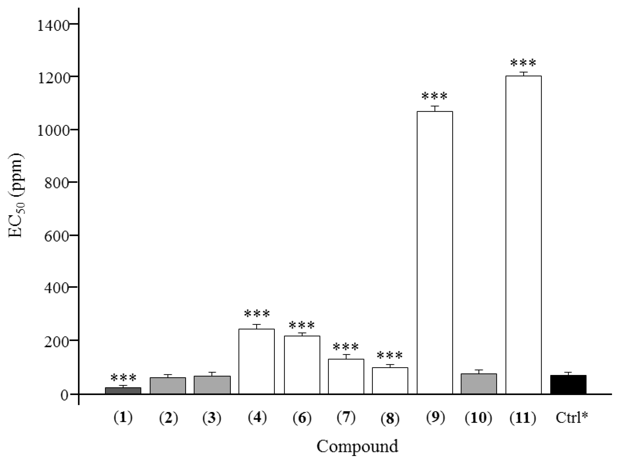
Figure 1. Chemical structures of compounds 111. * represents known but derived from the plant of Zanthoxylum armatum for the first time.
Molecules 23 00617 g001
Figure 2. Antifeedant EC50 of 10 lignans from Zanthoxylum armatum. * represents the positive control, toosendanin. *** represents the significant difference at p < 0.05.
Molecules 23 00617 g002
Figure 3. Chemical isolation scheme of Zanthoxylum armatum methanol extract.
Molecules 23 00617 g003
Table 1. Antifeedant activities of lignans from stem bark of Zanthoxylum armatum.
CompoundAntifeedant Index (%, Mean ± SD)EC50 (ppm)
25 ppm74 ppm222 ppm667 ppm2000 ppm
Asarinin (1)49.45 ± 3.7855.51 ± 1.7968.03 ± 4.2270.72 ± 2.8375.83 ± 3.6925.64 ± 3.21
Fargesin (2)40.65 ± 4.5152.12 ± 2.3160.71 ± 3.2175.21 ± 1.3981.96 ± 2.9363.24 ± 4.83
Horsfieldin (3)34.99 ± 5.3248.91 ± 3.3773.74 ± 3.0473.79 ± 6.0181.12 ± 3.3368.39 ± 5.58
Eudesmin (4)35.73 ± 4.3741.28 ± 3.0743.57 ± 2.1655.97 ± 3.4669.82 ± 3.56245.72 ± 3.74
Planispine (5)------
Planispine B (6)29.76 ± 3.1645.95 ± 5.1147.79 ± 3.2758.38 ± 3.1768.64 ± 5.23219.84 ± 4.69
Yangambin (7)37.80 ± 3.1249.71 ± 2.6150.82 ± 4.2760.95 ± 3.2666.96 ± 4.16132.50 ± 3.61
Acuminatolide (8)39.15 ± 1.2141.36 ± 3.8354.51 ± 2.1877.35 ± 3.6574.76 ± 3.93102.12 ± 2.40
Salicifoliol (9)6.82 ± 3.4712.52 ± 2.1524.78 ± 3.1730.46 ± 5.2339.06 ± 4.171069.20 ± 3.83
Magnolone (10)38.93 ± 4.2947.52 ± 1.9162.16 ± 3.9371.94 ± 3.1974.14 ± 3.9578.37 ± 3.44
Magnone A (11)21.66 ± 3.9128.99 ± 3.8532.76 ± 4.3343.53 ± 2.7756.60 ± 5.061203.53 ± 3.95
Toosendanin *32.32 ± 2.2852.45 ± 3.2769.52 ± 2.4776.54 ± 3.6286.27 ± 3.5171.69 ± 3.13
* represents the positive control. The antifeedant index of the blank control is 0.

