Lentinoids A–D, New Natural Products Isolated from Lentinus strigellus


1
Laboratory of Tropical Bioorganic Chemistry, Faculty of Natural, Exact Sciences and Technology, University of Panama, Panama 0824, Panama
2
Department of Microbiology, Faculty of Natural, Exact Sciences and Technology, University of Panama, Panama 0824, Panama
3
Centro de Investigaciones en Productos Naturales (CIPRONA), Universidad de Costa Rica, San José 2060, Costa Rica
4
Smithsonian Tropical Research Institute, Unit 0948, APO AA 34002–0948, Panama 0843, Panama
*
Author to whom correspondence should be addressed.
Molecules 2018, 23(4), 773; https://doi.org/10.3390/molecules23040773
Submission received: 26 February 2018
/
Revised: 18 March 2018
/
Accepted: 23 March 2018
/
Published: 28 March 2018
(This article belongs to the Section Natural Products Chemistry)
Abstract
:Four novel lentinoids (1–4), along with the known compounds striguellone A (5), isopanepoxydone (6) and panepoxydone (7), were isolated as part of our studies on Lentinus strigellus. The structures of 1–4 have been established by 1D- and 2D-NMR and MS analysis. Compounds (1–3) and (5–7) were tested against Listeria monocytogenes, Enterococcus faecalis, Pseudomonas aeruginosa and Klebsiella pneumoniae. These compounds showed inhibition diameters ranging from 7.5–9.5 mm, however, when the minimum inhibitory concentration (MIC) was determined, only compound 1 showed a significant activity of 200 μg/mL. Intermediates for the biosynthesis of the oxygenated cyclohexenyl derivatives isolated from lentinoid fungi (genera Lentinus and Panus) are proposed.
1. Introduction
Fungi represent one of the most extensive groups of organisms and it is estimated that there are between 1.5 and 5.1 million species [1]. Their capability to produce secondary metabolites with a wide spectrum of biological activities causes a high level of interest in the scientific community [2]. Some of them are edible and/or have been used in traditional medicine, such as the fungus Lentinus strigellus [3]. In their initial stage of growth, they can sometimes be manipulated to generate new compounds, and they could have interesting properties for biological applications.
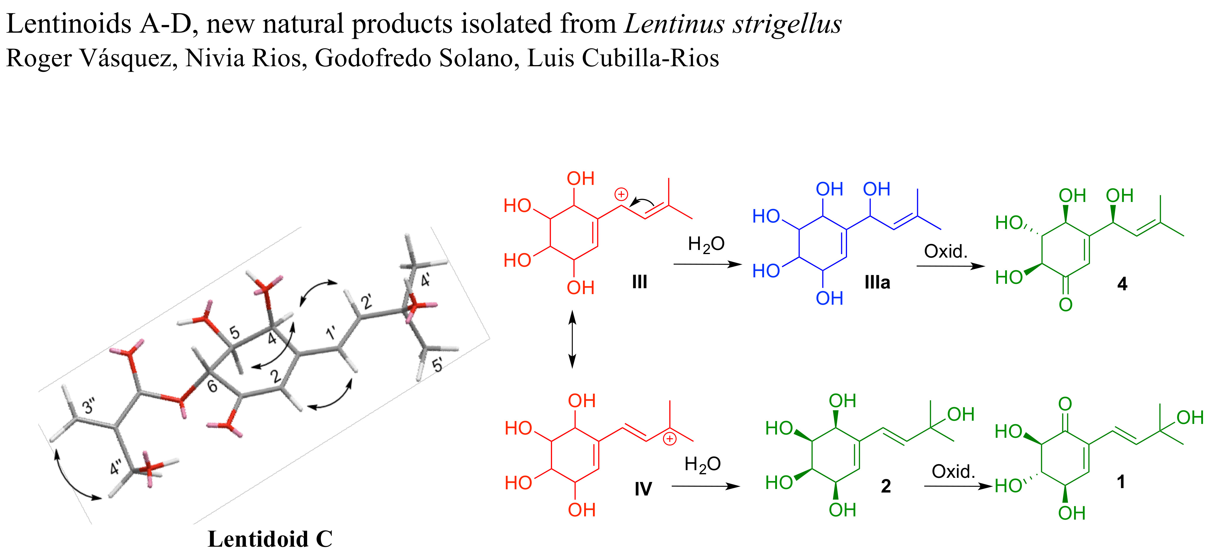
The genera Lentinus and Panus have some similarities that have generated controversy in some cases [4]. L. strigellus has also been described as Panus strigellus and Panus rudis var. strigellus [5]. Compounds such as strigellone A (5) [3], isopanepoxydone (6) [6], panepoxydone (7) [6], lentinoid E (8) [7] and other analogues have been reported from L. strigellus [8]. The lentinoids reported here have structural similarities with compounds isolated from species of the genera Panus (rudis and conchatus [6]) and Lentinus (strigosus [9] and connatus [10]). The fundamental structural scaffold of these compounds consists of an isoprenyl residue connected to a polyoxygenated cyclohexenyl unit. Lentinoid E (8) was previously isolated from the dichloromethane fraction obtained from a culture of L. strigellus grown in Sabouraud Dextrose Agar (SDA) adjusted to pH = 4.6, and using CaCl2 as elicitor at 26 °C for 15 days [7].
Continuing our study of L. strigellus, we undertook the analysis of the ethyl acetate fraction of the original extract using flash column chromatography and high performance liquid chromatography (HPLC). Thus, we describe the isolation and structural determination of four new lentinoids (1–4) along with known compounds (5–7) (Figure 1) that showed antibacterial properties.
2. Results and Discussion
Compound 1 was obtained as a brown viscous liquid. Its molecular formula, C11H16O5, was determined by High-resolution electrospray ionisation time-of-flight mass spectrometry (HR-ESI-TOF-MS) analysis on the basis of its cationized molecular peak [M + Na]+ at m/z 251.0891. The ion at m/z 269.0568 (100%) corresponded to [M + H2O + Na+] and the tandem mass spectrometry (MS2) spectrum showed that this ion is derived from the cluster [2M + H2O + Na+], m/z 497.0824. The Infrared (IR) spectrum showed bands at 3350 and 1690 cm−1 corresponding to a hydroxyl and carbonyl group, respectively. The nuclear magnetic resonance (NMR) data (Table 1) revealed the existence of an isoprenyl unit: two methyl groups (δH 1.30 (s); δC 29.7), a quaternary sp3 oxycarbon (δC 71.4), and two methines sp2 (δH 6.36, 6.44 (d); δC 120.1, 143.1). These signals have been observed in striguellone A (5) [3], a compound previously isolated from a culture of L. strigellus. This was confirmed by the correlation spectroscopy (COSY) experiment.
In the 13C-NMR spectrum, there was a quaternary carbon at δC 191.8 indicative of an α,β-unsaturated ketone. The chemical shift for the olefinic proton at C-3 displayed low-field shift at 6.89 ppm and appeared as a doublet, due to the coupling between H-3 and H-4, while in striguellone A, this appears as a singlet at 6.04 ppm (C-2) due the absence of this coupling.
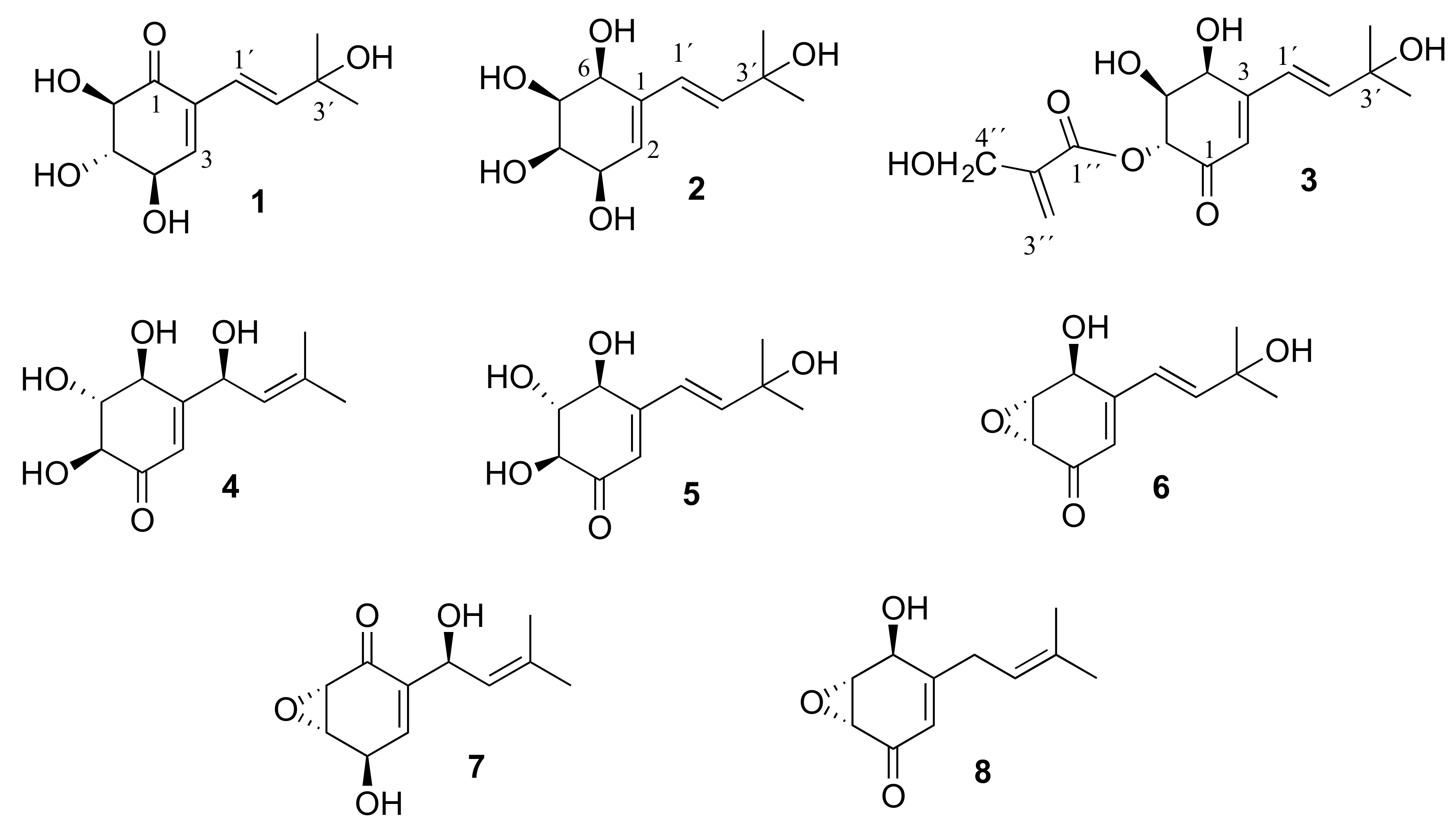
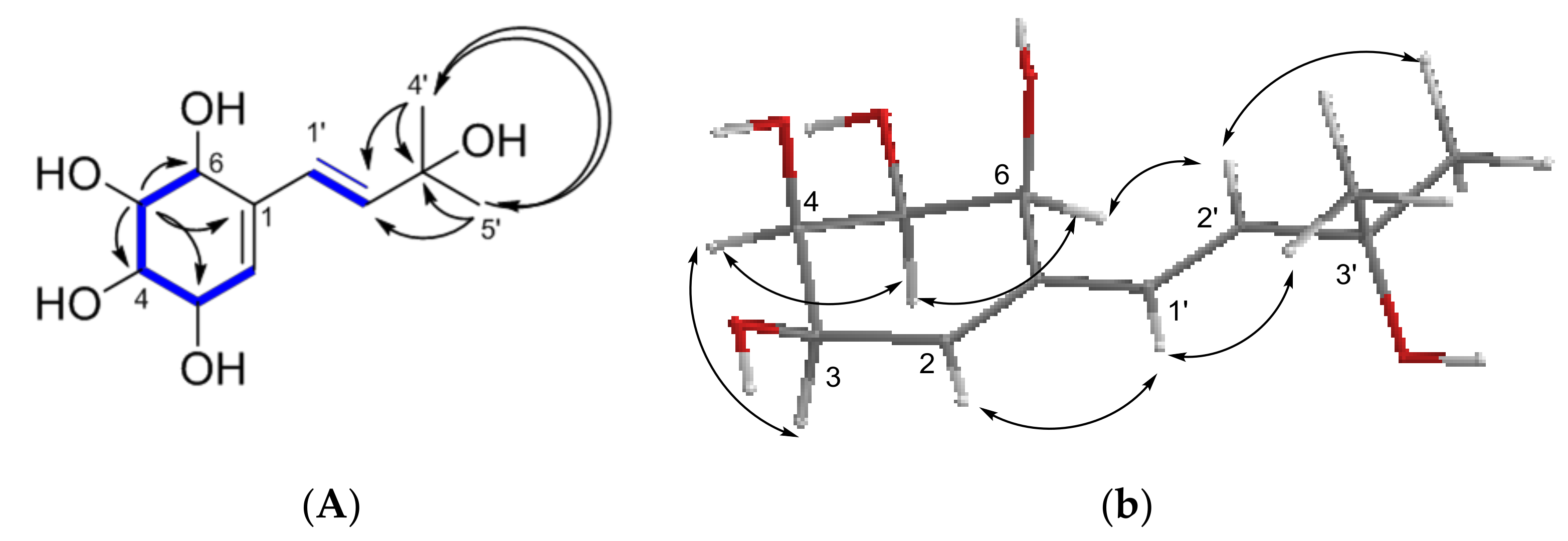
The quaternary olefinic carbon (δC 135.2) at the α-position of the α,β-unsaturated ketone displayed a high-field shift in comparison to C-3 (δC 155.7) in striguellone A; additionally, the methine carbon (δC 145.4) appeared at low-field shift occupying the β-position. The COSY spectrum revealed correlations between H-3/H-4, H-4/H-5, H-5/H-6 and H-1′/H-2′ (Figure 2A). The position of the carbonyl group was confirmed by the heteronuclear multiple-bond correlation (HMBC) experiment (Figure 2A). The nuclear Overhauser-effect spectroscopy (NOESY) experiment showed, clearly, correlation of H-1′/H-2′ with H-3 and H-4′/H-5′. On the other hand, a correlation between H-3 and H-4 was observed (Figure 2B). No interaction was detected for H-4/H-5 and H-5/H-6, placing them, in both cases, on opposite sides, which was confirmed by the coupling constants (3.6 and 10.8 Hz, respectively). Thereafter, we have assigned the new name lentinoid A to compound 1.
For compound 2, a brown viscous liquid, the molecular formula C11H18O5 was assigned, which has three degrees of unsaturation according to its HR-ESI-TOF-MS data ([M + Na]+, m/z 253.1034, calcd. 253.1046 for C11H18O5Na). The ion at m/z 235.0940 (100%) in the mass spectrum corresponded to the loss of water [M + Na − H2O]+, which was confirmed by an MS2 experiment. The IR spectrum showed a band at 3300 cm−1 corresponding to hydroxyl groups, although no signal for a carbonyl group was observed. The COSY and HMBC experiments (Figure 3A) confirmed the presence of an isoprenyl unit as in compound 1 (δH 1.30 (s); δC 29.9), a quaternary sp3 oxycarbon (δC 71.4) and two sp2 methines (δH 6.15, 6.24 (d); δC 126.5, 140.9). Above, 2 showed a quaternary sp2 carbon (δC 136.5), an sp2 methine (δH 5.67 (br t); δC 125.9) and four sp3 oxymethines (δH 3.37 (t), 3.49 (br m), 4.35 (d), 4.59 (br d); δC 57.1, 56.0, 63.9, 65.3).
The union of the oxymethines was established based on the COSY experiment (Figure 3A), observing correlations between H-2/H-3, H-3/H-4, H-4/H-5 and H-5/H-6, and the corresponding coupling constants. The relative stereochemistry was determined taking into account the following NOESY correlations: H-3/H-4, H-4/H-5 and H-5/H-6 (Figure 3B and Figure S2.6), as well as the values of the coupling constants (all bellow 4.6 Hz) between the hydrogens of the cyclohexyl unit. Therefore, all the hydroxyl groups were determined to be on the same site of the molecule. See 1H- and 13C-NMR data in Table 1. We have assigned the new name lentinoid B to compound 2.
For lentinoid C (3), the molecular formula C15H20O7 was assigned with six degrees of unsaturation according to its HR-ESI-TOF-MS data, [M + Na]+, m/z 335.1109 (calcd. 335.1101 for C15H20O7Na), with the bimolecular ion presented at m/z 647.2311. The IR spectrum showed a band at 3400 cm−1 corresponding to an OH group, and two bands for carbonyl groups: 1632 cm−1 for an α,β-unsaturated ketone and 1649 cm−1 for an α,β-unsaturated ester. Compound 3 showed similar NMR data (Table 2) to striguellone A, including the prenyl unit (two methyls (δC 29.6 and 29.7), an oxygenated quaternary carbon (δC 71.6), two sp2 carbons (δC 126.6 and 149.5)) and the α,β-unsaturated ketone (carbonyl group (δC 194.9), an sp2 quaternary carbon (δC 156.9) and sp2 methyne (δC 125.9)). Moreover, three oxygenated sp3 carbons were present (δC 76.6, 71.8 and 67.9). Altogether, the signals resemble the prenyl group and the cyclohexenyl ketone characteristic of lentinoids.
Additionally, the NMR data revealed the existence of a terminal methylene (δC 125.5, δH 5.96 and 6.35), a hydroxymethylene (δC 61.6, δH 4.34) and two quaternary carbons (δC 141.6 and δC 167.0), with the last one characteristic of a carboxyester group. The 2D NMR data (Figure 4A) showed the correlations between all protons and carbons for this fragment of compound 3. Moreover, the connection of the carboxy group and the cyclohexenyl ring was established through the correlation of H-6 (δH 5.72, δC 76.6) to both carbonyl groups (δC 167.0 and 194.9) in equal intensity.
The rotating frame overhauser effect spectroscopy (ROESY) experiment (Figure 4B) showed correlation between H-4 and H-5, indicating that they are on the same site of the molecule, and this was confirmed by the coupling constant of 3.6 Hz. No correlation between H-5 and H-6 was observed, indicating that they are in axial positions with a coupling constant of 10.8 Hz. Thus, the relative stereochemistry of compound 3 was established.
Due to possible instability and the small amount of compound 4 obtained, the structure was characterized by its 1H-NMR spectrum. Compound 4 showed signals for the isoprenyl group as in panepoxydone (7) characterized by two methyl groups attached to an sp2 carbon (δH 1.69 and 1.70), a methyne (δH 5.29, d, J = 8.8 Hz) and a hydroxymethyne (δH 5.00, d, J = 8.8 Hz). Based on the chemical shift for the other three hydroxymethynes, the coupling constants (H-6: δH 3.96, d, 12.0; H-5: δH 3.63, dd, 8.4 and 12.0; H-4: δH 4.36, d, 8.4), and the olefin proton (δH 6.89) with a chemical shift characteristic of an α-proton in an α,β-unsaturated ketone, we proposed a structure for the newly discovered lentinoid D (4).
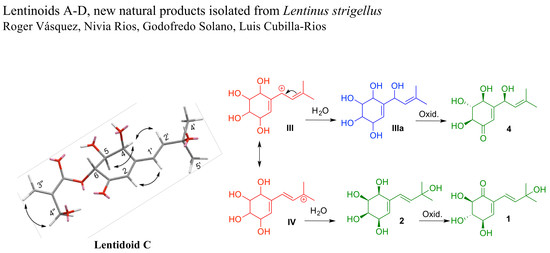
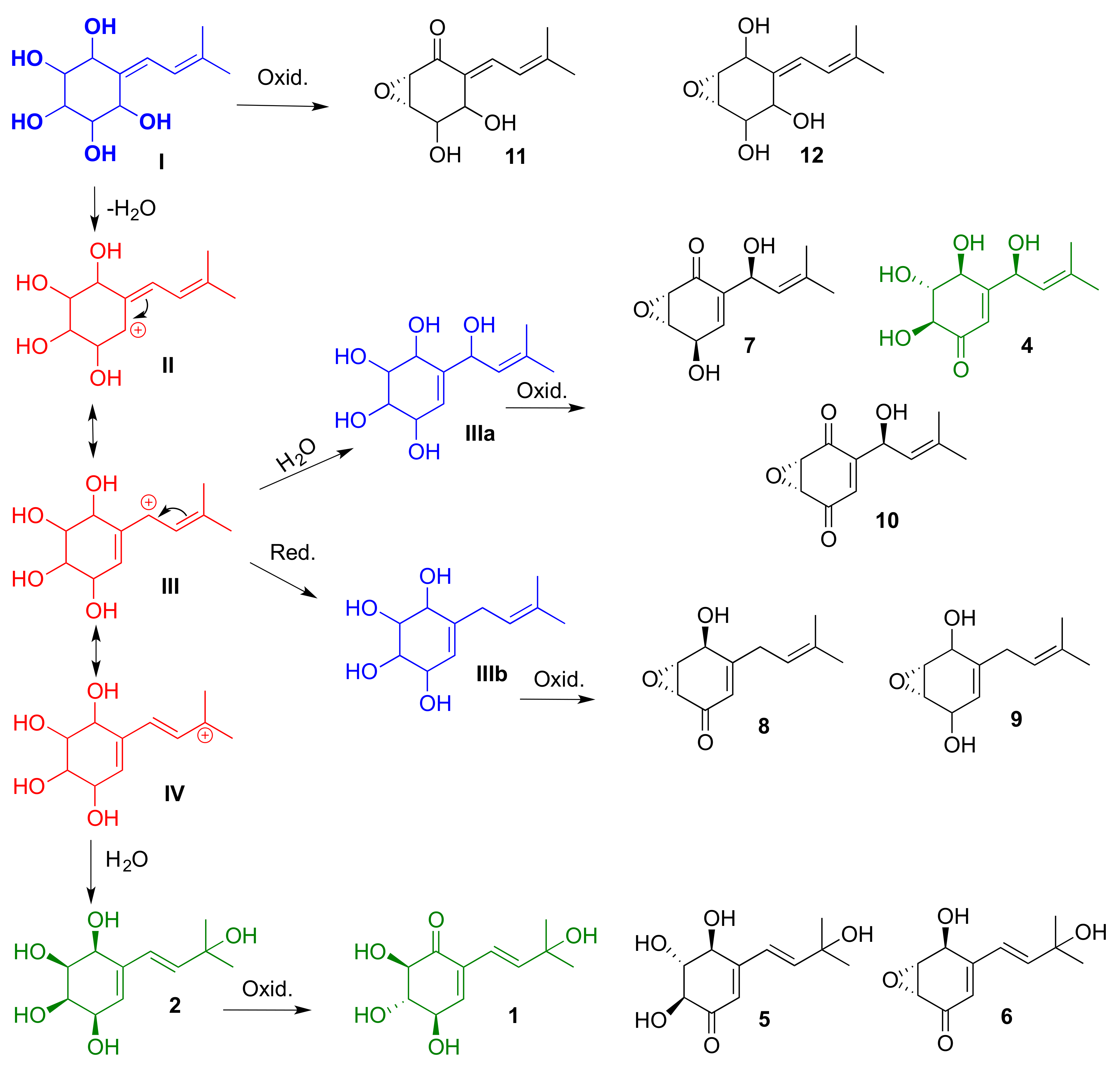
This group of compounds, isolated from lentinoid species (Lentinus and Panus), is characterized by an isoprenyl residue and an oxygenated cyclohexenyl ring, and could have similar biogenetic precursors. In Scheme 1, we proposed a not-yet-isolated precursor (I), and the structure of three possible intermediates (II–IV, in red) for the generation of this series of molecules. The biosynthesis may include two subsequent oxidation reactions of the not-yet-isolated compounds I, IIIa, IIIb and the newly reported lentinoid B (2). Compounds I, IIIa and IIIb (in blue) could be produced by some species of the genus Lentinus under different culture conditions.
Antibacterial Activity
The bacterial growth inhibition was evaluated in vitro by the disc diffusion method [11]. Compounds 1–3 and 5–7 were tested at a concentration of 6.25 µg/µL against Listeria monocytogenes, Enterococcus feacalis, Pseudomonas aeruginosa and Klebsiella pneumoniae. The first three bacteria were inhibited by at least one of the compounds with an inhibition diameter (ID) ranging from 7.5–9.5 mm. No antibacterial activity was detected against K. pneumoniae. The positive control used was gentamycin sulfate (ID, 15–25 mm) at a concentration of 1.25 µg/µL (Table 3).
The minimum inhibitory concentration (MIC) was determined for compounds 1, 2 and 3 using E. faecalis and P. aeruginosa (Table 4); nevertheless, only compound 1 resulted in a significant MIC; to inhibit E. faecalis, twice the amount of lentinoid A will be required. This could indicate that the presence of the carbonyl group is necessary for the antibacterial activity of lentinoid A compared to lentinoid B.
3. Materials and Methods
3.1. General
General experimental procedures. Optical rotations were measured in methanol on an Autopol V Plus Automatic Polarimeter (Whashington, NJ, USA) using a 10 cm cell. Ultra violet spectra were determined on a Waters 2996 PDA detector (Waters, Milford, MA, USA). IR spectra were determined using an Agilent Cary 630 FTIR spectrophotometer (film) (Agilent, Santa Clara, CA, USA). NMR spectra including 1D and 2D experiments were recorded at 400 MHz (JEOL, Ltd. Tokyo, Japan) and 600 MHz (Bruker, Billerica, MA, USA) using CD3OD and CDCl3 as solvents with tetramethylsilane (TMS) as internal standard. HRMS spectra were acquired on a Bruker micrOTOF-QIII (ESI-TOF), direct injection (Bruker, Billerica, MA, USA). The flash columns were made using silica gel 100 (70–230 mesh ASTM, Merck, Darmstadt, Germany) and silica gel (200–400 mesh, 60 Å, Sigma-Aldrich, St. Louis, MO, USA), the fractions collected were monitored by Sigma-Aldrich TLC plates 3110 (Sigma-Aldrich, St. Louis, MO, USA) and spots were detected using p-anisaldehyde spray reagent after heating. HPLC separations (Waters Delta 600) were carried out using a normal-phase semi-preparative column (YMC-Pack Sil, S-5 μm, 12 nm, 150 × 10 mm ID), a chiral column (QuiralPack IA, 5 μm 150 × 4.6 mm ID, Daicel Corporation, Tokyo, Japan) and a photodiode array detector Waters 2996. The injector was equipped with a 200 μL loop. Chemical structures (1D and 3D (force field MM2)) were drawn using Chemdraw (Perkin Elmer, Waltham, USA), version: 16.0.1.4.
3.2. Fungal Material
Fruiting bodies of Lentinus strigellus (collection number LC46) [7] were collected on burned wood near La Nevera highway in the Boquete, a region of Chiriquí Province in Panamá. The specimen was identified as Lentinus strigellus using molecular phylogenetic analyses. The air-dried specimens were deposited at the National Herbarium of the University of Panamá (PMA115711) and in the Herbarium of the Autonomous University of Chiriquí (UCH7481), Panamá.
3.3. Cultivation and Crude Extract
Initially, L. strigellus was cultivated in potato dextrose agar (PDA), malt extract agar (MEA) and Sabouraud Dextrose Agar (SDA) using six petri dishes each for 15 days. The chemical compositions of these three extracts were analyzed by thin-layer chromatography and NMR spectroscopy. A greater chemical diversity in the SDA extract using this analysis was detected. Hence, SDA cultivation was evaluated further, including the addition of the elicitors: arginine, glutamic acid, CaCl2, CuSO4 and FeSO4, and at three different pH values: 4.6, 5.6 and 6.6. The period of 15 days and temperature at 26 °C were the same for all experiments. The biomass yield (221 mg) was the highest for SDA with CaCl2 and a pH of 4.6 (calcium in low concentration tends to increase the yield of biomass produced [11]; in some cases this could bind proteins causing conformational changes and activating or inactivating mechanisms of catalysis [12]).
Culturing involving 67 petri dishes and the optimal conditions resulted in 3.10 g of crude ethyl acetate extract. To prepare the extract, the content of 15 petri dishes was transferred into one 1 L glass flask, with 400 mL of ethyl acetate, and extracted by sonicating for 20 min. This was repeated four times. The extract was recovered by drying under reduced pressure at 30 °C [7].
3.4. Compound Isolation
3.0 g of crude extract was fractionated by flash chromatography (4 cm ID) using silica gel (200–400 mesh, 10 g) and 1 L each of the following solvents: n-hexane, CH2Cl2, EtOAc, acetonitrile and water. All fractions (A–E) were analyzed by TLC. Fraction C (EtOAc), having a mass of 1561.4 mg (50.4%), was subjected to flash chromatography (2.5 cm ID) using silica gel (200–400 mesh, 70 g), and eluted with a gradient system from 100% CH2Cl2 to 100% EtOAc followed by a gradient from 100% EtOAc to 100% MeOH to afford 8 fractions (C1 to C8).
Fraction C6 (74.1 mg, 4.7%) was purified by normal-phase HPLC using a linear gradient (from 75:25 n-hexane–isopropanol to 40:60 n-hexane–isopropanol at 1.00 mL/min) in 55 min, to give 4 fractions, where the peak at tR 17 min corresponded to panepoxydone (7) (8.1 mg, 10.9%) and the peak at tR 21.5 min corresponded to isopanepoxydone (6) (12.3 mg, 16.6%).
Fraction C7 (235.0 mg, 15.1%) was flash chromatographed (2 cm ID) with silica gel (200–400 mesh, 22 g), and n-hexane/EtOAc (v/v = 100:0–0:100) and EtOAc/MeOH (v/v = 100:0–0:100) as eluents, to give 14 fractions (C7.1 to C7.14). C7.9 (76.2 mg, 32.4%) was subjected to normal-phase HPLC using a linear gradient (from 75:25 n-hexane–isopropanol to 40:60 n-hexane–isopropanol at 1.00 mL/min) in 55 min, to give 3 fractions, where the peak at tR 18 min corresponds to striguellone A (5) and the peak at tR 23 min corresponds to lentinoid A (1).
Fraction C7.10 (55.0 mg, 23.4%) was purified by normal-phase HPLC using a linear gradient (of 75:25 n-hexane–isopropanol to 40:60 n-hexane–isopropanol at 1.00 mL/min) in 55 min, to give 6 fractions (I–VI), where the peak at tR 33 min (fraction VI) corresponds to lentinoid B (2) 7.4 mg (13.5%). Fraction V (26.0 mg, 47.3%) was purified by normal-phase HPLC using a linear gradient (85:15 to 30:70 n-hexane–isopropanol at 1.00 mL/min) in 55 min, to give 3 fractions (V.1 to V.3). V.1 (7.3 mg, 28.1%) was purified by HPLC (Chiral Pack IA, 5 µm, 150 × 4.6 mm ID) using the isocratic 85:15 n-hexane–isopropanol at 0.50 mL/min to give lentinoid C (3) (tR 17 min; 2.6 mg, 35.6%). V.2 (5.4 mg, 20.8%) was purified by HPLC (Chiral Pack IA, 5 µm, 150 × 4.6 mm ID) using the isocratic 85:15 n-hexane–isopropanol at 0.50 mL/min to give lentinoid D (4) (tR 12 min; 1 mg, 13.7%).
3.5. Lentinoid A (1)
Brown viscous liquid; −64.5 (c 0.0029, MeOH); UV (MeOH) λmax 273 and 212 nm; IR mmax: 3350, 2948, 2876, 1690, 1390 and 1040 cm−1; 1H- and 13C-NMR data, see Table 1; (+)-HR-ESI-TOF-MS m/z 251.0891 (calcd. 251.0890 for C11H16O5Na).
3.6. Lentinoid B (2)
Brown viscous liquid; +3.3 (c 0.0024, MeOH); UV (Hx/IP) λmax 233 nm; IR mmax: 3300, 2942, 2831 and 1021 cm−1; 1H- and 13C-NMR data, see Table 1; (+)-HR-ESI-TOF-MS m/z 253.1034 [M + Na]+ (calcd. 253.1046 for C11H18O5Na).
3.7. Lentinoid C (3)
Brown viscous liquid; −75.0 (c 0.0005, MeOH); UV (MeOH) λmax 278 and 208 nm; IR mmax: 3280, 2951, 2840, 1649, 1632 and 1013 cm−1; 1H- and 13C-NMR data, see Table 2; (+)-HR-ESI-TOF-MS m/z 335.1109 (calcd. 335.1101 for C15H20O7Na).
3.8. Antibacterial Activities [13,14]
The antibacterial activity was determined through the susceptibility test of the British Society for Antimicrobial Chemotherapy (BSAC).
3.8.1. Preparation of 0.5 McFarland Turbidity Standard
The turbidity standard was prepared using 0.5 mL of solution of BaCl2 (BaCl2.2H2O; 0.048 M) in a tube containing 99.5 mL of H2SO4 (0.18 M). The absorbance of the McFarland standard (between 0.08 and 0.10 at 625 nm) was verified using a spectrophotometer. The solution was stored in the dark at 24 ± 2 °C.
3.8.2. Preparation of Bacterial Inoculum
Each bacterium was cultivated in Tripticase Soy Agar (TSA) for 24 h; thereafter, five colonies were chosen and transferred into a tube containing saline and isotonic solution and then visually compared with the 0.5 McFarland standard.
3.8.3. Bacterial Growth Inhibition in Vitro
The bacterial growth inhibition was evaluated in vitro by the disc diffusion method using Trypticase Soy Agar (TSA) in Petri dishes (145 mm) and placing a disc (6 mm diameter) with 50 µg of each tested compound dissolved in DMSO. The plate was then incubated for 18 h at 37 °C, and the inhibition area were measured. For the positive control, gentamycin sulfate (10 μg/mL) was used and the negative control was DMSO. The diameter of the inhibition around each disk was measured, providing a measure of zone–of-inhibition (ZOI) in mm (including the diameter of the disc).
3.8.4. Minimum Inhibitory Concentration (MIC)
The minimum inhibitory concentration (MIC) was determined using a stock solution prepared by adding 300 µg of each compound in 3 mL of Trypticase Soy Broth (TSB). Serial dilutions of the evaluated compounds and control were performed to determine the MIC. The evaluated concentrations of each compound were 100.0, 40.0, 33.3, 11.1 and 5.6 μg/mL. Each solution was thereafter inoculated with 50 μL (0.5 McFarland) of a culture of E. faecalis and P. aeruginosa, and incubated at 37 °C for 18 h. The positive control (gentamycin sulfate 10 mg/mL) concentrations were similar to those of the compounds. Additionally, two blanks were used, one with culture medium alone and one with DMSO. Each assay was performed in duplicate.
4. Conclusions
In summary, we have characterized four new lentinoids produced by L. strigellus as a response to changes in culture conditions. They represent new additions to the chemical diversity produced by this fungus. The compounds represented in Scheme 1, isolated from the genera Lentinus and Panus, could have similar precursors. Additional improvements to the experimental culture conditions and the use of additional analytical tools could lead to the isolation of new compounds of this class and therefore new active molecules. Moreover, three of these compounds showed antibacterial activity in a bacterial growth inhibition assay (inhibition zone in mm). However, when the minimum inhibitory concentration (MIC) was determined, only compound 1 showed a significant activity of 200 μg/mL.
Supplementary Materials
The following are available online, 1H- and 13C-NMR, 2D NMR and MS data for compounds 1–3 and 1H-NMR for compound 4. The isolations chromatograms for each novel compound are present.
Acknowledgments
This work was supported by grants from SENACYT–Panama (COL10-060 and FID11-051), the Project of Nagoya Protocols’ Application in Panamá and the National Research System (SNI). We also give our thanks to the personnel of Panamá’s Ministry of Environment for their assistance in obtaining the collect permit.
Author Contributions
Roger Vásquez carried out the experimental work (isolation and structural elucidation of compounds) and drafted the manuscript. Nivia Rios culture of fungi and development of the antibacterial assays. Godofredo Solano assisted in the acquisition of some NMR data and its interpretation. Luis Cubilla-Rios design the project, contributed in the structural characterization and wrote the manuscript.
Conflicts of Interest
The authors declare no conflict of interest.
References
- Raja, H.A.; Miller, A.N.; Pearce, C.J.; Oberlies, N.H. Fungal Identification Using Molecular Tools: A Primer for the Natural Products Research Community. J. Nat. Prod. 2017, 80, 756–770. [Google Scholar] [CrossRef] [PubMed]
- Hoffmeister, D.; Keller, N.P. Natural products of filamentous fungi: Enzymes, genes, and their regulation. Nat. Prod. Rep. 2007, 24, 393–416. [Google Scholar] [CrossRef] [PubMed]
- Zheng, Y.; Zhao, B.; Lu, C.; Lin, X.; Zheng, Z.; Su, W. Isolation, structure elucidation and apoptosis-inducing activity of new compounds from the edible fungus Lentinus striguellus. Nat. Prod. Commun. 2009, 4, 501–506. [Google Scholar] [PubMed]
- Senthilarasu, S. The lentinoid fungi (Lentinus and panaus) from Western Ghats, India. IMA Fungus 2015, 6, 119–128. [Google Scholar] [CrossRef] [PubMed]
- Guzmán, G.; Piepenbring, M. Los hongos de Panamá—Introducción a la Identificación de los Macroscópicos, 1st ed.; Instituto de Ecología, A.C.: Xalapa, Veracruz, México, 2011; p. 150. [Google Scholar]
- Kis, Z.; Closse, A.; Sigg, H.P.; Hruban, L.; Snatzke, G. Die Structure von Panepoxydon und verwandten Pilzmetaboliten. Helv. Chim. Acta 1970, 53, 1577–1597. [Google Scholar] [CrossRef]
- Julca-Canto, M.; Aguilar-Pérez, M.M.; Rios, N.; Sousa, J.P.B.; Cubilla-Rios, L. Additional new natural products produced by Lentinus strigellus: A biotechnological approach. Tetrahedron Lett. 2016, 57, 650–653. [Google Scholar] [CrossRef]
- Barros-Filho, B.A.; de Oliveira, M.C.; Mafezoli, J.; Barbosa, F.G.; Rodrigues-Filho, E. Secundary metabolite production by the basidiomycete, Lentinus strigellus, under different culture conditions. Nat. Prod. Commun. 2012, 7, 771–773. [Google Scholar] [PubMed]
- Souza-Fagundes, E.M.; Cota, B.B.; Rosa, L.H.; Romanha, A.J.; Correa-Oliveira, R.; Rosa, C.A.; Zani, C.L.; Teixeira-Carvalho, A.; Martins-Filho, O.A. In vitro activity of hypnophilin from Lentinus strigosus: A potential prototype for Chagas disease and leishmaniasis chemotherapy. Braz. J. Med. Biol. Res. 2010, 43, 1054–1061. [Google Scholar] [CrossRef] [PubMed]
- Rukachaisirikul, V.; Tansakul, C.; Saithong, S.; Pakawatchai, C.; Isaka, M.; Suvannakad, R. Hirsutane Sesquiterpenes from the Fungus Lentinus connatus BCC 8996. J. Nat. Prod. 2005, 68, 1674–1676. [Google Scholar] [CrossRef] [PubMed]
- Swainsbury, D.J.K.; Zhou, L.; Oldroyd, G.E.D.; Bornemann, P. Calcium Ion Binding Properties of Medicago truncatula Calcium/Calmodulin-Dependent Protein Kinase. J. Biochem. 2012, 51, 6895–6907. [Google Scholar] [CrossRef] [PubMed]
- Radha Krishna, P.; Srivastava, A.K.; Ramaswamy, N.K.; Suprasanna, P.; D’Souza, S.F. Banana peel as substrate for α-amylase production using Aspergillus niger. NCIM 616 and process optimization. Indian J. Biotechnol. 2012, 11, 314–319. [Google Scholar]
- Andrews, J.M. BSAC standardized disc susceptibility testing method (version 8). J. Antimicrob. Chemother. 2009, 64, 454–489. [Google Scholar] [CrossRef] [PubMed]
- Wotton, M. Methods for Antimicrobial Susceptibility Testing; British Society of Antimicrobial Chemotherapy (BSAC): Birmingham, UK, 2013. [Google Scholar]
Sample Availability: Samples of the compounds are not available from the authors. |
Figure 1.
Structures of compounds 1–8.

Figure 2.
(A) Correlation spectroscopy (COSY) (in blue boldface), heteronuclear multiple-bond correlation (HMBC) (black arrows) and (B) Overhauser-effect spectroscopy (NOESY) (black arrows) of 1.
Figure 2.
(A) Correlation spectroscopy (COSY) (in blue boldface), heteronuclear multiple-bond correlation (HMBC) (black arrows) and (B) Overhauser-effect spectroscopy (NOESY) (black arrows) of 1.

Figure 3.
(A) COSY (in boldface), HMBC (black arrows) and (B) NOESY (black arrows) of 2.

Figure 4.
(A) COSY (in blue boldface), HMBC (black arrows) and (B) ROESY (black arrows) of 3.

Scheme 1.
Proposed intermediates for the biosynthesis of the lentinoids, oxygenated cyclohexenyl derivatives, isolated from species of the genera Lentinus and Panus. Intermediates are in red. Compounds 1–2 and 4 are in green, the structure of not-yet-isolated compounds are in blue, and previously isolated compounds are in black: striguellone A (5), isopanepoxydone (6), panepoxydone (7), lentinoid E (8), 7-desoxypanepoxydol (9), panepoxydione (10), neopanepoxydone (11), and neopanepoxydol (12).


Table 1.
NMR data for compounds 1–2 in MeOH-d4, (J in Hz; 1H-NMR at 400 MHz; 13C-NMR at 100 MHz).
| Pos. | 1 | 2 | ||
|---|---|---|---|---|
| δC | δH | δC | δH | |
| 1 | 191.8, C | 136.5, C | ||
| 2 | 135.2, C | 125.9, CH | 5.67 (br t; 4.0) | |
| 3 | 145.4, CH | 6.89 (d; 2.4) | 63.9, CH | 4.35 (d; 4.4) |
| 4 | 73.1, CH | 4.41 (dd; 2.4, 8.4) | 57.1, CH | 3.37 (t; 4.0) |
| 5 | 79.3, CH | 3.63 (dd; 8.4, 11.6) | 56.0, CH | 3.49 (br m) |
| 6 | 68.8, CH | 4.60 (d; 11.6) | 65.3, CH | 4.59 (br d; 4.6) |
| 1′ | 120.1, CH | 6.36 (d; 16.0) | 126.5, CH | 6.15 (d; 16.0) |
| 2′ | 143.1, CH | 6.44 (d; 16.0) | 140.9, CH | 6.24 (d; 16.0) |
| 3′ | 71.4, C | 71.4, C | ||
| 4′ | 29.7, CH3 | 1.30 (s) | 29.9, CH3 | 1.30 (s) |
| 5′ | 29.7, CH3 | 1.30 (s) | 29.9, CH3 | 1.30 (s) |
Table 2.
NMR data for compound 3 in MeOH-d4, (J in Hz; 1H-NMR at 600 MHz; 13C-NMR at 150 MHz).
| Pos. | δC | δH | Pos. | δC | δH |
|---|---|---|---|---|---|
| 1 | 194.9 | 3´ | 71.6 | ||
| 2 | 125.9 | 6.02 (1H, s) | 4´ | 29.7 | 1.37 (3H, s) |
| 3 | 156.9 | 5´ | 29.6 | 1.36 (3H, s) | |
| 4 | 67.9 | 4.72 (1H, d; 3.6) | 1´´ | 167.0 | |
| 5 | 71.8 | 4.05 (1H, dd; 3.6, 10.8) | 2´´ | 141.6 | |
| 6 | 76.6 | 5.72 (1H, d; 10.8) | 3´´ | 125.5 | 5.96 (1H, m) 6.35 (1H, m) |
| 1′ | 126.6 | 6.46 (1H, d; 16,0) | 4´´ | 61.6 | 4.34 (2H, m) |
| 2′ | 149.5 | 6.64 (1H, d; 16,0) |
Table 3.
Antibacterial activity of compounds 1–3 and 5–7, inhibition diameter (mm).
| Bacteria Species | Compounds | Positive Control | |||||
|---|---|---|---|---|---|---|---|
| 1 | 2 | 3 | 5 | 6 | 7 | Gentamycin Sulfate | |
| Listeria monocytogenes | I | I | I | I | 7.5 | I | 25 |
| Enterococcus faecalis | 9 | 9 | 8 | 8 | 9.5 | I | 15 |
| Pseudomonas aeruginosa | I | 9.5 | I | 7.5 | 8.5 | I | 20 |
I = inactive.
Table 4.
Antibacterial activities of compounds 1–3, MIC (μg/mL).
| Bacteria Species | Compounds | Positive Control | ||
|---|---|---|---|---|
| 1 | 2 | 3 | Gentamycin Sulfate | |
| E. faecalis | 200 | ----- | ----- | 100 |
| P. aeruginosa | ----- | 160 | ----- | 33 |
---- No activity detected.
© 2018 by the authors. Licensee MDPI, Basel, Switzerland. This article is an open access article distributed under the terms and conditions of the Creative Commons Attribution (CC BY) license (http://creativecommons.org/licenses/by/4.0/).
Share and Cite
MDPI and ACS Style
Vásquez, R.; Rios, N.; Solano, G.; Cubilla-Rios, L. Lentinoids A–D, New Natural Products Isolated from Lentinus strigellus. Molecules 2018, 23, 773. https://doi.org/10.3390/molecules23040773
AMA Style
Vásquez R, Rios N, Solano G, Cubilla-Rios L. Lentinoids A–D, New Natural Products Isolated from Lentinus strigellus. Molecules. 2018; 23(4):773. https://doi.org/10.3390/molecules23040773
Chicago/Turabian StyleVásquez, Roger, Nivia Rios, Godofredo Solano, and Luis Cubilla-Rios. 2018. "Lentinoids A–D, New Natural Products Isolated from Lentinus strigellus" Molecules 23, no. 4: 773. https://doi.org/10.3390/molecules23040773