
Novel Neuroprotective Lead Compound Ligustrazine Derivative Mass Spectrometry Fragmentation Rule and Metabolites in Rats by LC/LTQ-Orbitrap MS

 and
and
Abstract
:1. Introduction
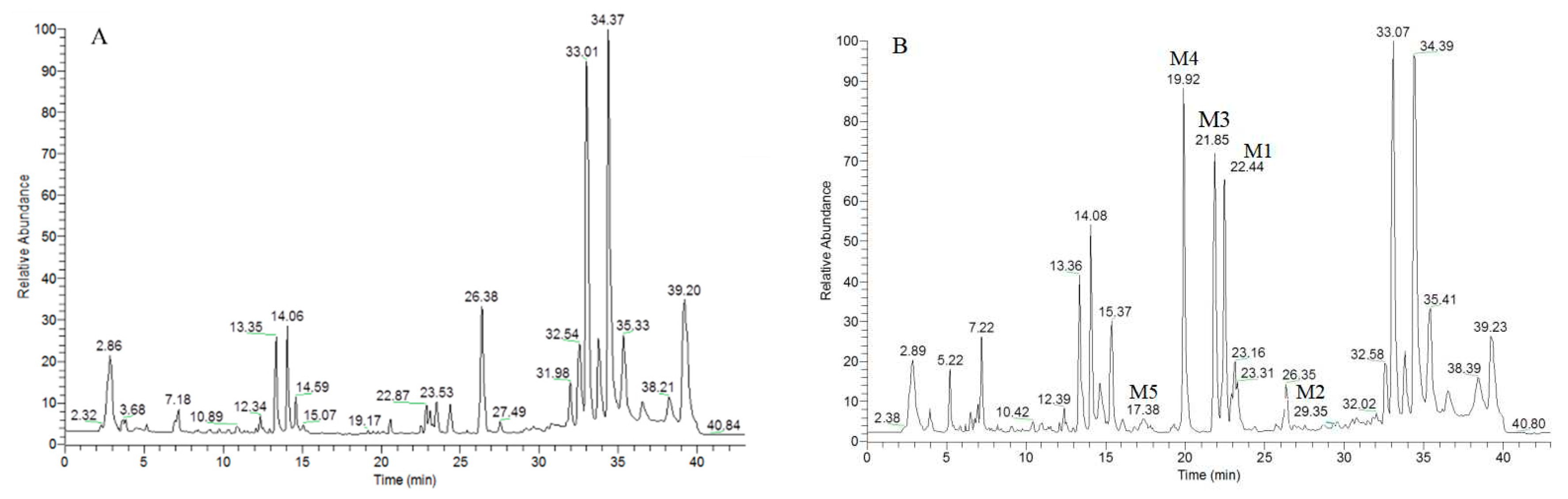
2. Results
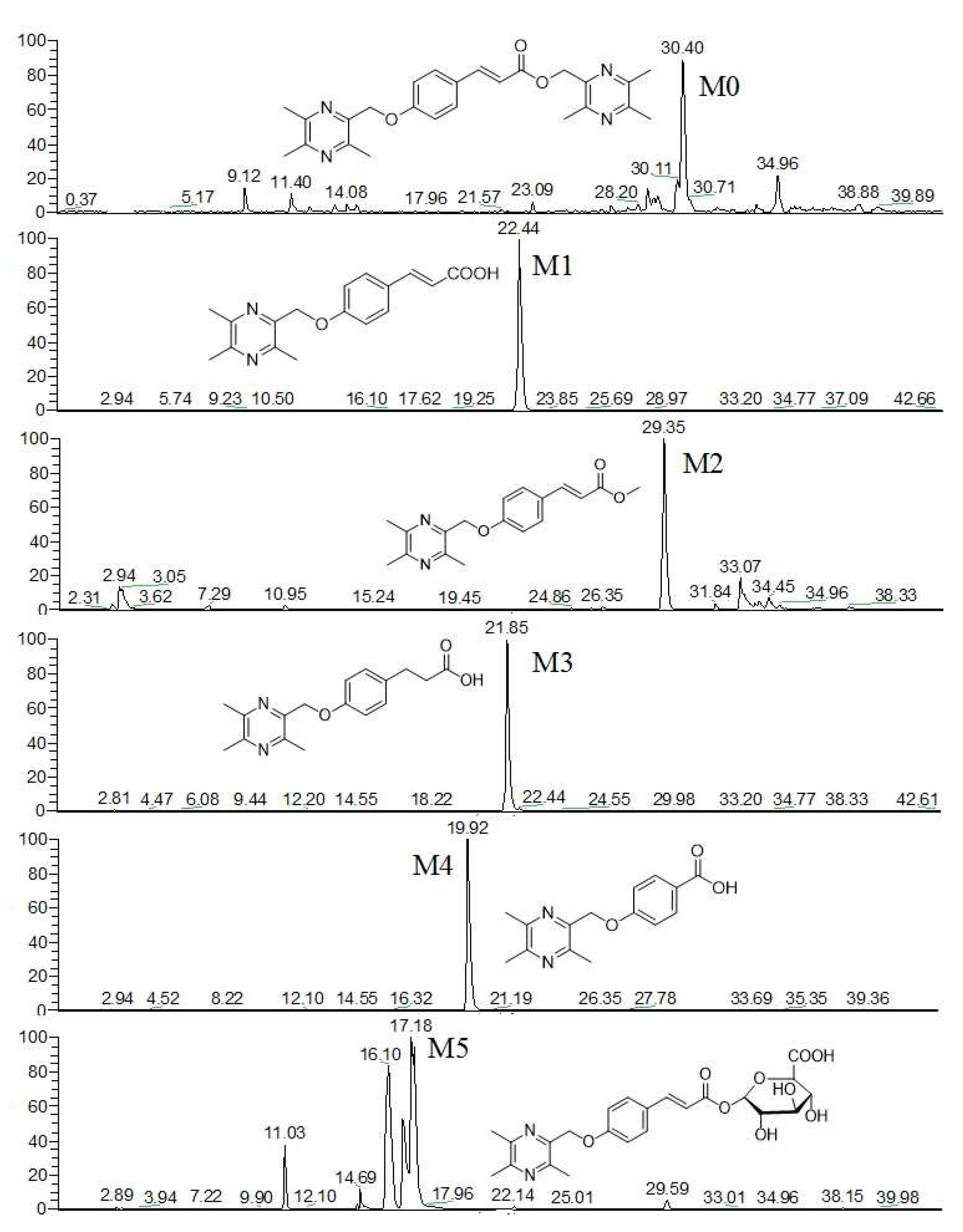
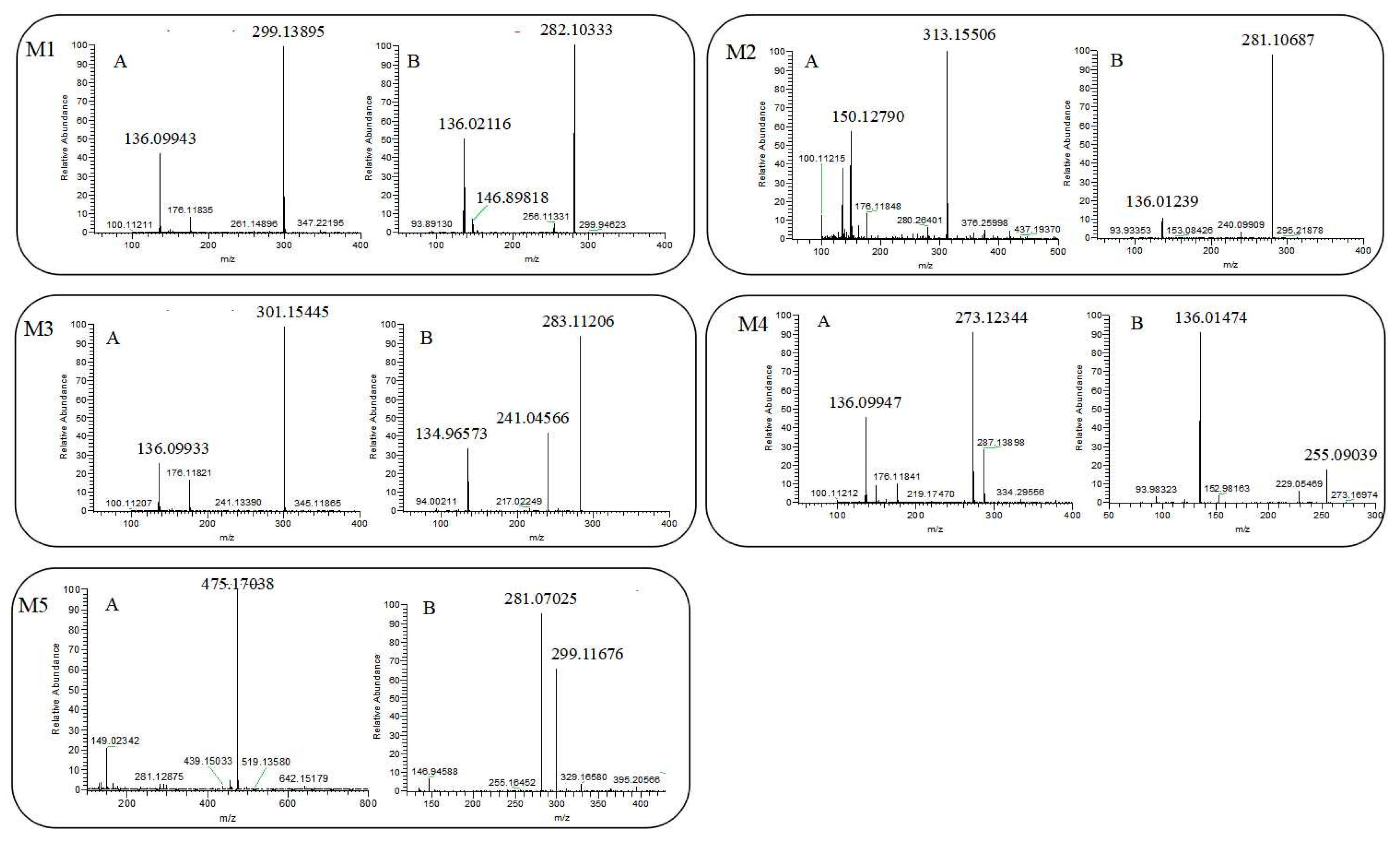
2.1. The Mass Spectrometry and Lysis of T-CA
2.2. Identification of Plasma Metabolites of T-CA
2.2.1. Identification of Plasma Metabolites M1
2.2.2. Identification of Plasma Metabolites M2
2.2.3. Identification of Plasma Metabolites M3
2.2.4. Identification of Plasma Metabolites M4
2.2.5. Identification of Plasma Metabolites M5
2.3. Neuroprotective Activity Test of Metabolites M1, M2, M4
3. Materials and Methods
3.1. Reagents, Chemicals and Animals
3.2. Instrumentations
3.3. Sample Preparation
3.4. Liquid Chromatographic and Mass Spectrometer Conditions
3.5. Experimental Procedure
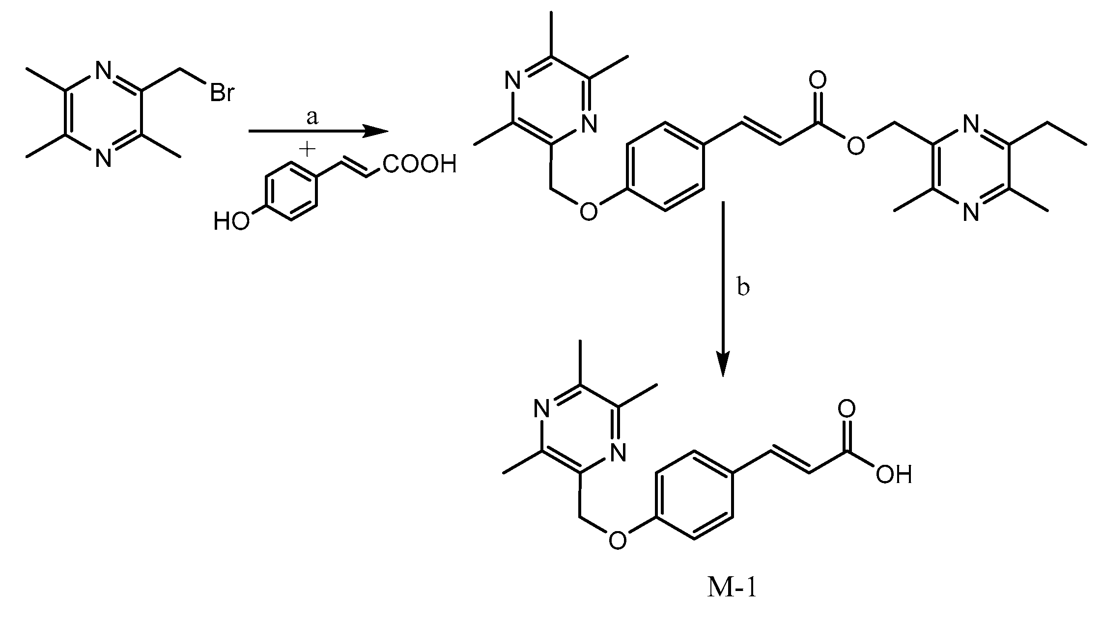
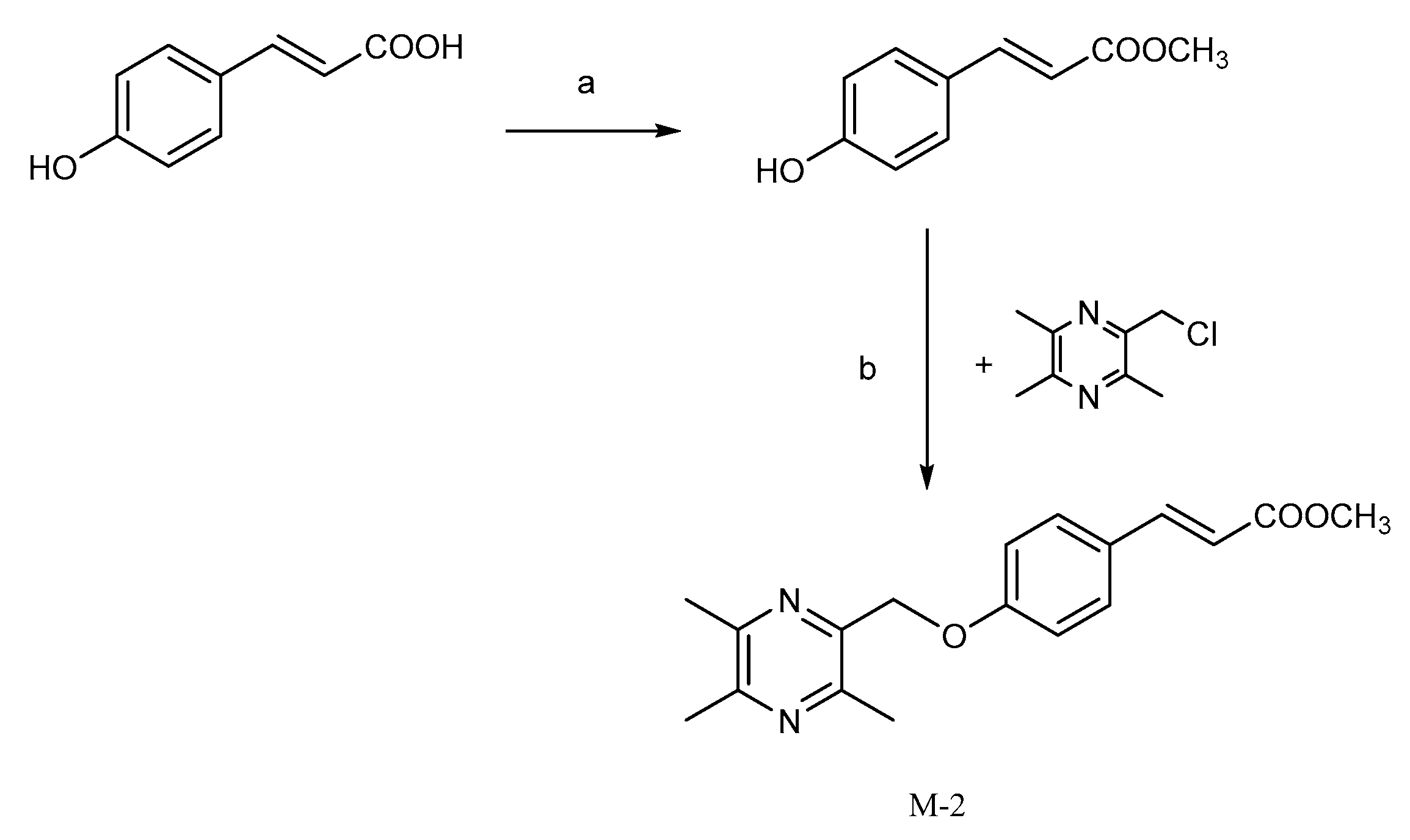
3.5.1. Preparation of M1 ((E)-3-(4-((3,5,6-trimethylpyrazin-2-yl)methoxy)phenyl)acrylic acid)
3.5.2. Preparation of M2 (methyl (E)-3-(4-((3,5,6-trimethylpyrazin-2-yl)methoxy)phenyl)acrylate))
3.5.3. Preparation of M4 (4-((3,5,6-trimethylpyrazin-2-yl)methoxy)benzoic acid)
3.6. Protective Effect on Injured PC12 Cells
4. Conclusions
Author Contributions
Acknowledgments
Conflicts of Interest
References
- Henz, B.M. The pharmacologic profile of desloratadine: A review. Allergy 2001, 56, 7–13. [Google Scholar] [CrossRef] [PubMed]
- Kong, R.; Liu, T.; Zhu, X.; Ahmad, S.; Williams, A.L.; Phan, A.T. Old drug new use—Amoxapine and its metabolites as potent bacterial β-glucuronidase inhibitors for alleviating cancer drug toxicity. Clin. Cancer Res. 2014, 20, 3521–3530. [Google Scholar] [CrossRef] [PubMed]
- Saha, S.; Rajpal, D.K.; Brown, J.R. Human microbial metabolites as a source of new drugs. Drug Discov. Today 2016, 21, 692–698. [Google Scholar] [CrossRef] [PubMed]
- Xu, K.; Wang, P.; Xu, X.; Chu, F.; Lin, J.; Zhang, Y.; Lei, H. An overview on structural modifications of ligustrazine and biological evaluation of its synthetic derivatives. Res. Chem. Intermed. 2015, 41, 1385–1411. [Google Scholar] [CrossRef]
- Zeng, Z.; Zhu, W.; Zhou, X.; Jin, Z.; Liu, H.; Chen, X. Tetramethylpyrazine, a Chinese drug, blocks coronary vasoconstriction by endothelin-1 and decreases plasma endothelin-1 levels in experimental animals. J. Cardiovasc. Pharmacol. 1998, 31, 313–316. [Google Scholar] [CrossRef]
- Zhang, Z.; Wei, T.; Hou, J.; Li, G.; Yu, S.; Xin, W. Tetramethylpyrazine scavenges superoxide anion and decreases nitric oxide production in human polymorphonuclear leukocytes. Life Sci. 2003, 72, 2465–2472. [Google Scholar] [CrossRef]
- Wong, K.L.; Chan, P.; Huang, W.C.; Yang, T.L.; Liu, I.M.; Lai, T.Y. Effect of tetramethylpyrazine on potassium channels to lower calcium concentration in cultured aortic smooth muscle cells. Clin. Exp. Pharmacol. Physiol. 2003, 30, 793–798. [Google Scholar] [CrossRef] [PubMed]
- Zhao, T.; Fu, Y.; Sun, H.; Liu, X. Ligustrazine suppresses neuron apoptosis via the Bax/Bcl-2 and caspase-3 pathway in PC12 cells and in rats with vascular dementia. IUBMB Life 2018, 70, 60–70. [Google Scholar] [CrossRef] [PubMed]
- Li, G.; Xu, X.; Xu, K.; Chu, F.; Song, J.; Zhou, S. Ligustrazinyl amides: A novel class of ligustrazine-phenolic acid derivatives with neuroprotective effects. Chem. Cent. J. 2015, 9, 9. [Google Scholar] [CrossRef] [PubMed]
- Xu, B.; Gong, Y.; Xu, X.; Zhang, C.; Zhang, Y.; Chu, F.; Liu, H.; Wang, P.; Lei, H. Synthesis and protective effect of new ligustrazine derivatives against CoCl2-induced neurotoxicity in differentiated PC12 cells. Part 2. MedChemComm 2015, 6, 806–809. [Google Scholar] [CrossRef]
- Wang, P.; Zhao, R.; Yan, W.; Zhang, X.; Zhang, H.; Xu, B. Neuroprotection by new ligustrazine-cinnamon acid derivatives on CoCl2-induced apoptosis in differentiated PC12 cells. Bioorg. Chem. 2018, 77, 360–369. [Google Scholar] [CrossRef] [PubMed]
- Li, G.; Wang, P.; Xu, X.; Lin, J.; Chu, F.; Song, J.; Zhou, S.; Wang, M.; Zhang, Y.; Lei, H. Synthesis and protective effect of ligustrazine intermediates against CoCl2-induced neurotoxicity in differentiated PC12 cell. Zhongguo Zhong Yao Za Zhi 2014, 39, 2679–2683. [Google Scholar] [PubMed]
- Xu, B.; Xu, X.; Zhang, C.; Zhang, Y.; Wu, G.; Yan, M. Synthesis and protective effect of new ligustrazine-vanillic acid derivatives against CoCl2-induced neurotoxicity in differentiated PC12 cells. Chem. Cent. J. 2017, 11, 20. [Google Scholar] [CrossRef] [PubMed]
- Li, G.; Tian, Y.; Zhang, Y.; Hong, Y.; Hao, Y.; Chen, C. A Novel Ligustrazine Derivative T-VA Prevents Neurotoxicity in Differentiated PC12 Cells and Protects the Brain against Ischemia Injury in MCAO Rats. Int. J. Mol. Sci. 2015, 16, 21759–21774. [Google Scholar] [CrossRef] [PubMed]
- Zhang, C.; Yan, W.; Zhao, R.; Xu, B.; Fang, X.; Yan, M. Design, synthesis and evaluation of new ligustrazine derivatives as potential plasma-stable neuroprotective agents. MedChemComm 2017, 8, 652–656. [Google Scholar] [CrossRef]
- Li, G. Design, synthesis and activity characterization of series tetramethylpyrazine compounds. Part 3. Master’s Thesis, Beijing University of Chinese Medicine, Beijing, China, June 2015. [Google Scholar]
- Zou, J.; Gao, P.; Hao, X.; Xu, H.; Zhan, P.; Liu, X. Recent progress in the structural modification and pharmacological activities of ligustrazine derivatives. Eur. J. Med. Chem. 2018, 147, 150–162. [Google Scholar] [CrossRef] [PubMed]
- Labat, L.; Goncalves, A.; Marques, A.R.; Duretz, B.; Granger, B.; Declèves, X. Liquid chromatography high resolution mass spectrometry for the determination of baclofen and its metabolites in plasma: Application to therapeutic drug monitoring. Biomed. Chromatogr. 2017, 31. [Google Scholar] [CrossRef] [PubMed]
- Hogenboom, A.C.; Van Leerdam, J.A.; De Voogt, P. Accurate mass screening and identification of emerging contaminants in environmental samples by liquid chromatography–hybrid linear ion trap Orbitrap mass spectrometry. J. Chromatogr. A 2009, 1216, 510–519. [Google Scholar] [CrossRef] [PubMed]
- Bar-Am, O.; Amit, T.; Youdim, M.B.H. Aminoindan and hydroxyaminoindan, metabolites of rasagiline and ladostigil, respectively, exert neuroprotective properties in vitro. J. Neurochem. 2007, 103, 500–508. [Google Scholar] [CrossRef] [PubMed]
- Li, W.; Liu, H.; Wei, C.; Wei, X.; Tang, M.; Kong, X. Pharmacokinetics and metabolism of Chf197, a ligustrazine derivative, in rats. Fundam. Clin. Pharmacol. 2015, 29, 478–487. [Google Scholar] [CrossRef] [PubMed]
- Zeng, Z.; Xie, R.Q.; Zhang, T.; Tan, L.X.; Xiu-Ying, Y.U. The regular patterns of mass spectrometry of lac-tones from Ligusticum chuanxiong Hort. J. Chin. Mass Spectrom. Soc. 2011, 32, 293–300. [Google Scholar]
- Thibert, V.; Legeay, P.; Chapuishugon, F.; Pichon, V. Molecularly imprinted polymer for the selective extraction of cocaine and its metabolites, benzoylecgonine and ecgonine methyl ester, from biological fluids before LC–MS analysis. J. Chromatogr. B 2014, 949, 16–23. [Google Scholar] [CrossRef] [PubMed]
- Wu, J.Y.; Gao, F.Y.; Ye, X.L.; Liu, H.; Fan, G.R.; He, G.W. Chuan’agelei and its flatten structure ferulic acid and ligustrazine: Mass fragmentation pathway. Acad. J. Second Mil. Med. Univ. 2012, 33, 755–758. [Google Scholar] [CrossRef]
- Kaever, V.; Martin, M.; Fauler, J.; Marx, K.H.; Resch, K. A novel metabolic pathway for leukotriene B4 in different cell types: Primary reduction of a double bond. Biochim. Biophys. Acta (BBA)-Lipids Lipid Metab. 1987, 922, 337–344. [Google Scholar] [CrossRef]
- Shi, Y.; Xia, Y.; Wang, J.; He, J.; Feng, F.; Liu, W. Metabolic profile of 5-hydroxy-4-methoxycanthin-6-one, a typical canthinone alkaloid, in rats determined by liquid chromatography-quadrupole time-of-flight tandem mass spectrometry together with multiple data processing techniques. J. Pharm. Biomed. Anal. 2016, 129, 60–69. [Google Scholar] [CrossRef] [PubMed]
- Zhao, X.; Jiang, J.; Yang, G.; Huang, J.; Yang, G.; He, G. Profiling and Preparation of Metabolites from Pyragrel in Human Urine by Online Solid-Phase Extraction Coupled with High Performance Liquid Chromatography Tandem Mass Spectrometry Followed by a Macroporous Resin-Based Purification Approach. Molecules 2017, 22, 494. [Google Scholar] [CrossRef] [PubMed]
- Wang, P.; Zhang, H.; Chu, F.; Xu, X.; Lin, J.; Chen, C. Synthesis and protective effect of new ligustrazine-benzoic acid derivatives against CoCl2-induced neurotoxicity in differentiated PC12 cells. Molecules 2014, 18, 13027–13042. [Google Scholar] [CrossRef] [PubMed]
Sample Availability: Samples of the compounds are not available from the authors. |


| Compound | tR/min | Species | Elemental Composition | m/z | RSD | Delta mmu |
|---|---|---|---|---|---|---|
| M0 | 30.40 | [M + H]+ | C25H29O3N4 | 433.22418 | 13.5 | 0.213 |
| M1 | 22.44 | [M + H]+ | C17H19O3N2 | 299.13895 | 9.5 | −0.199 |
| M2 | 29.35 | [M + H]+ | C18H21O3N2 | 313.15506 | 9.5 | 0.481 |
| M3 | 21.85 | [M + H]+ | C17H21O3N2 | 301.15445 | 8.5 | −0.039 |
| M4 | 19.92 | [M + H]+ | C15H17O3N2 | 273.12344 | 8.5 | −0.109 |
| M5 | 16.98 | [M + H]+ | C23H27O9N2 | 475.17038 | 11.5 | 0.063 |
| Compound | Cell Proliferation Rate% | EC50 | ||||
|---|---|---|---|---|---|---|
| 60 | 30 | 15 | 7.5 | 3.75 | ||
| T-CA | 102.94 | 89.83 | 71.09 | 27.89 | 39.02 | 7.97 |
| M1 | 77.77 | 45.80 | 45.64 | 16.46 | 2.90 | 40.68 |
| M2 | 89.95 | 82.06 | 75.97 | 42.04 | 20.69 | 9.67 |
| M4 | 42.37 | 39.62 | 14.67 | 9.48 | 8.98 | 39.42 |
© 2018 by the authors. Licensee MDPI, Basel, Switzerland. This article is an open access article distributed under the terms and conditions of the Creative Commons Attribution (CC BY) license (http://creativecommons.org/licenses/by/4.0/).
Share and Cite
Zhang, X.; Zhao, R.; Chen, M.; Ma, T.; Wu, G.; Xue, N.; Li, G.; Wang, H.; Fang, K.; Zhang, W.; et al. Novel Neuroprotective Lead Compound Ligustrazine Derivative Mass Spectrometry Fragmentation Rule and Metabolites in Rats by LC/LTQ-Orbitrap MS. Molecules 2018, 23, 1154. https://doi.org/10.3390/molecules23051154
Zhang X, Zhao R, Chen M, Ma T, Wu G, Xue N, Li G, Wang H, Fang K, Zhang W, et al. Novel Neuroprotective Lead Compound Ligustrazine Derivative Mass Spectrometry Fragmentation Rule and Metabolites in Rats by LC/LTQ-Orbitrap MS. Molecules. 2018; 23(5):1154. https://doi.org/10.3390/molecules23051154
Chicago/Turabian StyleZhang, Xinyu, Rui Zhao, Meng Chen, Tao Ma, Gaorong Wu, Nannan Xue, Guoliang Li, Hui Wang, Kang Fang, Wenxi Zhang, and et al. 2018. "Novel Neuroprotective Lead Compound Ligustrazine Derivative Mass Spectrometry Fragmentation Rule and Metabolites in Rats by LC/LTQ-Orbitrap MS" Molecules 23, no. 5: 1154. https://doi.org/10.3390/molecules23051154
APA StyleZhang, X., Zhao, R., Chen, M., Ma, T., Wu, G., Xue, N., Li, G., Wang, H., Fang, K., Zhang, W., Wang, P., & Lei, H. (2018). Novel Neuroprotective Lead Compound Ligustrazine Derivative Mass Spectrometry Fragmentation Rule and Metabolites in Rats by LC/LTQ-Orbitrap MS. Molecules, 23(5), 1154. https://doi.org/10.3390/molecules23051154