
Studies on the Design and Synthesis of Marine Peptide Analogues and Their Ability to Promote Proliferation in HUVECs and Zebrafish

Abstract
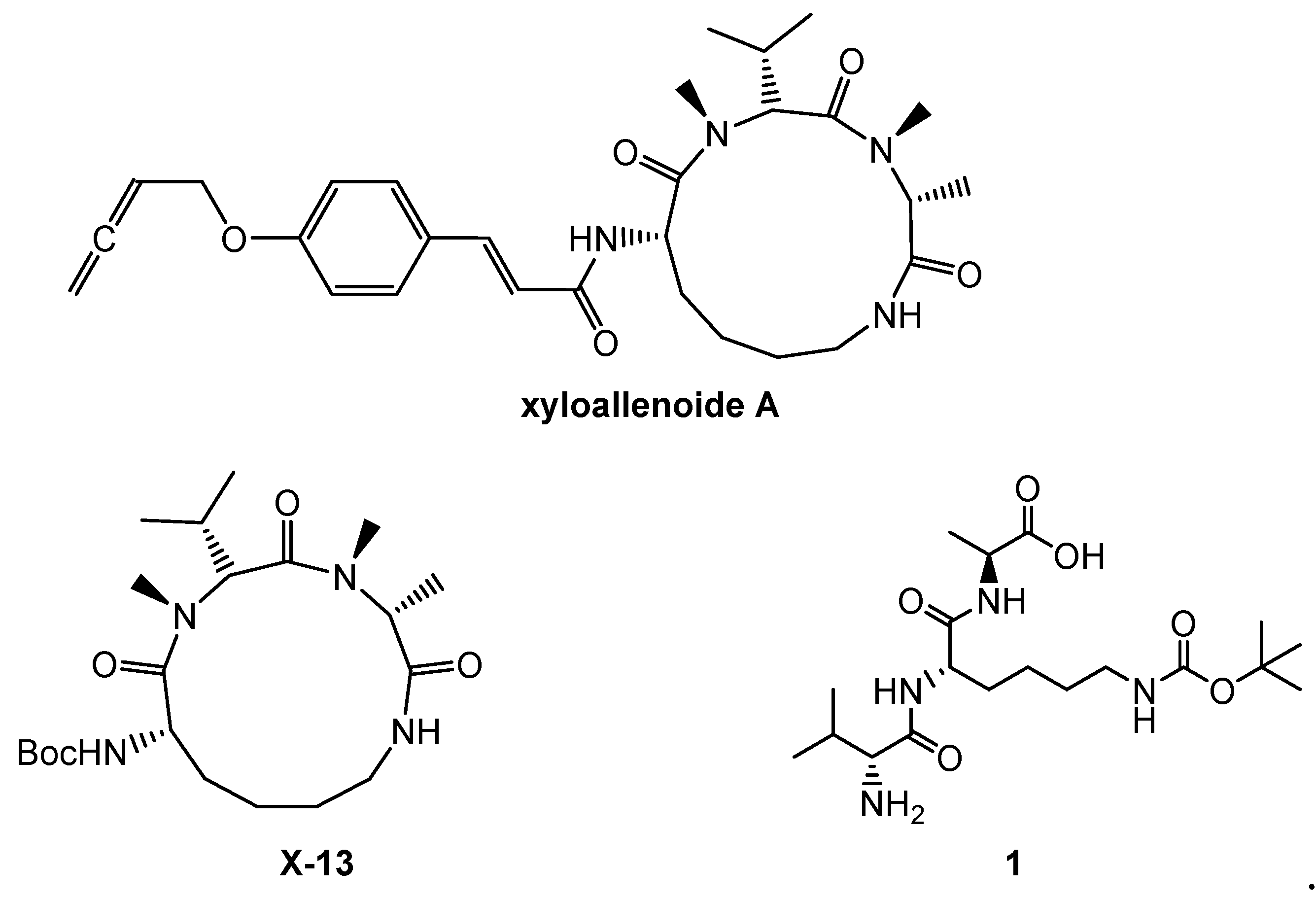
:1. Introduction
2. Results and Discussion
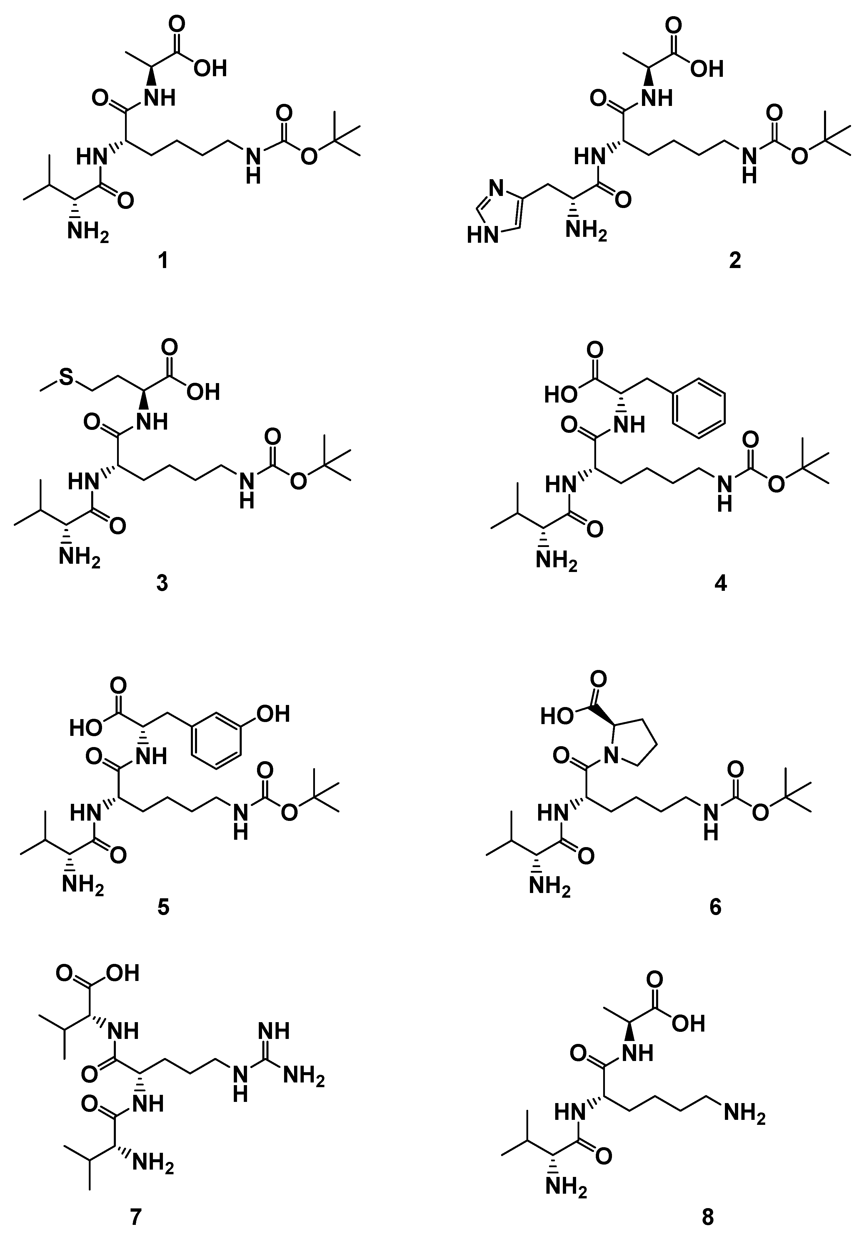
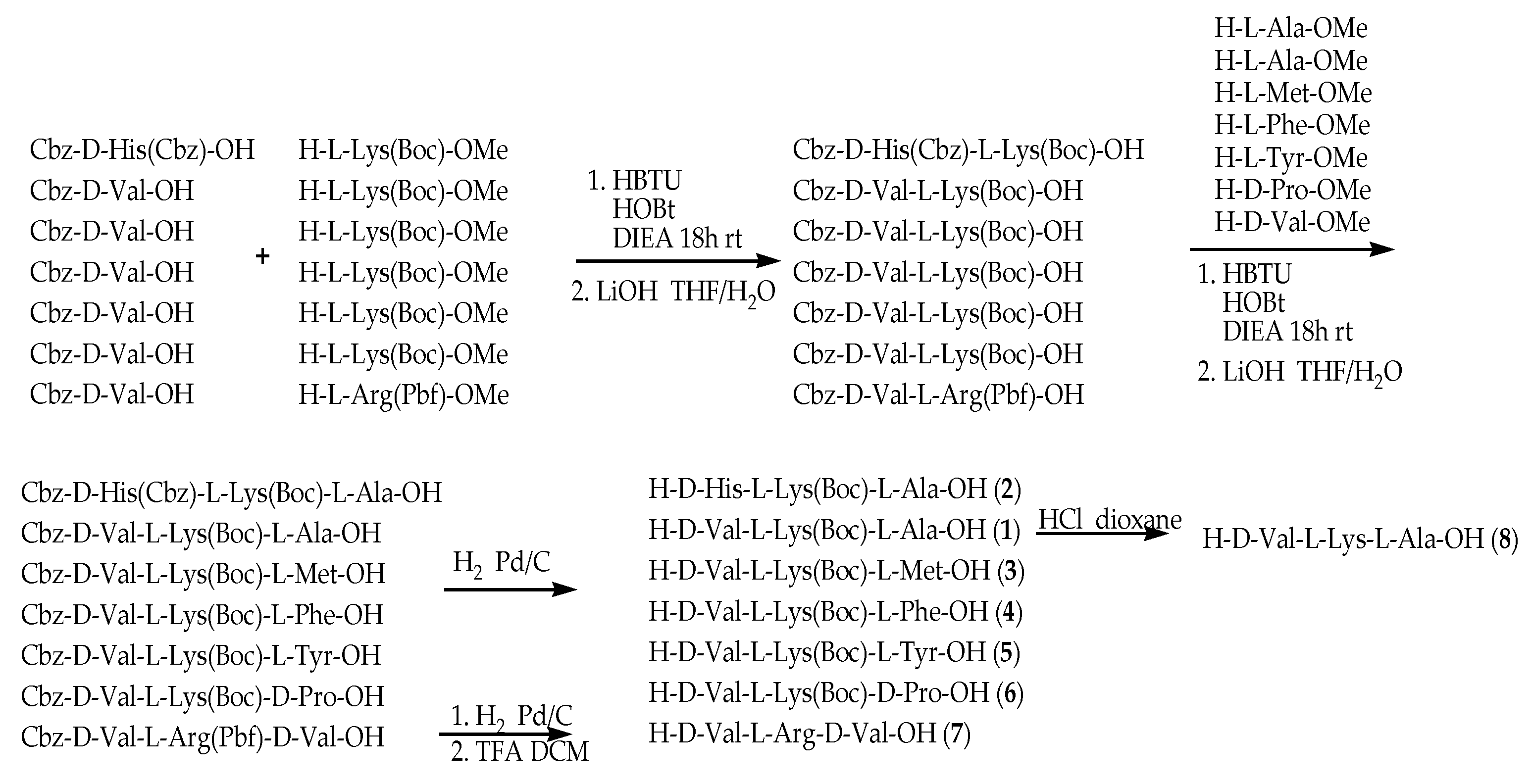
2.1. Chemistry
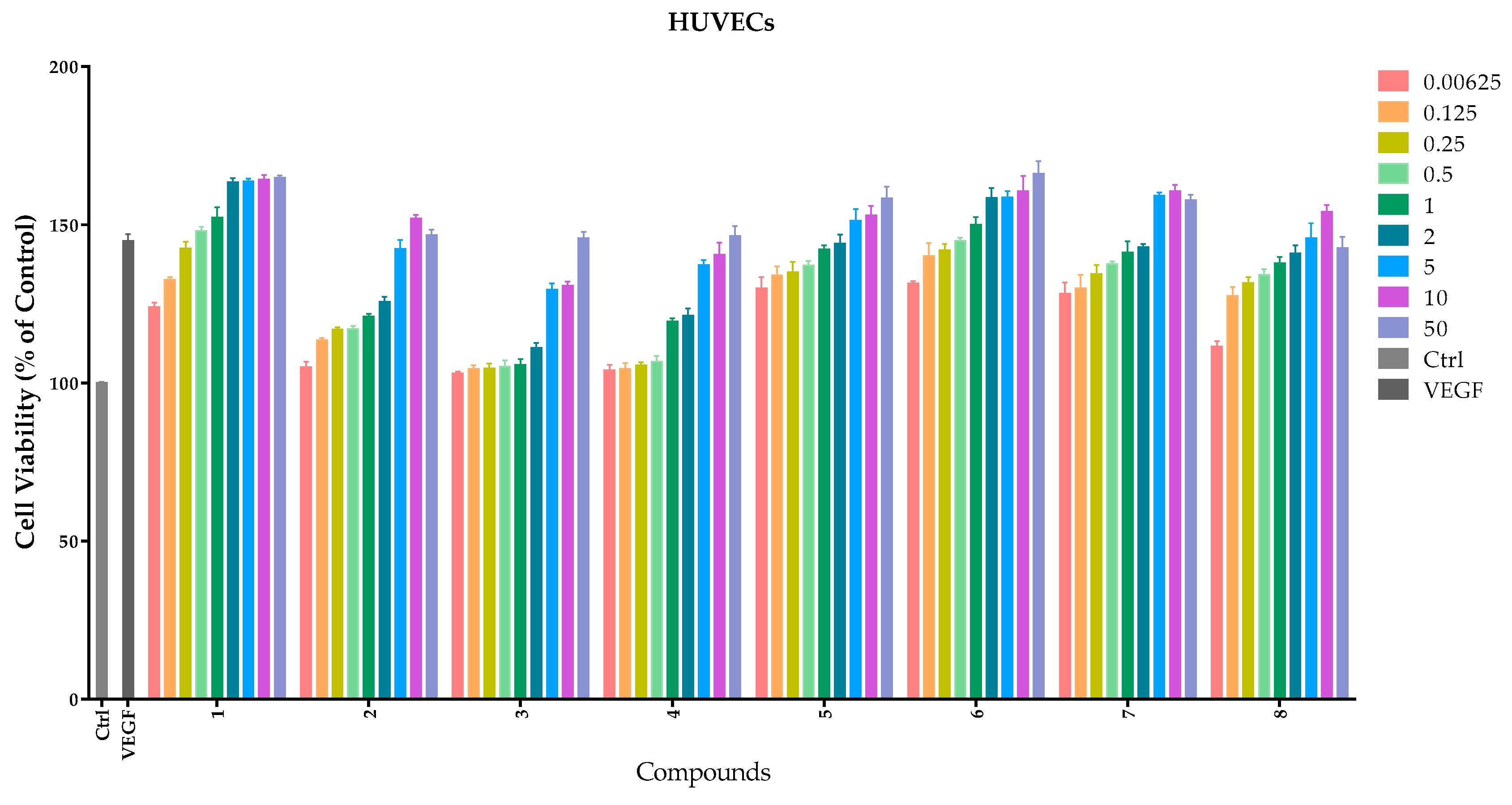
2.2. Effects of Compounds 1–8 on HUVEC Proliferation
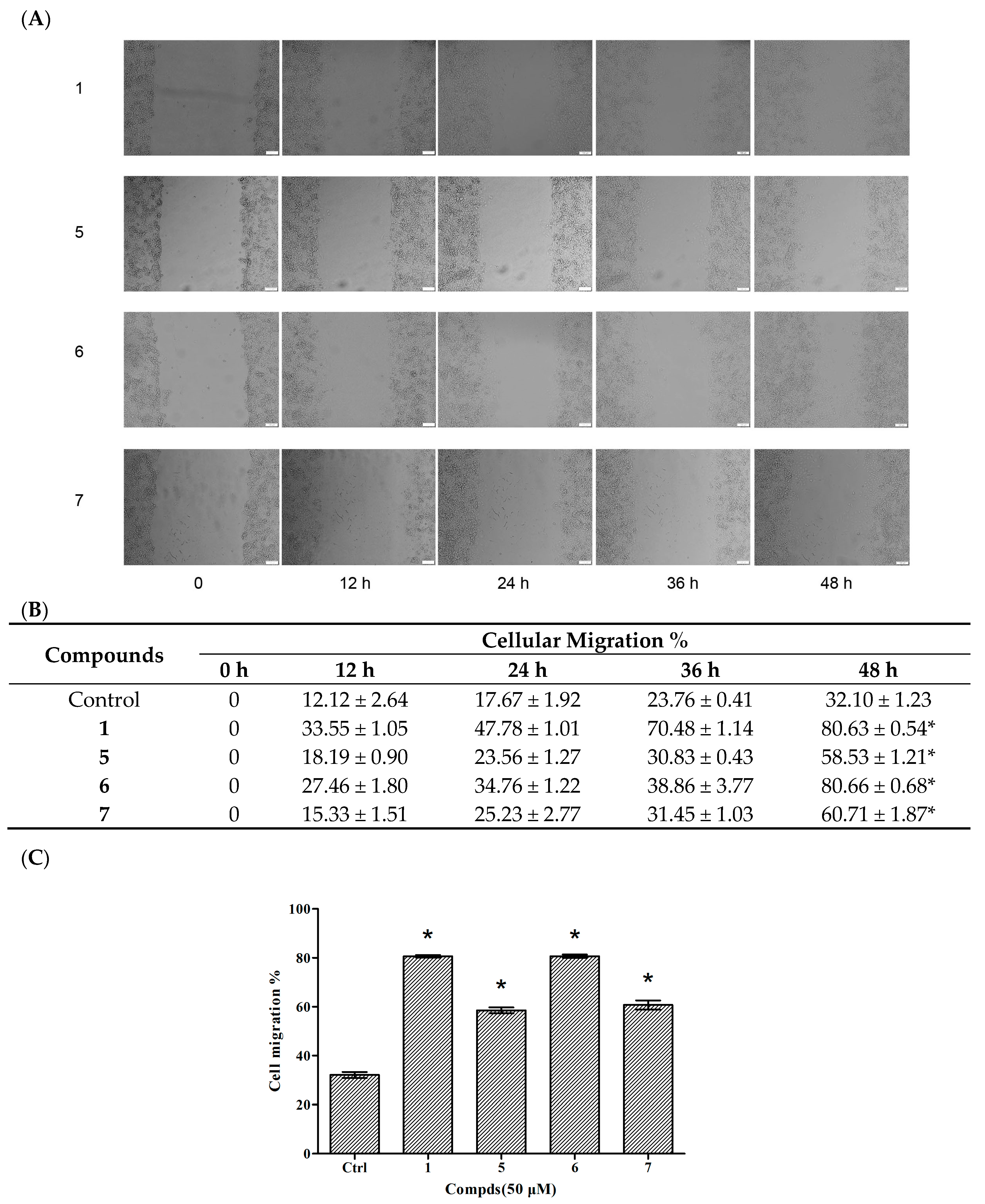
2.3. Migration Assays–Wound Healing of Compounds 5–7
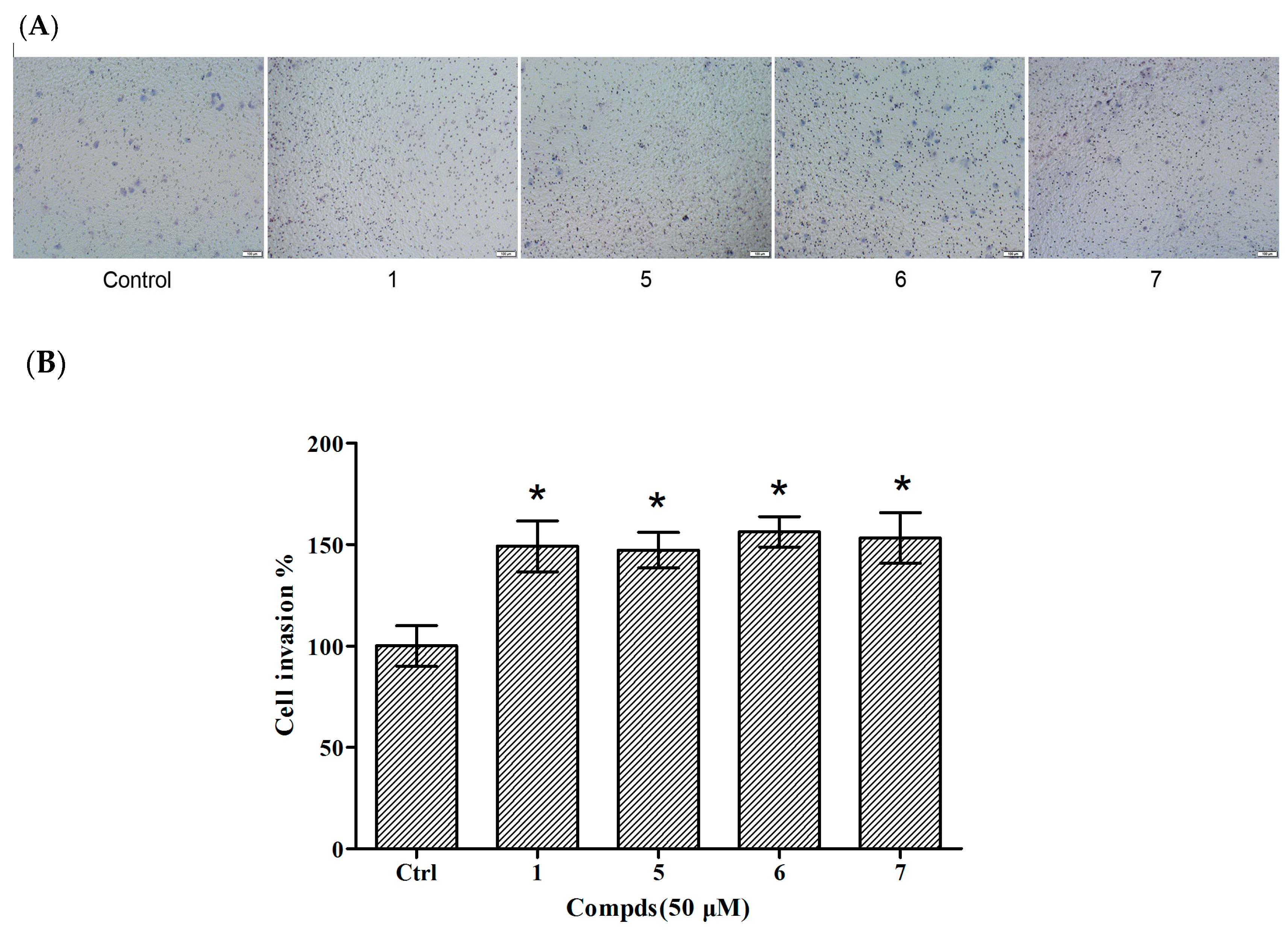
2.4. Invasion Assays of Compounds 5–7 in HUVECs
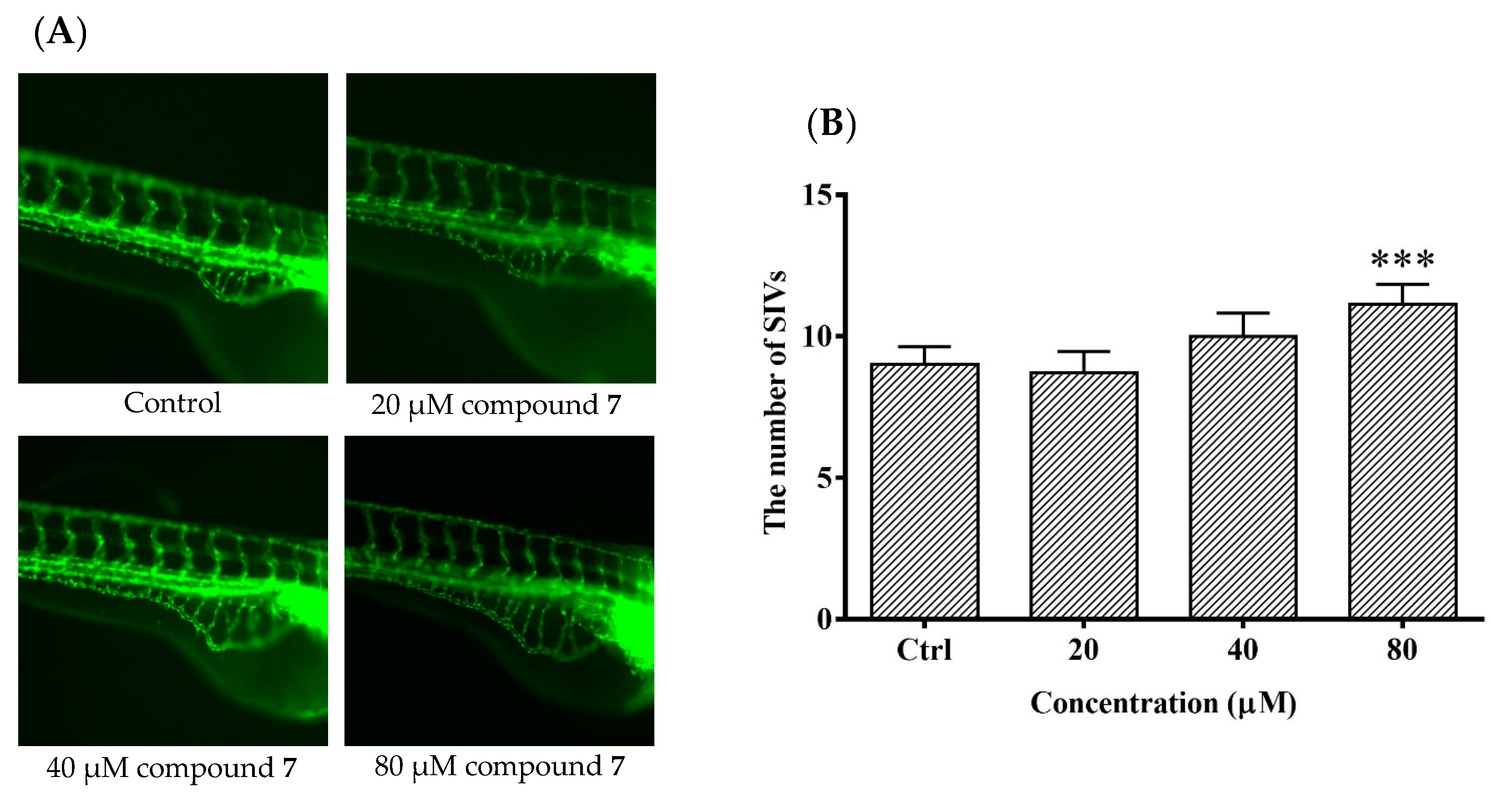
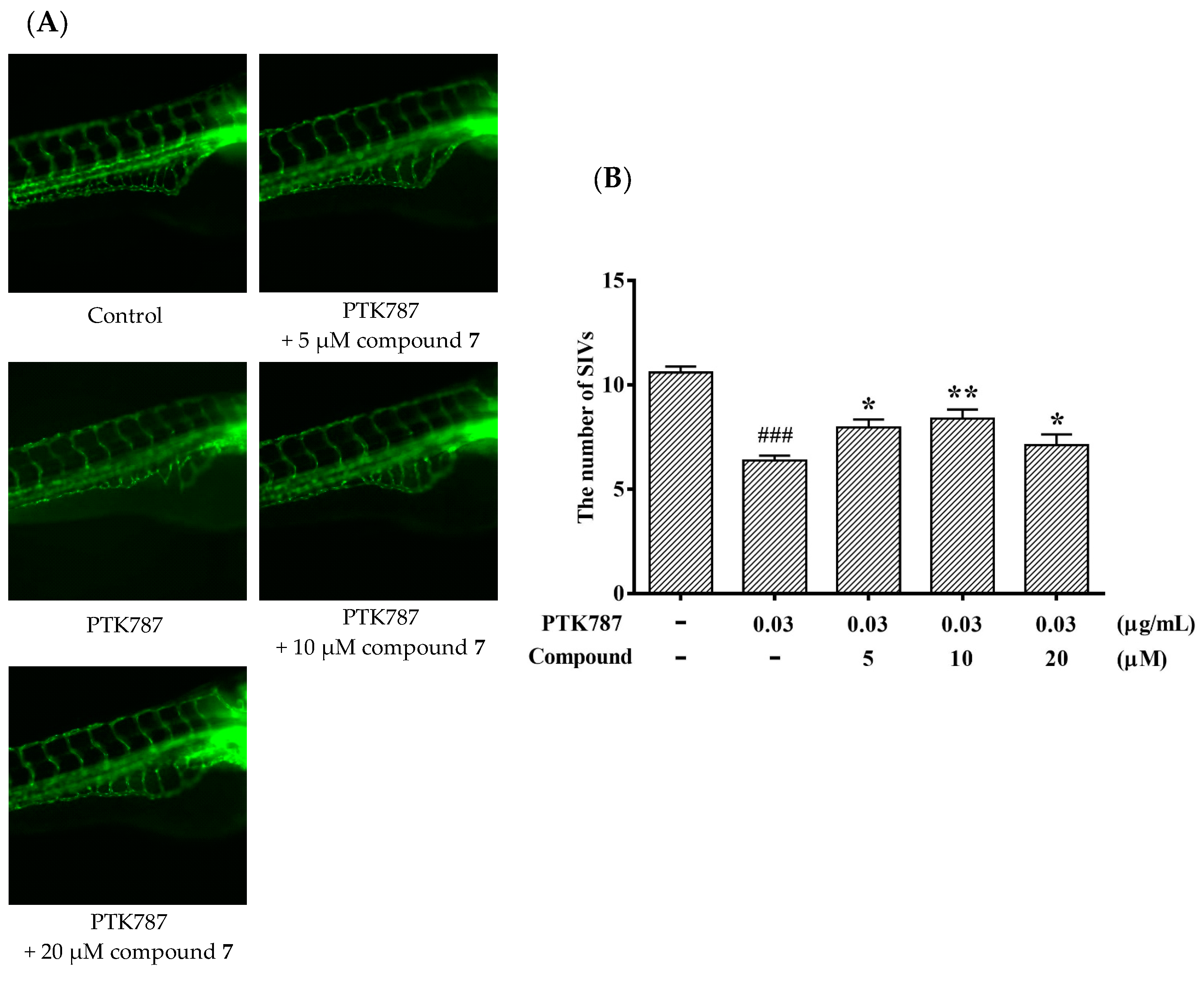
2.5. The Angiogenic Activity of Compound 6 in Zebrafish
3. Experimental Section
3.1. Chemistry
3.2. General Procedure for the Synthesis of Compounds 1–8
3.3. Cellular Culture and Drug Treatment
3.4. Proliferative Assays
3.5. Migration Assays
3.6. Invasion Assay
3.7. Zebrafish Assay of Compound 7
3.8. Statistical Analysis
4. Conclusions
Supplementary Materials
Author Contributions
Funding
Acknowledgments
Conflicts of Interest
References
- Zhou, X.; Liu, J.; Yang, B.; Lin, X.; Yang, X.W.; Liu, Y. Marine natural products with anti-HIV activities in the last decade. Curr. Med. Chem. 2013, 20, 953–973. [Google Scholar] [PubMed]
- Edwards, D.J.; Marquez, B.L.; Nogle, L.M. Structure and biosynthesis of the jamaicamides, new mixed polyketide-peptide neurotoxins from the marine cyanobacterium Lyngbya majuscula. Am. Math. Soc. 2004, 11, 817–833. [Google Scholar] [CrossRef] [PubMed]
- Huang, H.N.; Rajanbabu, V.; Pan, C.Y. A cancer vaccine based on the marine antimicrobial peptide pardaxin (GE33) for control of bladder-associated tumors. Biomaterials 2013, 34, 10151–10159. [Google Scholar] [CrossRef] [PubMed]
- Jang, I.S.; Sun, J.P. Hydroxyproline-containing collagen peptide derived from the skin of the Alaska pollack inhibits HIV-1 infection. Mol. Med. Rep. 2016, 14, 5489–5494. [Google Scholar] [CrossRef] [PubMed] [Green Version]
- Chopra, L.; Singh, G.; Choudhary, V. Sonorensin: An antimicrobial peptide, belonging to the heterocycloanthracin subfamily of bacteriocins, from a new marine isolate, Bacillus sonorensis MT93. Appl. Environ. Microbiol. 2014, 80. [Google Scholar] [CrossRef] [PubMed]
- Ko, S.C.; Kim, D.; Jeon, Y.J. Protective effect of a novel antioxidative peptide purified from a marine Chlorella ellipsoidea protein against free radical-induced oxidative stress. Food Chem. Toxicol. 2012, 50, 2294–2302. [Google Scholar] [CrossRef] [PubMed]
- Hyder, S.M.; Stancel, G.M. Regulation of angiogenic growth factors in the female reproductive tract by estrogens and progestins. Mol. Endocrinol. 1999, 13, 806–811. [Google Scholar] [CrossRef] [PubMed]
- Singla, R.; Soni, S.; Patial, V. Cytocompatible anti-microbial dressings of Syzygium cumini cellulose nanocrystals decorated with silver nanoparticles accelerate acute and diabetic wound healing. Sci. Rep. 2017, 7. [Google Scholar] [CrossRef]
- Suzane, C.P.; Marcell, C.M.; Sybele, S.; Joni, A.C.; Raquel, M.S.C. Role of osteogenic growth peptide (OGP) and OGP (10-14) in bone regeneration: A review. Mol. Sci. 2016, 17. [Google Scholar] [CrossRef]
- Guo, D.; Murdoch, C.E.; Xu, H. Vascular endothelial growth factor signaling requires glycine to promote angiogenesis. Sci. Rep. 2017, 7. [Google Scholar] [CrossRef] [PubMed]
- Lin, Y.; Wu, X.; Feng, S.; Jiang, G.; Zhou, S.; Vrijmoedb, L.L.P.; Gareth Jonesb, E.B. A novel N-cinnamoylcyclopeptide containing an allenic ether from the fungus Xylaria sp. (strain #2508) from the South China Sea. Tetrahedron. Lett. 2001, 42, 449–451. [Google Scholar]
- Wang, S.Y.; Xu, Z.L.; Wang, H.; Li, C.R.; Fu, L.W.; Pang, J.Y.; Li, J.; She, Z.G.; Lin, Y.C. Total Synthesis, absolute configuration, and biological activity of xyloallenoide A. Helv. Chim. Acta 2012, 95, 973–982. [Google Scholar] [CrossRef]
- Lu, X.L.; Xu, Z.L.; Yao, X.L. Marine cyclotripeptide X-13 promotes angiogenesis in zebrafish and human endothelial cells via PI3K/Akt/eNOS signaling pathways. Mar. Drugs 2012, 10, 1307–1320. [Google Scholar] [CrossRef] [PubMed]
- Li, J.; Lu, X.; Wu, Q.; Yu, G.; Xu, Z.; Qiu, L.; Pei, Z.; Lin, Y.; Pang, J. Design, SAR, angiogenic activities evaluation and pro-angiogenic mechanism of new marine cyclopeptide analogs. Curr. Med. Chem. 2013, 20, 1183–1194. [Google Scholar] [CrossRef] [PubMed]
- Byung, C.L.; Hyun, J.S.; Ji, S.K.; Kyung-Ho, J.; Yearn, S.C.; Kyung-Han, L.; Dae, Y.C. Synthesis of Tc-99m labeled glucos amino-Asp-cyclic (Arg-Gly-Asp-D-Phe-Lys) as a potential angiogenesis imaging agent. Bioorg. Med. Chem. 2007, 15, 7755–7764. [Google Scholar]
- Park, H.J.; Zhang, Y.; Georgescu, S.P.; Johnson, K.L.; Kong, D.; Galper, J.B. Human umbilical vein endothelial cells and human dermal microvascular endothelial cells offer new insights into the relationship between lipid metabolism and angiogenesis. Stem Cell Rev. 2006, 2, 93–102. [Google Scholar] [CrossRef] [PubMed]
- Yi, F.; Hao, Y.; Chong, X. Overexpression of microRNA-506-3p aggravates the injury of vascular endothelial cells in patients with hypertension by downregulating Beclin1 expression. Exp. Ther. Med. 2018, 15, 2844–2850. [Google Scholar] [CrossRef] [PubMed]
- Tal, T.L.; Mccollum, C.W.; Harris, P.S. Immediate and long-term consequences of vascular toxicity during zebrafish development. Reprod. Toxicol. 2014, 48, 51–61. [Google Scholar] [CrossRef] [PubMed]
Sample Availability: Samples of the compounds 1–8 are available from the authors. |


| Compounds | EC50 (μM) |
|---|---|
| 1 | 1.31 ± 0.0926 |
| 2 | 57.55 ± 6.10 |
| 3 | >200.00 |
| 4 | 76.02 ± 0.205 |
| 5 | 1.00 ± 0.002 |
| 6 | 1.00 ± 0.0005 |
| 7 | 0.88 ± 0.0972 |
| 8 | 1.33 ± 0.201 |
© 2018 by the authors. Licensee MDPI, Basel, Switzerland. This article is an open access article distributed under the terms and conditions of the Creative Commons Attribution (CC BY) license (http://creativecommons.org/licenses/by/4.0/).
Share and Cite
Zheng, Y.; Tong, Y.; Wang, X.; Zhou, J.; Pang, J. Studies on the Design and Synthesis of Marine Peptide Analogues and Their Ability to Promote Proliferation in HUVECs and Zebrafish. Molecules 2019, 24, 66. https://doi.org/10.3390/molecules24010066
Zheng Y, Tong Y, Wang X, Zhou J, Pang J. Studies on the Design and Synthesis of Marine Peptide Analogues and Their Ability to Promote Proliferation in HUVECs and Zebrafish. Molecules. 2019; 24(1):66. https://doi.org/10.3390/molecules24010066
Chicago/Turabian StyleZheng, Yinglin, Yichen Tong, Xinfeng Wang, Jiebin Zhou, and Jiyan Pang. 2019. "Studies on the Design and Synthesis of Marine Peptide Analogues and Their Ability to Promote Proliferation in HUVECs and Zebrafish" Molecules 24, no. 1: 66. https://doi.org/10.3390/molecules24010066