1.1. Antitumoral Activity of Cryptolepine
Cryptolepine (
1), neocryptolepine (
2), and isocryptolepine (
3) are typical indoloquinoline alkaloids isolated from the roots of
Cryptolepis sanguinolenta [
1,
2].
These alkaloids are composed of the tetracyclic indoloquinoline ring system, which only differ with respect to the orientation and site of their indole and quinoline ring junctures (
Figure 1). By introducing the appropriate motif at the certain positions, their derivatives can increase better biological activity than the mother core [
3].
Many researchers have achieved the structural modification of cryptolepine scaffold for the purpose of improving antitumor activity, while the pharmacological properties of the analogues are being studied deeply.
The bioactivities of many antitumoral agents are related to their interactions with the DNA molecule, which is regarded as a classical target for these drugs in clinical use. The basic mechanism of antitumoral activity of these drugs is to affect the replication, expression, transcription and other physiological functions of the DNA, which causes the tumor cell death [
4].
In 1990, Yamato et al. synthesized the indoloquinoline derivatives
4 (
Figure 2), and screened its biological properties in vitro and in vivo. The compounds
4 showed potential antitumor activity (P388 leukemia in mice), DNA intercalative property, and ability to induce topoisomerase II dependent DNA cleavage [
5].
In 1997, Deady et al. studied a series of cryptolepine derivatives
5 and evaluated their antitumoral activity in a series of murine and human tumor cell lines such as the mice lung cancer cells (LLC), mice leukemia cells (P388), human leukocyte cells (JL). These compounds appear to be mixed topoisomerase I/II inhibitors in the human leukemia cell lines studied [
6].
In 1998, Bonjean et al. verified the cryptolepine alkaloids bound tightly to DNA as a typical intercalating agent by various means of absorption, such as fluorescence, circular, and linear dichroism, as well as by a relaxation assay using DNA topoisomerases. At the same time, they provided direct evidence that DNA is the primary target of cryptolepine. The mechanism of the compounds inhibiting tumor cell proliferation is mainly based on the synthesis of DNA inhibition, not the inhibition of proteins and RNA [
7].
In 2002, Lisgarten, John N., reported that cryptolepine interacts with the DNA fragment d(CCTAGG)
2 in a base-stacking intercalation mode by using X-ray crystallography. It was found that cryptolepine intercalated between pyrimidine bases of the fragment in the form of π-π accumulation. This is the first single crystal structure of DNA intercalator complex, which is the small molecule to bind a non-alternating (pyrimidine-pyrimidine) DNA sequence [
8].
In 2012, Boddupally et al. have synthesized a series of 11-substituted cryptolepine derivatives
6 (
Figure 2). The compound
6 showed the most potent anticancer activity with IC
50 = 0.97 μM against HCT-116 colon cancer cell line and IC
50 = 2.33 μM against Raji lymphoma cells in further cytotoxic test in vitro. At the same time, this compound showed a strong inhibition of c-MYC expression [
9].
Gu and Lu demonstrated the binding of aniline-substituted cryptolepine derivatives with calf-thymus DNA presumably via an intercalation mechanism and studied the binding mode of these derivatives with duplex DNA by Surflex-dock software. They reported that these derivatives intercalated into the base-pairs, and reacted with DNA via mainly π-π interaction with medium, moreover the functional groups substituted on aniline ring affected the binding abilities [
10,
11].
The aim of this review is to present an overview of the potential of neocryptolepine and isocryptolepine as scaffolds for the design and development of new anticancer drugs. Both compounds have also the linearly arranged tetracyclic plane as same as cryptolepine, so they can be expected as candidates of antitumoral agent, although they have a slightly weaker capability to intercalate into DNA and inhibit human topoisomerase II [
12].
1.2. Antitumoral Activity of Neocryptolepines and Isocryptolepines
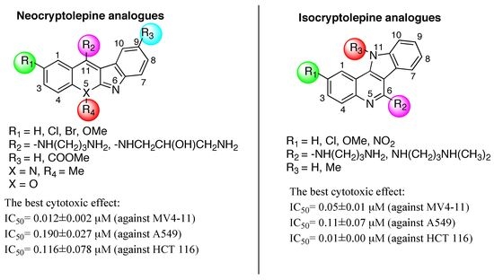
In our group, we have synthesized an array of novel neocryptolepine derivatives
7, and their congeners
8, 11-aminoalkylamino-substituted chromeno[2,3-
b]indoles
9, and isocryptolepine derivatives
10 (
Figure 3). Then the antiproliferative activities of these compounds were tested in vitro against MV4-11 (human leukemia), HCT116 (human colon cancer), and A549 (human non-small cell lung cancer) and BALB/3T3 (normal murine fibroblasts) cell lines.
We focused our attention on modifying the neocryptolepine structure at four positions: (1) the side chain at C11, introducing the various amino groups, e.g., -NH(CH
2)
nNH
2 (n = 2,3,4), -NH(CH
2)
3N(C
2H
5)
2, -NH(CH
2)
3OH, -NHCH
2CH(OH)CH
2NH
2, -NHCH
2CH(CH
3)CH
2NH
2, -NHCH
2CH(CH
3)NH
2, -NHCH
2C(CH
3)
2NH
2, and so on; (2) replacing the H atom at C2 with MeO, Br, Cl, Me, and replacing H at C9 with COOMe; (3) altering the Me from N5 to N6, (4) changing the N5 to O atom and (5) incorporating metal ion coordinated with diamine side chain [
13,
14,
15,
16,
17,
18].
In addition, we have also engaged in modifying isocryptolepine derivatives in three ways: the amino substituent effect at C6, and N11 methyl localization effect, and the substituent group effect at C2 of quinoline moiety [
19].
Some compounds with higher antiproliferative activity are listed in
Table 1 and
Table 2. The SAR (structure activity relationships) studies reveal that the most necessary strategy is introducing ω-aminoalkylamino group in the side chain. Among the tested 84-neocryptolepine derivatives and 44-isocryptolepine derivatives, the most potent compounds with higher activity contain the -NH(CH
2)
3NH
2 side chain.
Among them, 11-(3-amino-2-hydroxy)propylamino derivatives 7e and 7f were the most cytotoxic with a mean IC50 value of 0.042 μM and 0.057 μM against the MV4-11 cell line, 0.197 μM and 0.190 μM against the A549 cell line, and 0.138 μM and 0.117 μM against the HCT116 cell line.
We propose the reason that the amino terminus domain in the side chain would react with negative charged phosphate groups in DNA, which increases the insertion ability of the complex to DNA.
The methyl localization effect is an important additional contributor to increase the activity. The activity is greatly reduced (10~30 fold lower) by changing the Me group from N5 to N6 (i.e., 8) of neocryptolepine derivatives. The Me group can increase the activity of isocryptolepine derivatives, as found by a comparison of the compounds 10e and 10c.
Replacing H atom at the C2 with MeO, Br, Cl, Me, or replacing H at C9 by COOMe can improve the antiproliferactive activity of neocryptolepine derivatives. Each group contributes differently to the antiproliferactive activity of different tumor cells. For example, 7c with Br at C2 shows the best antiproliferactive activity against MV4-11 leukemia cells, and this modification did not increase antiproliferactive activity against normal BALB/3T3 cells, but unexpectedly it decreases 2-fold the antiproliferactive activity against A549 lung cancer cells. It can be suggested that this modification increases the selectivity of the tested compound, improving their effect on leukemic cells, without increasing possible adverse effects on normal cells. The substituent effect at C2 of isocryptolepine core also can change the activity; NO2 is the effective group to increase the activity against cancer but unfortunately also normal cells (10c and 10e). Therefore, we must examine our best to find an effective substituent for further improving the activity.
We boldly tried to replace the 5-nirtogen atom with oxygen atom, forming
9, which significantly drop the antiproliferactive activity [
16].
From all the assay data, isocryptolepine analogues are more potential as the anticancer drug candidates in comparison with the neocrytolepine analogues, compound 10e (or 10a) shows the higher antiproliferactive activity against A549 and HCT116 cancer cell, and lower antiproliferactive activity against normal cells comparing 7a, when amino-substituent is NH(CH2)3NH2 and substituent group is H at C2.
The mode of neocryptolepine and isocryptolepine binding to DNA was studied using UV-VIS absorption spectroscopy with salmon fish sperm DNA. From the DNA binding studies, it can be proven that the methyl localization effect and the substituent group effect at C2 of quinoline moiety influence the capability to intercalate into DNA. Two effects improve the activity of isocryptolepine to interact with DNA, the binding constant of
10f-DNA was 1.05 × 10
6 L/mol and
10g-DNA was 4.84 × 10
6 L/mol [
19]. The activity of neocryptolepine to interact with DNA varied; the binding constant of
7c-DNA was 2.93 × 10
5 and
8-DNA was 3.28 × 10
5 L/mol [
13].
In a series of neocrytolepine analogues,
7c shows the best antiproliferactive activity against MV4-11 leukemia with IC
50 0.012 ± 0.002 μM. Compounds
7f and
7i show almost the same highest antiproliferactive activity against A549 lung cancer cells with IC
50 0.190 ± 0.027 μM and 0.194 ± 0.063 μM, and the antiproliferative activity against HCT116 colon cancer cells with IC
50 0.117 ± 0.055 μM and 0.116 ± 0.078 μM. Analogue
7b unfortunately shows 2-fold higher cytotoxic activity against normal BALB/3T3 cells with the IC
50 0.401 ± 0.015 μM than other studied neocrytolepine analogues and 20-fold higher than cisplatin [
13].
In series of isocryptolepine analogues, 10e shows the best antiproliferactive activity against cancer cell lines with IC50 0.05 ± 0.01 μM (MV4-11), IC50 0.11 ± 0.07 μM (A549), and IC50 0.01 ± 0.00 μM (HCT116). Unfortunately, this compound had also 4~15 fold higher cytotoxic activity against normal BALB/3T3 cells with the IC50 0.08 ± 0.02 μM. These two simultaneous modifications at C2-NO2 and at N11-Me showed 15- and 100-fold increased cytotoxicity of 10e in comparison to doxorubicin and cisplatin, cytostatic used as a control.
In 2016, in vitro antiproliferative activities of neocryptolepine derivatives were evaluated by Okada (
Table 3). The compounds
7a and
7d showed the highest anticancer activity, having IC
50 values of 0.50 μM and 0.20 μM against the breast cancer MDA-MB-453 cell line, at the same time
7a having 0.64 μM and 2.7 μM against WiDr (colon adenocarcinoma) and SKOv3 (ovarian cancer) cell lines,
7d having 0.37 μM and 1.3 μM against WiDr (colon adenocarcinoma) and SKOv3 (ovarian cancer) cell lines.
Recently, we reported the in vitro antiproliferative activity of 11-substituted neocryptolepines with branched ω-aminoalkylamino chain. These 2-substituted 5-methyl-indolo[2,3-
b]quinoline derivatives were prepared by nucleophilic aromatic substitution (S
NAr) of 11-chloroneocryptolepines
2 with appropriate 1,2- and 1,3-diamines (
Scheme 1). Many of the prepared neocryptolepine derivatives showed submicromolar antiproliferative activity of less than μM against the human leukemia MV4-11 cell line (
Table 4).
For further studying cytotoxic and growth inhibitory activities of neocryptolepine derivatives, the anticancer screening was performed across the JFCR39 cancer cell line panel, evaluated at five concentration levels (100, 10, 1.0, 0.1, and 0.01 µM) (
Table 5). The results (the means of GI
50, TGI, and LC
50 values) showed that
7d have potent anticancer activity against the melanoma (LOX-IMVI) and lung (NCIH522, A549, and DMS273) cancer cell lines in the JFCR39 panels. Compound
7d shows a notable cytotoxicity against the breast (BSY-1, LC
50 = 0.82 µM), CNS (SF-539, LC
50 = 0.60 µM, SNB-75, LC
50 = 0.91 µM), colon (HCC2998, LC
50 = 0.74 µM), lung (NCI-H522, LC
50 = 0.71 µM, A549, LC
50 = 0.85, DMS273, LC
50 = 0.70, DMS114, LC
50 = 0.72 µM), and ovarian (VCAR-4, LC
50 = 0.48 µM) cancer cell lines [
20].
Table 5 shows that all of the compounds are highly potent against the melanoma (LOX-IMVI) and lung (NCI-H522, A549, and DMS273) cancer cell lines in the JFCR39 panels. Compounds
7d and
7k are more potent than compound
7q towards the cancer cell lines in the JFCR39 panel. Compounds
7d and
7k show a similar growth inhibitory activity (GI
50 > 0.01 µM) against the 31 and 28 cell lines, respectively, of the JFCR39 panel. Compound
7k shows a notable cytotoxicity against the breast (BSY-1, LC
50 = 0.82 µM), CNS (SF-539, LC
50 = 0.60 µM, SNB-75, LC
50 = 0.91 µM), colon (HCC2998, LC
50 = 0.74 µM), lung (NCI-H522, LC
50 = 0.71 µM, A549, LC
50 = 0.85, DMS273, LC
50 = 0.70, DMS114, LC
50 = 0.72) and ovarian (VCAR-4, LC
50 = 0.48 µM) cancer cell lines. Compound
7q shows a mentionable cytotoxicity for the CNS (SF-539, LC
50 = 0.63 µM, SNB-75, LC
50 = 0.93 µM), colon (HCC2998, LC
50 = 0.69 µM), lung (NCI-H522, IC
50 = 0.57 µM, DMS273, LC
50 = 0.61 µM), and melanoma (LOX-IMVI, LC
50 = 0.75 µM) cancer cell lines. Compound
7q displayed a strong growth inhibitory activity (GI
50 < 1 µM) for 28 panels in the JFCR39 panel of the various cell lines. DMS273 (lung cancer) and NCI-H522 (lung cancer) are the most sensitive cell lines of the 39 cell lines (GI
50 = 0.42 µM for both and LC
50 = 4.7 µM and 5.5 µM respectively). Compound
7q also showed a strong antitumor effect on LOX-IMVI (melanoma, GI
50 = 0.3 µM, LC
50 = 7.6 µM).
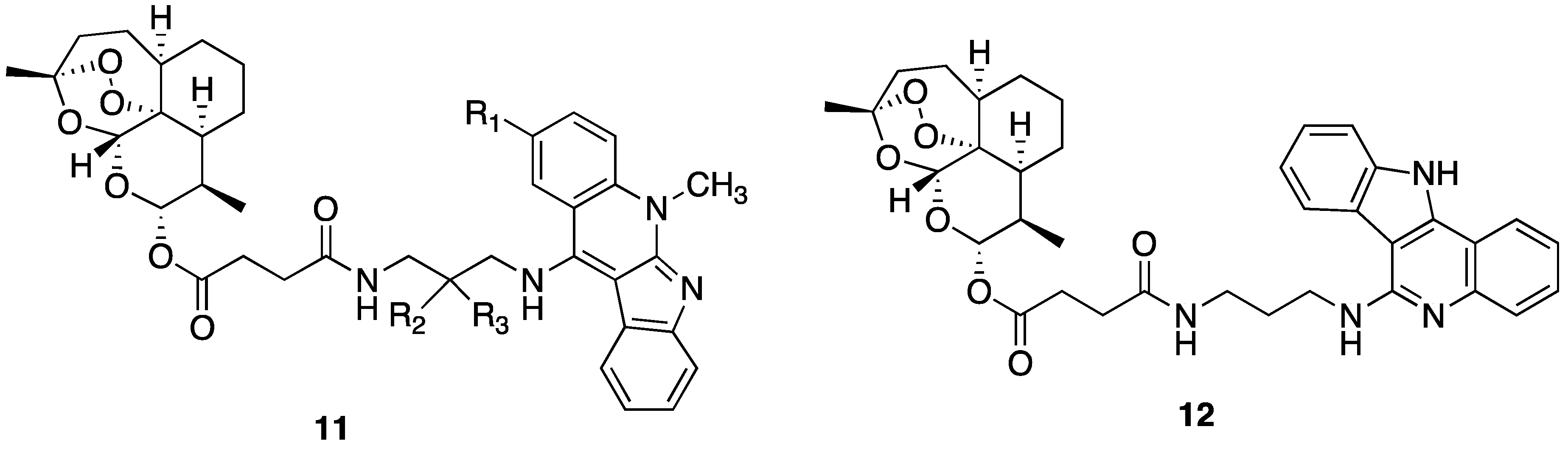
In our group, we achieved the synthesis of the artemisinin-indoloquinoline hybrids and studied their antiproliferative activity (
Figure 4). The artemisinin-neocryptolepine hybrids
11a,
11b and the artemisinin-isocryptolepine hybrids
12 showed the potent antiproliferative activity, based on the data in
Table 6.
In 2017, Li Wang synthesized the novel tacrine-neocryptolepine hybrids, and evaluated as inhibitors of human AChE (hAChE) and human BuChE (hBuChE) (
Figure 5). Compound
13 was a highly potent hAChEI having IC
50 = 0.95 ± 0.04 nM, and a highly potent hBuChE having IC
50 = 2.29 ± 0.14 nM [
22].


 ,
, 


