Article

Anomeric Spironucleosides of β-d-Glucopyranosyl Uracil as Potential Inhibitors of Glycogen Phosphorylase

by
Aggeliki Stathi
1,
Michael Mamais
1,2,
Evangelia D. Chrysina
2 and
Thanasis Gimisis
1,*
1
Organic Chemistry Laboratory, Department of Chemistry, National and Kapodistrian University of Athens, 10571 Athens, Greece
2
Institute of Chemical Biology, National Hellenic Research Foundation, 11635 Athens, Greece
*
Author to whom correspondence should be addressed.
Molecules 2019, 24(12), 2327; https://doi.org/10.3390/molecules24122327
Submission received: 13 May 2019 / Revised: 14 June 2019 / Accepted: 17 June 2019 / Published: 25 June 2019
(This article belongs to the Special Issue Biomimetic Radical Chemistry and Applications)

Abstract

:
In the case of type 2 diabetes, inhibitors of glycogen phosphorylase (GP) may prevent unwanted glycogenolysis under high glucose conditions and thus aim at the reduction of excessive glucose production by the liver. Anomeric spironucleosides, such as hydantocidin, present a rich synthetic chemistry and important biological function (e.g., inhibition of GP). For this study, the Suárez radical methodology was successfully applied to synthesize the first example of a 1,6-dioxa-4-azaspiro[4.5]decane system, not previously constructed via a radical pathway, starting from 6-hydroxymethyl-β-d-glucopyranosyluracil. It was shown that, in the rigid pyranosyl conformation, the required [1,5]-radical translocation was a minor process. The stereochemistry of the spirocycles obtained was unequivocally determined based on the chemical shifts of key sugar protons in the 1H-NMR spectra. The two spirocycles were found to be modest inhibitors of RMGPb.

Graphical Abstract

1. Introduction

Despite the prevalence of type 2 diabetes worldwide, no sufficient treatment has been identified; therefore, a molecular approach based on the three-dimensional structure of enzymes directly involved in glycogen metabolism has received increasing attention. Glycogen phosphorylase (GP) is an allosteric enzyme with a regulatory role in glycogen breakdown to glucose [1]. Since glucose is the physiological substrate of GP, it promotes the inactive form of the enzyme acting synergistically with insulin towards reducing the rate of glycogen degradation and shifting the equilibrium towards glycogen synthesis. GP three-dimensional structure in the T state (GPb) has been exploited as a target for the design of glucose-based compounds that inhibit enzymic action preventing glycogenolyis and acting as regulators of glucose levels in the bloodstream [2,3]. The glucose specificity for the catalytic site of the enzyme has been utilized to drive glucose derivatives to the active site of the enzyme, exploiting the catalytic channel by adding a variety of structural features to these compounds in terms of rigidity and functional groups [4]. One of the early lead inhibitors of rabbit muscle glycogen phosphorylase b (RMGPb) was pyranosyl spironucleosides [5] (2a,b, Figure 1), the structure of which was inspired from hydantocidin (1), a natural spiro compound with herbicidal and plant growth regulatory activity. To this end, a number of attempts to synthesize more potent inhibitors were made leading to other spiro-heterocycles that exhibited stronger affinity for RMGPb [6]. Similar studies in our laboratory led to N4-aryl-N1-(β-d-glucopyranosyl)cytidine nucleosides which exhibit RMGPb inhibition in the nanomolar range [7].
We present here a methodology, that involves a key [1,5]-radical translocation step for the synthesis of anomeric spironucleosides 4a,b (Figure 1), which were found to be modest inhibitors of rabbit muscle glycogen phosphorylase (RMGPb). Spirocyclic nucleosides present a rich synthetic chemistry and important biological function [8,9]. The targets in this paper contain a rare 1,6-dioxa-4-azaspiro[4.5]decane structure [10], which has not been previously constructed via a radical pathway [11]. In the similar 1,6-dioxa-4-azaspiro[4.4]nonane system (3, Figure 1), we reported an efficient protocol, using a 6-lithiation strategy for generating a 6-hydroxymethyluridine intermediate followed by oxidative cyclization through a [1,5]-radical translocation strategy [12,13]. We were interested in applying this protocol to the related “decane” system.

2. Results

2.1. Synthesis

Initially, we attempted to access compound 7 (Scheme 1) by direct lithiation of the known 2,3,4,6-tetra-O-benzyl-β-d-glucopyranosyluracil [14]. The protocol was based on the previous well-established 6-lithiation of protected 2-deoxy- and ribouridines, followed by the reaction with dimethylformamide or ethyl formate to generate the corresponding 6-formyluridines [12,15,16]. Although a major product formed under these conditions, spectral analysis revealed that it was the product of a selective mono-debenzylation and did not contain a formyl group. Although we could not unequivocally determine the position of debenzylation, we hypothesized that this had occurred in position-3, proximal to the 6-position where the initial lithiation is expected to occur (data not shown). This reductive debenzylation could be reminiscent of a previous method employing lithium naphthalenide [17].
The above result prompted us to change our strategy and include a previously reported [18] N1-(β-d-glucopyranosyl)-6-methyluracil (6a) intermediate in our synthesis, as exemplified in Scheme 1. Under optimized conditions, the N-glycosylation reaction of persilylated 6-methyluracil, in the presence of an excess of TMSOTf in DCE, led to the formation of three products, namely, 6ac, isolated after column chromatography in 67%, 30%, and 3% yield, respectively. As determined by ESI-MS and NMR, the major product was the expected N1-glycosylated 6-methyluridine 6a, whereas the N3-glycosylated analogue 6b was isolated in 30% yield, together with a small amount of N1,N3-bisglycosylated isomer 6c.
The main feature that differentiated 6b from 6a in the 1H-NMR spectrum was a substantial downfield shift of H-2′ (δ∆ = 0.8 ppm) in 6b, induced by the magnetic anisotropy effect of the second vicinal amidic 4-carbonyl. A similar effect was observed in the more complex spectra of the bis-substituted analogue 6c, and the [Μ + H]+ peak at 786.3 amu (ESI-MS) clearly differentiated 6c from the other two isomers ([M + H]+ at 457.2 amu).
The allylic methyl group of 6a was oxidized to the corresponding aldehyde 7 in the presence of selenium dioxide (3 equivs) in dioxane: acetic acid [19], in 67% yield. Apart from the aldehyde 7, isolated in 67% yield and recognized in 1H-NMR by its characteristic aldehydic proton at 9.90 ppm, a second more polar product was isolated in low yield, identified as the allylic alcohol 8. Its formation is expected by the mechanism of the reaction which follows an electrophilic allylic addition to selenium followed by a [2,3]-sigmatropic rearrangement [20,21]. Application of stoichiometric amounts of SeO2 also led to aldehyde 7 as the major product, although full conversion was not observed even after prolonged reaction times. When the reaction was performed with 3 equivs of SeO2, under strictly anhydrous conditions, the yield of aldehyde 7 was maximized. Reduction of aldehyde 7 with NaBH4 in CHCl3/isopropanol, in the presence of silica gel, at 0 °C [22] led to partial removal of acetate groups. By lowering the reaction temperature to −30 °C, exclusive formation of the allylic alcohol 8 was observed, and the product was isolated in 90% yield.
The key step photolysis of 8, under the standard optimized Suárez conditions [23], utilized for alkoxy radical generation in hydrogen atom transfer (HAT) reactions [11], in the presence of DIB and I2, in DCM and under visible light (150 W) irradiation, led to the isolation of three products in 50%, 10%, and 8% yield. The major product was, surprisingly, the aldehyde 7, whereas the two minor products corresponded to the expected isomeric spironucleosides 9a,b. The reaction is expected to proceed through the generation of an alkoxyl radical intermediate, followed by a [1,5]-radical translocation [24] to generate a C-1′ radical intermediate which, after oxidation and ionic cyclization, provides the spirocycles 9a,b [12,13]. The main formation of aldehyde 7 can be explained by a possible disproportionation reaction of the above alkoxy radical intermediate to aldehyde 7 and alcohol 8, with the latter re-entering the reaction cycle (see discussion below). The application of the same conditions as in the similar 1,6-dioxa-4-azaspiro[4.4]nonane system [12,13] aids in the comparison of the two systems and indicates that the rigidity of the β-d-glucopyranosyl ring renders the [1,5]-hydrogen atom transfer less favorable in this system than in the previously observed flexible ribosyl system. It should be noted that when the same reaction was attempted with a similar photolysis in the presence of Pb(OAc)4, I2, and CaCO3 [25], a complex mixture of products was obtained that could not be further characterized. Final removal of the acetate protection of 9a,b with ammonia in methanol led to the isolation of the target spirocycles 4a,b in quantitative yield.

2.2. Kinetic Experiments

RMGPb was isolated, purified, and recrystallized according to previously established protocols [26]. Compounds 4a,b were assayed in the direction of glycogen synthesis for their inhibitory effect on RMGPb as described before [26,27]. They both exhibited competitive inhibition with respect to the substrate glucose 1-phosphate (Glc-1-P), at constant concentrations of glycogen (0.2% w/v) and AMP (1 mM). Compound 4b was found to be a stronger inhibitor of RMGPb (35% inhibition at 1 mM) than 4a (26% inhibition at 1 mM).

3. Discussion

The stereochemistry of the two spirocyclic products could be inferred unequivocally, from the 1H-NMR spectra, as can be seen in Figure 2. Both spectra contain features that can be explained by the magnetic anisotropy induced by the 2-C=O group onto the sugar α- or β- hydrogens depending on the stereochemistry of the new spiro-center. As the new spirocycle locks the configuration of the pyrimidine ring with respect to the sugar ring, the 2-C=O is spaced in the vicinity of H-2′ in the R-anomer 9a and on the other hand, in the vicinity of H-3′ and H-5′, in the S-anomer 9b. This results in significant downfield shifts of the corresponding sugar Hs in the 1H-NMR spectra. Specifically, there is a 0.55 ppm shift of H-2′ going from the S- to R-anomer (5.62 to 6.17 ppm), whereas there is a 0.48 and 0.84 ppm shift for H-5′ (4.18 to 4.65 ppm) and H-3′ (5.47 to 6.31 ppm), respectively, going from the R- to the S-anomer (Figure 2). The remaining Hs (H-4′, H-6′, H-5, and H-7) have similar chemical shifts in the two spectra, with the difference that the pairs of H-6′ and H-7 protons appear as AB quartets in the case of the more congested S-anomer 9b, whereas they collapse to singlets in the case of the R-anomer 9a.
The same trends reported above for the protected derivatives were also observed in the case of the final compounds 4a,b. Specifically, there was a 0.61 ppm shift of H-2′ going from the S- (4b) to R- (4a) anomer (3.95 to 4.56 ppm), whereas there is a 0.37 and 0.79 ppm shift for H-5′ (3.90 to 4.27 ppm) and H-3′ (3.83 to 4.62 ppm), respectively, going from the R- (4a) to the S- (4b) anomer. The remaining Hs (H-4′, H-6′, H-5, and H-7) had similar chemical shifts in the two spectra, with the difference, again in this case, being that the pairs of H-6′ and H-7 protons appear as AB quartets in the case of the more congested S-anomer 4b, whereas they collapse to singlets in the case of the R-anomer 4a. It should be noted that 2D NOESY spectroscopy did not provide any additional information for the above systems, as the only correlations that were observed were those between either protons H-2′ and H-4′ or H-3′ and H-5′, above and below the plane of the glucopyranosyl ring, respectively.
The above analysis allowed us to better interpret the 1H-NMR spectrum of compound 8 (in DMSO-d6) which appeared as a mixture of rotamers, indicating a slow, on the NMR time scale, rotation around the glycosidic C1′-N1 bond, due to the new 6-hydroxymethyl substituent. The existence of rotamers was indicated by the presence of two amidic hydrogens at 11.48 and 11.32 ppm in a ≈1:2 ratio. Two more characteristic low field signals were a doublet at 6.43 ppm and a triplet at 6.08 ppm exhibiting the same 1:2 ratio. By applying the analysis above, performed for the final spirocyclic products, one can assign the doublet to the H-2′ of the rotamer with the 6-CH2OH group in α-position and the triplet to the H-3′ of the conformer with the 6-CH2OH group in β-position. When the 1H-NMR spectrum of 8 was obtained at a higher temperature (75 °C), the above signals collapsed to broad singlets, confirming the above hypothesis. The observed ratio of the two rotamers at equilibrium is significant in the next key step, as explained below.
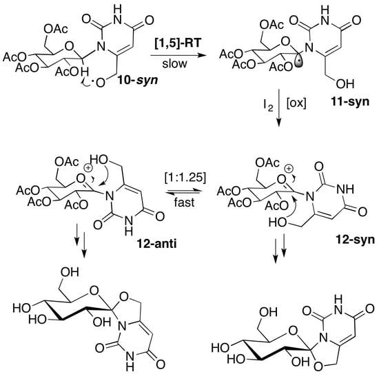
Regarding the mechanism of the key step, it is expected that, under the Suárez conditions, an alkoxy radical intermediate is produced that may exist in two possible conformations (10-syn, 10-anti, Scheme 2). These two conformers are similar to those observed above for alcohol 8 and are expected to be formed in a similar 1:2 ratio (10-syn:10-anti). The rigidity of the pyranosyl chair conformation may not allow a fast interconversion between the conformers for steric reasons, and the product distribution may be affected directly by these two conformer populations and their corresponding reactivity. Specifically, 10-anti cannot undergo a [1,5]-hydrogen shift and an alternative [1,6]-hydrogen atom transfer from the 2-position of the sugar is known to be disfavored in the presence of acetyl protection [23]. The only available pathway for conformer 10-anti is a radical disproportionation reaction leading to 7 and 8, of which the latter re-enters the reaction cycle, while the former accumulates (Scheme 2).
On the other hand, conformer 10-syn possesses a suitable conformation for a [1,5]-radical translocation leading to 11-syn intermediate. After oxidation of 11-syn, in the presence of I2, the 12-syn oxonium ion may exist in equilibrium with conformer 12-anti, through rotation of the C1′-N1 bond, and also with a possible Vorbrüggen-type intermediate, formed through the anchimeric assistance of the 2′-acetyl group. The Vorbrüggen intermediate is the main species that determines the stereochemistry of the final product in N-glycosylation reactions [28], and if the same was true in our system, exclusive formation of the S-anomer 9b would be expected. Nevertheless, in our system, a 1:1.25 S:R mixture of anomers 9b:9a was obtained. This result allows us to draw two major conclusions regarding the mechanism. First, rotation around the C1′-N1 bond in the oxonium ion 12-syn to produce 12-anti has to be faster than cyclization in order to allow the formation of the second, prior to cyclization, and there must be no major thermodynamic difference between the two conformers. Second, the formation of the Vorbrüggen intermediate must not be favored in this system, for steric reasons, and even if it is formed, through conformer 12-anti, the process rate is comparable to that of the cyclization of 12-syn conformer to the observed S-anomer 9a (Scheme 2).
Kinetics determined that the S anomeric spirocycle 4b exhibited 1.25 times higher inhibition than the R anomer 4a. The difference could be associated with the locked syn conformation of the pyrimidine ring with regard to the β-d-glucose moiety and possible unfavorable interactions of 4a within the catalytic site between the backbone CO of His377 with the uracil 2-C=O, as has been observed previously with protein crystallography (unpublished results). Although we attempted to obtain X-ray crystallographic data by soaking crystals of RMGPb with either 4a or 4b, the rather low affinity of both spirocycles did not provide sufficient data for establishing their binding in the catalytic site and studying their interactions. Both spironucleosides are stronger binders than the natural inhibitors of GP, β- and α-d-glucose [29]. For example, 4b is about 7 and 1.5 times stronger than β- and α-d-glucose, respectively. The new compounds, nevertheless, exhibit a rather low inhibition profile compared with the known spirohydantoin derivative of glucopyranose [5] and other known strong catalytic site inhibitors of RMGPb [7,30]. We established previously that anti is the desirable conformation of the pyrimidine ring at the anomeric position of β-d-glucose leading to strong inhibition [7], and the current results confirm this finding. Anomeric spironucleosides are rigid structures and, given that they possess the correct conformation, are expected to bind strongly to the catalytic site of GP. Our current studies are therefore directed towards anomeric spironucleosides with locked anti conformations, and these results will be reported in due course.

4. Materials and Methods

All reagents and solvents were purchased from commercial sources (Sigma–Aldrich, Merck, NJ, USA; Alfa-Aesar, Fisher Scientific, MA, USA) and used without further purification, unless otherwise stated. All reactions were carried out under an argon atmosphere on a magnetic stirrer (IKA®-Werke GmbH & Co. KG, Staufen, Germany) and monitored by thin-layer chromatography. Compounds were purified by flash chromatography on silica gel 40–60 μm, 60 Å. NMR measurements were performed with a Varian Mercury 200 Nuclear Magnetic Resonance Spectrometer (Varian Inc., Agilent Technologies, Palo Alto, CA, USA) at 200 MHz for 1H and at 50 MHz for 13C, respectively. The deuterated solvents used for NMR spectroscopy were CDCl3 and D2O. Chemical shifts are given in ppm and were referenced on residual solvent peaks for CDCl3 (δ 7.26 ppm for 1H-NMR and 77.16 ppm for 13C-NMR), whereas for D2O an external reference of 3-(trimethylsilyl)-1-propanesulfonic acid sodium salt was used. Coupling constants were measured in Hz. Hydrogen atom assignments, when given, are based on COSY spectra. Melting points were obtained by using a Gallenkamp Sanyo apparatus (Fisher Scientific, MA, USA) and are uncorrected. Mass spectrometry experiments were carried out in a Thermo Finnigan Surveyor MSQ plus Mass Spectrometer (ThermoFisher Scientific, MA, USA), using the Electron Spray Ionization technique (ESI-MS). High-resolution mass spectrometry experiments were carried out in a Q-TOF Bruker MaXis Impact HR-Mass Spectrometer (Bruker, MA, USA). 1,2,3,4,6-Penta-O-acetyl-β-d-glucopyranose was synthesized using standard synthetic protocols [31]. AMP, Glc-1-P (dipotassium salt), and oyster glycogen were obtained from Sigma-Aldrich (Merck, NJ, USA) and used without further purification. Oyster glycogen was freed of AMP according to Helmreich and Cori [32]. 1H and 13C-NMR spectra of new compounds are available in the Supplementary Materials.
2,4-Di-(trimethylsilyloxy)-(6-methylpyrimidine) (5). A suspension of 6-methyluracil (1 g, 7.93 mmol) and well-grinded ammonium sulphate (80 mg, 0.60 mmol 0.076 eq) in HMDS (8.4 mL, 39.7 mmol, 5 eq) was heated to 120 °C under anhydrous conditions until full dissolution occurred. Upon completion, the excess of HMDS was removed through distillation, toluene was added twice (5 mL) followed by distillation to remove all traces of excess HMDS to yield 2.1 g (7.8 mmol, 97%) of the title compound which was characterized without further purification. 1H-NMR (200 MHz, CDCl3): δ = 0.34 (s, 18H), 1.95 (3H, s), 5.81 ppm (1H, s). 13C-NMR (50 MHz, CDCl3): δ = 0.00 (3C), 0.03 (3C), 23.4, 102.6, 162.6, 169.8, 170.0 ppm.
N-Glycosylation of 6-methyluridine. To a solution of 5 (1.5 g, 5.6 mmol, 1.5 eq) in dry 1,2-dichloroethane (7 mL) at r.t., a solution of TMSOTf (1.61 mL, 8.33 mmol, 2.25 eq) and 2,3,4,6-tetra-O-acetyl-β-d-glucopyranose[31] (1.44 g, 3.7 mmol) in dry DCE (3.5 mL) was added. The reaction mixture was heated at reflux until full consumption of the sugar (1 h). The mixture was then cooled, diluted with DCM, and washed successively twice with saturated aq. NaHCO3 solution, water, and brine. The organic layer was dried over anhydrous Na2SO4, filtered, the solvents evaporated, and the crude product was purified by column chromatography (30–70% Et2O in EtOAc) to give, in order of elution, 6c as a white solid (86 mg, 0.11 mmol, 3%), 6a as a white solid (1.13 g, 2.48 mmol, 67%) and 6b as a white solid (0.51 g, 1.11 mmol, 30%).
1-(Tetra-O-acetyl-β-d-glucopyranosyl)-6-methyluracil (6a): Rf = 0.40 (70:30 Et2O:EtOAc). 1H-NMR (200 MHz, CDCl3): δ = 2.00 (s, 3H), 2.02 (s, 3H), 2.06 (s, 3H), 2.08 (s, 3H), 2.55 (s, 3H), 3.90 (bd, J = 9.5 Hz, 1H), 4.16 (dd, 1H, J = 12.2, 1.7 Hz), 4.27 (dd, 1H, J = 12.6, 4.2 Hz), 5.14 (t, 1H, J = 9.5 Hz), 5.35 (t, 1H, J = 9.5 Hz), 5.46 (t, 1H, J = 9.4 Hz), 5.57 (bs, 2H), 6.27 (d, 1H, J = 9.1 Hz), 8.62 ppm (bs, 1H). 13C-NMR (50 MHz, CDCl3): δ = 20.60, 20.78, 20.80 (2C), 20.97, 61.8, 67.9, 69.5, 72.8, 75.1, 80.5, 104.1, 139.3, 150.5, 162.7, 169.7, 169.8, 170.0, 170.7 ppm. HRMS (ESI): calcd. for C19H25N2O11+ [M + H]+ 457.1458 found 457.1465.
3-(Tetra-O-acetyl-β-d-glucopyranosyl)-6-methyluracil (6b): Rf = 0.35 (70:30 Et2O:EtOAc). 1H-NMR (200 MHz, CDCl3): δ = 1.95 (s, 3H), 2.01 (s, 3H), 2.04 (s, 3H), 2.06 (s, 3H), 2.17 (s, 3H), 3.84 (ddd, J = 10.1, 5.3, 2.5 Hz, 1H), 4.30–4.10 (m, 2H), 5.15 (t, J = 9.7 Hz, 1H), 5.30 (dd, J = 9.4, 8.6 Hz, 1H), 5.52 (s, 1H), 6.08 (d, J = 9.4 Hz, 1H), 6.18 (dd, J = 9.4, 8.4 Hz, 1H), 9.70 ppm (s, 1H). 13C-NMR (50 MHz, CDCl3): δ = 18.6, 20.4, 20.5 (2C), 20.6, 62.0, 67.9, 68.0, 73.7, 74.5, 78.3, 98.9, 151.5, 152.1, 162.5, 169.4, 169.6, 169.9, 170.5 ppm. ESI-MS: 457.2 [M + H]+.
1,3-Bis-(tetra-O-acetyl-β-d-glucopyranosyl)-6-methyluracil (6c): Rf = 0.50 (70:30 Et2O:EtOAc). 1H-NMR (200 MHz, CDCl3): δ = 2.01 (s, 6H), 2.02 (s, 6H), 2.04 (s, 6H), 2.06 (s, 6H), 2.57 (s, 1H), 3.96–3.74 (m, 2H), 4.37–4.10 (m, 4H), 5.52–5.02 (m, 5H), 5.55 (s, 1H), 6.02 (dd, J = 9.3, 9.3 Hz, 1H), 6.10 (d, J = 9.3 Hz, 1H), 6.34 ppm (d, J = 9.9 Hz, 1H). 13C-NMR (50 MHz CDCl3): δ = 20.0, 20.3, 20.6 (4C), 20.7 (3C), 61.4, 61.9, 67.5, 67.8, 68.1, 69.2, 73.3, 73.5, 74.7, 75.0, 79.2, 81.3, 103.4, 150.5, 152.9, 160.8, 169.40, 169.49, 169.62, 169.96, 170.20, 170.40, 170.60, 170.78 ppm. ESI-MS: 786.3 [M + H]+.
1-(Tetra-O-acetyl-β-d-glucopyranosyl)-6-formyluracil (7). To a solution of 6a (2.1 g, 4.63 mmol) in dry dioxane (40 mL), selenium oxide was added (1.5 g, 13.9 mmol, 3 eq) and acetic acid (1.32 mL, 23.1 mmol). The reaction mixture was heated at reflux until full consumption of the starting material (5 h). The mixture was then cooled, diluted with ethyl acetate, and washed successively with saturated aq. NaHCO3 solution, water, and brine. The organic layer was dried over anhydrous Na2SO4, filtered, the solvents evaporated, and the crude product was purified by column chromatography (97:3, EtOAC:Et2O) to yield the title compound as a white solid (1.5 g, 3.1 mmol, 67%). Rf = 0.50 (100% EtOAc). 1H-NMR (200 MHz, CDCl3): δ = 2.01 (s, 3H), 2.04 (s, 3H), 2.06 (s, 3H), 2.10 (s, 3H), 3.92 (ddd, J = 9.9, 3.3, 3.0 Hz, 1H), 4.19 (m, 2H), 5.24 (t, J = 9.7 Hz, 1H), 5.41 (t, J = 9.4 Hz, 1H), 5.61 (t, J = 9.3 Hz, 1H), 6.11 (d, J = 9.4 Hz, 1H), 6.28 (d, J = 2.3 Hz, 1H), 8.53 (s, 1H), 9.90 ppm (s, 1H). 13C-NMR (50 MHz, CDCl3): δ = 20.2, 20.4 (2C), 20.5, 61.0, 67.2, 70.3, 72.2, 74.9, 81.3, 110.0, 147.0, 150.2, 161.6, 169.4, 169.7, 170.0, 170.4, 183.4 ppm. HRMS (ESI): calcd. for C19H23N2O12+ [M + H]+ 471.1251 found 471.1255.
1-(Tetra-O-acetyl-β-d-glucopyranosyl)-6-hydroxymethyluracil (8). To a solution of 7 (0.2 g, 0.425 mmol) in 2-propanol (2.5 mL) and chloroform (0.6 mL), dry silica gel was added (43 mg) and the suspension was cooled to −30 °C. Then, ΝaBH4 (0.161 g, 4.25 mmol, 10 eq) was added and the reaction mixture was stirred until full consumption of the starting material (1 h). Then, the mixture was diluted with DCM, filtered through Celite®, and the filtrate washed successively with saturated aq. NaHCO3 solution, water, and saturated sodium chloride solution. The organic layer was dried over anhydrous Na2SO4, filtered, the solvents were evaporated, and the crude product was purified by column chromatography (EtOAc) to give the title compound as a white solid (146 mg, 0,38 mmol, 90%). Rf = 0.40 (70:30, Et2O:EtOAc). 1H-NMR (200 ΜHz, DMSO-d6): (mixture of tautomers) δ = 1.92 (s, 3H), 1.96 (s, 3H), 2.01 (s, 6H), 4.30 (m, 2H), 4.92 (t, J = 9.6 Hz, 1H), 5.26–5.62 (m, 2H), 5.69 (s, 1H), 5.84 (s, 2H), 6.08 (t, J = 9.0 Hz, 1H), 6.42 (d, J = 9.4 Hz, 1H), 11.32 ppm (s, 1H), 11.48 ppm (s, 1H). 13C-NMR (50 MHz, CDCl3): δ = 20.4, 20.5 (2C), 20.7, 60.1, 61.2, 67.5, 69.8, 72.6, 75.2, 81.5, 102.9, 151.3, 157.2, 162.8, 169.5, 169.8, 170.4, 170.7 ppm. HRMS (ESI): calcd. for C19H25N2O12+ [M + H]+ 473.1407 found 473.1410.
Spirocyclization of 8. A solution of 8 (150 mg, 0.32 mmol) in dichloromethane (16 mL) was degassed by argon gas bubbling for 10 min. Then, diacetoxyiodobenzene (155 mg, 0.48 mmol, 1.5 eq) and iodine (91 mg, 0.32 mmol, 1 eq) were added. Photolysis was carried out at r.t., with two 75 W Philips Standard 230 V visible light lamps, for 2.5 h. Afterwards, the reaction was quenched by 10% aq. Na2S2O3 solution and then extracted with dichloromethane. The organic layer was collected, dried over anhydrous Na2SO4, and then filtered, the solvent was evaporated, and the crude product was purified by column chromatography (EtOAc: Et2O gradient) to give, in order of elution, compound 7 (75 mg, 0.16 mmol, 50%), 9a as a white solid, (12 mg, 0.026 mmol, 8%), and 9b as a white solid (15 mg, 0.032 mmol, 10%).
(3R,3′R,4′S,5′R,6′R)-6′-(Acetoxymethyl)-5,7-dioxo-1,3′,4′,5,5′,6,6′,7-octahydrospiro[oxazolo[3,4-c]pyrimidine-3,2′-pyran]-3′,4′,5′-triyl triacetate (9a): Rf = 0.60 (70:30 Et2O:EtOAc). 1H-NMR (200 MHz, CDCl3): δ = 2.00 (s, 3H), 2.01 (s, 3H), 2.05 (s, 3H), 2.09 (s, 3H), 4.17–4.10 (m, 1H), 4.20 (s, 2H), 5.04 (m, 2H), 5.34 (t, J = 9.5 Hz, 1H), 5.48 (t, J = 10.0 Hz, 1H), 5.59 (s, 1H), 6.16 ppm (d, J = 9.8 Hz, 1H). 13C-NMR (50 MHz, CDCl3): δ = 20.52, 20.60, 20.62, 20.72, 61.2, 67.4, 68.2, 68.6, 71.3, 71.4, 93.8, 112.1, 145.7, 151.3, 163.1, 168.7, 169.2, 170.1, 170.7 ppm. HRMS (ESI): calcd. for C19H23N2O12+ [M + H]+ 471.1246 found 471.1239.
(3S,3′R,4′S,5′R,6′R)-6′-(Acetoxymethyl)-5,7-dioxo-1,3′,4′,5,5′,6,6′,7-octahydrospiro[oxazolo[3,4-c]pyrimidine-3,2′-pyran]-3′,4′,5′-triyl triacetate (9b): Rf = 0.40 (70:30 Et2O:EtOAc). 1H-NMR (200 MHz, CDCl3): δ = 1.99 (s, 3H), 2.03 (s, 3H), 2.05 (s, 3H), 2.09 (s, 3H), 4.10 (dd, J = 12.7, 2.5 Hz, 1H), 4.26 (dd, J = 12.7, 3.6 Hz, 1H), 4.65 (ddd, J = 9.6, 3.6, 2.5 Hz, 1H), 4.87 (dd, J = 14.5, 1.0 Hz, 1H), 5.06 (dd, J = 14.5, 1.6 Hz, 1H), 5.33 (dd, J = 10.1, 8.9 Hz, 1H), 5.63 (d, J = 9.3 Hz, 1H), 5.65 (s, 1H), 6.31 (t, J = 9.2 Hz, 1H), 8.61 ppm (s, 1H). 13C-NMR (50 MHz, CDCl3): δ = 20.52, 20.60, 20.62, 20.70, 61.4, 67.2, 67.3, 71.0, 72.4, 72.6, 94.2, 114.5, 148.3, 152.7, 163.3, 169.1, 169.6, 170.0, 170.6 ppm. HRMS (ESI): calcd. for C19H23N2O12+ [M + H]+ 471.1246 found 471.1241.
(3R,3′R,4′S,5′S,6′R)-3′,4′,5′-Trihydroxy-6′-(hydroxymethyl)-3′,4′,5′,6′-tetrahydrospiro[oxazolo[3,4-c]pyrimidine-3,2′-pyran]-5,7(1H,6H)-dione (4a). A solution of 9a (20 mg, 0.043 mmol) in methanolic ammonia (7 N, 0.35 mL) was stirred for 12 h at r.t., until full conversion to a single compound. Then, the solvent was evaporated and the compound was dried under high vacuum to yield the title compound as a white solid (13 mg, 0.043 mmol, 100%). 1H-NMR (200 MHz, D2O): δ = 3.64 (t, J = 9.1 Hz, 1H), 3.97–3.75 (m, 4H), 4.56 (d, J = 9.7 Hz, 1H), 5.20 (s, 2H), 5.88 ppm (s, 1H). 13C-NMR (50 MHz, D2O): δ = 63.0, 70.8, 71.6, 72.5, 76.5, 78.3, 96.7, 116.8, 142.5, 157.1, 169.6 ppm. HRMS (ESI): calcd. for C11H15N2O8+ [M + H]+ 303.0823 found 303.0830.
(3S,3′R,4′S,5′S,6′R)-3′,4′,5′-Trihydroxy-6′-(hydroxymethyl)-3′,4′,5′,6′-tetrahydrospiro[oxazolo[3,4-c]pyrimidine-3,2′-pyran]-5,7(1H,6H)-dione (4b). A solution of 9b (29 mg, 0.062 mmol) in methanolic ammonia (7 N, 0.51 mL) was stirred for 12 h at r.t., until full conversion to a single compound. Then, the solvent was evaporated and the compound was dried under high vacuum to yield the title compound as a white solid (19 mg, 0.062 mmol, 100%). 1H-NMR (200 MHz, D2O): δ = 3.58 (t, J = 9.6 Hz, 1H), 3.70 (dd, J = 12.5, 5.5 Hz, 2H), 3.85 (dd, J = 12.4, 2.1 Hz, 1H), 3.95 (d, J = 9.5 Hz, 2H), 4.26 (ddd, J = 10.1, 5.3, 2.2 Hz, 1H), 4.62 (t, J = 9.2 Hz, 2H), 5.08 (d, J = 14.9 Hz, 1H), 5.18 (d, J = 15.0 Hz, 1H), 5.81 ppm (s, 1H). 13C-NMR (50 MHz, D2O): δ = 169.5, 158.2, 149.4, 119.6, 96.4, 79.5, 77.3, 76.2, 71.6, 70.2, 63.4 ppm. HRMS (ESI): calcd. for C11H15N2O8+ [M + H]+ 303.0823 found 303.0828.

5. Conclusions

In conclusion, we successfully applied the Suárez radical methodology to synthesize the first example of a 1,6-dioxa-4-azaspiro[4.5]decane system starting from 6-hydroxymethyl-β-d- glucopyranosyluracil. We showed that, in the rigid pyranosyl conformation, the required [1,5]-radical translocation is a minor process. The stereochemistry of the spirocycles obtained was unequivocally determined by the chemical shifts of key sugar protons in the 1H-NMR spectra. Finally, the two spirocycles were found to be modest inhibitors of RMGPb, corroborating the finding that anti should be the desired conformation of the pyrimidine ring of future anomeric spironucleosides, which may lead to strong inhibition of GP.

Supplementary Materials

The following are available online. 1H and 13C-NMR spectra of new compounds (Figures S1–10). Tables of kinetic measurements (Tables S1 and S2).

Author Contributions

Conceptualization, T.G.; methodology, T.G. and E.D.C.; investigation, A.S. and M.M.; writing—original draft preparation, T.G. and M.M.; writing—review and editing, T.G.; supervision, T.G. and E.D.C.; project administration, T.G.; funding acquisition, T.G. and E.D.C.

Funding

This work was funded by Heracleitus II (M.M.) and the Special Account of N.K.U.A. (T.G.).

Conflicts of Interest

The authors declare no conflict of interest.

References

  1. Rines, A.K.; Sharabi, K.; Tavares, C.D.J.; Puigserver, P. Targeting hepatic glucose metabolism in the treatment of type 2 diabetes. Nat. Rev. Drug Discov. 2016, 15, 786–804. [Google Scholar] [CrossRef] [PubMed] [Green Version]
  2. Gimisis, T. Synthesis of N-Glucopyranosidic Derivatives as Potential Inhibitors that Bind at the Catalytic Site of Glycogen Phosphorylase. Mini-Rev. Med. Chem. 2010, 10, 1127–1138. [Google Scholar] [CrossRef] [PubMed]
  3. Donnier-Maréchal, M.; Vidal, S. Glycogen phosphorylase inhibitors: A patent review (2013–2015). Expert Opin. Ther. Pat. 2016, 26, 199–212. [Google Scholar] [CrossRef] [PubMed]
  4. Chrysina, E.D.; Chajistamatiou, A.; Chegkazi, M. From structure-based to knowledge-based drug design through X-ray protein crystallography: Sketching glycogen phosphorylase binding sites. Curr. Med. Chem. 2011, 18, 2620–2629. [Google Scholar] [CrossRef] [PubMed]
  5. Bichard, C.J.F.; Mitchell, E.P.; Wormald, M.R.; Watson, K.A.; Johnson, L.N.; Zographos, S.E.; Koutra, D.D.; Oikonomakos, N.G.; Fleet, G.W.J. Potent inhibition of glycogen phosphorylase by a spirohydantoin of glucopyranose: First pyranose analogues of hydantocidin. Tetrahedron Lett. 1995, 36, 2145–2148. [Google Scholar] [CrossRef]
  6. Goyard, D.; Kónya, B.; Chajistamatiou, A.S.; Chrysina, E.D.; Leroy, J.; Balzarin, S.; Tournier, M.; Tousch, D.; Petit, P.; Duret, C.; et al. Glucose-derived spiro-isoxazolines are anti-hyperglycemic agents against type 2 diabetes through glycogen phosphorylase inhibition. Eur. J. Med. Chem. 2016, 108, 444–454. [Google Scholar] [CrossRef] [PubMed] [Green Version]
  7. Mamais, M.; Degli Esposti, A.; Kouloumoundra, V.; Gustavsson, T.; Monti, F.; Venturini, A.; Chrysina, E.D.; Markovitsi, D.; Gimisis, T. A New Potent Inhibitor of Glycogen Phosphorylase Reveals the Basicity of the Catalytic Site. Chem. A Eur. J. 2017, 23, 8800–8805. [Google Scholar] [CrossRef] [PubMed]
  8. Soto, M.; Rodríguez-Solla, H.; Soengas, R. Recent Advances in the Chemistry and Biology of Spirocyclic Nucleosides. In Topics in Heterocyclic Chemistry; Springer: Berlin, Heidelberg, Germany, 2019; pp. 1–43. [Google Scholar]
  9. Chatgilialoglu, C.; Ferreri, C.; Gimisis, T.; Roberti, M.; Balzarini, J.; De Clercq, E. Synthesis and Biological Evaluation of Novel 1’-Branched and Spiro-Nucleoside Analogues. Nucleosides Nucleotides Nucleic Acids 2004, 23, 1565–1581. [Google Scholar] [CrossRef] [PubMed]
  10. Gómez-García, O.; Gómez, E.; Toscano, R.; Salgado-Zamora, H.; Álvarez-Toledano, C. One-Pot Synthesis of Spirotetrahydrooxino [3,4-c] pyridines and Spirotetrahydrofuro [3,2-b] pyridin-2-ones via Lactonization from Activated Pyridyldihydroozaxoles and Bis(trimethylsilyl)ketene Acetals. Synthesis 2016, 48, 1371–1380. [Google Scholar] [CrossRef]
  11. Martín, A.; Suárez, E. Carbohydrate Spiro-heterocycles via Radical Chemistry. In Topics in Heterocyclic Chemistry; Springer: Berlin/Heidelberg, Germany, 2019; pp. 1–54. [Google Scholar]
  12. Chatgilialoglu, C.; Gimisis, T.; Spada, G.P. C-1’ Radical-Based Approaches for the Synthesis of Anomeric Spironucleosides. Chem. A Eur. J. 1999, 5, 2866–2876. [Google Scholar] [CrossRef]
  13. Gimisis, T.; Chatgilialoglu, C.; Gimisis, T.; Castellari, C. A new class of anomeric spironucleosides. Chem. Commun. 1997, 2089–2090. [Google Scholar] [CrossRef]
  14. Liao, J.; Sun, J.; Yu, B. Effective synthesis of nucleosides with glycosyl trifluoroacetimidates as donors. Tetrahedron Lett. 2008, 49, 5036–5038. [Google Scholar] [CrossRef]
  15. Groziak, M.P.; Koohang, A. Facile addition of hydroxylic nucleophiles to the formyl group of uridine-6-carboxaldehydes. J. Org. Chem. 1992, 57, 940–944. [Google Scholar] [CrossRef]
  16. Tanaka, H.; Hayakawa, H.; Miyasaka, T. “Umpulong” of reactivity at the C-6 position of uridine: A simple and general method for 6-substituted uridines. Tetrahedron 1982, 38, 2635–2642. [Google Scholar] [CrossRef]
  17. Liu, H.-J.; Yip, J.; Shia, K.-S. Reductive cleavage of benzyl ethers with lithium naphthalenide. A convenient method for debenzylation. Tetrahedron Lett. 1997, 38, 2253–2256. [Google Scholar] [CrossRef]
  18. Wittenburg, E. Nucleoside und verwandte Verbindungen. VII. Alkylierung und Glykosidierung der Silyl-derivate 6-substituierter Uracile. Collect. Czechoslov. Chem. Commun. 1971, 36, 246–261. [Google Scholar] [CrossRef]
  19. Felczak, K.; Drabikowska, A.K.; Vilpo, J.A.; Kulikowski, T.; Shugar, D. 6-Substituted and 5,6-Disubstituted Derivatives of Uridine: Stereoselective Synthesis, Interaction with Uridine Phosphorylase, and In Vitro Antitumor Activity. J. Med. Chem. 1996, 39, 1720–1728. [Google Scholar] [CrossRef] [PubMed]
  20. Warpehoski, M.A.; Chabaud, B.; Sharpless, K.B. Selenium dioxide oxidation of endocyclic olefins. Evidence for a dissociation-recombination pathway. J. Org. Chem. 1982, 47, 2897–2900. [Google Scholar] [CrossRef]
  21. Młochowski, J.; Brząszcz, M.; Giurg, M.; Palus, J.; Wójtowicz, H. Selenium-Promoted Oxidation of Organic Compounds: Reactions and Mechanisms. Eur. J. Org. Chem. 2003, 2003, 4329–4339. [Google Scholar] [CrossRef]
  22. Florent, J.-C.; Dong, X.; Gaudel, G.; Mitaku, S.; Monneret, C.; Gesson, J.-P.; Jacquesy, J.-C.; Mondon, M.; Renoux, B.; Andrianomenjanahary, S.; et al. Prodrugs of Anthracyclines for Use in Antibody-Directed Enzyme Prodrug Therapy. J. Med. Chem. 1998, 41, 3572–3581. [Google Scholar] [CrossRef]
  23. Francisco, C.G.; Freire, R.; Herrera, A.J.; Pérez-Martín, I.; Suárez, E. Intramolecular 1,5-versus 1,6-hydrogen abstraction reaction promoted by alkoxyl radicals in pyranose and furanose models. Tetrahedron 2007, 63, 8910–8920. [Google Scholar] [CrossRef]
  24. Barton, D.H.R.; Beaton, J.M.; Geller, L.E.; Pechet, M.M. A New Photochemical Reaction 1. J. Am. Chem. Soc. 1961, 83, 4076–4083. [Google Scholar] [CrossRef]
  25. Kittaka, A.; Kato, H.; Tanaka, H.; Nonaka, Y.; Amano, M.; Nakamura, K.T.; Miyasaka, T. Face selective 6,1’-(1-oxo) ethano bridge formation of uracil nucleosides under hypoiodite reaction conditions. Tetrahedron 1999, 55, 5319–5344. [Google Scholar] [CrossRef]
  26. Oikonomakos, N.G.; Kontou, M.; Zographos, S.E.; Watson, K.A.; Johnson, L.N.; Bichard, C.J.F.; Fleet, G.W.J.; Acharya, K.R. N-acetyl-β-d-glucopyranosylamine: A potent T-state inhibitor of glycogen phosphorylase. A comparison with α-d-glucose. Protein Sci. 1995, 4, 2469–2477. [Google Scholar] [CrossRef] [PubMed]
  27. Saheki, S.; Takeda, A.; Shimazu, T. Assay of inorganic phosphate in the mild pH range, suitable for measurement of glycogen phosphorylase activity. Anal. Biochem. 1985, 148, 277–281. [Google Scholar] [CrossRef]
  28. Vorbrüggen, H.; Krolikiewicz, K.; Bennua, B. Nucleoside syntheses, XXII1) Nucleoside synthesis with trimethylsilyl triflate and perchlorate as catalysts. Chem. Ber. 1981, 114, 1234–1255. [Google Scholar] [CrossRef]
  29. Martin, J.L.; Veluraja, K.; Ross, K.; Johnson, L.N.; Fleet, G.W.J.; Ramsden, N.G.; Bruce, I.; Orchard, M.G.; Oikonomakos, N.G. Glucose analog inhibitors of glycogen phosphorylase: The design of potential drugs for diabetes. Biochemistry 1991, 30, 10101–10116. [Google Scholar] [CrossRef] [PubMed]
  30. Bokor, É.; Kun, S.; Docsa, T.; Gergely, P.; Somsák, L. 4(5)-Aryl-2-C-glucopyranosyl-imidazoles as New Nanomolar Glucose Analogue Inhibitors of Glycogen Phosphorylase. ACS Med. Chem. Lett. 2015, 6, 1215–1219. [Google Scholar] [CrossRef] [PubMed] [Green Version]
  31. Grugel, H.; Minuth, T.; Boysen, M. Novel Olefin-Phosphorus Hybrid and Diene Ligands Derived from Carbohydrates. Synthesis 2010, 19, 3248–3258. [Google Scholar]
  32. Helmreich, E.; Cori, C.F. The role of adenylic acid in the activation of phsphorylase. Proc. Natl. Acad. Sci. USA 1964, 51, 131–138. [Google Scholar] [CrossRef]
Sample Availability: Samples of compounds 4, 69 are available from the authors.
Figure 1. Natural and synthetic anomeric spironucleosides.
Molecules 24 02327 g001
Scheme 1. (i) TMSOTf, DCE, reflux, 1 h, 67%, (ii) SeO2, dioxane, AcOH, reflux, 5 h, 67%, (iii) NaBH4, silica gel, CHCl3, propanol, −30 °C, 1 h, 90%, (iv) DIB, I2, CH2Cl2, hv, r.t., 2.5 h, 18% (9a:9b = 1.25:1), and 50% (7), (v) NH3 (7N in MeOH), r.t., 16 h, 100%.
Molecules 24 02327 sch001
Figure 2. Comparison of 1H-NMR spectral shifts in compounds 9a,b.
Molecules 24 02327 g002
Scheme 2. Proposed mechanism for the formation of 9a,b.
Molecules 24 02327 sch002