Article

Investigation of the Phase Transition Mechanism in LiFePO4 Cathode Using In Situ Raman Spectroscopy and 2D Correlation Spectroscopy during Initial Cycle

1
Department of Chemistry, Institute for Molecular Science and Fusion Technology, Kangwon National University, Chuncheon 24341, Korea
2
Department of Nano Applied Engineering, Kangwon National University, Chuncheon 24341, Korea
3
Department of Materials Science and Engineering, University of Delaware, Newark, DE 19716, USA
*
Authors to whom correspondence should be addressed.
The authors (Y. Park and S.M. Kim) contributed equally to this work.
Molecules 2019, 24(2), 291; https://doi.org/10.3390/molecules24020291
Submission received: 10 December 2018 / Revised: 27 December 2018 / Accepted: 8 January 2019 / Published: 14 January 2019
(This article belongs to the Special Issue Raman Spectroscopy: A Spectroscopic 'Swiss-Army Knife')

Abstract

:
The phase transition of the LiFePO4 and FePO4 in Li-ion cell during charging-discharging processes in the first and second cycles is elucidated by Raman spectroscopy in real time. In situ Raman spectroscopy showed the sudden phase transition between LiFePO4 and FePO4. Principal component analysis (PCA) results also indicated that the structural changes and electrochemical performance (charge-discharge curve) are correlated with each other. Phase transition between LiFePO4 and FePO4 principally appeared in the second charging process compared with that in the first charging process. 2D correlation spectra provided the phase transition mechanism of LiFePO4 cathode which occurred during the charging-discharging processes in the first and second cycles. PCA and 2D correlation spectroscopy are very helpful methods to understand in situ Raman spectra for the Li-ion battery.

Graphical Abstract

1. Introduction

Battery performance, duration, and stability can be determined during the initial cycle. Therefore, it is important to understand structural changes in cathode and anode materials, formation of solid electrolyte interphase (SEI) film, electrical performance, etc. in real time. In the past few years, many researchers have tried to monitor in real time reactions inside batteries during electrochemical performance by using XRD [1,2,3,4], XAS [5,6,7], Raman [8,9,10,11,12,13,14,15,16,17], NMR [18], and so forth [19,20]. However, the studies for electrochemical reactions during the initial cycle is still limited.
LiFePO4 is one of the cathode materials having lower cost, environmental compatibility, high theoretical specific capacity of 170 mAhg−1 and especially a superior safety performance [21,22]. Therefore, it is spotlighted for large size lithium-ion (Li-ion) batteries, such as the energy sources in hybrid-electric vehicles (HEVs) or electric vehicles (EVs), and energy grid storage. Nevertheless, application of LiFePO4 cathode material is limited owing to low electronic conductivity and poor Li-ion diffusion [23]. To overcome these problems, LiFePO4 cathode materials have been alternated using carbon coating [13,22] or extending the surface area [23].
LiFePO4 cathode material has flat charging and discharging profiles at ~3.4 V vs. Li/Li+ resulting from LiFePO4 phase transition [24]. Many researchers studied the phase transition of LiFePO4 structure during charging-discharging processes. T. Yamanaka et al. [25] and other groups [26,27,28] reported that LiFePO4 separates to LiFePO4 and FePO4 phases during the charging process, having a miscibility gap at the Li concentration between them. The so-called ‘core shrinking’ theoretical model of the phase transition has been described by A.S. Andersson et al. and V. Srinivasan et al. [29,30]. Recently, C. Delmas et al. reported another phase transition mechanism, which is named the ‘domino-cascade’ model [31]. They assume that phase transformations at particle-level are fast, and small particles appear either fully charged (FePO4 phase) or discharged (LiFePO4 phase) due to a very rapid two-phase front [4,31]. The phase transition mechanism of LiFePO4 has been also investigated through in situ Raman spectroscopy [4,13]. Many models of phase transformation in LiFePO4 were reported; however the understanding is still limited. The interpretation of the highly overlapped spectra obtained during the charging–discharging processes remains difficult because the electrochemical reactions in the batteries are very complicated [32].
Nowadays generalized two-dimensional correlation spectroscopy (2D-COS) is accepted as one of the tools applicable to the in-depth analysis of various spectral data. 2D-COS can sort out important information in systematic variations of almost any reasonable analytical signals observed with a variety of probes under various forms of applied external perturbations. The perturbation-induced spectral variation of systems can be analyzed by a form of cross correlation analysis in 2D-COS. This approach has been applied to better understand complex systems, such as Li-ion batteries, because 2D-COS provides better information that is not readily detected with conventional spectroscopic measurements. In our previous study, electrochemical reactions of electrode materials using 2D-COS have successfully elucidated [33,34,35,36,37,38,39,40,41] analysis of in situ Raman spectra of LiCoO2/Li cell [32].
Principal component analysis (PCA) is a very popular analytical technique for spectroscopy. It can decompose the original data into the products of scores and loading vectors and provides a precise mathematical estimation of changes along sample and variable vectors. From PCA results, we can capture the dynamics of systems in response to perturbations and to display relationships among variables or among variables and systems [42,43,44].
In this study, PCA and 2D-COS were applied to in situ Raman spectroscopy for deeper understanding of phase transition of LiFePO4 cathode. PCA results adequately explain the correlation of spectral changes and electrochemical performance. We also deduced the phase transition mechanism during the first and second charging-discharging processes using 2D correlation spectroscopy.

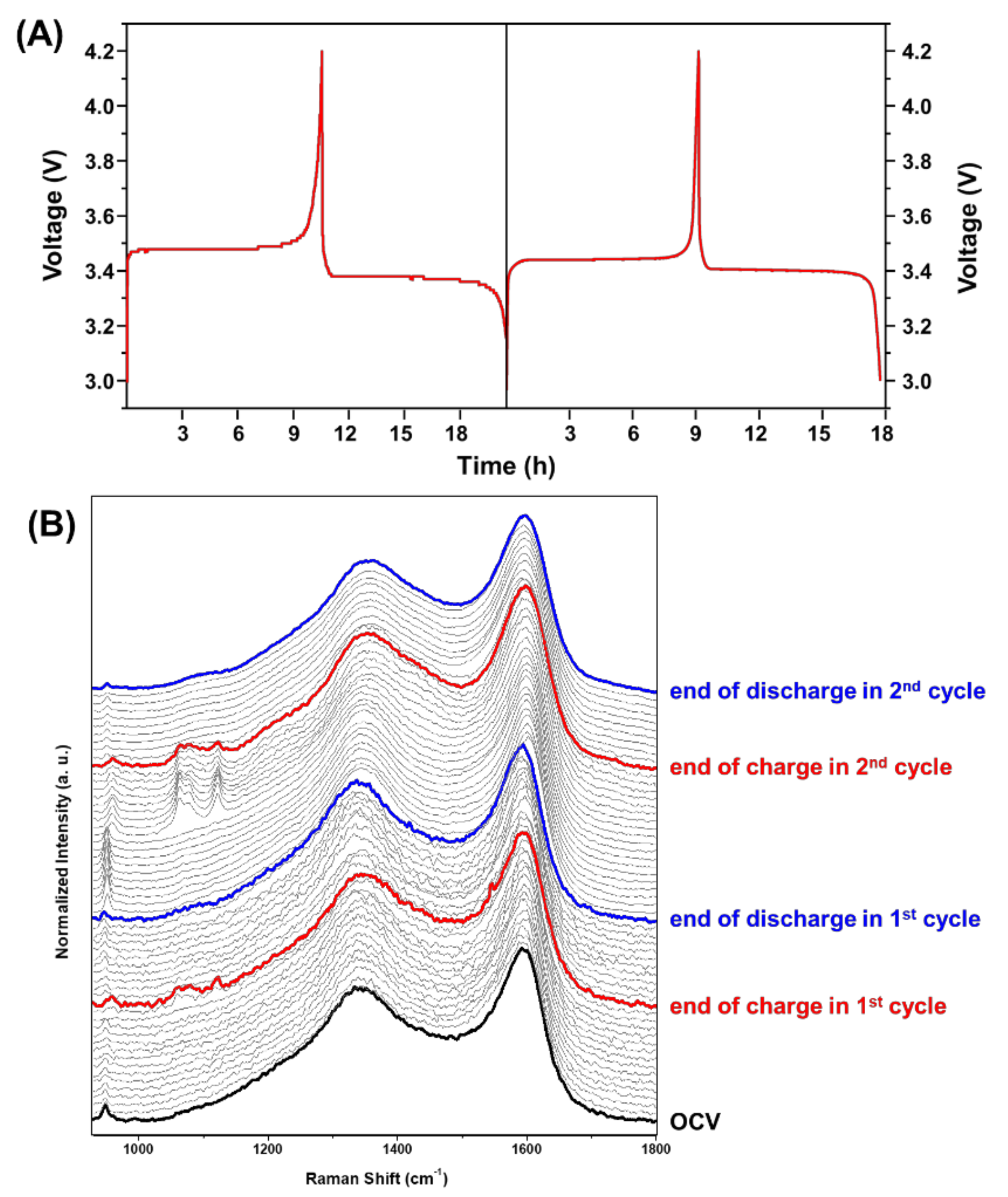
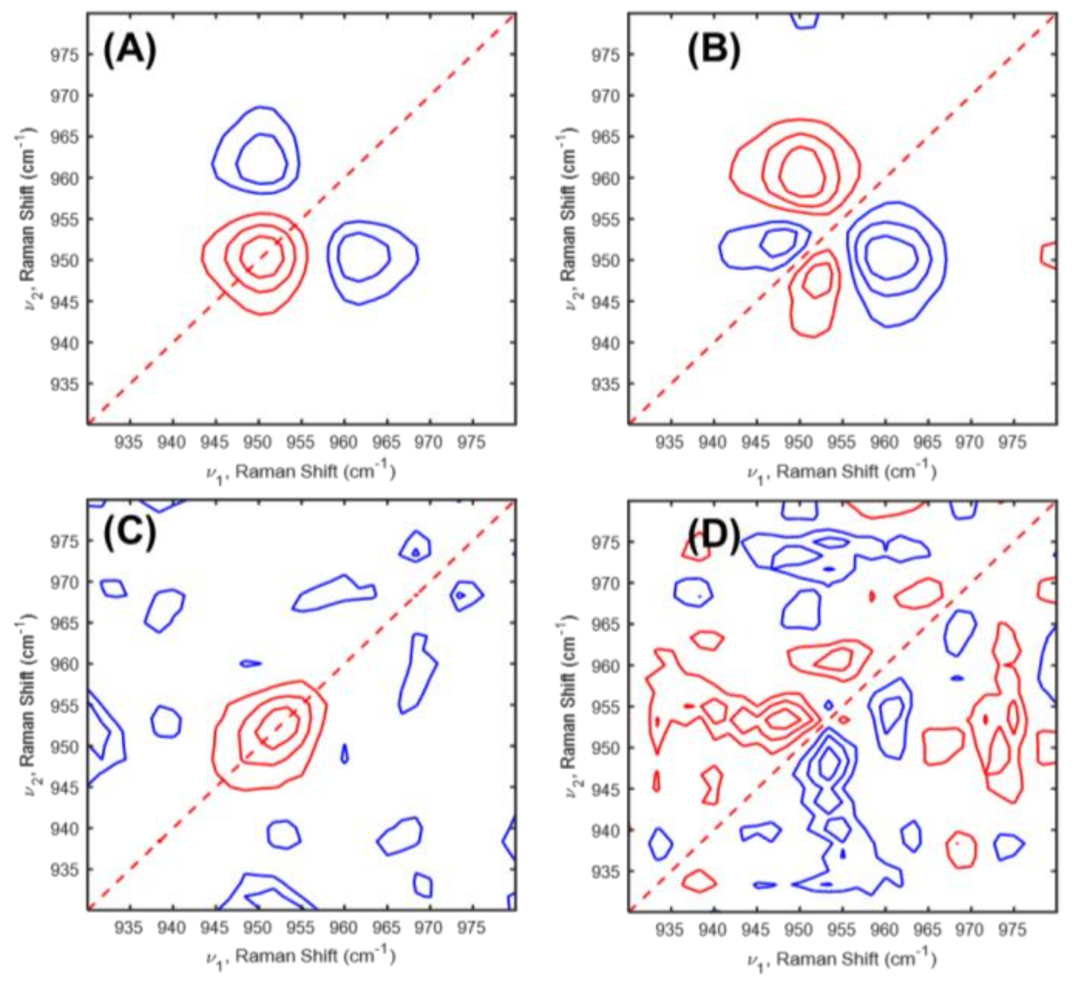
2. Results and Discussion

We measured in situ Raman spectra during charging-discharging processes in the first and second cycles. Figure 1A shows charging and discharging profiles of the first and second cycles of a Li-ion cell with LiFePO4 cathode. There are plates region at 3.48 and 3.38 V and 3.44 and 3.40 V during the first and second charging-discharging processes, respectively. This is the most noticeable feature of LiFePO4 material [24]. The sharp changes were observed in the other potential near the end of charging and near the end of discharging in the first and second cycles. From this result, we predict that lattices structure, Li ion diffusion rate, phase transition suddenly changed near the end of charging and discharging in the first and second cycles. In situ Raman spectra were collected during the first charging-discharging processes every 1 h, especially those that were recorded every 5 min and 2 min during near the end of charge in the first and second cycles, respectively, as shown in Figure 1B.
Unexpected spectral changes were detected near the end of charging in the first (10 h) and second cycles (9 h), respectively. The bands at 960 and 1020–1130 cm−1, assigned to symmetric and asymmetric stretching of PO43− in FePO4 phase [13], suddenly appeared at the end of charging then suddenly disappeared after charging. On the other hand, a band at 950 cm−1, corresponds to the symmetric stretching of PO43− in LiFePO4 phase, abruptly fades at the end of charging then abruptly came out after charging. As shown in in situ Raman spectra recorded near the end of charging of the first and second cycles, the variations of the bands at 950, 960 and 1020–1130 cm−1 also suddenly occurred, which were not gradual changes. This observation means that phase transition happened promptly near the end of charge. We also observed that the intensity of these bands decreased during the first and second charging-discharging processes. This result indicates that optical skin depth for the LiFePO4 cathode may change without any thermal damages because of laser exposure. If the electrode is harmed by laser, a new band at 1003 cm−1 assigned to a disordered phase of FePO4 should have appeared [45].
Because the spectral region of PO43− stretching between 930 and 980 cm−1 is only presented as the existence of both LiFePO4 and FePO4 [13], we only focus this region as shown in Figure 2. In Figure 2, phase transition between LiFePO4 and FePO4 phases were obviously monitored. During the first and second cycles, the band at 950 cm−1, observed in open circuit voltage (OCV), changed to 960 cm−1 at the end of charging and then repositioned at 950 cm−1 after charging. The band shift from 950 to 948 cm−1 was observed only in the end of discharging of the first cycle, not in that of the second cycle. This difference clearly indicates that the more stable structure of LiFePO4 may be formed after the first cycle. Therefore, we examined when the phase changes happened during the second charging process. Through Table S1, we speculate that the potential range that appeared during phase transition is between 3.6 and 3.7 V during the second charging process.
To understand the correlation between the structural changes and the performances of the Li-ion cell, we also carried out PCA for the in situ Raman spectra of LiFePO4/Li cell during charging-discharging processes in the first and second cycles. We applied PCA to in situ Raman spectra obtained from Figure 1. Figure S1A–C presented plots of the first four scores, the loading values, and the loading vectors in 930–980 cm−1 region, respectively, of the in situ Raman spectra for LiFePO4 cathode in Li-ion cell during the charging-discharging processes in the first and second cycles. Interestingly, the variation trends of score plots for the first principal component (PC 1) are in good agreement with those of CV curve of LiFePO4/Li cell during the charging-discharging processes in the first and second cycles, as shown in Figure S2. As shown in loading vectors of Figure S1C, the first four principal components (PCs) of the bands at 948 and 951 cm−1 have positive, positive, negative, and positive intensities, respectively, indicating that LiFePO4 phase mainly changed at 1 h between 8 h 40 min in the charging process of the second cycle. In contrast, the band at 960 cm−1 has negative, positive, positive, and positive intensities in the first four PCs, respectively, indicating that FePO4 phase principally varied at 8 h 50 min and 8 h 52 min in charging process of the second cycle. PCA results clearly identify the LiFePO4 and FePO4 phases. The phase transition between LiFePO4 and FePO4 occurred even during the first charging process, but they are not clearly detected in PCA results. This result shows that phase transition of LiFePO4 and FePO4 in the first charging process was not as strong as the second charging process.
To deeper understand the structural changes in LiFePO4 cathode in Li-ion cell during the charging-discharging processes, 2D-COS was applied to in situ Raman spectra of LiFePO4/Li cell during the charging-discharging processes in the first and second cycles. Figure 3 and Figure 4 displayed 2D correlation in situ Raman spectra during the charging-discharging processes in the first and second cycles.
We analyzed firstly in situ Raman spectra of LiFePO4/Li cell during the charging-discharging processes of the first cycle. As shown in Figure 3, 2D correlation spectra of the charging-discharging processes of the first and second cycles were different. During the first charging process, two auto peaks at 946 and 960 cm−1 were observed in the synchronous 2D correlation spectrum as shown in Figure 3A. Three positive cross peaks at the (950,946), (958,948), and (961,958) cm−1 were observed in the asynchronous 2D correlation spectrum. They have positive, negative, and positive cross peaks, respectively, in the corresponding synchronous 2D correlation spectrum. From the sign of the cross peaks in the synchronous and asynchronous 2D correlation spectra, we determine the sequential order of the band intensity changes with time as follows; 950 (due to LiFePO4 phase) → 946 (due to LiFePO4 phase) → 958 (due o FePO4 phase) → 961 (due to FePO4 phase) cm−1. This result means that LiFePO4 phase distorted, then FePO4 phase appeared, and finally FePO4 phase distorted during the first charging process. During the first discharging process, one auto peak at 945 cm−1 is detected in the synchronous 2D correlation spectrum as shown in Figure 3C. In the asynchronous spectrum (Figure 3D), a lot of cross peaks appeared associated with noise. This observation means that the intensity of a band corresponded distorted LiFePO4 phase only changed in the first discharging process. In other words, phase transition in LiFePO4 structure did not happen during the first discharging process.
2D correlation spectra during the charging-discharging processes in the second cycle are displayed as shown in Figure 4. In the second charging process, two cross peaks at (951,946) and (960,951) cm−1 appeared in the asynchronous spectrum (Figure 4B). Based on the sign of the cross peaks in 2D correlation spectra, we deduced the following sequence of the intensity variations: 960 (due to FePO4 phase) → 951 (due to LiFePO4 phase) → 946 (due to LiFePO4 phase). Variation of FePO4 phase occurred before changes of LiFePO4 phase and finally LiFePO4 phase distorted. Interestingly, FePO4 phase changes appeared in advance unlike the first charging process. This indicates that the FePO4 phase does not appear abruptly in near the end of charging during the second cycle. This observation means that variations of the FePO4 phase were formed during the first cycle taking place before the LiFePO4 phase change. During the second discharging process, two cross peaks at (953,948) and (960,953) cm−1 appeared in the asynchronous spectrum (Figure 4D). A band at 960 cm−1 is not detectable in 1D in situ Raman spectra during the second discharging process. These results indicate that the FePO4 phase existed in a small of amount but not disappeared suddenly. From the 2D correlation analysis, the sequential order of the band intensity changes can be determined as follows: 948 (due to LiFePO4 phase) → 953 (due to LiFePO4 phase) → 960 (due to FePO4 phase) cm−1. The event of the band intensity changes during the second discharging process represented exactly opposite trend with that during the second charging process. In other words, reversible phase transition was shown after the first cycle. From this result we conclude that LiFePO4 structure is getting stable after the first cycle. As shown in Figure S3, the power spectra, which were extracted along the diagonal line on the synchronous 2D correlation spectra of Figure 3A,C, and Figure 4A,B, indicate that the phase transition between LiFePO4 and FePO4 phases appeared more sensitively during the second charging process. This result is in a good agreement with the PCA result.

3. Materials and Methods

The electrochemical behavior of LiFePO4 was investigated using a modified coin cell (2032 type). The modified coin cell was fabricated using the method described by Wu et al. [13]. In this study, a 1 mm hole was drilled in the back of a 2032 type coin cell (Wellcos Corporation, Gunpo, Korea), and the cell was used as the cathode can. A slurry consisting of 90 wt % LiFePO4 (Nippon Chemical Co., Tokyo, Japan) powder, 5 wt % Super P (conductor), and 5 wt % polyvinylidene fluoride (PVdF, binder) dissolved in 1-methyl-2-pyrrolidinone (NMP) was prepared. Cathodes were made by coating the slurry (thickness was 200 μm) onto a mesh-type aluminum foil substrate (Dexmet Corporation, Wallingford, CT, USA). Using these electrodes, the test cells were fabricated with metallic Li anodes and two separators (Celgard 2400, Celgard Co., NC, USA and Amotech Co., Incheon, South Korea) in an Ar-filled glove box. The electrolyte was a 1.0 M solution of LiPF6 in EC: DEC = 1:1 by volume (PANAX ETEC Co., Busan, Korea). To evaluate the cell performance, the test cells were aged for 4 h at 40 °C in a vacuum oven. Then, the first and second charge-discharge cycles were galvanostatically performed at rates of 0.1 C, at a constant current of 3.0–4.2 V at room temperature using a CH660E electrochemical analyzer (CH Instruments Inc., Bee Cave, TX, USA).
The in situ Raman spectra were obtained at room temperature during the first and second charging-discharging processes using a Jobin Yvon/HORIBA HR evolution Raman spectrometer equipped with an integral BX 41 confocal microscope. Radiation from an air-cooled frequency-doubled Nd:YAG laser (532 nm) was used as the excitation source. Raman scattering was detected at a 180° geometry using a multichannel air-cooled (−60 °C) charge-coupled device (CCD) camera (1024 × 256 pixels). We performed Raman shift calibration with Si wafer, which has a characteristic Raman shift at 520 cm−1. The in situ Raman spectra were collected with 4 s exposure time and 10 accumulations using 50× (NA 0.8) objective during the charging-discharging processes in the first and second cycles. The power and spot size of the laser were about 12.5 mW and 8 μm, respectively. All spectra were baseline corrected, smoothed and normalized using the PLS_Toolbox 8.1 software (Eigenvector Research, Inc., Wenatchee, WA, USA) for MATMAB R2017b (The Mathworks Inc., Natick, MA, USA). Smoothing was performed by Saviski-Golay method with 3 points filter width. Area normalization carried out in the 1150–1800 cm−1 region corresponding to carbon of conductor (Super P) because we only focused on the spectral variation in 930–980 cm−1 in this study. We also performed PCA analysis with PLS_Toolbox 8.1 software (Eigenvector Research, Inc., Wenatchee, WA, USA) for MATMAB R2017b (The Mathworks Inc., Natick, MA, USA). Synchronous and asynchronous 2D correlation in situ Raman spectra were obtained using MATLAB. The red and blue lines represent positive and negative cross peaks, respectively.

4. Conclusions

In situ Raman spectroscopy is a useful method to monitor the phase transition of LiFePO4 cathode during the first and second charging-discharging processes. By applying the PCA and 2D-COS to in situ Raman spectra, we can get more information, which was not easily detected using only 1D spectra. As PC 1 score plots are in good agreement with the trend of charging and discharging profiles, spectral changes affect electrochemical performance. The phase transition mechanism of the LiFePO4 cathode in LiFePO4/Li ion cell can be examined using 2D correlation spectra obtained from in situ Raman spectra during the first and second charging-discharging processes.

Supplementary Materials

The following are available online. Figure S1. PAC results for in situ Raman spectra of LiFePO4 cathode during the charging-discharging processes in the first and second cycles. The plots of the first four (A) scores, (B) loading vectors, and (C) loading vectors of 930–980 cm−1 region, Table S1. Structural changes appeared voltage and time during the second charging-discharging processes., Figure S2. (A) Charging and discharging profiles during the first and second cycles of a LiFePO4/Li cell; a–f: the first cycle (a: 1 h, b: 7 h, c: 10 h 30 min (end of charge), d: 11 h, e: 18 h, f: 20 h (end of discharge)) and g–i: the second cycle (g: 1 h, h: 7 h, i: 9 h 8 min (end of charge), j: 10 h, k: 15 h, l: 18 h (end of discharge)). (B) Score plot of PC 1. Figure S3. Power spectra extracted along the diagonal line on the synchronous 2D correlation spectra as shown in Figure 3A,C and Figure 4A,C.

Author Contributions

Conceptualization and methodology, Y.M.J.; performed experiments, S.M.K.; Resources, S.M.L.; data curation, Y.P., S.M.K., and S.J.; 2D correlation analysis, I.N.; writing—original draft preparation, Y.P.; writing—review and editing, Y.M.J. and I.N.; supervision, Y.M.J. All authors discussed and approved the final version.

Funding

This research was supported by the National Research Foundation of Korea (NRF) grants funded by the Ministry of Science, NRF-2015R1D1A3A01015885.

Acknowledgments

The authors thank Dexmet Corporation of Wallingford, CT, USA, for providing the Microgrid expanded metal current conducting foils and their support for our experiments.

Conflicts of Interest

The funders had no role in the design of the study; in the collection, analyses, or interpretation of data; in the writing of the manuscript, or in the decision to publish the results.

References

  1. Wang, X.-J.; Jaye, C.; Nam, K.-W.; Zhang, B.; Chen, H.-Y.; Bai, J.; Li, H.; Huang, X.; Fischer, D.A.; Yang, X.-Q. Investigation of the structural changes in Li1−xFePO4 upon charging by synchrotron radiation techniques. J. Mater. Chem. 2011, 21, 11406–11411. [Google Scholar] [CrossRef]
  2. Dong, H.; Guo, H.; He, Y.; Gao, J.; Han, W.; Lu, X.; Yan, S.; Yang, K.; Li, H.; Chen, D. Structural stability and Li-ion transport property of LiFePO4 under high-pressure. Solid State Ion. 2017, 301, 133–137. [Google Scholar] [CrossRef]
  3. Zhang, L.; Guo, X.; Huang, J.; Qu, Y.; Niu, C.; Du, Z.; Li, D.; Chen, Y. Coin-Cell-Based In Situ Characterization Techniques for Li-Ion Batteries. Front. Energy Res. 2018, 6, 16. [Google Scholar] [CrossRef]
  4. Siddique, N.A.; Salehi, A.; Wei, Z.; Liu, D.; Sajjad, S.D.; Liu, F. Length-Scale-Dependent Phase Transformation of LiFePO4: An In situ and Operando Study Using Micro-Raman Spectroscopy and XRD. ChemPhysChem 2015, 16, 2383–2388. [Google Scholar] [CrossRef] [PubMed]
  5. Ruther, R.E.; Zhou, H.; Dhital, C.; Saravanan, K.; Kercher, A.K.; Chen, G.; Huq, A.; Delnick, F.M.; Nanda, J. Synthesis, Structure, and Electrochemical Performance of High Capacity Li2Cu0.5Ni0.5O2 Cathodes. Chem. Mater. 2015, 27, 6746–6754. [Google Scholar] [CrossRef]
  6. Li, B.; Shao, R.; Yan, H.; An, L.; Zhang, B.; Wei, H.; Ma, J.; Xia, D.; Han, X. Understanding the Stability for Li-Rich Layered Oxide Li2RuO3 Cathode. Adv. Funct. Mater. 2016, 26, 1330–1337. [Google Scholar] [CrossRef]
  7. Kim, S.; Zhang, Z.; Wang, S.; Yang, L.; Cairns, E.J.; Penner-Hahn, J.E.; Deb, A. Electrochemical and Structural Investigation of the Mechanism of Irreversibility in Li3V2(PO4)3 Cathodes. J. Phys. Chem. C 2016, 120, 7005–7012. [Google Scholar] [CrossRef]
  8. Dalgleish, T.; Williams, J.M.G.; Golden, A.-M.J.; Perkins, N.; Barrett, L.F.; Barnard, P.J.; Au Yeung, C.; Murphy, V.; Elward, R.; Tchanturia, K. In-situ Raman Microscopy of Idividual LiNi0.8Co0.15Al0.05O2 Particles in the Li-ion Battery Composite Cathode. J. Exp. Psychol. Gen. 2007, 136, 23–42. [Google Scholar] [CrossRef]
  9. Fukumitsu, H.; Omori, M.; Terada, K.; Suehiro, S. Development of In Situ Cross-Sectional Raman Imaging of LiCoO2 Cathode for Li-ion Battery. Electrochemistry 2015, 83, 993–996. [Google Scholar] [CrossRef]
  10. Hardwick, L.J.; Hahn, M.; Ruch, P.; Holzapfel, M.; Scheifele, W.; Buqa, H.; Krumeich, F.; Novák, P.; Kötz, R. An in situ Raman study of the intercalation of supercapacitor-type electrolyte into microcrystalline graphite. Electrochim. Acta 2006, 52, 675–680. [Google Scholar] [CrossRef]
  11. Hy, S.; Chen, Y.-H.; Liu, J.; Rick, J.; Hwang, B.-J. In situ surface enhanced Raman spectroscopic studies of solid electrolyte interphase formation in lithium ion battery electrodes. J. Power Sources 2014, 256, 324–328. [Google Scholar] [CrossRef]
  12. Zhu, W.; Liu, D.; Trottier, J.; Gagnon, C.; Howe, J.; Mauger, A.; Julien, C.M.; Zaghib, K. In-situ Raman spectroscopic investigation of LiMn1.45Ni0.45M0.1O4 (M=Cr, Co) 5 V cathode materials. J. Power Sources 2015, 298, 341–348. [Google Scholar] [CrossRef]
  13. Wu, J.; Dathar, G.K.P.; Sun, C.; Theivanayagam, M.G.; Applestone, D.; Dylla, A.G.; Manthiram, A.; Henkelman, G.; Goodenough, J.B.; Stevenson, K.J. In situ Raman spectroscopy of LiFePO4: size and morphology dependence during charge and self-discharge. Nanotechnology 2013, 24, 424009. [Google Scholar] [CrossRef] [PubMed]
  14. Huang, J.X.; Li, B.; Liu, B.; Liu, B.J.; Zhao, J.B.; Ren, B. Structural evolution of NM (Ni and Mn) lithium-rich layered material revealed by in-situ electrochemical Raman spectroscopic study. J. Power Sources 2016, 310, 85–90. [Google Scholar] [CrossRef]
  15. Stancovski, V.; Badilescu, S. In situ Raman spectroscopic-electrochemical studies of lithium-ion battery materials: A historical overview. J. Appl. Electrochem. 2014, 44, 23–43. [Google Scholar] [CrossRef]
  16. Ruch, P.W.; Hardwick, L.J.; Hahn, M.; Foelske, A.; Kötz, R.; Wokaun, A. Electrochemical doping of single-walled carbon nanotubes in double layer capacitors studied by in situ Raman spectroscopy. Carbon N. Y. 2009, 47, 38–52. [Google Scholar] [CrossRef]
  17. Gross, T.; Giebeler, L.; Hess, C. Novel in situ cell for Raman diagnostics of lithium-ion batteries. Rev. Sci. Instrum. 2013, 84, 073109/1–073109/7. [Google Scholar] [CrossRef] [PubMed]
  18. Tang, W.; Goh, B.M.; Hu, M.Y.; Wan, C.; Tian, B.; Deng, X.; Peng, C.; Lin, M.; Hu, J.Z.; Loh, K.P. In Situ Raman and Nuclear Magnetic Resonance Study of Trapped Lithium in the Solid Electrolyte Interface of Reduced Graphene Oxide. J. Phys. Chem. C 2016, 120, 2600–2608. [Google Scholar] [CrossRef]
  19. Harks, P.P.R.M.L.; Mulder, F.M.; Notten, P.H.L. In situ methods for Li-ion battery research: A review of recent developments. J. Power Sources 2015, 288, 92–105. [Google Scholar] [CrossRef] [Green Version]
  20. Seidl, L.; Martens, S.; Ma, J.; Stimming, U.; Schneider, O. In situ scanning tunneling microscopy studies of the SEI formation on graphite electrodes for Li+-ion batteries. Nanoscale 2016, 8, 14004–14014. [Google Scholar] [CrossRef]
  21. Yang, J.; Wang, J.; Li, X.; Wang, D.; Liu, J.; Liang, G.; Gauthier, M.; Li, Y.; Geng, D.; Li, R. Hierarchically porous LiFePO4/nitrogen-doped carbon nanotubes composite as a cathode for lithium ion batteries. J. Mater. Chem. 2012, 22, 7537–7543. [Google Scholar] [CrossRef]
  22. Nagpure, S.C.; Bhushan, B.; Babu, S.S. Raman and NMR studies of aged LiFePO4 cathode. Appl. Surf. Sci. 2012, 259, 49–54. [Google Scholar] [CrossRef]
  23. Hu, L.; Zhang, T.; Liang, J.; Zhu, Y.; Zhang, K.; Qian, Y. Trace Fe3+ mediated synthesis of LiFePO4 micro/nanostructures towards improved electrochemical performance for lithium-ion batteries. RSC Adv. 2016, 6, 456–463. [Google Scholar] [CrossRef]
  24. Zhu, Y.; Wang, C. Strain accommodation and potential hysteresis of LiFePO4 cathodes during lithium ion insertion/extraction. J. Power Sources 2011, 196, 1442–1448. [Google Scholar] [CrossRef]
  25. Yamanaka, T.; Abe, T.; Nishio, K.; Ogumi, Z. Diffusion of Li-deficient phases in large LiFePO4 single crystals during chemical delithiation. J. Mater. Chem. A 2018, 6, 11005–11011. [Google Scholar] [CrossRef]
  26. Yamada, A.; Koizumi, H.; Nishimura, S.; Sonoyama, N.; Kanno, R.; Yonemura, M.; Nakamura, T.; Kobayashi, Y. Room-temperature miscibility gap in LixFePO4. Nat. Mater. 2006, 5, 357–360. [Google Scholar] [CrossRef]
  27. Meethong, N.; Huang, H.-Y.S.; Carter, W.C.; Chiang, Y.-M. Size-Dependent Lithium Miscibility Gap in Nanoscale Li1-xFePO4. Electrochem. Solid-State Lett. 2007, 10, A134–A138. [Google Scholar] [CrossRef]
  28. Wagemaker, M.; Singh, D.P.; Borghols, W.J.H.; Lafont, U.; Haverkate, L.; Peterson, V.K.; Mulder, F.M. Dynamic Solubility Limits in Nanosized Olivine LiFePO4. J. Am. Chem. Soc. 2011, 133, 10222–10228. [Google Scholar] [CrossRef]
  29. Andersson, A.S.; Kalska, B.; Häggström, L.; Thomas, J.O. Lithium extraction/insertion in LiFePO4: An X-ray diffraction and Mössbauer spectroscopy study. Solid State Ion. 2000, 130, 41–52. [Google Scholar] [CrossRef]
  30. Srinivasan, V.; Newman, J. Discharge Model for the Lithium Iron-Phosphate Electrode. J. Electrochem. Soc. 2004, 151, A1517–A1529. [Google Scholar] [CrossRef]
  31. Delmas, C.; Maccario, M.; Croguennec, L.; Le Cras, F.; Weill, F. Lithium deintercalation in LiFePO4 nanoparticles via a domino-cascade model. Nat. Mater. 2008, 7, 665–671. [Google Scholar] [CrossRef] [PubMed]
  32. Park, Y.; Kim, Y.; Kim, S.M.; Jin, S.; Han, I.K.; Lee, S.M.; Jung, Y.M. Reaction at the Electrolyte–Electrode Interface in a Li-Ion Battery Studied by In Situ Raman Spectroscopy. Bull. Korean Chem. Soc. 2017, 38, 511–513. [Google Scholar] [CrossRef]
  33. Park, Y.; Shin, S.H.; Lee, S.M.; Kim, S.P.; Choi, H.C.; Jung, Y.M. 2D Raman correlation analysis of formation mechanism of passivating film on overcharged LiCoO2 electrode with additive system. J. Mol. Struct. 2014, 1069, 183–187. [Google Scholar] [CrossRef]
  34. Park, Y.; Kim, N.H.; Kim, J.Y.; Eom, I.-Y.; Jeong, Y.U.; Kim, M.S.; Lee, S.M.; Choi, H.C.; Jung, Y.M. Surface characterization of the high voltage LiCoO2/Li cell by X-ray photoelectron spectroscopy and 2D correlation analysis. Vib. Spectrosc. 2010, 53, 60–63. [Google Scholar] [CrossRef]
  35. Park, Y.; Kim, N.H.; Kim, J.M.; Kim, Y.C.; Jeong, Y.U.; Lee, S.M.; Choi, H.C.; Jung, Y.M. Surface Reaction of LiCoO2/Li System under High-Voltage Conditions by X-ray Spectroscopy and Two-Dimensional Correlation Spectroscopy (2D-COS). Appl. Spectrosc. 2011, 65, 320–325. [Google Scholar] [CrossRef] [PubMed]
  36. Park, Y.; Kim, N.H.; Choi, H.C.; Lee, S.M.; Hwang, H.; Jeong, Y.U.; Jung, Y.M. Two-dimensional hetero-spectral Raman/XAS correlation analysis of Li[Ni0.45Co0.18Mn0.25Al0.12]O2 cathode in the overcharged state. Vib. Spectrosc. 2012, 60, 226–230. [Google Scholar] [CrossRef]
  37. Park, Y.; Shin, S.H.; Hwang, H.; Lee, S.M.; Kim, S.P.; Choi, H.C.; Jung, Y.M. Investigation of solid electrolyte interface (SEI) film on LiCoO2 cathode in fluoroethylene carbonate (FEC)-containing electrolyte by 2D correlation X-ray photoelectron spectroscopy (XPS). J. Mol. Struct. 2014, 1069, 157–163. [Google Scholar] [CrossRef]
  38. Choi, H.C.; Bin Kim, S.; Jung, Y.M. Application of soft X-ray absorption spectroscopy and two-dimensional correlation spectroscopy to the electrochemical reaction in the Li1+xV3O8/Li cell. J. Mol. Struct. 2006, 799, 91–95. [Google Scholar] [CrossRef]
  39. Choi, H.C.; Jung, Y.M.; Kim, S. Bin Characterization of the Electrochemical Reactions in the Li1+xV3O8/Li Cell by Soft X-ray Absorption Spectroscopy and Two-Dimensional Correlation Analysis. Appl. Spectrosc. 2003, 57, 984–990. [Google Scholar] [CrossRef]
  40. Choi, H.C.; Jung, Y.M.; Noda, I.; Kim, S. Bin A Study of the Mechanism of the Electrochemical Reaction of Lithium with CoO by Two-Dimensional Soft X-ray Absorption Spectroscopy (2D XAS), 2D Raman, and 2D Heterospectral XAS−Raman Correlation Analysis. J. Phys. Chem. B 2003, 107, 5806–5811. [Google Scholar] [CrossRef]
  41. Park, Y.; Kim, N.H.; Cho, S.B.; Kim, J.M.; Kim, G.C.; Kim, M.S.; Lee, S.M.; Eom, I.-Y.; Choi, H.C.; Jung, Y.M. Characterization of the passivating layer on Li[Ni0.31Co0.32Mn0.28Al0.09]O2 cathode in the overcharge state. J. Mol. Struct. 2010, 974, 139–143. [Google Scholar] [CrossRef]
  42. Jolliffe, I.T. Principal Component Analysis, 1st ed.; Springer: New York, NY, USA, 1986. [Google Scholar]
  43. Martens, H.; Næs, T. Multivariate Calibration; John Wiley and Sons: New York, NY, USA, 1991. [Google Scholar]
  44. Vandeginste, B.G.M.; Massart, D.L. Handbook of Chemometrics and Qualimetrics: Part B; Elsevier Science B. V.: Amsterdam, The Netherlands, 1998. [Google Scholar]
  45. Burba, C.M.; Palmer, J.M.; Holinsworth, B.S. Laser-induced phase changes in olivine FePO4: A warning on characterizing LiFePO4-based cathodes with Raman spectroscopy. J. Raman Spectrosc. 2009, 40, 225–228. [Google Scholar] [CrossRef]
Sample Availability: Samples of the compounds are not available from the authors.
Figure 1. (A) Charging and discharging profiles during the first and second cycles of a LiFePO4/Li cell. (B) In situ Raman spectra of LiFePO4 cathode in the 930–1800 cm−1 region measured during the charging-discharging processes of the first and second cycles of LiFePO4/Li cell.
Molecules 24 00291 g001
Figure 2. In situ Raman spectra in the PO43− stretching region (930–980 cm−1) of OCV, a–f: the first cycle (a: 1 h, b: 7 h, c: 10 h 30 min (end of charge), d: 11 h, e: 18 h, f: 20 h (end of discharge)) and g–i: the second cycle (g: 1 h, h: 7 h, i: 9 h 8 min (end of charge), j: 10 h, k: 15 h, l: 18 h (end of discharge)).
Molecules 24 00291 g002
Figure 3. (A,C) Synchronous and (B,D) asynchronous 2D correlation spectra of in situ Raman spectra during the (A,B) charging and (C,D) discharging processes in the first cycle obtained Figure 1.
Molecules 24 00291 g003
Figure 4. (A,C) Synchronous and (B,D) asynchronous 2D correlation spectra of in situ Raman spectra during the (A,B) charging and (C,D) discharging processes in the second cycle obtained Figure 1.
Molecules 24 00291 g004

