Article

Phenolic Profile and Bioactive Properties of Carissa macrocarpa (Eckl.) A.DC.: An In Vitro Comparative Study between Leaves, Stems, and Flowers

by
Fedia Souilem
1,2,†,
Maria Inês Dias
1,†,
Lillian Barros
1,
Ricardo C. Calhelha
1,
Maria José Alves
1,
Fethia Harzallah-Skhiri
2 and
Isabel C.F.R. Ferreira
1,*
1
Centro de Investigação de Montanha (CIMO), Instituto Politécnico de Bragança, Campus de Santa Apolónia, 5300-253 Bragança, Portugal
2
Laboratoire de Recherche “Bioressources”: Biologie Intégrative & Valorisation (BIOLIVAL) LR14ES06, Institut Supérieur de Biotechnologie de Monastir, Avenue Tahar Hadded, BP 74,5000, Université de Monastir, Monastir 5000, Tunisia
*
Author to whom correspondence should be addressed.
These authors contributed equally to this work.
Molecules 2019, 24(9), 1696; https://doi.org/10.3390/molecules24091696
Submission received: 16 April 2019 / Revised: 25 April 2019 / Accepted: 28 April 2019 / Published: 30 April 2019
(This article belongs to the Collection Bioactive Compounds)

Abstract

:
The present work aimed to characterize leaves, stems, and flowers of Carissa macrocarpa (Eckl.) A.DC., by performing an analysis of the phenolic compounds by HPLC-DAD/ESI-MS, correlating them with bioactive properties, such as antioxidant, anti-inflammatory, cytotoxic, and antimicrobial activities. Thirty polyphenols were identified in the hydroethanolic extract, including phenolic acids, flavan-3-ols, and flavonol glycosides derivatives (which presented the highest number of identified compounds). However, flavan-3-ols showed the highest concentration in stems (mainly owing to the presence of dimers, trimmers, and tetramers of type B (epi)catechin). Leaves were distinguished by their high antioxidant and anti-inflammatory activities, as well as their bactericidal effect against E. coli, while stems presented a higher cytotoxic activity and bactericidal effect against Gram-positive bacteria. Moreover, a high correlation between the studied bioactivities and the presence of phenolic compounds was also verified. The obtained results bring added value to the studied plant species.

Graphical Abstract

1. Introduction

The genus Carissa belongs to the family of Apocynaceae, which normally has a high content of phenolic compounds, such as flavonoids, as well as lignans and sesquiterpenes. Accordingly, this plant genus presents several therapeutic applications such as antioxidant, analgesic, anti-inflammatory, hypolipidemic, wound healing, antimicrobial, antidiabetic, antiepileptic, anti-cancer, diuretic, hepatoprotective, and improvement of nephrotoxicity [1].
Carissa macrocarpa (Eckl.) A.DC. (syn: C. grandiflora (E.Mey.) A.DC.) is a native plant from South Africa, KwaZulu-Natal, commonly known as Natal plum [2,3]. It is an ornamental shrub, characterized by large, green, lush, and persistent leaves; star-shaped and white flowers; and edible oval fruits [3,4]. The ripe fruits are delicious and can be used for the preparation of jams, sauces, desserts, yogurt, jellies, and ice cream, while this plant is also used in traditional medicine for the treatment of diarrhea in livestock, cough, and venereal diseases [5,6]. C. macrocarpa leaves, stems, and roots present antioxidant and antimicrobial activities [3,4], and the leaves present cytotoxic activity [7], owing to the presence of various secondary metabolites, such as flavonoids, saponins, triterpenoids, anthraquinones, and tannins
The presence of different phenolic compounds, such as flavonoids, hydroxycinnamic and hydroxybenzoic acids, coumarins, xanthones, chalcones, stilbenes, lignins, and lignans have been described in numerous publications regarding the phenolic profile of the genus Carissa. These compounds are known for their important role in cancer treatment [8], hepatoprotective effects [9], and antifungal properties [10]. Some of these biological properties are directly related to the antioxidant activity of these compounds, in accordance with that described by Martins et al. [10].
To the author’s best knowledge, there are no previous studies describing the anti-inflammatory activity of C. macrocarpa leaves, stems, or flowers, and only few studies could be found regarding antioxidant, cytotoxic, and antibacterial properties of different extracts obtained from leaves and stems. Moreover, there is no report on C. macrocarpa flowers. Therefore, the aim of the present study was to established the individual phenolic profile of the hydroethanolic extracts obtained from leaves, stems, and flowers of C. macrocarpa, being further correlated with their antioxidant, antibacterial, anti-inflammatory, and cytotoxic properties.

2. Results and Discussion

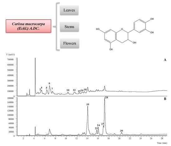
2.1. Phenolic Profile of the Aerial Parts of C. Macrocarpa

Table 1 presents the chromatographic characteristics (obtained by HPLC-DAD/ESI-MS) and tentative identification of the phenolic compounds present in the hydroethanolic extracts obtained from leaves, stems, and flowers of C. macrocarpa. The quantification of each identified compound is presented in Table 2. An exemplificative phenolic profile of the hydroethanolic extract prepared from leaves is presented in Figure 1.
Thirty phenolic compounds were tentatively identified in the hydroethanolic extracts prepared from C. macrocarpa leaves, stems, and flowers: nine phenolic acids (chlorogenic, coumaric, and syringic coumaric acid derivatives), thirteen flavonols (kaempherol and quercetin derivatives), and eight flavan-3-ols ((epi)-catechin derivatives). To the author’s best knowledge, all the phenolic compounds were tentatively identified for the first time in the hydroethanolic extracts of C. macrocarpa.
Phenolic acids were found in higher amounts in the hydroethanolic extract of flowers, and in very close amounts to flavanols in the same extract, while leaves showed the lowest amount of phenolic acids. Four chlorogenic acid derivatives (peaks 1, 5, 6, and 8), four p-coumaric acid derivatives (peaks 2, 4, 13, and 15), and one syringic acid derivative (peak 3) were tentatively identified. Peaks 1, 5, 6, and 8 ([M − H] at m/z 353) were tentatively identified as 3-O-caffeoylquinic acid (peak 1), 4-O-caffeoylquinic acid (cis and trans, peak 5 and 6, respectively), and 5-O-caffeoylquinic acid (peak 8). The assignments of the different caffeoylquinic acid isomers were made using the hierarchical key system previously reported by Clifford et al. (2003) and Clifford et al. (2005) [11,12]. Moreover, 5-O-caffeoylquinic acid (peak 8) was positively identified in comparison with the available commercial standard. Peaks 2, 4, 13, and 15 ([M − H] at m/z 337) were tentatively identified according to their MS2 fragmentation as cis and trans 3-p-coumaroylquinic acid, and cis and trans 5-p-coumaroylquinic acid, respectively, as previously reported by Clifford et al. (2003) and Clifford et al. (2005) [11,12]. The identification of the compounds 2/4, 5/6, and 13/15 as cis/trans isomers, was based on experimental results previously published by our research group [13], also following the information described by Clifford and coworkers [14,15] regarding these isomers. The hydroxycinnamoyl cis and trans derivatives were distinguished, after UV irradiation (366 nm, 24 h) of these acids in our laboratory [13]. Compound 3 with MS2 fragments at m/z 197 ([syringic acid-H]) resulting from the loss of a hexosyl moiety (−162 u) was tentatively assigned as syringic acid hexoside.
Flavonol glycoside derivatives of quercetin (MS2 fragment at m/z 301) and kaempferol (MS2 fragment at m/z 285) were the main molecules present in this family of phenolic compounds. Quercetin-3-O-rutinoside (rutin; peak 28) and kaempferol-3-O-rutinoside (peak 30) were identified upon comparison of their chromatographic characteristics with available commercial standards. These compounds have already been identified in the Apocyneaceae family [16].
Peaks 18, 21, 25, and 26 ([M − H] at m/z 739) and 20 ([M − H] at m/z 755) provided the same consequently fragmentation losses of a deoxyhexosyl unit (146 u) and deoxyhexosyl-hexosyl unit (308 u), indicating the location of each residue on different positions of the aglycones of kaempferol and quercetin, respectively, being tentatively identified as kaempferol-O-deoxyhexoside-O-deoxyhexosyl-hexoside isomers 1, 2 3, and 4, and quercetin-O-deoxyhexoside-O-deoxyhexosyl-hexoside, respectively. Peak 23 also showed a pseudomolecular ion [M − H] at m/z 739, presenting a unique MS2 fragment at m/z 285 (−454 u, which corresponded to 146 + 146 u + 162 u) indicating that the three sugar units were linked together in the same oxygen position of the aglycone, and in this particular case, kaempferol, being tentatively identified as kaempferol-O-di-deoxyhexosyl- hexoside. Peak 24 presented a pseudomolecular ion [M − H] at m/z 575, releasing an MS2 fragment at m/z 285, corresponding to the loss of acetyl (42 u), malonyl (86 u), and hexosyl (162 u) residues, being tentatively identified as acetylkaempherol-O-malonylhexoside. Peaks 22 and 27 ([M − H] at m/z 609; MS2 at m/z 301) and 29 ([M − H] at m/z 593; at m/z 301) showed a single MS2 fragment that indicated the loss of 308 u, corresponding to two sugar units linked together (deoxyhexosyl-hexoside moieties), being tentatively identified as quercetin-O-deoxyhexosyl-hexoside isomer 1 and 2, and kaempherol-O-deoxyhexosyl-hexoside, respectively. Peak 10 ([M − H] at m/z 755) fragmented at m/z 593 (−162 u, hexosyl unit) and 285 (−308 u, deoxyhexosyl-hexoside unit), being tentatively identified as kaempherol-O-hexoside-O-deoxyhexosyl-hexoside.
Flavan-3-ols were the main family of phenolic compounds present in the hydroethanolic extracts of stems of C. macrocarpa; not being detected in the flower samples. Peaks 7, 9, 11, 12, 14, 16, 17, and 19 were tentatively identified based on their chromatographic characteristics (pseudomolecular analysis and MS2 fragmentation pattern) coherent with B-type (epi)catechin derivatives. Thus, they were tentatively identified as B-type (epi)catechin dimers (peaks 7 and 9, [M − H] at m/z 577), trimers (peaks 11 and 16, [M − H] at m/z 865), and tetramers (peaks 12, 14, and 19, [M − H] at m/z 1153) [17]. Peak 17 was tentatively identified based on the information found in the literature as a (epi)catechin trimer with a type-A linkage, thus being assigned as an A-type (epi)catechin trimer [18].
The phenolic profile of the three plant parts was relatively similar; nevertheless, they all presented different major compounds. For the leaves, cis 5-p-coumaroylquinic acid (peak 13, 2.9 mg/g extract) was the major molecule, while trans 5-p-coumaroylquinic acid (peak 15, 7.3 mg/g extract) was the major compound found in stems, and cis 4-O-caffeolyquinic acid (peak 5, 10.35 μg/g extract) was the main compound found in flowers.

2.2. Bioactivities of the Hydroethanolic Extracts of Leaves, Stems, and Flowers

Results regarding in vitro antioxidant activity of the ethanol/water (80:20 v/v) extracts prepared from leaves, stems, and flowers of C. macrocarpa are described in Table 3.
Overall, all samples presented high antioxidant activity, but the statistical analysis did not show significant differences that could allow to particularize a more potent plant part. However, observing the four used methodologies, the leaves presented the lowest EC50 values for DPPH scavenging activity, reducing power, and β-carotene bleaching inhibition (EC50 = 26 ± 1, 36 ± 1, and 300 ± 1 μg/mL, respectively), while for the TBARS assay, the stems showed the lowest EC50 value (12.1 ± 0.1 μg/mL). The antioxidant activity of methanolic extracts from leaves and stems of C. grandiflora originated in Pakistan was previously reported, presenting similar results for DPPH scavenging activity of leaves (0.2089 ± mg/mL), but lower results for stems (0.0615 mg/mL) [4]. The cytotoxic effects of the hydroethanolic extracts prepared from C. macrocarpa were evaluated on non-tumour (PLP2) and four human tumour cell lines, with the results being summarized in Table 3. It can be observed that all samples exhibited anti-proliferative activity on the four tested tumour cell lines with GI50 values ranging between 52.1 ± 0.3 and 167 ± 2 μg/mL, with the exception of the hydroethanolic extract of the flowers against HepG2. Unlike the antioxidant activity, the stems showed the lowest GI50 values in all the cell lines studied. It is worthwhile mentioning that any of the samples exhibited toxicity against the normal liver cell line (PLP2), up to the maximal tested concentration (GI50 > 400 μg/mL). Khatun et al. (2017) [19] investigated C. macrocarpa methanolic extract, which reduced the viability of adenocarcinoma cell lines (SW-480 and SW-48) with IC50 values of 140.6 and 376.6 μg/mL, respectively, after 24 h of treatment, and 108.4 and 290.0 μg/mL, respectively, after 48 h of treatment. Sehar et al. (2011) [20] studied the aqueous extract of C. spinarum stems and its n-butanol fraction, which exhibited a potential cytotoxic effect on a wide range of human tumour cell lines, with apoptotic activity in human leukaemia HL-60 cells, through the mitochondrial dependent pathway in HL-60 cells. In addition, the methanolic extract of Carissa opaca Stapf ex Haines leaves and their fractions were tested against MCF-7 breast cancer cell line, indicating that the fractions were more active than the crude extracts [21]. Carandinol extracted from the leaves of C. carandas exhibited significant in vitro cytotoxicity against HeLa, PC-3, and 3T3 cell lines [22]. Lignans, carissanol, carinol, and nortrachelogenin extracted from the stems of C. spinarum displayed cytotoxicity against breast (MCF-7) and lung (A549) tumour cell lines [23].
The results regarding the anti-inflammatory activity of C. macrocarpa hydroethanolic extracts are also summarized in Table 3. All the samples revealed inhibition of the NO production with higher GI50 values in comparison with the positive control, and it can be verified that the highest activity was observed in the leaves (GI50 = 179 ± 6 μg/mL). To the author’s best knowledge, there are no previous reports regarding the anti-inflammatory activity of C. macrocarpa. However, the anti-inflammatory activity of naringin isolate from the leaves of C. carandas was investigated in vivo by a carrageenan induced hind rat pawedema model and in vitro by measuring its inhibitory effect on LPS induced release of NO from RAW 264.7 macrophages. The results showed that naringin (compound not found in our samples) exhibited potent inhibition of inflammation and inhibited LPS induced release of NO from macrophages (IC50 = 6.4 μM) [24].
The hydroethanolic extracts of leaves, stems, and flowers of C. macrocarpa were tested for their antimicrobial activity against selected clinical isolates, representing both Gram-positive and Gram-negative bacteria. The results of the obtained minimum inhibitory concentration (MIC) values of the hydroethanolic extracts of C. macrocarpa are presented in Table 3. Gram-positive bacteria were more sensitive to the extracts presenting lower MIC values ranging from 0.625 to 20 mg/mL, and were more susceptible to both leaves and stems. The best results among Gram-negative bacteria were observed for Escherichia coli ESBL and Morganella morganii. Regarding Klebsiella pneumoniae and Klebsiella pneumoniae ESBL, the extracts did not express any activity up to the maximal tested concentration. The antibacterial activity of C. macrocarpa extracts is supported by the studies performed by Abbas et al. (2014) [4], which reported a higher MIC value in the methanolic extracts of C. macrocarpa stems and leaves against three pathogenic microorganisms (E. coli, Staphylococcus aureus, and Staphylococcus epidermidis), with MIC values ranging between 0.39 and 1.88 mg/mL.
Table 3 also presents the Pearson’s correlation analysis between the bioactivities and the sum of total phenolic acid derivatives, total flavan-3-ols, total flavonols, and total phenolic compounds. The results with a confidence level below 70% are not shown in the table, being classified as a moderate correlation (confidence level between 50% and 70%) and weak and negligible correlations (confidence level between 50% and 30% and between 30% and 0%, respectively). Regarding antioxidant activity, it can be observed that flavan-3-ols had the highest correlations with β-carotene bleaching inhibition and TBARS assay (r2 = 0781, and 0794, respectively), while DPPH scavenging activity presented a higher correlation to the phenolic acids (r2 = 0.887), and reducing power to the total phenolic compounds (r2 = 0.995). The antioxidant potential of flavan-3-ols derivatives has been widely reported [25]; however, because of the structural diversity of these type of compounds, their in vivo activity can be very different, which is the reason they did not correlate so strongly with the biochemical assay of β-carotene bleaching inhibition and TBARS. Regarding the cytotoxic activity, phenolic acids showed correlations with all the studied cell lines, although showing a higher correlation with MCF-7 and NCI-H460 (r2 = 0.862 and 0.911, respectively). For HeLa and HepG2 cell lines, the highest correlation levels were observed with total phenolic compounds (r2 = 0.781) and flavonols (r2 = 0.953), respectively. Finally, the correlations on the antimicrobial activity revealed that it is with Gram-negative bacteria that phenolic compounds correlate more, with values that ranged between r2 = 0.720 and 0.938. However, the results obtained for E. coli showed a correlation level of r2 = 0.961 and r2 = 0.952 with the phenolic acids and flavonols, respectively, a much higher correlation than those observed for the other Gram-negative bacteria. In the groups of phenolic acids and flavonols, no compound stood out and, therefore, perhaps synergetic effects of all compounds are responsible for the high correlations observed. However, the biological properties of hydroxycinnamic acids and quercetin and kaempferol derivatives have already been abundantly observed and described; notwithstanding, the synergies that occur in natural extracts may favour the biological properties.

3. Materials and Methods

3.1. Plant Material and Preparation of the Hydroethanolic Extracts

The samples of Carissa macrocarpa (Eckl.) A.DC. (leaves, stems, and flowers) were collected in Monastir, Tunisia during 2016. The samples were dried until at a constant weight in an incubator at 35 °C. Then, the plant material was ground to approximately 40 mesh, and the homogeneous samples were stored in a desiccator protected from light.
The hydroalcoholic extract was obtained by maceration using aqueous ethanolic solution (80%, v/v; 30 g/mL) as the extraction solvent, applying the previous conditions reported by the authors (Barros et al., 2013). After filtration (Whatman n°4 filter), the solvent was first evaporated at 40 °C, under reduced pressure, in a rotary evaporator (Büchi R-210, Flawil, Switzerland) and the residual solvent was removed in a freeze drier (−49 °C, 0.089 bar, during 48 h, FreeZone 4.5, Labconco, Kansas City, MO, USA).

3.2. Phenolic Profile of the Hydroethanolic Extracts of Leaves, Stems, and Flowers

The dry extracts were re-suspended at a concentration of 5 mg/mL using aqueous ethanol (80%, v/v) and filtered (0.2 µm disposable LC filter disk, 30 mm, nylon). Afterwards, the phenolic profile was found by liquid chromatography with a diode-array detector (280, 330, and 370 nm wavelengths) coupled to an electrospray ionization mass spectrometry operating in negative mode (Dionex Ultimate 3000 UPLC and Linear Ion Trap LTQ XL, Thermo Scientific, San Jose, CA, USA), as previously described by the authors [26]. The phenolic compounds were identified according to their chromatographic characteristics by comparison with those obtained with standard compounds and with literature. Calibration curves of appropriate standards were obtained in the range of 200–5 µg/mL for the quantitative analysis. The results were expressed in mg per g of extract (mg/g).

3.3. Bioactivities of the Hydroethanolic Extracts of Leaves, Stems, and Flowers

3.3.1. Antioxidant Activity

The extracts were diluted in distilled water at a concentration of 10 mg/mL; then, successive dilutions were carried out (5000 and 6.25 µg/mL). The DPPH radical-scavenging activity, reducing power, inhibition of β-carotene bleaching, and TBARS assay were the methodologies applied to determine the antioxidant activity [27]. The results were expressed as EC50 values (sample concentration providing 50% of antioxidant activity) and Trolox was used as a positive control.

3.3.2. Cytotoxic Activity

The extracts were re-dissolved in water at a 8 mg/mL concentration and further diluted in the range of 400 to 6.25 µg/mL. The cytotoxic properties were evaluated using four human tumor cell lines: MCF-7 (breast adenocarcinoma), NCI-H460 (non-small cell lung cancer), HeLa (cervical carcinoma), and HepG2 (hepatocellular carcinoma). A non-tumor cell line (PLP2) was also evaluated using a procedure previously described in Abreu et al. (2011) [28]. Sulforhodamine B assay was carried out [29], with Ellipticine used as positive control, and a negative control was provided by each suspension of cells. The results were expressed in GI50 values (concentration that inhibited 50% of the cell proliferation).

3.3.3. Anti-Inflammatory Activity

The extracts were re-dissolved in water at a concentration of 8 mg/mL and then diluted in the range of 400 to 6.25 µg/mL. A mouse macrophage-like cell line RAW 264.7 was used in this study and the Griess Reagent System (GRS) kit was applied to determine the nitric oxide, measured at 515 nm (ELx800 microplate reader, Bio-Tek Instruments, Inc; Winooski, VT, USA), as described previously [30]. The results were expressed in EC50 values (sample concentration providing 50% of inhibition of NO production) and Dexamethasone was used as a positive control, while in negative controls, no LPS was added.

3.3.4. Antibacterial Activity

The extracts were re-dissolved in water to obtain a stock solution of 100 mg/mL and then diluted in the range of 20 to 1.25 µg/mL. The antimicrobial potential of the extracts was assessed using five Gram-negative bacteria and three Gram-positive bacteria. For each bacteria, the minimum inhibitory concentration (MIC) and minimum bactericidal concentration (MBC) were determined using a colorimetric assay, as described by Svobodova et al. (2017) [31].

3.4. Statistical Analysis

Triplicates of the samples were assayed and three repetitions of each methodology were performed, with the results being expressed as mean values and standard deviations (SD). The significant differences between samples were evaluated using one-way analysis of variance (ANOVA) followed by Tukey’s HSD Test with p = 0.05 (SPSS v. 23.0 program); when there were less than three samples, a Student´s t-test was applied (p = 0.05). Furthermore, a Pearson’s correlation analysis between the bioactivities and all the sum contents of the analysed compounds (total phenolic acid derivatives, total flavan-3-ols, total flavonols, and total phenolic compounds) was carried out, with a 95% confidence level.

4. Conclusions

The results obtained showed that the different parts of C. macrocarpa can be used as sources of phenolic compounds, with high bioactive potential to be exploited in the development of novel pharmaceutical formulations, for example. Overall, among the thirty phenolic compounds identified in the hydroethanolic extracts prepared from leaves, stems, and flowers of C. macrocarp, three distinct families were found: phenolic acids, flavan-3-ols, and flavonols. The last-mentioned group presented the highest number of identified compounds; thus, flavan-3-ols showed the highest concentration in stems (mainly owing to the presence of dimers, trimmers, and tetramers of (epi)-catechin). The phenolic acids were found in higher amounts in flowers, mainly owing to the presence of 4-O-caffeoylquinic acid. Leaves were distinguished by their high antioxidant and anti-inflammatory activity, as well as bactericidal activity against E. coli. Stems presented a high cytotoxic activity and bactericidal effect against Gram-positive bacteria. The high correlation between the bioactivities studied and the presence of phenolic compounds was also proven, which meets consumer expectations about their health and well-being. Overall, the underexplored parts of C. macrocarpa presented high intrinsic bioactive properties, such as antioxidant, cytotoxic, anti-inflammatory, and antimicrobial activities, potentiated by the presence of phenolic compounds. The added value of this plant can lead to its application in several industries, with different outputs.

Author Contributions

Formal analysis, L.B.; Investigation, F.S.; Methodology, R.C.C. and M.J.A.; Supervision, F.H.-S. and I.C.F.R.F.; Writing—original draft, M.I.D.; Writing—review & editing, I.C.F.R.F.

Funding

The authors are grateful to the Foundation for Science and Technology (FCT, Portugal) and FEDER under Programmer PT2020 for financial support to CIMO (UID/AGR/00690/2019); L. Barros and M.I. Dias thank the national funding by FCT, P.I., through the institutional scientific employment program-contract for their contracts. The authors are also grateful to FEDER-Interreg España-Portugal programme for financial support through the project 0377_Iberphenol_6_E.

Conflicts of Interest

The authors declare no conflict of interest.

References

  1. Patel, S. Food, pharmaceutical and industrial potential of Carissa genus: An overview. Rev. Environ. Sci. Biotechnol. 2013, 12, 201–208. [Google Scholar] [CrossRef]
  2. Moodley, R.; Koorbanally, N.; Jonnalagadda, S.B. Elemental composition and fatty acid profile of the edible fruits of Amatungula (Carissa macrocarpa) and impact of soil quality on chemical characteristics. Anal. Chim. Acta 2012, 730, 33–41. [Google Scholar] [CrossRef]
  3. Khalil, E.; Aljeshi, Y.M.; Saleh, F.A. Authentication of Carissa macrocarpa Cultivated in Saudi Arabia; Botanical, Phytochemical and Genetic Study. J. Pharm. Sci. Res. 2015, 7, 497–508. [Google Scholar]
  4. Abbas, M.; Rasool, N.; Riaz, M.; Zubair, M.; Abbas, M.; Ul-Haq, N.; Hayat, N. GC-MS profiling, antioxidant, and antimicrobial studies of various parts of Carissa grandiflora. Bulg. Chem. Commun. 2014, 46, 831–839. [Google Scholar]
  5. Council, N.R. Lost Crops of Africa: Volume III: Fruits; National Academies Press: Washington, DC, USA, 2008. [Google Scholar]
  6. Moodley, R.; Chenia, H.; Jonnalagadda, S.B. Antibacterial and anti-adhesion activity of the pentacyclic triterpenoids isolated from the leaves and edible fruits of Carissa macrocarpa. J. Med. Plant Res. 2011, 5, 4851–4858. [Google Scholar]
  7. Khalil, H.; Mohamed, M.; Morsy, M.; Kandeel, M. Flavonoid and Phenolic Compounds from Carissa macrocarpa: Molecular Docking and Cytotoxicity Studies. Pharmacogn. Mag. 2018, 14, 304. [Google Scholar] [CrossRef]
  8. Carocho, M.; Ferreira, I.C.F.R. The role of phenolic compounds in the fight against cancer—A review. Anticancer. Agents Med. Chem. 2013, 13, 1236–1258. [Google Scholar] [CrossRef]
  9. Pereira, C.; Barros, L.; Ferreira, I.C. Extraction, identification, fractionation and isolation of phenolic compounds in plants with hepatoprotective effects. J. Sci. Food Agric. 2016, 96, 1068–1084. [Google Scholar] [CrossRef]
  10. Martins, N.; Barros, L.; Henriques, M.; Silva, S.; Ferreira, I.C.F.R. Activity of phenolic compounds from plant origin against Candida species. Ind. Crops Prod. 2015, 74, 648–670. [Google Scholar] [CrossRef]
  11. Clifford, M.N.; Johnston, K.L.; Knight, S.; Kuhnert, N. Hierarchical scheme for LC-MSnidentification of chlorogenic acids. J. Agric. Food Chem. 2003, 51, 2900–2911. [Google Scholar] [CrossRef] [PubMed]
  12. Clifford, M.N.; Knight, S.; Kuhnert, N. Discriminating between the six isomers of dicaffeoylquinic acid by LC-MSn. J. Agric. Food Chem. 2005, 53, 3821–3832. [Google Scholar] [CrossRef] [PubMed]
  13. Barros, L.; Dueñas, M.; Carvalho, A.M.; Ferreira, I.C.F.R.; Santos-Buelga, C. Characterization of phenolic compounds in flowers of wild medicinal plants from Northeastern Portugal. Food Chem. Toxicol. 2012, 50, 1576–1582. [Google Scholar] [CrossRef]
  14. Clifford, M.N.; Zheng, W.; Kuhnert, N. Profiling the chlorogenic acids of aster by HPLC–MSn. Phytochem. Anal. 2006, 17, 384–393. [Google Scholar] [CrossRef] [PubMed]
  15. Clifford, M.N.; Wu, W.; Kirkpatrick, J.; Kuhnert, N. Profiling the chlorogenic acids and other caffeic acid derivatives of herbal chrysanthemum by LC-MSn. J. Agric. Food Chem. 2007, 55, 929–936. [Google Scholar] [CrossRef] [PubMed]
  16. Heneidak, S.; Grayer, R.J.; Kite, G.C.; Simmonds, M.S.J. Flavonoid glycosides from Egyptian species of the tribe Asclepiadeae (Apocynaceae, subfamily Asclepiadoideae). Biochem. Syst. Ecol. 2006, 34, 575–584. [Google Scholar] [CrossRef]
  17. Dias, M.I.; Barros, L.; Fernandes, I.P.; Ruphuy, G.; Oliveira, M.B.P.; Santos-Buelga, C.; Barreiro, M.F.; Ferreira, I.C.F.R. A bioactive formulation based on Fragaria vesca L. vegetative parts: Chemical characterisation and application in κ-carrageenan gelatin. J. Funct. Foods 2015, 16, 243–255. [Google Scholar] [CrossRef]
  18. Zhang, S.; Zhu, M. Characterization of Polyphenolics in Grape Pomace Extracts Using ESI Q-TOF MS/MS. HSOA J. Food Sci. Nutr. 2015, 1, 1–10. [Google Scholar]
  19. Khatun, M.; Habib, M.R.; Rabbi, M.A.; Amin, R.; Islam, M.F.; Nurujjaman, M.; Karim, M.R.; Rahman, M.H. Antioxidant, cytotoxic and antineoplastic effects of Carissa carandas Linn. leaves. Exp. Toxicol. Pathol. 2017, 69, 469–476. [Google Scholar] [CrossRef]
  20. Sehar, I.; Pal, H.C.; Shukla, S.; Bhushan, S.; Hamid, A.; Gupta, B.D.; Saxena, A.K. Cytotoxic evaluation and induction of mitochondria-mediated apoptosis in human leukaemia HL-60 cells by Carissa spinarum stem isolate. J. Pharm. Pharmacol. 2011, 63, 1078–1090. [Google Scholar] [CrossRef]
  21. Nisa, S.; Bibi, Y.; Zia, M.; Waheed, A.; Pharm Sci, P.J.; Fayyaz Chaudhary, M. Anticancer investigations on Carissa opaca and Toona ciliata extracts against human breast carcinoma cell line Biological evaluation and chemical fingerprint of plant-derived materials View project medicinal plant View project Anticancer inv. Pak. J. Pharm. Sci. 2013, 26, 1009–1012. [Google Scholar]
  22. Begum, S.; Syed, S.A.; Siddiqui, B.S.; Sattar, S.A.; Choudhary, M.I. Carandinol: First isohopane triterpene from the leaves of Carissa carandas L. and its cytotoxicity against cancer cell lines. Phytochem. Lett. 2013, 6, 91–95. [Google Scholar] [CrossRef]
  23. Wangteeraprasert, R.; Lipipun, V.; Gunaratnam, M.; Neidle, S.; Gibbons, S.; Likhitwitayawuid, K. Bioactive Compounds from Carissa spinarum. Phyther. Res. 2012, 26, 1496–1499. [Google Scholar] [CrossRef]
  24. El-desoky, A.H.; Abdel-Rahman, R.F.; Ahmed, O.K.; El-Beltagi, H.S.; Hattori, M. Anti-inflammatory and antioxidant activities of naringin isolated from Carissa carandas L.: In vitro and in vivo evidence. Phytomedicine 2018, 42, 126–134. [Google Scholar] [CrossRef]
  25. Monagas, M.; Garrido, I.; Lebrón-Aguilar, R.; Gómez-Cordovés, M.C.; Rybarczyk, A.; Amarowicz, R.; Bartolomé, B. Comparative Flavan-3-ol Profile and Antioxidant Capacity of Roasted Peanut, Hazelnut, and Almond Skins. J. Agric. Food Chem. 2009, 57, 10590–10599. [Google Scholar] [CrossRef]
  26. Bessada, S.M.F.; Barreira, J.C.M.; Barros, L.; Ferreira, I.C.F.R.; Oliveira, M.B.P.P. Phenolic profile and antioxidant activity of Coleostephus myconis (L.) Rchb.f.: An underexploited and highly disseminated species. Ind. Crops Prod. 2016, 89, 45–51. [Google Scholar] [CrossRef]
  27. Sarmento, A.; Barros, L.; Fernandes, Â.; Carvalho, A.M.; Ferreira, I.C. Valorization of traditional foods: Nutritional and bioactive properties of Cicer arietinum L. and Lathyrus sativus L. pulses. J. Sci. Food Agric. 2015, 95, 179–185. [Google Scholar] [CrossRef]
  28. Abreu, R.M.V.; Ferreira, I.C.F.R.; Calhelha, R.C.; Lima, R.T.; Vasconcelos, M.H.; Adega, F.; Chaves, R.; Queiroz, M.-J.R.P. Anti-hepatocellular carcinoma activity using human HepG2 cells and hepatotoxicity of 6-substituted methyl 3-aminothieno[3,2-b]pyridine-2-carboxylate derivatives: In vitro evaluation, cell cycle analysis and QSAR studies. Eur. J. Med. Chem. 2011, 46, 5800–5806. [Google Scholar] [CrossRef]
  29. Barros, L.; Pereira, E.; Calhelha, R.C.; Dueñas, M.; Carvalho, A.M.; Santos-Buelga, C.; Ferreira, I.C.F.R. Bioactivity and chemical characterization in hydrophilic and lipophilic compounds of Chenopodium ambrosioides L. J. Funct. Foods 2013, 5, 1732–1740. [Google Scholar] [CrossRef]
  30. Souilem, F.; Fernandes, Â.; Calhelha, R.C.; Barreira, J.C.M.; Barros, L.; Skhiri, F.; Martins, A.; Ferreira, I.C.F.R. Wild mushrooms and their mycelia as sources of bioactive compounds: Antioxidant, anti-inflammatory and cytotoxic properties. Food Chem. 2017, 230, 40–48. [Google Scholar] [CrossRef]
  31. Svobodova, B.; Barros, L.; Calhelha, R.C.; Heleno, S.; Alves, M.J.; Walcott, S.; Bittova, M.; Kuban, V.; Ferreira, I.C.F.R. Bioactive properties and phenolic profile of Momordica charantia L. medicinal plant growing wild in Trinidad and Tobago. Ind. Crops Prod. 2017, 95, 365–373. [Google Scholar] [CrossRef]
Sample Availability: The lyophilized samples are available in our laboratory.
Figure 1. Phenolic profile of the hydroethanolic extract of C. macrocarpa leaves recorded at 280 nm (A) and 370 nm (B) obtained by HPLC-DAD/ESI-MS.
Molecules 24 01696 g001
Table 1. Retention time (Rt), wavelengths of maximum absorption in the visible region (λmax), mass spectral data, and tentative identification of the phenolic compounds present in the hydroethanolic extracts of C. macrocarpa leaves, stems, and flowers.
PeakRt (min)λmax (nm)[M − H] (m/z)MS2 (m/z)Tentative Identification
14.65324353191(100),179(45),161(15),135(10)3-O-Caffeolyquinic acid
25.16310337191(100),173(3),161(5)cis 3-p-Coumaroylquinic acid
35.21284359239(95),197(100),181(6),153(10),137(5)Syringic acid hexoside
45.34290337191(100),173(5),161(5)trans 3-p-Coumaroylquinic acid
56.31287353191(32),173(100),161(5),135(5)cis 4-O-Caffeolyquinic acid
66.8325353191(75),173(100),161(12),135(5)trans 4-O-Caffeolyquinic acid
77.35281577425(100),289(13)Type B (epi)catechin dimer
88.1325353191(100),179(35),161(5),135(5)5-O-Caffeolyquinic acid *
99.01280577425(100),289(19)Type B (epi)catechin dimer
109.45266,347755593(20),285(100)Kaempherol-O-hexoside-O-rutinoside
1110.26280865451(14),425(16),407(12),289(11)Type B (epi)catechin trimer
1211.472801153865(78),577(35),575(43),289(5)Type B (epi)catechintetramer
1310.77284337191(100),173(5),161(5)cis 5-p-Coumaroylquinic acid
1411.492801153865(82),577(24),575(36),289(5)Type B (epi)catechin tetramer
1512.15310337191(100),173(3),161(3)trans 5-p-Coumaroylquinic acid
1612.41280865451(15),425(13),407(17),289(7)Type B (epi)catechin trimer
1712.9280863711(26),573(61),451(12),411(5),289(22)Type A (epi)catechin trimer
1813.1267,347739593(100),285(25)Kaempferol-O-deoxyhexoside-O-deoxyhexosyl-hexoside isomer 1
1913.352801153865(54),577(23),575(24),289(5)Type B (epi)catechin tetramer
2013.96265,352755609(100),301(25)Quercetin-O-deoxyhexoside-O-deoxyhexosyl-hexoside
2114.07266,357739593(100),285(25)Kaempferol-O-deoxyhexoside-O-deoxyhexosyl-hexoside isomer 2
2214.7257,352609301(100)Quercetin-O-deoxyhexosyl-hexoside isomer 1
2315.62280,339739285(100)Kaempferol-O-di-deoxyhexoside-hexoside
2415.95271,344575285(100)Acetylkaempherol-O-malonylhexoside
2516.09266,357739593(100),285(25)Kaempferol-O-deoxyhexoside-O-deoxyhexosyl-hexoside isomer 3
2616.39257,348739593(100),285(25)Kaempferol-O-deoxyhexoside-O-deoxyhexosyl-hexoside isomer 4
2716.88266,352609301(100)Quercetin-O-deoxyhexosyl-hexoside isomer 2
2817.1257,354609301(100)Quercetin-3-O-rutinoside *
2919.17266,346593285(100)Kaempherol-O-deoxyhexosyl-hexoside
3020.32266,346593285(100)Kaempherol-3-O-rutinoside *
* Compounds identified and quantified according to their chromatographic characteristics by comparison to those obtained with standard compounds.
Table 2. Quantification (mg/g of extract) of the phenolic compounds present in the hydroethanolic extracts of C. macrocarpa leaves, stems, and flowers.
PeakLeavesStemsFlowers
1nd0.49 ± 0.01nd
20.25 ± 0.01ndnd
3nd0.34 ± 0.01nd
40.34 ± 0.01 bnd0.6 ± 0.01 a
50.26 ± 0.004 c0.61 ± 0.02 b3.1 ± 0.1 a
60.5 ± 0.02 b2.28 ± 0.02 and
72 ± 0.1 b6.4 ± 0.2 and
8ndnd0.17 ± 0.01
9nd2.88 ± 0.01nd
10ndnd0.48 ± 0.02
111.9 ± 0.1 b6.09 ± 0.02 and
122.9 ± 0.1ndnd
13nd0.97 ± 0.01 b1.21 ± 0.02 a
14nd7.3 ± 0.2nd
15ndnd0.49 ± 0.02
161.9 ± 0.1 b3.7 ± 0.1 and
171.57 ± 0.05 b4.9 ± 0.2 and
18ndndtr
192.2 ± 0.1 b3.9 ± 0.1 and
201.79 ± 0.031.03 ± 0.01nd
21ndndtr
22ndndtr
231.03 ± 0.01ndnd
241.1 ± 0.03ndnd
25ndndtr
26ndnd1.6 ± 0.1
27trndnd
281.86 ± 0.04 b2.6 ± 0.1 a1.48 ± 0.01 c
29ndndtr
30trnd1.73 ± 0.03
TPA1.35 ± 0.04 c4.68 ± 0.01 b5.5 ± 0.1 a
TF3O12.5 ± 0.2 b35.1 ± 0.2 and
TF5.7 ± 0.1 a3.6 ± 0.1 c5.3 ± 0.1 b
TPC19.6 ± 0.3 b43.4 ± 0.1 a10.8 ± 0.2 c
tr—traces; nd—not detected. Standard calibration curves: caffeic acid (y = 168823x − 161172, R2 = 0.9939; peaks 1, 5, 6, and 8); catechin (y = 84950x − 23200, R2 = 0.9999; peaks 7, 9, 11, 12, 14, 16, 17, and 19); p-coumaric acid (y = 301950x + 6966.7, R2 = 0.9999; peaks 2, 4, 13, and 15); quercetin-3-O-glucoside (y = 34843x − 160173, R2 = 0.9998; peaks 20, 21, 24, and 25); quercetin-3-O-rutinoside (y = 13343x + 76751, R2 = 0.9998; peaks 10, 18, 22, 23, 26, 27, 28, 29, 30, and 31). TPA—total phenolic acids; TF3O—total flavan-3-ols; TF—total flavonols; TPC—total phenolic compounds.
Table 3. Antioxidant, cytotoxic, anti-inflammatory, and antibacterial activities of hydroethanolic extracts of C. macrocarpa leaves, stems, and flowers, and their correlation with the families of the phenolic compounds identified (mean ± SD).
LeavesStemsFlowersCorrelation Factor r2
TPATF3OTFTPC
Antioxidant activity EC50 values (μg/mL) A
DPPH scavenging activity26 ± 1 b281 ± 1 a223 ± 6 a0.887w/n0.862m
Reducing power36 ± 1 b33 ± 1 a279 ± 4 bw/n0.940w/n0.995
β-carotene bleaching inhibition300 ± 1 b270 ± 10 b1107 ± 47 am0.7810.719m
TBARS inhibition15.4 ± 0.1 b12.1 ± 0.1 c92.5 ± 0.1 am0.7940.718m
Cytotoxicity GI50 values (μg/mL) B
MCF-7 (breast carcinoma)167 ± 2 a70.38 ± 0.03 c95.25 ± 0.01 b0.862w/n0.846m
NCI-H460 (non-small cell lung carcinoma)120 ± 1 a58.7 ± 0.2 c68 ± 1 b0.911w/n0.898m
HeLa (cervical carcinoma)101 ± 1 a52.1 ± 0.3 c75 ± 1 b0.721mm0.781
HepG2 (hepatocellular carcinoma)152 ± 3 a89 ± 1 b>4000.943w/n0.953w/n
PLP2 (non-tumour porcine liver primary cells)>400>400>400----
Anti-inflammatory activity IC50 values (μg/mL) C
NO production179 ± 6 c208 ± 9 a196 ± 4 bmw/nw/nw/n
Antibacterial activity MIC values (mg/mL)
Gram-negative bacteria
Escherichia coliD510100.961w/n0.952w/n
Escherichia coli ESBL E202020----
Klebsiella pneumoniaeD>20>20>20----
Klebsiella pneumoniae ESBL D>20>20>20----
Morganella morganiiD1010200.7200.7720.774w/n
Pseudomonas aeruginosa202020----
Gram-positive bacteria
Enterococcus faecalisF1.251.2550.7200.7720.774w/n
Listeria monocytogenesG2.50.62520m0.825mm
MRSA *,F2.52.5200.7200.7720.742m
MSSA F52.510w/n0.938m0.852
A Trolox EC50 values: 43.03 ± 1.71 μg/mL (DDPH), 29.62 ± 3.15 μg/mL (reducing power), 2.63 ± 0.14 μg/mL (β-carotene bleaching inhibition), and 3.73 ± 1.9 μg/mL (TBARS inhibition); B Ellipticine GI50 value: 0.91 ± 0.04 μg/mL (MCF-7), 1.03 ± 0.09 μg/mL (NCI-H460), 1.91 ± 0.06 μg/mL (HeLa), 1.1 ± 0.2 μg/mL (HepG2), and 3.2 ± 0.7 μg/mL (PLP2); C Dexamethaxone EC50 value: 16 ± 1 μg/mL. S—susceptible; I—intermediate; R—resistant. This classification was made according to the interpretative breakpoints suggested by Clinical and Laboratory Standards Institute (CLSI) and the European Committee on Antimicrobial Susceptibility Testing (EUCAST): D Amoxicillin/Clavulanic acid (E. coli ≤ 8/4, S; K. pneumoniae ≤ 8/4, S; K. pneumoniae ESBL ≥ 32, R; M. morganii > 16/8, R); E Amikacin (E. coli ESBL 16, I); F Vancomycin (E. faecalis, MRSA, and MSSA ≤ 2, S); G Ampicillin (L. monocytogenes ≤ 0.2, S). In each row and for the different extraction procedure, different letters mean significant differences (p < 0.05). Data shown on correlation factor only considered the strong and very strong correlations ((0.7–0.9) and > 0.9, respectively); m—moderate correlations ((0.5–0.7); w/n—weak and negligible correlations ((0.3–0.5) and (0–0.3), respectively). MIC—minimum inhibitory concentration.

