Article

Antimicrobial Efficacy of Glass Ionomer Cement in Incorporation with Biogenic Zingiber officinale Capped Silver-Nanobiotic, Chlorhexidine Diacetate and Lyophilized Miswak

by
Amal Adnan Ashour
1,
Sakeenabi Basha
2,
Nayef H. Felemban
3,
Enas T. Enan
4,
Amal Ahmed Alyamani
5 and
Sanaa M. F. Gad El-Rab
6,*
1
Department of Oral and Maxillofacial Surgery and Diagnostic Sciences, Oral Pathology Division, Faculty of Dentistry, Taif University, Taif 26571, Saudi Arabia
2
Department of Preventive and Community Dentistry, Faculty of Dentistry, Taif University, Taif 26571, Saudi Arabia
3
Preventive Dentistry Department, Faculty of Dentistry, Taif University, Taif 26571, Saudi Arabia
4
Department of Dental Biomaterials, Faculty of Dentistry, Mansoura University, Mansoura 35511, Egypt
5
Department of Biotechnology, Faculty of Science, Taif University, Taif 21974, Saudi Arabia
6
Department of Botany and Microbiology, Faculty of Science, Assiut University, Assiut 71516, Egypt
*
Author to whom correspondence should be addressed.
Molecules 2022, 27(2), 528; https://doi.org/10.3390/molecules27020528
Submission received: 30 November 2021 / Revised: 4 January 2022 / Accepted: 9 January 2022 / Published: 14 January 2022

Abstract

:
In the present study, Zingiber officinale is used for the synthesis of Zingiber officinale capped silver nanoparticles (ZOE-AgNPs) and compares the antimicrobial efficacy and compressive strength of conventional glass ionomer cement (GIC) combined with ZOE-AgNPs, lyophilized miswak, and chlorhexidine diacetate (CHX) against oral microbes. Five groups of the disc-shaped GIC specimens were prepared. Group A: lyophilized miswak and GIC combination, Group B: ZOE-AgNPs and GIC combinations, Group C: CHX and GIC combination, Group D: ZOE-AgNPs + CHX + GIC; Group E: Conventional GIC. Results confirmed the successful formation of ZOE-AgNPs that was monitored by UV-Vis sharp absorption spectra at 415 nm. The X-ray diffractometer (XRD) and transmission electron microscope (TEM) results revealed the formation of ZOE-AgNPs with a mean size 10.5–14.12 nm. The peaks of the Fourier transform infrared spectroscopy (FTIR) were appearing the involvement of ZOE components onto the surface of ZOE-AgNPs which played as bioreducing, and stabilizing agents. At a 24-h, one-week and three-week intervals, Group D showed the significantly highest mean inhibitory zones compared to Group A, Group B, and Group C. At microbe-level comparison, Streptococcus mutans and Staphylococcus aureus were inhibited significantly by all the specimens tested except group E when compared to Candida albicans. Group D specimens showed slightly higher (45.8 ± 5.4) mean compressive strength in comparison with other groups. The combination of GIC with ZOE-AgNPs and chlorhexidine together enhanced its antimicrobial efficacy and compressive strength compared to GIC with ZOE-AgNPs or lyophilized miswak or chlorhexidine combination alone. The present study revealed that The combination of GIC with active components of ZOE-AgNPs and chlorhexidine paves the way to lead its effective nano-dental materials applications.

1. Introduction

Glass ionomer cement (GIC) is widely accepted and is the most commonly used acid-based cement in the restoration of carious lesions due to its distinctive properties like chemical adhesion to the tooth structure, good biocompatibility, fluoride release, and reduced thermal expansion [1]. The antibacterial efficacy of GIC is attributed to its fluoride release property; however, it is not effective enough to inhibit microbial growth leading to secondary caries beneath the GIC restoration [2,3]. Along with low antimicrobial efficacy, GIC also exhibits high dissolution in water sorption, low wear-resistance, and low fracture toughness leading to restoration failures [3]. To overcome these limitations, various antimicrobial agents were incorporated into GIC to enhance its antimicrobial efficacy without affecting its mechanical properties [4,5,6,7]. Inorganic antimicrobial agents such as silver nanoparticles (AgNPs) offer long term antibacterial activity, high surface to volume ratio, low bacterial resistance, high thermal stability, and low volatility [8,9]. Due to their nano size, they can penetrate the microbial cell wall/membranes leading to cell death by damaging the microbial DNA [10]. Biosynthesized AgNPs have enhanced antimicrobial efficacy due to the added medicinal properties of plant extract used to synthesize the nanoparticles [11,12]. Previous studies have shown the incorporation of AgNPs enhanced antimicrobial efficacy and mechanical properties of conventional GIC without any cytotoxic effects on pulpal cells [13,14,15,16]. Miswak (Salvadora persica) is one of the most commonly used tooth cleaning tools since ancient times in many parts of the world. It is used as a toothbrush stick for treatment of gum inflammation and in oral hygiene. Extract of miswak possesses different antimicrobial and antifungal properties due to the presence of chlorides, trimethylamine, fluoride, silica, saponins, sulfur, flavonoids, and phenols [17]. Also, its antimicrobial efficacy is related to its b-sitosterol, and m-anisi acid [18]. Previous studies have shown significant antimicrobial efficacy of miswak extract against cariogenic microbes when is used alone and combined with conventional GIC [19,20]. Chlorhexidine gluconate is a gold standard antimicrobial agent, and previous in-vitro and in-vivo studies showed the combination of GIC with chlorhexidine enhanced its antimicrobial efficacy [6,21,22,23]. The study by Charannya et al. [24] showed the combination of AgNPs with 2% chlorhexidine enhanced its antimicrobial efficacy against K. pneumonia, E. faecalis, and Candida albicans (C. albicans) when compared to each of those solutions used individually. However, the past scientific literature did not offer any publication investigating the effect of the combination of AgNPs and chlorhexidine together in GIC. Hence, the present study aims to compare the antimicrobial efficacy and compressive strength of conventional GIC combined with biosynthesized AgNPs, miswak extract, or chlorhexidine and a combination of AgNPs and chlorhexidine together with GIC (Scheme 1) against Streptococcus mutans (S. mutans), Staphylococcus aureus (S. aureus), and C. albicans because oral microorganisms like S. mutans, S. aureus and C. albicans with other microbes forms dental plaque [25].

2. Results and Discussion

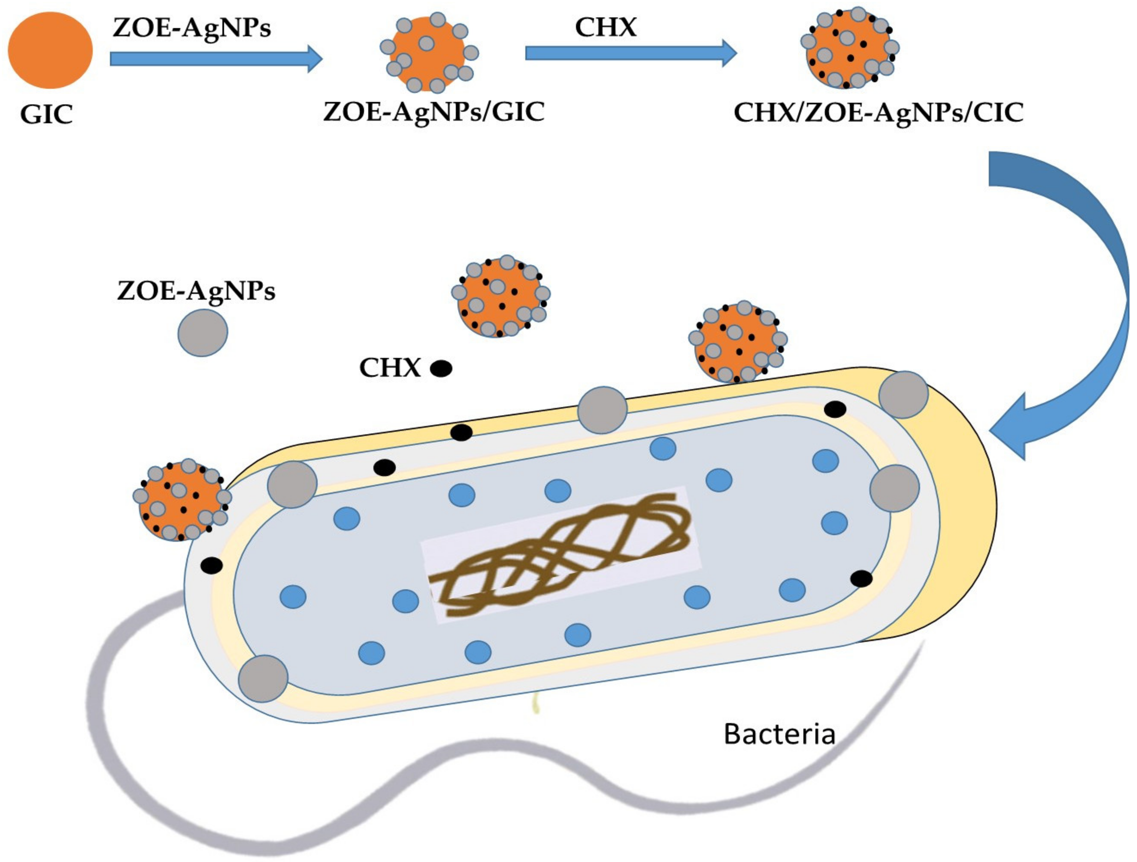
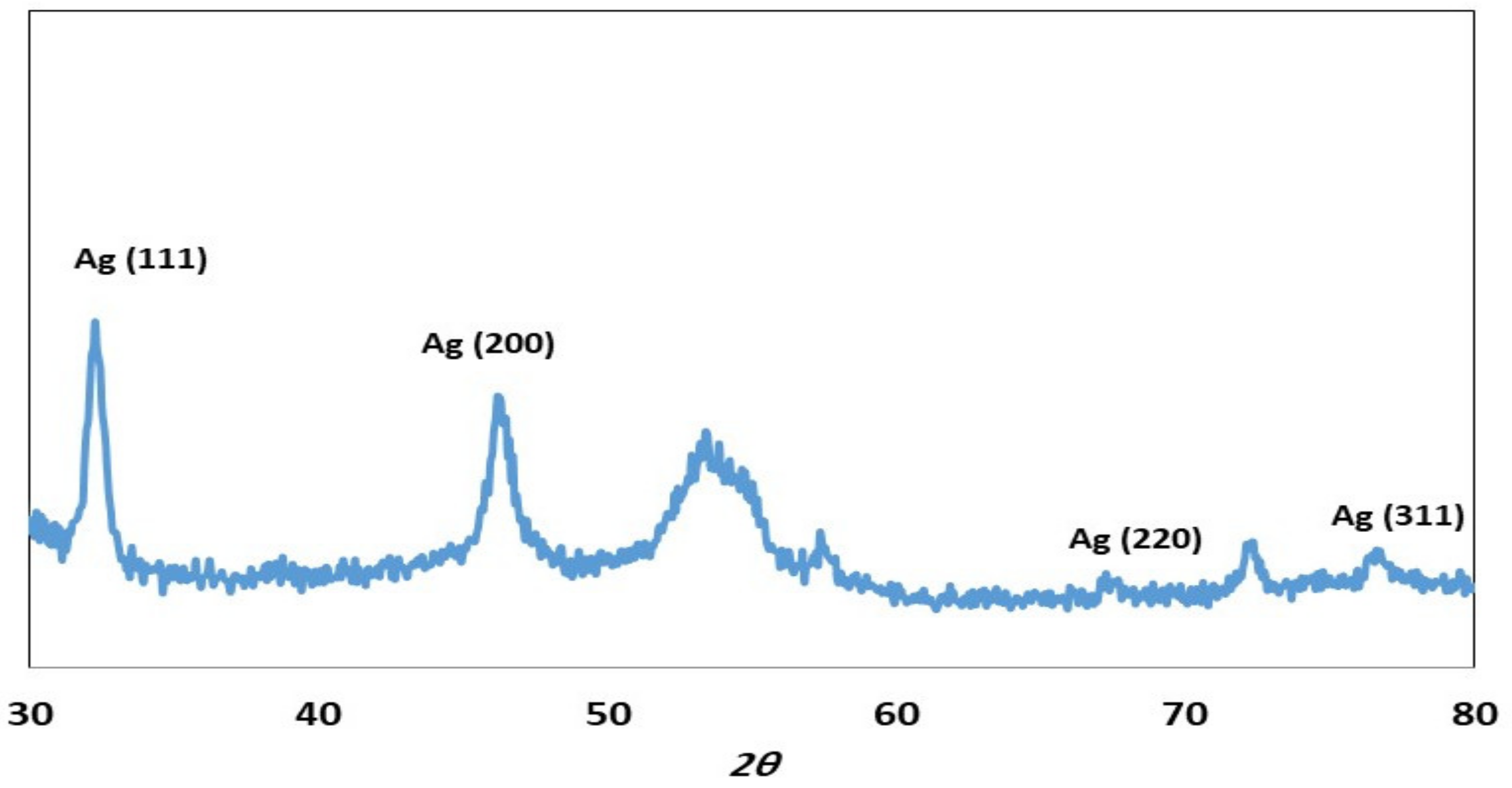
2.1. Characterization of ZOE-AgNPS

The UV-Vis spectrophotometric analysis peak displayed at 415 nm for ZOE-AgNPs (Figure 1). The colorless solution of AgNO3 turned into yellow in the presence ZOE and then to dark brown during the reaction. The color modification in the solution mixture suggested ZOE-AgNPs formation. Enan et al., studied the effect of leaf extract on the formation of AgNPs. The results obtained by them were very similar to what we found [26].
TEM image of the ZOE-AgNPs (Figure 2) displayed a spherical shape with average size from 10.5 to 14.12 nm. Our finding results were almost in agreement with previous findings [27].
The XRD analysis is commonly used to display the crystal structure of the synthesized ZOE-AgNPs. Figure 3 shows the XRD pattern of ZOE-AgNPs synthesized by using ZOE. XRD pattern reveals the 2ϴ peaks obtained at 32.26°, 46.24°, 67. 24°, and 76.72° indexed with (111), (200), (220), and (311) planes, respectively, which confirmed the formation of ZOE-AgNPs with face-centered cubic structure of pure silver metal that was exhibited by all reflectance. Other peaks at 2θ values in ZOE-AgNPs pattern can be ascribed to the residues of the organic content of the ZOE. These peaks reveal the crystallization of some plant metabolite moieties on the surface of the ZOE-AgNPs, which is in agreement with Halawani et al.’s results [28].
The FTIR of ZOE-AgNPs was displayed as shown in Figure 4. The absorption peak at 3395 cm−1 represents the hydroxyl (OH) group, and alkyl CH stretch was recorded at 2917.89 cm−1. A peak at 1631.6 cm−1 can be attributed to the stretching vibrations of –COC (alkane). Sharp peak at 1075.34 cm−1, represents stretching vibrations of –COO. These peaks are present both in ZOE-AgNPs and ZOE. The bands at 916 and other peaks at 3395.34, 2917.89, 1604.55, 1075.34 cm−1, indicate the presence of various compounds such as phenolic compounds, carbohydrate, alkanoids, flavonoids and alkaloids, the active compounds of ZOE, which acts as the reducing and stabilizing agents [29].

2.2. Charcterization of Miswak

Miswak FTIR analysis and its corresponding functional groups related to benzyl isothiocyanate, alkaloids (salvadorine), sulfur, and benzyl cyanates (Data not shown) are all responsible for the growth inhibition of bacteria and fungus strains [30].

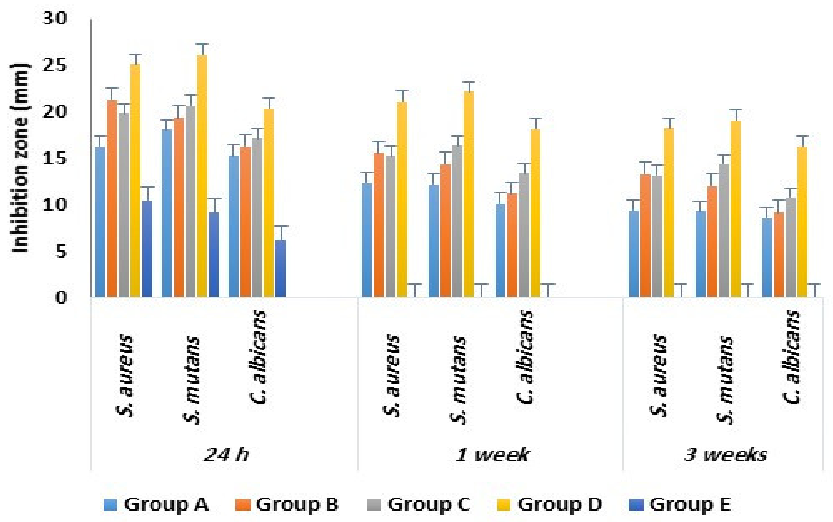
2.3. Antimicrobial Activity

Glass ionomer cement is the most commonly used restorative material due to its biocompatibility with the tooth structure [1]. The present study was the first of a kind that tested antimicrobial efficacy of GIC combined with miswak, ZOE-AgNPs, and chlorhexidine against Streptococcus mutans, Staphylococcus aureus, and Candida albicans. These microorganisms were selected in the present study due to their association with dental caries [31,32,33].
Table 1 shows the mean inhibitory zones of tested specimens. The mean inhibitory zones for all tested microbes were significantly high for Group D with GIC, AgNPs and chlorhexidine, Group A with GIC and miswak, Group B with GIC and AgNPs, and Group C with GIC and chlorhexidine combinations than Group E with GIC alone at a 24-h interval. At one week and three weeks intervals, Group D showed significantly higher mean inhibitory zones compared to Group A, Group B, and Group C.
At a microbe-level comparison, Streptococcus mutan, and Staphylococcus aureus were inhibited significantly by all the specimens tested compared to Candida Albicans (Figure 5).
Previously, authors have studied the antimicrobial efficacy of GIC combined with miswak, AgNPs, and chlorhexidine alone and showed that a combination of GIC with antimicrobial agents enhanced its antimicrobial efficacy without altering its mechanical properties [6,13,14,15,16,19,20,21,22,23,24]. However, in the present study, and for the first time, as shown in Table 2, GIC was combined with ZOE-AgNPs and chlorhexidine, and the result showed significantly higher antimicrobial efficacy against tested microbes compared to GIC with a single antimicrobial agent as well as conventional GIC without any combinations. Previous studies showed that chlorhexidine encapsulated in silica nanoparticles enhanced its antimicrobial efficacy [34,35]. The study by Charannya et al. [24] showed that a combination of AgNPs with 2% chlorhexidine enhanced its antimicrobial efficacy when compared to each of those solutions used individually.
The present study showed that an addition of lyophilized miswak to GIC enhanced its antimicrobial efficacy. This is in agreement with previous in vitro and in vivo studies which showed that the addition of miswak extract enhanced the antimicrobial efficacy of conventional GIC [19,20]. According to the present study result, the mean inhibitory zone was significantly higher for ZOE-AgNPs and chlorhexidine combined GIC at one week and three week intervals compared to lyophilized miswak combined GIC, ZOE-AgNPs combined, GIC, and chlorhexidine combined GIC. This might be due to the substantive property of ZOE-AgNPs and chlorhexidine together enhancing its antimicrobial efficacy at one- and three-week intervals [9,23].
The present study result showed that GIC combined with antimicrobial agents showed superior antibacterial activity against Streptococcus mutans and Staphylococcus aureus than antifungal activity against Candida albicans. This result is in line with previous studies [9,19,25], which showed different zones of inhibition against different oral microbes due to variance in the membrane permeability of the studied microorganism [37]. However, a combination of GIC with chlorhexidine and ZOE-AgNPs together enhanced its antifungal activity compared to GIC with chlorhexidine or ZOE-AgNPs alone. This result is in line with a previous study by Charannya et al., [24] who showed that a combination of nano-silver with chlorhexidine enhanced its antifungal activity.

2.4. Compressive Strength Measurement

Group D specimens’ slightly higher (45.8 ± 5.4) mean compressive strength compared to Group A (43.2 ± 3.1), Group B (44.7 ± 4.8), Group C (43.9 ± 3.6), and Group E (43.4 ± 3.2) (Table 3).
The present study result showed that a combination of conventional GIC with AgNPs and chlorhexidine together and AgNPs alone showed slightly higher mean compressive strength compared to conventional and miswak extract combined GIC, however, the difference was not statistically significant. This is in agreement with the previous studies [6,13,14] which showed the addition of silver nanoparticles enhanced mechanical properties of conventional GIC including its compressive strength.
The study limitation included it’s in vitro type, as it was not possible to simulate all the oral conditions in the lab setting. Future in vivo studies can confirm the result of the present study.

3. Materials and Methods

3.1. Study Design, Microbial Strains Used, and Ethical Approval

An in-vitro study was conducted at the Department of Microbiology and University Dental Hospital, Taif University, KSA. Ethical clearance was obtained from the Institutional review board, Taif University (Ethical clearance number-41-1107-00152). To evaluate antimicrobial efficacy, one strain of candida and two bacterial strains were used in this study (Candida albican, Streptococcus mutans, and Staphylococcus aureus) [38].
Materials used:
(a)
The roots of miswak/Salvadora persica (SP) that were at least six months old, purchased from a local market, Taif City, KSA.
(b)
The Zingiber officinale plant, purchased from a local market, Taif City, KSA.
(c)
Conventional GIC (GC Fuji IX, Tokyo, Japan).
(d)
Chlorhexidine diacetate powder (RM1659-25G, HiMedia Laboratories, Mumbai, India).

3.2. Preparation of Test Specimens

Miswak (Salvadora persica) extract preparation: The miswak roots were ground in a blender (Tomado Chopper, model No. TM-1273,). The 10 g of grounded miswak powder was mixed with 100 mL of deionized water in a sterilized screw-capped bottle. The mixture was kept in the refrigerator for 48 h below 4 °C and then centrifuged at 313 rpm for 10 min. The 100% aqueous miswak extract was obtained by filtering the supernatant using 0.45 mm filter paper, lyophilized and stored at −20 °C till further use.

3.3. Biosynthesis of Silver Nanoparticles

Silver nanoparticles were biosynthesized using Zingiber officinale extract (ZOE, 10%) [39] with 1 mM AgNO3 and characterized using transmission electron microscopy (TEM), Fourier transform infrared analysis (FT-IR), UV-visible spectrum, and X-ray diffraction analysis [40,41,42].

3.4. Preparation of GIC Combination Specimens

The disc-shaped GIC specimens were prepared using split Teflon moulds (3 mm height and 6 mm diameter). The modified GIC specimens were portioned and mixed according to the manufacturer’s instructions, placed in the molds, allowed to sit for 20 min, and then were removed from the mold and were sterilized using ultraviolet radiation for 30 min. A total of 150 specimens were divided into five groups. Then, each of the main groups was tested for antimicrobial efficacy against tested bacteria at three-time intervals of one day, one week, and three weeks. Each of the main groups was performed in triplicates (Table 4).

3.5. Determination of Antimicrobial Activity

A disc diffusion method was used to determine the antimicrobial activity [43]. The Streptococcus mutan, and Staphylococcus aureus were activated in Mueller-Hinton broth and Candida albican in Sabouraud dextrose. The 0.1 mL (1.5 × 10⁸) of activated bacteria and Candida albicans were inoculated in Mueller-Hinton agar medium and Sabouraud dextrose agar media, respectively. The GIC specimen discs were placed in inoculated medium. The inoculated plates were incubated at 37 °C for 24 h. The diameter of the inhibition zone in mm (DIZ) of the tested microbes determined the antimicrobial activity specimen groups. All tests were performed in triplets. The antimicrobial efficacy was tested at three intervals: 24-h, one week, and three weeks. The GIC specimens were stored in distilled water at 37 °C during the testing intervals.

3.6. Compressive Strength Measurement

To test the compressive strength (CS), cylindrical specimens (6 mm height and 4 mm diameter) were prepared using a Teflon mold. Before removal from the Teflon mold, the specimens were allowed to set at room temperature for 20 min. The CS of set cement was measured using a Material Test System (810 MTS Co., Minneapolis, MN, USA) at a crosshead speed of 0.5 mm/min, after 24 h of mixing. For each specimen group, ten specimens were tested. The maximum recorded force at the fracture was measured and CS (N/mm2) was determined using the following Equation (1) [29].
CS = 4P/πd2
where P represents the failure load, and d represents the diameter of the specimen.

3.7. Statistical Analysis

Mean difference was tested using One-way Analysis of Variance (ANOVA) followed by Tukey’s Post Hoc using the Statistical Package for Social Science version 17 (SPSS Inc., Chicago, IL, USA). All statistical tests were two-sided, and the significance level was set at p < 0.05.

4. Conclusions

Within the limitation of the present study, the ZOE-AgNPs was synthesized using ZOE and characterized using means of innumerable analytical methods. It can be concluded that the combination of GIC with synthesized ZOE-AgNPs and chlorhexidine together enhanced its antimicrobial efficacy and compressive strength compared to GIC with ZOE-AgNPs, miswak extract or chlorhexidine combination alone. Antimicrobial efficacy was higher against Streptococcus mutans and Staphylococcus aureus in comparison to Candida albicans.

Author Contributions

Conceptualization, A.A.A. (Amal Adnan Ashour), S.B., N.H.F., E.T.E., A.A.A. (Amal Ahmed Alyamani) and S.M.F.G.E.-R.; methodology, S.B., A.A.A. (Amal Adnan Ashour), N.H.F., E.T.E. and S.M.F.G.E.-R.; software, S.B. and S.M.F.G.E.-R.; validation, S.B., A.A.A. (Amal Adnan Ashour), A.A.A. (Amal Ahmed Alyamani), N.H.F., E.T.E. and S.M.F.G.E.-R.; formal analysis, S.B., A.A.A. (Amal Ahmed Alyamani); investigation, S.B., A.A.A. (Amal Adnan Ashour), N.H.F. and. E.T.E.; resources, A.A.A. (Amal Ahmed Alyamani) and S.M.F.G.E.-R.; data curation, A.A.A. (Amal Ahmed Alyamani); writing—original draft preparation, S.B., A.A.A. (Amal Ahmed Alyamani) and S.M.F.G.E.-R.; writing—review and editing, S.M.F.G.E.-R.; visualization and A.A.A. (Amal Ahmed Alyamani); supervision, S.B. and A.A.A. (Amal Adnan Ashour); project administration, S.B. and S.M.F.G.E.-R.; funding acquisition, S.B. and A.A.A. (Amal Adnan Ashour). All authors have read and agreed to the published version of the manuscript.

Funding

This work was funded by Deanship of Scientific Research, Taif University (research group project No. 1/439/6084), Taif, Saudi Arabia. The authors declare that the funding bodies had no role in the design of the study, the collection, analysis, and interpretation of data, or in writing the manuscript.

Institutional Review Board Statement

The study was conducted according to the guidelines of the Declaration of Helsinki, and approved by the Institutional Review Board (or Ethics Committee) of Taif University (protocol code-41-1107-00152 and 1/12/2019).

Informed Consent Statement

Written Informed consent was obtained from all subjects involved in the study.

Data Availability Statement

Data will be made available on request from corresponding author.

Acknowledgments

We would like to express our gratitude to Deanship of Scientific Research, Taif University, Taif, Saudi Arabia for financial support under the research project number (1/439/6084).

Conflicts of Interest

The authors declare that they have no conflict of interest.

Sample Availability

Samples of the compounds are not available from the authors.

References

  1. Sidhu, S.K.; Nicholson, J.W. A Review of Glass-Ionomer Cements for Clinical Dentistry. J. Funct. Biomater. 2016, 7, 16. [Google Scholar] [CrossRef] [PubMed]
  2. Mickenautsch, S.; Yengopal, V. Absence of carious lesions at margins of glass-ionomer cement and amalgam restorations: An update of systematic review evidence. BMC Res. Notes 2011, 4, 58. [Google Scholar] [CrossRef] [PubMed] [Green Version]
  3. Scholtanus, J.D.; Huysmans, M.C. Clinical failure of class-II restorations of a highly viscous glass-ionomer material over a 6-year period: A retrospective study. J. Dent. 2007, 35, 156–162. [Google Scholar] [CrossRef] [PubMed]
  4. do Amaral, G. Restorative materials containing antimicrobial agents: Is there evidence for their antimicrobial and anticaries effects? A systematic review. Br. Dent. J. 2016, 220, 568. [Google Scholar] [CrossRef] [Green Version]
  5. Tüzüner, T.; Dimkov, A.; Nicholson, J.W. The effect of antimicrobial additives on the properties of dental glass-ionomer cements: A review. Acta Biomater. Odontol. Scand. 2019, 5, 9–21. [Google Scholar] [CrossRef] [Green Version]
  6. Mittal, S.; Soni, H.; Sharma, D.K.; Mittal, K.; Pathania, V.; Sharma, S. Comparative evaluation of the antibacterial and physical properties of conventional glass ionomer cement containing chlorhexidine and antibiotics. J. Int. Soc. Prev. Community Dent. 2015, 5, 268–275. [Google Scholar] [CrossRef] [Green Version]
  7. Fernandez, C.C.; Sokolonski, A.R.; Fonseca, M.S.; Stanisic, D.; Araújo, D.B.; Azevedo, V.; Portela, R.D.; Tasic, L. Applications of Silver Nanoparticles in Dentistry: Advances and Technological Innovation. Int. J. Mol. Sci. 2021, 22, 2485. [Google Scholar] [CrossRef]
  8. Monowar, T.; Rahman, M.S.; Bhore, S.J.; Raju, G.; Sathasivam, K.V. Silver Nanoparticles Synthesized by Using the Endophytic Bacterium Pantoea ananatis are Promising Antimicrobial Agents against Multidrug Resistant Bacteria. Molecules 2018, 23, 3220. [Google Scholar] [CrossRef] [Green Version]
  9. Espinosa-Cristóbal, L.F.; Holguín-Meráz, C.; Zaragoza-Contreras, E.A.; Martínez-Martínez, R.E.; Donohue-Cornejo, A.; Loyola-Rodríguez, J.P.; Cuevas-González, J.C.; Reyes-López, S.Y. Antimicrobial and Substantivity Properties of Silver Nanoparticles against Oral Microbiomes Clinically Isolated from Young and Young-Adult Patients. J. Nanomater. 2019, 2019, 3205971. [Google Scholar] [CrossRef] [Green Version]
  10. Jaiswal, S.; Mishra, P. Antimicrobial and antibiofilm activity of curcumin-silver nanoparticles with improved stability and selective toxicity to bacteria over mammalian cells. Med. Microbiol. Immunol. 2018, 207, 39–53. [Google Scholar] [CrossRef]
  11. Vijayaraghavan, K.; Nalini, S.P. Biotemplates in the green synthesis of silver nanoparticles. Biotechnol. J. 2010, 5, 1098–1110. [Google Scholar] [CrossRef]
  12. Sharma, V.K.; Yngard, R.A.; Lin, Y. Silver nanoparticles: Green synthesis and their antimicrobial activities. Adv. Colloid Interface Sci. 2009, 145, 83–96. [Google Scholar] [CrossRef]
  13. Paiva, L.; Fidalgo, T.K.S.; da Costa, L.P.; Maia, L.C.; Balan, L.; Anselme, K.; Ploux, L.; Thiré, R.M.S.M. Antibacterial properties and compressive strength of new one-step preparation silver nanoparticles in glass ionomer cements (NanoAg-GIC). J. Dent. 2018, 69, 102–109. [Google Scholar] [CrossRef]
  14. Jowkar, Z.; Jowkar, M.; Shafiei, F. Mechanical and dentin bond strength properties of the nanosilver enriched glass ionomer cement. J. Clin. Exp. Dent. 2019, 11, e275–e281. [Google Scholar] [CrossRef]
  15. Porenczuk, A.; Grzeczkowicz, A.; Maciejewska, I.; Gołaś, M.; Piskorska, K.; Kolenda, A.; Gozdowski, D.; Kopeć-Swoboda, E.; Granicka, L.; Olczak-Kowalczyk, D. An initial evaluation of cytotoxicity, genotoxicity and antibacterial effectiveness of a disinfection liquid containing silver nanoparticles alone and combined with a glass-ionomer cement and dentin bonding systems. Adv. Clin. Exp. Med. 2019, 28, 75–83. [Google Scholar] [CrossRef]
  16. Siqueira, P.C.; Magalhães, A.P.; Pires, W.C.; Pereira, F.C.; Silveira-Lacerda, E.P.; Carrião, M.S.; Bakuzis, A.F.; Souza-Costa, C.A.; Lopes, L.G.; Estrela, C. Cytotoxicity of glass ionomer cements containing silver nanoparticles. J. Clin. Exp. Dent. 2015, 7, e622–e627. [Google Scholar] [CrossRef]
  17. Singer, L.; Bierbaum, G.; Kehl, K.; Bourauel, C. Evaluation of the antimicrobial activity and compressive strength of a dental cement modified using plant extract mixture. J. Mater. Sci. Mater. Med. 2020, 31, 116. [Google Scholar] [CrossRef]
  18. Naseem, S.; Hashmi, K.; Fasih, F.; Sharafat, S.; Khanani, R. In vitro evaluation of antimicrobial effect of miswak against common oral pathogens. Pak. J. Med. Sci. 2014, 30, 398–403. [Google Scholar] [CrossRef]
  19. El Tatari, A.; de Soet, J.J.; Abou Shelib, M.; van Amerongen, W.E. Influence of Salvadora persica (miswak) extract on physical and antimicrobial properties of glass ionomer cement. Eur. Arch. Paediatr. Dent. 2011, 12, 22–24. [Google Scholar] [CrossRef] [Green Version]
  20. Kabil, N.S.; Badran, A.S.; Wassel, M.O. Effect of the addition of chlorhexidine and miswak extract on the clinical performance and antibacterial properties of conventional glass ionomer: An in vivo study. Int. J. Paediatr. Dent. 2017, 27, 380–387. [Google Scholar] [CrossRef]
  21. Duque, C.; Aida, K.L.; Pereira, J.A.; Teixeira, G.S.; Caldo-Teixeira, A.S.; Perrone, L.R.; Caiaffa, K.S.; Negrini, T.C.; Castilho, A.R.F.; Costa, C.A.S. In vitro and in vivo evaluations of glass-ionomer cement containing chlorhexidine for Atraumatic Restorative Treatment. J. Appl. Oral Sci. 2017, 25, 541–550. [Google Scholar] [CrossRef]
  22. Bellis, C.A.; Addison, O.; Nobbs, A.H.; Duckworth, P.F.; Holder, J.A.; Barbour, M.E. Glass ionomer cements with milled, dry chlorhexidine hexametaphosphate filler particles to provide long-term antimicrobial properties with recharge capacity. Dent. Mater. 2018, 34, 1717–1726. [Google Scholar] [CrossRef]
  23. Joshi, J.S.; Roshan, N.M.; Sakeenabi, B.; Poornima, P.; Nagaveni, N.B.; Subbareddy, V.V. Inhibition of Residual Cariogenic Bacteria in Atraumatic Restorative Treatment by Chlorhexidine: Disinfection or Incorporation. Pediatr. Dent. 2017, 39, 308–312. [Google Scholar]
  24. Charannya, S.; Duraivel, D.; Padminee, K.; Poorni, S.; Nishanthine, C.; Srinivasan, M.R. Comparative Evaluation of Antimicrobial Efficacy of Silver Nanoparticles and 2% Chlorhexidine Gluconate When Used Alone and in Combination Assessed Using Agar Diffusion Method: An in-vitro Study. Contemp. Clin. Dent. 2018, 9 (Suppl. S2), S204–S209. [Google Scholar] [CrossRef]
  25. Berger, D.; Rakhamimova, A.; Pollack, A.; Loewy, Z. Oral Biofilms: Development, Control, and Analysis. High Throughput 2018, 7, 24. [Google Scholar]
  26. Enan, E.T.; Ashour, A.A.; Basha, S.; Felemban, N.H.; Gad El-Rab, S.M.F. Antimicrobial activity of biosynthesized silver nanoparticles, amoxicillin, and glass-ionomer cement against Streptococcus mutans and Staphylococcus aureus. Nanotechnology 2021, 32, 215101. [Google Scholar] [CrossRef]
  27. Gad El-Rab, S.M.F.; Basha, S.; Ashour, A.A.; Enan, E.T.; Alyamani, A.A.; Felemban, N.H. Green Synthesis of Copper Nano-Drug and Its Dental Application upon Periodontal Disease-Causing Microorganisms. J. Microbiol. Biotechnol. 2021, 31, 1656–1666. [Google Scholar] [CrossRef]
  28. Halawani, E.M.; Hassan, A.M.; El-Rab, S.M.G. Nanoformulation of Biogenic Cefotaxime-Conjugated-Silver Nanoparticles for Enhanced Antibacterial Efficacy against Multidrug-Resistant Bacteria and Anticancer Studies. Int. J. Nanomed. 2020, 15, 1889–1901. [Google Scholar] [CrossRef] [PubMed] [Green Version]
  29. Mehata, M.S. Green route synthesis of silver nanoparticles using plants/ginger extracts with enhanced surface plasmon resonance and degradation of textile dye. Mater. Sci. Eng. B 2021, 273, 115418. [Google Scholar] [CrossRef]
  30. Basha, S.; Ashour, A.A.; Felemban, N.H.; Enan, E.T.; Felemban, M.F.; Alyamani, A.A.; Gad El-Rab, S.M.F. Antimicrobial and synergistic compassion of miswak, nano-silver drug, and chlorhexidine alone and their combinations upon Streptococcus mutans, Staphylococcus aureus, and Candida albicans. J. Pure Appl. Microbiol. 2021, in press. [Google Scholar]
  31. Koo, H.; Bowen, W.H. Candida albicans and Streptococcus mutans: A potential synergistic alliance to cause virulent tooth decay in children. Future Microbiol. 2014, 9, 1295–1297. [Google Scholar] [CrossRef]
  32. Falsetta, M.L.; Klein, M.I.; Colonne, P.M.; Scott-Anne, K.; Gregoire, S.; Pai, C.H.; Gonzalez-Begne, M.; Watson, G.; Krysan, D.J.; Bowen, W.H.; et al. Symbiotic relationship between Streptococcus mutans and Candida albicans synergizes virulence of plaque biofilms in vivo. Infect. Immun. 2014, 82, 1968–1981. [Google Scholar] [CrossRef] [PubMed] [Green Version]
  33. Xiao, J.; Huang, X.; Alkhers, N.; Alzamil, H.; Alzoubi, S.; Wu, T.T.; Castillo, D.A.; Campbell, F.; Davis, J.; Herzog, K.; et al. Candida albicans and Early Childhood Caries: A Systematic Review and Meta-Analysis. Caries Res. 2018, 52, 102–112. [Google Scholar] [CrossRef] [Green Version]
  34. Seneviratne, C.J.; Leung, K.C.; Wong, C.H.; Lee, S.F.; Li, X.; Leung, P.C.; Lau, C.B.; Wat, E.; Jin, L. Nanoparticle-encapsulated chlorhexidine against oral bacterial biofilms. PLoS ONE 2014, 9, e103234. [Google Scholar] [CrossRef] [Green Version]
  35. Leung, K.C.; Seneviratne, C.J.; Li, X.; Leung, P.C.; Lau, C.B.; Wong, C.H.; Pang, K.Y.; Wong, C.W.; Wat, E.; Jin, L. Synergistic Antibacterial Effects of Nanoparticles Encapsulated with Scutellaria baicalensis and Pure Chlorhexidine on Oral Bacterial Biofilms. Nanomaterials 2016, 6, 61. [Google Scholar] [CrossRef] [Green Version]
  36. Shahriari, S.; Barekatain, M.; Shahtalebi, M.A.; Farhad, S.Z. Evaluation of antibacterial properties of a glass-ionomer cement containing purified powder of Salvia officinalis: An in vitro study. Int. J. Prev. Med. 2019, 10, 110. [Google Scholar]
  37. Mozayeni, M.A.; Hadian, A.; Bakhshaei, P.; Dianat, O. Comparison of antifungal activity of 2% chlorhexidine, calcium hydroxide, and nanosilver gels against Candida albicans. J. Dent. 2015, 12, 109–117. [Google Scholar]
  38. Koneman, E.W.; Allen, S.D.; Janda, W.M.; Schreckenberger, P.C.; Winn, W.C., Jr. Color Atlas and Textbook of Diagnostic Microbiology, 5th ed.; Lippincott-Raven Publishers: Philadelphia, PA, USA, 1997. [Google Scholar]
  39. Gad El-Rab, S.M.F.; Halawani, E.M.; Alzahrani, S.S.S. Biosynthesis of silver nano-drug using Juniperus excelsa and its synergistic antibacterial activity against multidrug-resistant bacteria for wound dressing applications. 3 Biotech 2021, 11, 255. [Google Scholar] [CrossRef]
  40. Gad El-Rab, S.M.F.; Halawani, E.M.; Hassan, A.M. Formulation of Ceftriaxone Conjugated Gold Nanoparticles and Their Medical Applications against Extended-Spectrum β-Lactamase Producing Bacteria and Breast Cancer. J. Microbiol. Biotechnol. 2018, 28, 1563–1572. [Google Scholar] [CrossRef]
  41. Gad El-Rab, S.M.F.; Abo-Amer, A.E.; Asiri, A.M. Biogenic Synthesis of ZnO Nanoparticles and Its Potential Use as Antimicrobial Agent against Multidrug-Resistant Pathogens. Curr. Microbiol. 2020, 77, 1767–1779. [Google Scholar] [CrossRef]
  42. Gad El-Rab, S.M.F.; Ashour, A.A.; Basha, S.; Alyamani, A.A.; Felemban, N.H.; Enan, E.T. Well-Orientation Strategy Biosynthesis of Cefuroxime-Silver Nanoantibiotic for Reinforced Biodentine™ and Its Dental Application against Streptococcus mutans. Molecules 2021, 26, 6832. [Google Scholar] [CrossRef] [PubMed]
  43. Hudzicki, J. Kirby-Bauer Disk Diffusion Susceptibility Test Protocol; American Society of Microbiology: Washington, DC, USA, 2016. [Google Scholar]
Scheme 1. Schema of synthetic diagram for ZOE-AgNPs/CHX/GIC and their synergistic antimicrobial efficacy.
Molecules 27 00528 sch001
Figure 1. UV-Vis spectral range showing a peak of ZOE-AgNPs at 415 nm.
Molecules 27 00528 g001
Figure 2. TEM image ZOE-AgNPs.
Molecules 27 00528 g002
Figure 3. XRD pattern of AgNPs.
Molecules 27 00528 g003
Figure 4. FT-IR spectra of AgNPs synthesized with ZOE.
Molecules 27 00528 g004
Figure 5. Antimicrobial efficacy of tested GIC specimens at different time intervals. GIC—Glass ionomer cement, Group A—GIC + lyophilized miswak, Group B—GIC + ZOE-AgNPS, Group C—GIC + chlorhexidine diacetate, Group D—GIC + ZOE-AgNPs + Chlorhexidine, Group E—control (glass ionomer alone).
Molecules 27 00528 g005
Table 1. Antimicrobial efficacy of tested GIC specimens at different time intervals.
Study Groups with Tested MicrobesInhibition Zone in mm
InZ
24 h
InZ
1 Week
InZ
3 Week
S. aureus
a- Group A (n = 30) 16.3 ± 2.112.4 ± 1.19.4 ± 1.1
b- Group B (n = 30) 21.3 ± 1.415.6 ± 1.213.3 ± 1.1
c- Group C (n = 30) 19.8 ± 1.915.3 ± 1.213.2 ± 1.1
d- Group D (n = 30)25.2 ± 2.221.2 ± 1.818.3 ± 1.3
e- Group E (n = 30)10.5 ± 1.600
ANOVA, F value16.7127.1726.341
ANOVA, p value0.00010.0030.03
Tukey post Hocd > e, d > a, d > c, d > b, b > e, b > a, c > e, a > ed > a, d > c, d > b d > a, d > b, d >c
S. mutans
a- Group A (n = 30) 18.1 ± 1.912.2 ± 1.29.3 ± 1.1
b- Group B (n = 30) 19.4 ± 2.114.4 ± 1.112.1 ± 0.9
c- Group C (n = 30) 20.7 ± 1.516.4 ± 1.614.4 ± 1.5
d- Group D (n = 30)26.2 ± 2.622.2 ± 2.119.1 ± 1.7
e- Group E (n = 30)9.2 ± 1.200
ANOVA, F value17.8215.3286.723
ANOVA, p value0.00010.040.03
Tukey post Hocd > e, d > a, d > c, d > b, b > e, c > e, a > ed > a, d > b, d > c, c > ad > a, d > b, d >c, b > a, c > a
C. albicans
a- Group A (n = 30) 15.3 ± 1.110.2 ± 1.28.6 ± 0.9
b- Group B (n = 30) 16.3 ± 1.111.2 ± 1.59.2 ± 1.3
c- Group C (n = 30) 17.2 ± 1.213.4 ± 1.310.8 ± 1.1
d- Group D (n = 30)20.4 ± 2.118.2 ± 1.916.3 ± 1.3
e- Group E (n = 30)2.3 ± 1.100
ANOVA, F value14.2316.4216.723
ANOVA, p value0.00010.030.03
Tukey post Hocd > e, d > a, d > c, d > b, b > e, c > e, a > ed > a, d > b, d > c, c > a d > a, d > b, d >c, c > a
Group A—GIC + lyophilized miswak, Group B—GIC + ZOE-AgNPs, Group C—GIC + chlorhexidine diacetate, Group D—GIC + ZOE-AgNPs + Chlorhexidine, Group E—control (glass ionomer alone), InZ—inhibition zone, h—hour, n(a–e)—Number of speciment.
Table 2. Comparison of earlier research with restorative materials for antibacterial activity against oral bacteria.
MaterialsConcentrationBacteriaReference
  • GIC + CHX
  • 99.5% GIC + 0.5% CHX
  • S. mutans
  • Kabil et al. [20]
  • GIC + AgNPs
  • 99.5% GIC + 0.5% AgNPs
  • S. mutans
  • Paiva et al. [13]
  • GIC + miswak
  • GIC + 1.25% miswak
  • S. mutans and L. casei
  • Shahriari et al. [36]
  • AgNPs + CHX Solution
  • 15 µg/mL AgNPs + 2% CHX
  • E. faecalis, K. pneumoniae, and C. albicans
  • Charannya et al. [25]
  • GIC + ZOE-AgNPs + CHX
  • 98.5% GIC + 0.5% ZOE-AgNPs + 1% CHX
  • S. aureus, S. mutans and C. albicans
  • This study
Table 3. Compressive strength (MPa) of tested specimens.
Study GroupsnMean ± SDANOVA F ValueANOVA p Value
Group A1043.2 ± 3.12.3260.07
Group B1044.7 ± 4.8
Group C1043.9 ± 3.6
Group D1045.8 ± 5.4
Group E1042.4 ± 2.7
Group A—GIC + lyophilized miswak, Group B—GIC + ZOE-AgNPs, Group C—GIC + chlorhexidine diacetate, Group D—GIC + ZOE-AgNPs + Chlorhexidine, Group E—control (glass ionomer alone), SD—standard deviation, ANOVA—analysis of variance.
Table 4. Proportion of the Lyophilized miswak, ZOE-AgNPs and Chlorhexidine diacetate added to Conventional GIC.
GroupAbbreviation Components
Group A
  • Lyophilized miswak/GIC
  • (99% GIC with 1% lyophilized miswak, w/w).
Group B
  • ZOE-AgNPs/GIC
  • (99.5% GIC with 0.5% ZOE-AgNPs, w/w).
Group C
  • CHX/GIC
  • (99% GIC with 1% CHX, w/w).
Group D
  • CHX/ZOE-AgNPs/GIC
  • (98% GIC with 1% CHX, 0.5% ZOE-AgNPs (w/w).
Group E
  • GIC
  • Conventional GIC without any addition.
Publisher’s Note: MDPI stays neutral with regard to jurisdictional claims in published maps and institutional affiliations.

