Article

New Amino Acid-Based Thiosemicarbazones and Hydrazones: Synthesis and Evaluation as Fluorimetric Chemosensors in Aqueous Mixtures

by
Cátia I. C. Esteves
,
Maria Manuela M. Raposo
and
Susana P. G. Costa
*
Centre of Chemistry, University of Minho, Campus de Gualtar, 4710-057 Braga, Portugal
*
Author to whom correspondence should be addressed.
Molecules 2023, 28(21), 7256; https://doi.org/10.3390/molecules28217256
Submission received: 17 July 2023 / Revised: 25 September 2023 / Accepted: 20 October 2023 / Published: 25 October 2023
(This article belongs to the Special Issue ECSOC-26)

Abstract

:
Bearing in mind the interest in the development and application of amino acids/peptides as bioinspired systems for sensing, a series of new phenylalanine derivatives bearing thiosemicarbazone and hydrazone units at the side chain were synthesised and evaluated as fluorimetric chemosensors for ions. Thiosemicarbazone and hydrazone moieties were chosen because they are considered both proton-donor and proton-acceptor, which is an interesting feature in the design of chemosensors. The obtained compounds were tested for the recognition of organic and inorganic anions (such as AcO, F, Cl, Br, I, ClO4, CN, NO3, BzO, OH, H2PO4 and HSO4) and of alkaline, alkaline-earth, and transition metal cations, (such as Na+, K+, Cs+, Ag+, Cu+, Cu2+, Ca2+, Cd2+, Co2+, Pb2+, Pd2+, Ni2+, Hg2+, Zn2+, Fe2+, Fe3+ and Cr3+) in acetonitrile and its aqueous mixtures in varying ratios via spectrofluorimetric titrations. The results indicate that there is a strong interaction via the donor N, O and S atoms at the side chain of the various phenylalanines, with higher sensitivity for Cu2+, Fe3+ and F in a 1:2 ligand-ion stoichiometry. The photophysical and metal ion-sensing properties of these phenylalanines suggest that they might be suitable for incorporation into peptide chemosensory frameworks.

Graphical Abstract

1. Introduction

Amino acids in peptides and proteins are involved in many biochemical processes due to their coordinating ability towards metal ions due to the nitrogen, oxygen and sulphur electron donor atoms at the main and side chains [1,2]. Natural amino acids can coordinate anions via the amino and hydroxyl groups at the side chain of arginine, tryptophan, serine, threonine and tyrosine, and amide bond NHs [3].
Thus, the incorporation of suitable heterocycles or groups at the side chain of natural amino acids can result in increased complexing ability and enhanced photophysical properties, which are of great interest for biochemistry, cellular biology and imaging. Synthetic amino acid derivatives possess structural diversity and are functionally versatile, allowing for the assembly of peptides and, eventually, proteins with tuned properties. Using specially tailored amino acids bearing heterocycles in sensory applications has benefits compared to natural amino acids, such as the cooperative action of the extra hetero-atoms to exhibit a more effective binding process and overall sensing ability; enhanced optical response at longer wavelengths of absorption and fluorescence; and improved fluorescence properties (compared to tryptophan, the most fluorescent natural amino acid). These features allow for higher detection sensitivity and lower detection and quantification limits, and their intrinsic biological nature also allows for prospective use in fluorescence-based biological assays, such as targeting peptides for molecular imaging; enzyme activity; or site-specific protein labelling for the in vivo tracking of protein localization, dynamics and concentration [4,5,6,7,8]. From a survey of the most recent literature, examples of amino acids and peptides involved in solution- or material-based fluorescent sensing were found to be highly varied and included a tryptophan-quinoline conjugate for the turn-off detection of Fe2+ [9]; dipeptide receptors with aggregation-induced emission properties for the recognition of methylmercury and Hg2+ [10]; cellulose nanofibers modified with L-histidine for the detection of Cr6+ and Hg2+ [11]; gold–silver bimetallic nanoclusters capped with tryptophan for the detection of histamine [12]; a near-infrared fluorophore based on a methionine attached to a chromenylium-cyanine for the analysis of Hg2+ in the environment and living cells [13]; a phenylalanine-based dual-channel probe for the detection of Fe3+; Cu2+ and F [14]; pyrene-phenylalanine conjugates for Cu2+ analysis [15]; a derivative of tyrosine as a turn-off fluorescent sensor for Hg2+ [16]; carbon quantum dots functionalized with different amino acids (glutamine, histidine, arginine, lysine and proline), capable of monitoring multiple metal ions in water [17]; and a series of pentapetides bearing tyrosine for the detection of Cu2+ at the nanomolar level [18]. These are only a few examples that confirm the growing interest in and application of amino acids/peptides as bio-inspired systems for sensing.
There are various analytical techniques for the identification of anions and cations, such as inductively coupled plasma emission or mass spectroscopy (ICP-ES or ICP-MS), atomic absorption spectroscopy (AAS), anodic stripping voltammetry (ASV), and total reflection X-ray fluorimetry (TXRF), among others, but they require elaborate procedures, high-cost equipment, and trained technicians [19,20]. Therefore, a simpler methodology based on optical sensing is attractive for the fast and straightforward detection of ions [21].
Detecting cations of chemical and biochemical interest is a hot topic of research, as sodium, potassium, magnesium and calcium, among other cations, are involved in nervous impulse transmission, contraction of muscle fibres, control of cell activity, etc., and it is well known that heavy metals such as mercury, lead, and cadmium are toxic for organisms, and early and easy detection is desirable [21,22,23,24]. Trivalent metal cations are involved in many cellular processes, and Fe3+ is an essential element in biochemical routes at the cellular level [25]. Its imbalance is related to disorders such as anaemia; hemochromatosis; diabetes; and abnormal function of heart, pancreas and liver). Meanwhile, for anions, their selective recognition is also a hot topic, and fluoride stands out as one of particular importance due to its widespread use in dental care and treatment of osteoporosis [26,27,28,29].
Compared to cation receptors, there are far fewer reports on anion receptors due to the lower stability constants for host–anion interactions, which are related to the diversity of anion shapes, their strong pH dependence and the competition of water for receptor–anion complexes. Most of the receptors for anion binding are based on hydrogen bonding and electrostatic interactions. Hydrogen bonds are directional and allow for discrimination between anions with different shapes and geometries [26]. (Thio)ureas and hydrazones are well-known anion binding groups because their hydrogen-bonding ability results in the formation of stable complexes [30,31,32,33,34]. This ability is related to the NH protons′ acidity and the number of available binding sites, and it is possible to tune the acidity by introducing substituents of different electronic character (electron-donating or electron-withdrawing) [35]. Among the molecules that contain (thio)urea fragments, thiosemicarbazones have emerged as anion receptors, and they can easily be included in aromatic frameworks functionalized with π-conjugated heterocyclic moieties, with acceptor or donor characteristics, to adjust the acidity of the NH, having been reported as having the colorimetric and fluorimetric sensing of anions [35].
Bearing in mind our interest in exploring new designs of biomolecule-based fluorescent sensing for prospective use in biology and medicine, and our previous reports in the design of unnatural amino acids as optical probes for several metal cations and anions [36,37,38,39], we now report the synthesis and characterization of novel phenylalanine derivatives bearing thiosemicarbazone and hydrazone units at the side chain and their evaluation as amino acid-based fluorimetric chemosensors for ions in aqueous solution. The new compounds contained auxiliary electron donor (hetero)aromatic rings of different strengths (i.e., benzene, thiophene and a fused benzopyranothiophene) to modulate their response as chemosensors and the combination of the abovementioned moieties (thiosemicarbazone/hydrazone and sulphur/oxygen heterocycles) resulted in a dual sensor for detection of both anions and cations. The interaction of the newly reported probes with the ions was studied via absorption and fluorescence spectroscopy.

2. Results and Discussion

2.1. Synthesis of Phenylalanine Thiosemicarbazones and Hydrazones 3a–d

The new phenylalanines 3a–d with thiosemicarbazone and hydrazone moieties at the side chain were synthesized in quantitative yield via reaction of N-tert-butoxycarbonyl-4-formyl-L-phenylalanine methyl ester 1 [40,41] with the appropriate thiosemicarbazide 2a–b or hydrazide 2c–d, in methanol at room temperature (Scheme 1, Table 1). These new compounds were fully characterised, and their structure was confirmed via the usual spectroscopic techniques.
In the 1H NMR spectra, the characteristic signal for the methylene CH=N was visible at about δ 7.90–7.97 ppm and the more acidic thiosemicarbazone/hydrazone NH proton between δ 10.45–10.70 and δ 9.93–10.27 ppm, respectively. On the 13C spectra, the methylene CH=N carbon appeared at δ 142.92–144.36 ppm, the thiocarbonyl in compounds 3a,b was visible at δ 175.56–178.14 ppm, and the carbonyl in compounds 3c,d appeared at δ 162.42–162.80 ppm. No evidence for the loss of the integrity of the chiral centre in these reaction conditions was found via NMR.

2.2. Photophysical Study of Phenylalanines 3a–d

The UV-visible absorption and emission spectra of thiosemicarbazone/hydrazone phenylalanines (3a–d) were measured in acetonitrile (10−6–10−5 M) (Table 1). The nature of the pendant group at position 4 of the phenylalanine core had a clear influence on the position of the absorption and emission bands of compounds 3a–d. When compared to the unsubstituted phenylalanine (λabs = 258 nm and λem= 280 nm, in the same solvent), phenylalanine derivatives 3a–d displayed absorption and emission maxima at longer wavelengths (λabs = 316–359 nm and λem = 394–443 nm), with compound 3d bearing a fused benzopyranothiophene displaying the larger bathochromic shift. The relative fluorescence quantum yields (ΦF) were determined using a 10−6 M solution of 9,10-diphenylanthracene in ethanol as fluorescence standard (ΦF = 0.95) [42], and phenylalanine derivative 3d exhibited very high fluorescence quantum yield (0.86), whereas compounds 3a–c were less fluorescent (0.01–010). In terms of Stokes′ shifts, the studied compounds displayed values between 5282 and 6486 cm−1, which is an attractive property for fluorescent probes to decrease the probability of crosstalk between the excitation source and fluorescence emission [43]. Considering the nature of the compounds and their prospective application in biological assays, solubility in water or in high water content mixtures is highly desirable. Therefore, phenylalanines 3a–d were also dissolved in mixtures of this solvent with varying water proportions: 80:20, 50:50 and 20:80 (v/v) (10−6–10−5 M) and their absorption and fluorescence spectra were obtained (Table 2 and Figure 1, 3d as representative example).
Regarding the absorption wavelength, it was found that there was a small hypsochromic shift (≈5 nm) with increased water content for thiosemicarbazones 3a,b, while for hydrazones 3c,d, there was a small bathochromic shift (4–8 nm). As for the emission wavelength and its relationship with the water ratio, there was no change to thiosemicarbazones 3a,b, and for hydrazones 3c,d, there a slight shift to longer wavelengths (15–20 nm). As a general trend for all compounds, the relative fluorescence quantum yields decreased ca. 20% as the water content increased.

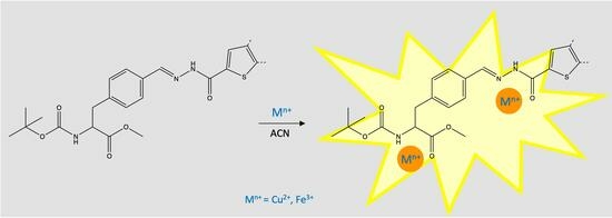
2.3. Preliminary Chemosensing Tests and Spectrofluorimetric Titrations of Phenylalanines 3a–d with Selected Ions

The modification of phenylalanine (the least fluorescent natural amino acid) at its side chain was intended to provide additional binding sites with the heterocycle′s donor atoms as well as enhanced photophysical properties (absorption and emission at longer wavelengths and higher fluorescence) due to fluorescent heterocycles.
Various organic and inorganic anions were chosen due to their biological, environmental and analytical relevance (such as AcO, F, Cl, Br, I, ClO4, CN, NO3, BzO, OH H2PO4 and HSO4), as well as various alkaline, alkaline–earth and transition metal cations (such as Na+, K+, Cs+, Ag+, Cu+, Cu2+, Ca2+, Cd2+, Co2+, Pb2+, Pd2+, Ni2+, Hg2+, Zn2+, Fe2+, Fe3+ and Cr3+). The interaction of phenylalanines 3a–d with the above-mentioned ions was studied via absorption and fluorescence spectroscopy in ACN and its mixture ACN/H2O (20:80).
A preliminary evaluation of sensing ability was performed by adding 100 equiv of each ion to the solutions of phenylalanines 3a–d in ACN and ACN/H2O (20:80). The position and intensity of the absorption bands did not change, but there was significant variation in the fluorescence intensity in the presence of some ions, especially with Fe3+, Cu2+ and F. Therefore, considering the fluorescence properties of hydrazones 3c,d, spectrofluorimetric titrations were performed in the presence of these selected ions.
In spectrofluorimetric titrations with Cu2+ in ACN, a strong increase in fluorescence intensity (a chelation-enhanced fluorescence, or CHEF, effect) was observed for phenylalanine hydrazones 3c and 3d, with a small number of equivalents being necessary to increase fluorescence until it reached a plateau (30 equiv for 3c and 8 equiv for 3d). The titration with Cu2+ in ACN/H2O (20:80) resulted in a loss of sensitivity for both receptors, requiring higher numbers of cations to induce a similar fluorimetric response, but for compound 3d, a chelation-enhanced quenching effect (CHEQ) with a slight hypsochromic shift of the band was seen.
The same trend was obtained from the titration of phenylalanine hydrazones 3c and 3d with Fe3+ in ACN, namely a CHEF effect upon addition of 80 equiv for 3c or 5 equiv for 3d. The titration of compound 3d with Fe3+ in ACN/H2O (20:80) resulted in a marked loss of sensitivity and the previously observed CHEQ effect. From this data, it could be concluded that hydrazone 3d had higher sensitivity for both cations when compared to hydrazine 3c.
It is known that hydrazones can create a cavity to accommodate the cation, involving the nonbonding electrons of amino nitrogen and the carbonyl oxygen. In the case of the described amino acids, one can expect that this binding occurs similarly at the side chain hydrazono group and also at the main chain via the amino nitrogen and carbonyl oxygen. This proposal agrees with the calculated ligand:metal ratio (1:2), which agrees with previous experimental and theoretical results obtained by the authors with other amino acid-based chemosensors [44,45]. In this proposed mode, the sulphur atom at the thiophene (electron rich heterocycle) may also be cooperative to the binding.
Contrarily to what was expected, both receptors proved to be less sensitive to F when compared to the cations; nevertheless, the sensitivity of hydrazone 3d was higher than that of 3c. With regard to the interaction with F, the titration in ACN revealed a significant fluorescence intensity decrease upon addition of increasing amounts of F, accompanied by a concomitant red shift of about 20 nm of the emission band, with roughly 250 equiv of F. In can/H2O (20:80), there was a very slight decrease of fluorescence (only 15%) upon addition of 70 equiv of F. This diminished response in aqueous mixtures may be related to the reduced basicity of F in aqueous environment due to efficient solvation by the water molecules [46].
Figure 2, Figure 3 and Figure 4 show the marked effects of the interaction with Cu2+, Fe3+ and F in the band centred at the maximum emission wavelength, in the spectrofluorimetric titrations of phenylalanines 3c–d in ACN and ACN/H2O (20:80).
The CHEF or “off–on” effect in hydrazones is related to the inhibition of the C=N double bond isomerization in the excited state due to the binding of the cation, thus locking a more favorable conformation for increased conjugation, with in turn allows a photoinduced electron transfer (PET) process along the conjugated system [30].
All the other tested cations induced a less pronounced CHEF or CHEQ effect in ACN for a much larger number of added equivalents, without increase or complete decrease of fluorescence (see Supporting Information). As a representative example, the addition of roughly 200 equiv of Cd2+ resulted in a 90% fluorescence increase for 3c, while for 3d, no change was seen. As for Cr3+, upon the addition of 800 equiv to 3c, an increase of 100% was seen, while for 3d no change was visible. Addition of 600 equiv of Cu+ to hydrazone 3c resulted in an increase of fluorescence of 90%. Also, for 3c, the addition of 75 equiv of Fe2+ and Pb2+ was accompanied by a 90% and 75% increase in intensity, respectively. Titration of 3c with 200 equiv of Ni2+ caused a 75% increase in intensity, whereas the addition of 400 equiv to 3d caused a 50% quenching of fluorescence. Addition of 350 equiv of Hg2+ to 3c resulted in an increase of fluorescence of 80% and addition of 70 equiv to 3d caused a 75% quenching of fluorescence.
No significant changes were seen in the spectrofluorimetric titrations of hydrazones 3c and 3d with the other anions in ACN and ACN/H2O (20:80).
The Stern–Volmer plot of relative fluorescence intensity (I0/I) for phenylalanine hydrazone 3d versus Cu2+ and Fe3+ concentration in ACN/H2O (20:80) and versus F concentration in ACN confirmed a linear relationship indicative of a dynamic quenching, except for F in ACN, which appears to be a combination of dynamic and static quenching (Figure 5) [47].
Previous studies showed that the unprotected amino acid C- and N-terminals of the amino acid did not significantly change coordination ability and that it could occur concurrently at the additional heteroatoms at the side chain [48]. Moreover, our previously reported works in synthetic fluorescent amino acids revealed that they maintain their sensing ability when integrated into short peptides, which also displayed sensing ability [45]. Further insight into the binding mode was attempted by 1H NMR titrations with compound 3d as representative example, with Cu2+ in ACN-d3, but the compound was insoluble in the required concentration. A new attempt in DMSO-d6 resulted in significant broadening of the signals after addition of the cations, and no reliable information could be collected.
Regarding the mode of interaction with anions, more specifically with F, NH-containing receptors such as hydrazine, thiosemicarbazone groups and nitrogen heterocycles such as imidazoles and pyrroles are known to interact via the formation of hydrogen-bonding complexes that eventually lead to deprotonation [49,50,51,52,53]. The formation of hydrogen bonding complexes in the fluorescence spectra can be reflected by changes in fluorescence intensity due to the formation of the complex, whereas the formation of the deprotonated species can be ascribed to further quenching and shift of the emission band [46,53]. In the present case of compound 3d, the fluorescence intensity decreased upon addition of increasing amounts of F, accompanied by a simultaneous red-shift of about 20 nm on the emission band.
The binding stoichiometry and the binding affinity of phenylalanines 3c–d with selected anions/cations were obtained from spectrofluorimetric titrations in ACN and ACN/H2O (20:80) by HypSpec software, based on the Benesi–Hildebrand equation [54]. The results suggest a 1:2 ligand–metal cation or ligand–anion stoichiometry (Table 3). The higher values obtained for binding of Cu2+ and Fe3+ in ACN and ACN/H2O (20:80) are most likely related to the similar ionic radius of the cations and the favorable stabilization of the complex considering the HSAB theory: O and N are hard bases, Fe3+ is a hard acid and Cu2+ is a borderline acid. The lower binding affinity in the aqueous mixture may be explained by the high water content and the competing solvation of the cations by the water molecules.
A comparison between the binding ability of phenylalanines 3c–d was made with recently published amino acid and peptide fluorescent chemosensors for Cu2+, shown in Table 4, which confirms that the newly reported phenylalanines show much higher binding affinity.

3. Materials and Methods

3.1. General

Commercially available thiosemicarbazides and hydrazides 2a–d (Sigma–Aldrich, St. Louis, MO, USA) were used as received. The synthesis of compound 1 was carried out as previously reported [23,24]. A Stuart SMP3 melting point apparatus was used for measuring melting points (Barloworld Scientific Ltd, Staffordshire, UK). Thin layer chromatography (TLC) was carried out on 0.25 mm-thick precoated silica plates (Merck Fertigplatten Kieselgel 60F254) with UV light visualisation. IR spectra (using KBr discs or as liquid film) were obtained in a BOMEM MB 104 spectrophotometer (ABB, Zurich, Switzerland). UV-vis absorption spectra were obtained in a Shimadzu UV/2501PC spectrophotometer (Shimadzu Europa GmbH, Duisburg, Germany) and fluorescence spectra were obtained in a Horiba FluoroMax-4 spectrofluorometer (HORIBA Europe GmbH, Darmstadt, Germany) in standard quartz cuvettes with 1 cm optical path. The relative fluorescence quantum yield for the various compounds was calculated using 9,10-diphenylanthracene as fluorescence standard (absolute fluorescence quantum yield in ethanol 0.95). The solutions of the compounds and the standard were excited at the same wavelength with identical absorbance, nearest to the wavelength of maximum absorption of the compound, and the quantum yield of the compound (Φcpd) was calculated using the equation Φcpd = Φstd (Astd/Acpd)(Fcpd/Fstd)(ncpd/nstd)2, where the subscript “std” denotes standard and “cpd” denotes compound, Φstd is the absolute quantum yield of the standard, A is the absorbance of the solution at the excitation wavelength, F is the integrated fluorescence intensity and n is the refractive index of the solvent [60].
NMR spectra were recorded on a Bruker Avance III 400 (Bruker, Billerica, MA, USA) at an operating frequency of 400 MHz for 1H and 100.6 MHz for 13C, using the solvent peak as an internal reference at 25 °C and the chemical shift values (δ relative to TMS) are given in ppm. Signal assignments were supported by heteronuclear correlation NMR. Low- and high-resolution mass spectra were recorded at the University of Vigo, Spain in the “C.A.C.T.I. Unidad de Espectrometria de Masas”.

3.2. Synthesis of Phenylalanine Thiosemicarbazones and Hydrazones 3a–d

General procedure: a mixture of N-tert-butoxycarbonyl-4-formyl-L-phenylalanine methyl ester 1 (1 equiv) and the appropriate thiosemicarbazide 2a–b or hydrazide 2c–d (1 equiv) in methanol (50 mL/mmol) was stirred for 12 h at room temperature, and the solvent was removed in a rotary evaporator. Column chromatography was carried out with silica gel using mixtures of petroleum ether 40–60 and dichloromethane of increasing polarity. The fractions containing the desired compound were combined and evaporated to dryness.

3.2.1. N-(tert-Butoxycarbonyl)-4-((2-carbamothioylhydrazono)methylene)-L-phenylalanine methyl ester 3a

Starting from 1 (0.050 g, 0.163 mmol) and thiosemicarbazide 2a (0.015 g, 0.163 mmol), afforded compound 3a as yellow oil (0.057 g, 0.151 mmol, 93%). IR (liquid film): ν = 3434, 3269, 3161, 2978, 1703, 1594, 1529, 1508, 1444, 1367, 1277, 1252, 1166, 1094, 1058, 1020, 939, 916, 873, 826, 735 cm1. 1H NMR (400 MHz, CDCl3): δ = 1.40 (s, 9H, C(CH3)3), 3.02–3.18 (m, 2H, β-CH2), 3.72 (s, 3H, OCH3), 4.61–4.64 (m, 1H, α-H), 5.18 (d, J 8.0 Hz, 1H, NH Boc), 6.75 (br s, 1H, NH2), 7.17 (d, J 8.4 Hz, 2H, H2 and H6), 7.24 (br s, 1H, NH2), 7.54 (d, J 8.4 Hz, 2H, H3 and H5), 7.91 (s, 1H, CH=N), 10.45 (s, 1H, =N-NH) ppm. 13C NMR (100.6 MHz, CDCl3): δ = 28.22 (C(CH3)3), 38.33 (β-CH2), 52.33 (OCH3), 54.29 (α-C), 80.05 (C(CH3)3), 127.54 (C3 and C5), 129.78 (C2 and C6), 131.85 (C4), 139.15 (C1), 143.82 (CH=N) 155.02 (C=O Boc), 172.24 (C=O ester), 178.14 (C=S) ppm. UV/Vis (ACN, nm): λmax (log ε) = 316 (4.21). MS m/z (ESI, %): 381 ([M+H]+, 100). HRMS: m/z (ESI) calcd for C17H25N4O4S 381.15910; found 381.15888.

3.2.2. N-(tert-Butoxycarbonyl)-4-((2-(phenylcarbamothioyl)hydrazono)methylene)-L-phenyl-alanine methyl ester 3b

Starting from 1 (0.050 g, 0.163 mmol) and 4-phenylthiosemicarbazide 2b (0.027 g, 0.163 mmol), afforded compound 3b as yellow oil (0.071 g, 0.156 mmol, 96%). IR (liquid film): ν = 3431, 3323, 3146, 3054, 2977, 2856, 1711, 1596, 1534, 1513, 1446, 1391, 1366, 1332, 1259, 1195, 1168, 1120, 1060, 1020, 959, 939, 912, 871, 735, 695, 646, 613 cm−1. 1H NMR (400 MHz, CDCl3): δ = 1.41 (s, 9H, C(CH3)3), 3.03–3.19 (m, 2H, β-CH2), 3.72 (s, 3H, OCH3), 4.63–4.65 (m, 1H, α-H), 5.14 (d, J 8.4 Hz, 1H, NH Boc), 7.19 (d, J 8.2 Hz, 2H, H2 and H6), 7.26 (dt, J 8.0 and 1.2 Hz, 1H, H4′), 7.41 (t, J 8.0 Hz, 2H, H3′ and H5′), 7.59 (d, J 8.2 Hz, 2H, H3 and H5), 7.66 (dd, J 8.0 and 1.2 Hz, 2H, H2′ and H6′), 7.97 (s, 1H, CH=N), 9.21 (s, 1H NH-Ph), 10.70 (s, 1H, =N-NH) ppm. 13C NMR (100.6 MHz, CDCl3): δ = 28.19 (C(CH3)3), 38.29 (β-CH2), 52.25 (OCH3), 54.23 (α-C), 79.96 (C(CH3)3), 124.52 (C2′ and C6′), 126.09 (C4′), 127.50 (C3 and C5), 128.69 (C3′ and C5′), 129.77 (C2 and C6), 131.90 (C4), 137.90 (C1′), 139.05 (C1), 142.92 (CH=N) 154.95 (C=O Boc), 172.11 (C=O ester), 175.56 (C=S) ppm. UV/Vis (ACN, nm): λmax (log ε) = 322 (4.24). MS m/z (ESI, %): 457 ([M+H]+, 100). HRMS: m/z (ESI) calcd for C23H29N4O4S 457.19040; found 457.19003.

3.2.3. N-(tert-Butoxycarbonyl)-4-((2-(5′-(4″-fluorophenyl)thiophene-2′-carbonyl)hydrazono) methylene)-L-phenylalanine methyl ester 3c

Starting from 1 (0.051 g, 0.166 mmol) and 5-(4′-fluorophenyl)thiophene-2-carbohydrazide 2c (0.039 g, 0.166 mmol), afforded compound 3c as a white solid (0.081 g, 0.157 mmol, 95%). Mp = 180.2–181.0 °C. IR (KBr 1%): ν = 3365, 3157, 3007, 2978, 2934, 1751, 1683, 1653, 1602, 1516, 1444, 1391, 1370, 1352, 1328, 1297, 1233, 1160, 1100, 1052, 1039, 989, 954, 934, 877, 859, 826, 798, 748, 716, 678, 641, 623 cm−1. 1H NMR (400 MHz, CDCl3): δ = 1.43 (s, 9H, C(CH3)3), 3.06–3.22 (m, 2H, β-CH2), 3.74 (s, 3H, OCH3), 4.63–4.65 (m, 1H, α-H), 5.08 (d, J 8.4 Hz, 1H, NH Boc), 7.13–7.17 (m, 2H, H-3″ and H-5″), 7.25 (d, J 7.7 Hz, 2H, H-2 and H-6), 7.30 (d, J 3.6 Hz, 1H, H-4′), 7.65–7.69 (m, 2H, H-2″ and H-6″), 7.74 (d, J 7.7 Hz, 2H, H-3 and H-5), 7.95 (s, 1H, CH=N), 8.20 (s, 1H, H-3′), 10.27 (s, 1H, =N-NH) ppm. 13C NMR (100.6 MHz, CDCl3): δ = 28.28 (C(CH3)3), 38.48 (β-CH2), 52.34 (OCH3), 54.35 (α-C), 80.09 (C(CH3)3), 116.12 (d, J 22.1 Hz, C-3″ and C-5″), 122.91 (C-4′), 127.83 (C-3 and C-5), 128.05 (d, J 8.0 Hz, C-2″ and C-6″), 129.88 (C-2 and C-6), 130.21 (d, J 8.0 Hz, C-1″), 131.43 (C-5′), 132.56 (C-4), 136.47 (C-3′), 138.71 (C-1), 144.22 (CH=N), 151.7 (C-2′), 155.04 (C=O Boc), 162.80 (C=O hydrazone), 163.00 (d, J 248.5, C-4″), 172.24 (C=O ester) ppm. UV/Vis (ACN, nm): λmax (log ε) = 325 (4.24). MS m/z (ESI, %): 470 (M+-55, 100), 526 ([M+H]+, 19). HRMS: m/z (ESI) calcd for C27H29FN3O5S 526.18065; found 526.18063.

3.2.4. N-(tert-Butoxycarbonyl)-4-((2-(8′-fluoro-4H-[1]-benzopyrano [4,3-b]thiophene-2′-carbonyl)hydrazono)methylene)-L-phenylalanine methyl ester 3d

Starting from 1 (0.051 g, 0.166 mmol) and 8-fluoro-4H-[1]-benzopyrano [4,3-b]thiophene-2-carboxylic acid hydrazide 2d (0.044 g, 0.166 mmol), afforded hydrazone 3d as a yellow solid (0.087 g, 0.156 mmol, 95%). Mp = 182.4–183.0 °C. IR (KBr 1%): ν = 3377, 3276, 3154, 3030, 2984, 2932, 2847, 1764, 1732, 1687, 1650, 1605, 1518, 1464, 1410, 1368, 1353, 1324, 1289, 1268, 1253, 1174, 1112, 1072, 1061, 1031, 1002, 939, 876, 860, 816, 734, 700 cm1. 1H NMR (400 MHz, CDCl3): δ = 1.44 (s, 9H, C(CH3)3), 3.07–3.25 (m, 2H, β-CH2), 3.77 (s, 3H, OCH3), 4.65–4.67 (m, 1H, α-H), 5.09 (d, J 7.6 Hz, 1H, NH Boc), 5.28 (s, 2H, H-4′), 6.92–6.94 (m, 2H, H-6′ and H-9′), 7.12–7.14 (m, 1H, H-7′), 7.29 (d, J 8.4 Hz, 2H, H-2 and H-6), 7.73 (d, J 8.4 Hz, 2H, H-3 and H-5), 7.90 (s, 1H, CH=N), 7.95 (s, 1H, H-3′) 9.93 (s, 1H, =N-NH) ppm. 13C NMR (100.6 MHz, CDCl3): δ = 28.29 (C(CH3)3), 38.48 (β-CH2), 52.39 (OCH3), 54.36 (α-C), 65.99 (C4′), 80.13 (C(CH3)3), 109.57 (d, J 24.1 Hz, C-7′), 116.41 (d J 23.1 Hz, C-9′), 118.10 (d, J 8.0 Hz, C-6′), 120.71 (C-9a′), 127.7 (C-3 and C-5), 130.00 (C-2 and C-6), 131.41 (C-3′), 131.70 (C2′), 132.32 (C-4), 138.98 (C-1), 140.20 (C-3a′), 144.36 (CH=N), 148.87 (C-5a′), 155.05 (C=O Boc), 155.15 (C3b′), 157.72 (d, J 230.4, C-8′), 162.42 (C=O hydrazone), 172.14 (C=O ester) ppm. UV/Vis (ACN, nm): λmax (log ε) = 359 (4.28). MS m/z (ESI, %): 498 (M+-55, 100), 554 ([M+H]+, 22). HRMS: m/z (ESI) calcd for C28H29FN3O6S 554.17556; found 554.17537.

3.3. Stock Solutions

Solutions of compounds 3a–d (1.0 × 10⁻⁵–1.0 × 10⁻⁶ M) and of the ions under study (1.0 × 10⁻¹–1.0 × 10⁻³ M) were prepared in UV-grade acetonitrile (in the form of hydrated tetrafluorborate salts for Cu+, Ag+, Pd2+ and Co2+, hydrated perchlorate salts for K+, Cd2+, Ca2+, Fe3+, Fe2+, Cr3+, Cu2+, Ni2+, Cs+, Na+, Hg2+, Pb2+, Zn2+ and hydrated tetrabutylammonium for AcO, F, Cl, Br, I, ClO4, CN, NO3, BzO, OH, H2PO4 and HSO4).

3.4. Preliminary Chemosensing Tests and Fluorimetric Titrations

Preliminary chemosensing studies were performed via the addition of up to 100 equivalents of each ion to a solution of compounds 3a–d, and the colorimetric responses were evaluated by naked eye and by recording the UV-vis spectra in the range between 200 and 700 nm. The fluorimetric response was evaluated in a Vilber Lourmat CN15 viewing cabinet under UV lamp at 365 nm.
Titration of the compounds with the different ions was performed via the sequential addition of ions to the compound′s solution, in a 10 mm path-length quartz cuvette, and fluorescence spectra were measured via excitation at the wavelength of maximum absorption for each compound, indicated in Table 2, with a 2 nm slit. The association constants and the binding stoichiometry were obtained from the fluorimetric titrations with HypSpec software, based on the Benesi–Hildebrand equation [54].

4. Conclusions

A series of new thiosemicarbazone and hydrazone phenylalanines were synthesized in excellent yields via a straightforward procedure with minimum work-up. The new compounds were characterized, and their photophysical properties revealed that the hydrazones 3c,d were more emissive, especially 3d, with its relative fluorescence quantum yield of 0.86 in ACN or 0.71 in ACN/H2O (20:80). The chemosensing ability of the thiosemicarbazones 3a,b and hydrazones 3c,d was evaluated via spectrophotometric and spectrofluorimetric titrations in ACN and ACN/H2O (20:80), in the presence of selected ions. Spectrofluorimetric titrations revealed the ability of compounds 3c,d to interact especially with Cu2+, Fe3+, and F (with higher sensitivity for the cations). Among these receptors, phenylalanine hydrazone 3d showed higher detection sensitivity for the above-mentioned ions, particularly in can, and the results suggest that there is an interaction with the ions via the donor N, O and S atoms at the side chain of the various phenylalanines. The stoichiometry of the complexes between compounds 3c,d and some selected ions was found to be a 1:2 ligand–ion stoichiometry. Considering the fluorescence properties, phenylalanines 3c,d appear to be very promising candidates as amino acid based fluorescent probes for chemosensing applications within a peptide framework, or as fluorescent markers and probes for conformational studies in peptides.

Supplementary Materials

The following supporting information can be downloaded at: https://www.mdpi.com/article/10.3390/molecules28217256/s1, Spectrofluorimetric titrations of phenylalanines 3c,d with several cations and anions in ACN and ACN/H2O (20:80).

Author Contributions

Conceptualization, C.I.C.E. and S.P.G.C.; methodology, C.I.C.E. and S.P.G.C.; validation, S.P.G.C. and M.M.M.R.; formal analysis, C.I.C.E., M.M.M.R. and S.P.G.C.; investigation, C.I.C.E.; resources, S.P.G.C. and M.M.M.R.; writing—original draft preparation, C.I.C.E.; writing—review and editing, C.I.C.E., M.M.M.R. and S.P.G.C.; supervision, S.P.G.C. and M.M.M.R.; project administration, S.P.G.C. and M.M.M.R.; funding acquisition, S.P.G.C. and M.M.M.R. All authors have read and agreed to the published version of the manuscript.

Funding

This research was funded by the Foundation for Science and Technology (FCT) for financial support to CQ-UM [PEst-C/QUI/UI0686/2013 (FCOMP-01-0124-FEDER-037302)], CQUM (UIDB/00686/2020) and a PhD grant to C.I.C. Esteves (SFRH/BD/68360/2010). The NMR spectrometer Bruker Avance III 400 is part of the National NMR Network and was purchased within the framework of the National Program for Scientific Re-equipment with funds from POCI 2010 (FEDER) and FCT.

Institutional Review Board Statement

Not applicable.

Informed Consent Statement

Not applicable.

Data Availability Statement

Not applicable.

Conflicts of Interest

The authors declare no conflict of interest.

Sample Availability

Not applicable.

References

  1. Qian, X.; Xu, Z. Fluorescence imaging of metal ions implicated in diseases. Chem. Soc. Rev. 2015, 44, 4487–4493. [Google Scholar] [CrossRef]
  2. Shimazaki, Y.; Takani, M.; Yamauchi, O. Metal complexes of amino acids and amino acid side chain groups. Structure and properties. Dalton Trans. 2009, 38, 7854–7869. [Google Scholar] [CrossRef]
  3. Kubik, S. Amino acid containing anion receptors. Chem. Soc. Rev. 2009, 38, 585–605. [Google Scholar] [CrossRef]
  4. Elia, N. Using unnatural amino acids to selectively label proteins for cellular imaging: A cell biologist viewpoint. FEBS J. 2021, 288, 1107–1117. [Google Scholar] [CrossRef]
  5. Won, Y.; Pagar, A.D.; Patil, M.D.; Dawson, P.E.; Yun, H. Recent advances in enzyme engineering through incorporation of unnatural amino acids. Biotech. Bioproc. Eng. 2019, 24, 592–604. [Google Scholar] [CrossRef]
  6. Pless, S.A.; Ahern, C.A. Unnatural amino acids as probes of ligand-receptor interactions and their conformational consequences. Annu. Rev. Pharmacol. Toxicol. 2013, 53, 211–229. [Google Scholar] [CrossRef] [PubMed]
  7. Niu, W.; Guo, J. Expanding the chemistry of fluorescent protein biosensors through genetic incorporation of unnatural amino acids. Mol. BioSyst. 2013, 9, 2961–2970. [Google Scholar] [CrossRef] [PubMed]
  8. Yamawaki, Y.; Yufu, T.; Kato, T. The effect of a peptide substrate containing an unnatural branched amino acid on chymotrypsin activity. Processes 2021, 9, 242. [Google Scholar] [CrossRef]
  9. Nagarajan, R.; Vanjare, B.D.; Lee, K.H. The first tryptophan based turn-off chemosensor for Fe2+ ion detection. Spectrochim. Acta. A Mol. Biomol. Spectrosc. 2021, 262, 120103. [Google Scholar] [CrossRef] [PubMed]
  10. Yu, H.; Ryu, K.; Park, J.; Subedi, S.; Lee, K.-H. Design and synthesis of fluorescent peptide-based probes with aggregation-induced emission characteristic for detecting CH3Hg+ and Hg2+ in aqueous environment: Tuning fluorescent detection for CH3Hg+ by replacing peptide receptors. Dyes Pigment. 2022, 204, 110461. [Google Scholar] [CrossRef]
  11. Kumar, V.; Rattan, G.; Tewatia, P.; Kaur, M.; Pathania, D.; Singhal, S.; Kaushik, A. Rice straw derived cellulose nanofibers modified with L-histidine for ultra-trace fluorometric assay of Cr(VI) and Hg(II) in aqueous medium. J. Clean. Prod. 2023, 391, 136106. [Google Scholar] [CrossRef]
  12. Swathy, S.; Pallam, G.S.; Kumar, K.G. Tryptophan capped gold–silver bimetallic nanoclusters-based turn-off fluorescence sensor for the determination of histamine. Talanta 2023, 256, 124321. [Google Scholar] [CrossRef]
  13. Alcay, Y.; Ozdemir, E.; Yildirim, M.S.; Ertugral, U.; Yavuz, O.; Aribuga, H.; Ozkilic, Y.; Tuzun, N.Ş.; Sert, A.B.O.; Kok, F.N.; et al. A methionine biomolecule-modified chromenylium-cyanine fluorescent probe for the analysis of Hg2+ in the environment and living cells. Talanta 2023, 259, 124471. [Google Scholar] [CrossRef]
  14. Drisya, V.; Shurooque, K.S.; Das, S.; Chakkumkumarath, L. Fmoc-phenylalanine-based fluorimetric and colorimetric dual-channel probes for the detection of Fe3+, Cu2+ and F. J. Photochem. Photobiol. A Chem. 2023, 442, 114796. [Google Scholar] [CrossRef]
  15. Tamrakar, A.; Nigam, K.K.; Maddeshiya, T.; Pandey, M.D. Pyrene functionalized luminescent phenylalanine for selective detection of copper (II) ions in aqueous media. J. Fluoresc. 2023, 33, 1175–1182. [Google Scholar] [CrossRef] [PubMed]
  16. Das, K.; Pandey, M.; Chakraborty, S.; Sen, S.; Halder, S. Development of colorimetric probe for the selective detection of HgII. Aust. J. Chem. 2023, 76, 581–589. [Google Scholar] [CrossRef]
  17. Qin, T.; Wang, J.; Liu, Y.; Guo, S. Carbon Quantum Dots based chemosensor array for monitoring multiple metal ions. Molecules 2022, 27, 3843. [Google Scholar] [CrossRef] [PubMed]
  18. Żamojć, K.; Kamrowski, D.; Zdrowowicz, M.; Wyrzykowski, D.; Wiczk, W.; Chmurzyński, L.; Makowska, J. A pentapeptide with tyrosine moiety as fluorescent chemosensor for selective nanomolar-level detection of copper(II) ions. Int. J. Mol. Sci. 2020, 21, 743. [Google Scholar] [CrossRef] [PubMed]
  19. Immanuel David, C.; Prabakaran, G.; Nandhakumar, R. Recent approaches of 2HN derived fluorophores on recognition of Al3+ ions: A review for future outlook. Microchem. J. 2021, 169, 106590. [Google Scholar] [CrossRef]
  20. Prabakaran, G.; Immanuel David, C.; Nandhakumar, R. A review on pyrene based chemosensors for the specific detection on d-transition metal ions and their various applications. J. Environ. Chem. Eng. 2023, 11, 109701. [Google Scholar] [CrossRef]
  21. Carter, K.P.; Young, A.M.; Palmer, A.E. Fluorescent sensors for measuring metal ions in living systems. Chem. Rev. 2014, 114, 4564–4601. [Google Scholar] [CrossRef]
  22. Wu, D.; Sedgwick, A.C.; Gunnlaugsson, T.; Akkaya, E.U.; Yoon, J.; James, T.D. Fluorescent chemosensors: The past, present and future. Chem. Soc. Rev. 2017, 46, 7105–7123. [Google Scholar] [CrossRef]
  23. Patil, N.S.; Dhake, R.B.; Ahamed, M.I.; Fegade, U. A mini review on organic chemosensors for cation recognition (2013–2019). J. Fluoresc. 2020, 30, 1295–1330. [Google Scholar] [CrossRef] [PubMed]
  24. Wagay, S.A.; Khan, L.; Ali, R. Recent advancements in ion-pair receptors. Chem. Asian J. 2023, 18, e202201080. [Google Scholar] [CrossRef] [PubMed]
  25. Li, S.; Zhang, D.; Xie, X.; Ma, S.; Liu, Y.; Xu, Z.; Gao, Y.; Ye, Y. A novel solvent-dependently bifunctional NIR absorptive and fluorescent ratiometric probe for detecting Fe3+/Cu2+ and its application in bioimaging. Sens. Actuators B 2016, 224, 661–667. [Google Scholar] [CrossRef]
  26. Gale, P.A.; Caltagirone, C. Fluorescent and colorimetric sensors for anionic species. Coord. Chem. Rev. 2018, 354, 2–27. [Google Scholar] [CrossRef]
  27. Kaur, N.; Kaur, G.; Fegade, U.A.; Singh, A.; Sahoo, S.K.; Kuwar, A.S.; Singh, N. Anion sensing with chemosensors having multiple NH recognition units. Trends Anal. Chem. 2017, 95, 86–109. [Google Scholar] [CrossRef]
  28. Deng, Z.; Wang, C.; Zhang, H.; Ai, T.; Kou, K. Hydrogen-bonded colorimetric and fluorescence chemosensor for fluoride anion with high selectivity and sensitivity: A review. Front. Chem. 2021, 9, 666450. [Google Scholar] [CrossRef]
  29. Han, J.; Kiss, L.; Mei, H.; Remete, A.M.; PonikvarSvet, M.; Sedgwick, D.M.; Roman, R.; Fustero, S.; Moriwaki, H.; Soloshonok, V.A. Chemical aspects of human and environmental overload with fluorine. Chem. Rev. 2021, 121, 4678–4742. [Google Scholar] [CrossRef]
  30. Pereira, T.M.; Kümmerle, A.E. Hydrazone-Based Small-Molecule Chemosensors. In Computational Biology and Chemistry; Behzadi, P., Bernabò, N., Eds.; IntechOpen: London, UK, 2020. [Google Scholar] [CrossRef]
  31. Jabeen, M. A comprehensive review on analytical applications of hydrazone derivatives. J. Turk. Chem. Soc. Sect. A Chem. 2022, 9, 663–698. [Google Scholar] [CrossRef]
  32. Chen, W.; Liang, H.; Wen, X.; Li, Z.; Xiong, H.; Tian, Q.; Yan, M.; Tan, Y.; Royal, G. Synchronous colorimetric determination of CN, F, and H2PO4 based on structural manipulation of hydrazone sensors. Inorg. Chim. Acta 2022, 532, 120760. [Google Scholar] [CrossRef]
  33. Al-Saidi, H.M.; Khan, S. Recent advances in thiourea based colorimetric and fluorescent chemosensors for detection of anions and neutral analytes: A review. Crit. Rev. Anal. Chem. 2022, 52, 1–17. [Google Scholar] [CrossRef]
  34. Okudan, A.; Erdemir, S.; Kocyigit, O. Naked-eye detection of fluoride and acetate anions by using simple and efficient urea and thiourea based colorimetric sensors. J. Mol. Struct. 2013, 1048, 392–398. [Google Scholar] [CrossRef]
  35. Santos-Figueroa, L.E.; Moragues, M.E.; Raposo, M.M.M.; Batista, R.M.F.; Ferreira, R.C.M.; Costa, S.P.G.; Sancenón, F.; Martínez-Máñez, R.; Ros-Lis, J.V.; Soto, J. Synthesis and evaluation of thiosemicarbazones functionalized with furyl moieties as new chemosensors for anion recognition. Org. Biomol. Chem. 2012, 10, 7418–7428. [Google Scholar] [CrossRef] [PubMed]
  36. Batista, P.M.R.; Martins, C.D.F.; Raposo, M.M.M.; Costa, S.P.G. Novel crown ether amino acids as fluorescent reporters for metal ions. Molecules 2023, 28, 3326. [Google Scholar] [CrossRef]
  37. Esteves, C.I.C.; Ferreira, R.C.M.; Raposo, M.M.M.; Costa, S.P.G. New fluoroionophores for metal cations based on benzo[d]oxazol-5-yl-alanine bearing pyrrole and imidazole. Dyes Pigm. 2018, 151, 211–218. [Google Scholar] [CrossRef]
  38. Esteves, C.I.C.; Raposo, M.M.M.; Costa, S.P.G. New 2,4,5-triarylimidazoles based on a phenylalanine core: Synthesis, photophysical characterization and evaluation as fluorimetric chemosensors for ion recognition. Dyes Pigm. 2016, 134, 358–368. [Google Scholar] [CrossRef]
  39. Esteves, C.I.C.; Raposo, M.M.M.; Costa, S.P.G. Non-canonical amino acids bearing thiophene and bithiophene: Synthesis by an Ugi multicomponent reaction and studies on ion recognition ability. Amino Acids 2017, 49, 921–930. [Google Scholar] [CrossRef]
  40. Morera, E.; Ortar, G.; Varani, A. An improved preparation of 4-hydroxymethyl-L-phenylalanine. Synth. Commun. 1998, 28, 4279–4285. [Google Scholar] [CrossRef]
  41. Shieh, W.-C.; Carlson, J.A. A simple asymmetric synthesis of 4-arylphenylalanines via palladium-catalyzed cross-coupling reaction of arylboronic acids with tyrosine triflate. J. Org. Chem. 1992, 57, 379–381. [Google Scholar] [CrossRef]
  42. Morris, J.V.; Mahaney, M.A.; Huber, J.R. Fluorescence quantum yield determinations-9,10-diphenylanthracene as a reference-standard in different solvents. J. Phys. Chem. 1976, 80, 969–974. [Google Scholar] [CrossRef]
  43. Li, X.; Zhang, T.; Diao, X.; Li, Y.; Su, Y.; Yang, J.; Shang, Z.; Liu, S.; Zhou, J.; Li, G.; et al. Mitochondria-targeted fluorescent nanoparticles with large stokes shift for long-term bioimaging. Molecules 2023, 28, 3962. [Google Scholar] [CrossRef] [PubMed]
  44. Oliveira, E.; Costa, S.P.G.; Raposo, M.M.M.; Faza, O.N.; Lodeiro, C. Synthesis, characterization, fluorescence and computational studies of new Cu2+, Ni2+ and Hg2+ complexes with emissive thienylbenzoxazolyl-alanine ligands. Inorg. Chim. Acta 2011, 366, 154–160. [Google Scholar] [CrossRef]
  45. Oliveira, E.; Genovese, D.; Juris, R.; Zaccheroni, N.; Capelo, J.L.; Raposo, M.M.M.; Costa, S.P.G.; Prodi, L.; Lodeiro, C. Bioinspired systems for metal-ion sensing: New emissive peptide probes based on benzo[d]oxazole derivatives and their gold and silica nanoparticles. Inorg. Chem. 2011, 50, 8834–8849. [Google Scholar] [CrossRef]
  46. Marín-Hernández, C.; Santos-Figueroa, L.E.; El Sayed, S.; Pardo, T.; Raposo, M.M.M.; Batista, R.M.F.; Costa, S.P.G.; Sancenón, F.; Martínez-Máñez, R. Synthesis and evaluation of the chromo-fluorogenic recognition ability of imidazoquinoline derivatives toward ions. Dyes Pigm. 2015, 122, 50–58. [Google Scholar] [CrossRef]
  47. Valeur, B.; Berberan-Santos, M.N. Molecular Fluorescence: Principles and Applications, 2nd ed.; Wiley-VCH Verlag GmbH & Co. KGaA: Weinheim, Germany, 2012. [Google Scholar]
  48. Esteves, C.I.C.; Raposo, M.M.M.; Costa, S.P.G. Synthesis and evaluation of benzothiazolyl and benzimidazolyl asparagines as amino acid based selective fluorimetric chemosensors for Cu2+. Tetrahedron 2010, 66, 7479–7486. [Google Scholar] [CrossRef]
  49. Amendola, V.; Fabbrizzi, L.; Mosca, L.; Schmidtchen, F.-P. Urea-, squaramide-, and sulfonamide-based anion receptors: A thermodynamic study. Chem. Eur. J. 2011, 17, 5972–5981. [Google Scholar] [CrossRef]
  50. Pérez-Casas, C.; Yatsimirsky, A.K. Detailing hydrogen bonding and deprotonation equilibria between anions and urea/thiourea derivatives. J. Org. Chem. 2008, 73, 2275–2284. [Google Scholar] [CrossRef] [PubMed]
  51. Caltagirone, C.; Mulas, A.; Isaia, F.; Lippolis, V.; Gale, P.A.; Light, M.A. Metal-induced pre-organisation for anion recognition in a neutral platinum-containing receptor. Chem. Commun. 2009, 7, 6279–6281. [Google Scholar] [CrossRef]
  52. Amendola, V.; Esteban-Gómez, D.; Fabbrizzi, L.; Licchelli, M. What anions do to N−H-containing receptors. Acc. Chem. Res. 2006, 39, 343–353. [Google Scholar] [CrossRef]
  53. Barišić, D.; Cindro, N.; Vidović, N.; Bregović, N.; Tomišić, V. Protonation and anion-binding properties of aromatic sulfonylurea derivatives. RSC Adv. 2021, 11, 23992–24000. [Google Scholar] [CrossRef]
  54. Gans, P.; Sabatini, A.; Vacca, A. Investigation of equilibria in solution. Determination of equilibrium constants with the HYPERQUAD suite of program. Talanta 1996, 43, 1739–1753. [Google Scholar] [CrossRef]
  55. Nigam, K.K.; Tamrakar, A.; Mishra, G.; Pandey, M.D. A luminescent pyrene-valine conjugates for the detection of copper (II) ions in aqueous media. Mater. Lett. X 2023, 19, 100212. [Google Scholar] [CrossRef]
  56. Hao, C.; Li, Y.; Fan, B.; Zeng, G.; Zhang, D.; Bian, Z.; Wu, J. A new peptide-based chemosensor for selective imaging of copper ion and hydrogen sulfide in living cells. Microchem. J. 2020, 154, 104658. [Google Scholar] [CrossRef]
  57. Hao, C.; Guo, X.; Lai, Q.; Li, Y.; Fan, B.; Zeng, G.; He, Z.; Wu, J. Peptide-based fluorescent chemical sensors for the specific detection of Cu2+ and S2−. Inorg. Chim. Acta 2020, 513, 119943. [Google Scholar] [CrossRef]
  58. Pundi, A.; Chang, C.-J.; Chen, J.; Hsieh, S.-R.; Lee, M.-C. A dimedone-phenylalanine-based fluorescent sensor for the detection of iron (III), copper (II), L-cysteine, and L-tryptophan in solution and pharmaceutical samples. Spectrochim. Acta-A Mol. Biomol. Spectrosc. 2022, 274, 121108. [Google Scholar] [CrossRef]
  59. Müller, L.K.; Duznovic, I.; Tietze, D.; Weber, W.; Ali, M.; Stein, V.; Ensinger, W.; Tietze, A.A. Ultrasensitive and selective copper(ii) detection: Introducing a bioinspired and robust sensor. Chem.—A Eur. J. 2020, 26, 8511–8517. [Google Scholar] [CrossRef] [PubMed]
  60. Rurack, K. Fluorescence Quantum Yields: Methods of Determination and Standards. In Standardization and Quality Assurance in Fluorescence Measurements I.; Resch-Genger, U., Ed.; Springer Series on Fluorescence; Springer: Berlin/Heidelberg, Germany, 2008; Volume 5. [Google Scholar] [CrossRef]
Scheme 1. Synthesis of phenylalanine thiosemicarbazones and hydrazones 3a–d.
Molecules 28 07256 sch001
Figure 1. Normalised UV-visible absorption and fluorescence spectra of phenylalanine hydrazone 3d in ACN and ACN/H2O mixtures in different proportion at T = 298 K (λexc = 359 nm) (absorption, broken line; emission, solid line).
Molecules 28 07256 g001
Figure 2. Fluorimetric titrations of phenylalanine hydrazones 3c and 3d with Cu2+ in ACN (left) and ACN/H2O (20:80) (right) [λexc 3c = 325 nm and λexc 3d = 359 nm]. Inset: normalised emission at 394 nm and 443 nm, respectively, as a function of added metal equivalents.
Molecules 28 07256 g002
Figure 3. Fluorimetric titrations of phenylalanine hydrazones 3c and 3d with Fe3+ in ACN (left) and ACN/H2O (20:80) (right) [λexc 3c = 325 nm, λexc 3d = 359 nm]. Inset: normalised emission at 394 nm and 443 nm, respectively, as a function of added metal equivalents.
Molecules 28 07256 g003
Figure 4. Fluorimetric titrations of phenylalanine hydrazone 3d with F in ACN (left) and ACN/H2O (20:80) (right) [λexc 3d = 359 nm]. Inset: normalised emission at 443 nm as a function of added fluoride equivalents.
Molecules 28 07256 g004
Figure 5. Stern–Volmer plots for the titration of phenylalanine hydrazone 3d in ACN/H2O (20:80) with Cu2+ and Fe3+ and with F in ACN and ACN/H2O (20:80).
Molecules 28 07256 g005
Table 1. Yields, UV-visible absorption and fluorescence data for phenylalanines 3a–d in ACN.
Cpd.Yield (%)UV/VisFluorescence
λabslog ελemStokes′ Shift (cm−1)ΦF
3a933164.2139960050.01
3b963224.2440764860.01
3c953254.2439453890.10
3d953594.2844352820.86
Table 2. UV-visible absorption and fluorescence data for phenylalanines 3a–d in ACN and ACN/H2O mixtures.
Cpd.SolventUV/VisFluorescence
λabslog ελemStokes′ Shift (cm−1)ΦF
3aACN3164.2139960050.01
ACN/H2O (80:20)3134.204006949<0.01
ACN/H2O (50:50)3124.204007051<0.01
ACN/H2O (20:80)3114.214007154<0.01
3bACN3224.2440764860.01
ACN/H2O (80:20)3214.244076583<0.01
ACN/H2O (50:50)3194.244076778<0.01
ACN/H2O (20:80)3174.244076976<0.01
3cACN3254.2439453890.10
ACN/H2O (80:20)3284.2440557960.06
ACN/H2O (50:50)3294.2340053950.07
ACN/H2O (20:80)3334.2341458750.08
3dACN3594.2844352820.86
ACN/H2O (80:20)3604.2845357030.85
ACN/H2O (50:50)3634.2845656180.78
ACN/H2O (20:80)3634.2845857140.71
Table 3. Association constants (Kass) for the interaction of phenylalanine hydrazones 3c–d with several anions/cations in ACN and ACN/H2O (20:80) (L:M or L:A stoichiometry obtained from HypSpec is 1:2).
Cpd.Ionlog Kass
ACNACN/H2O (20:80)
3cCd2+11.34 ± 0.04---
Cr3+5.08 ± 0.01---
Cu2+12.306 ± 0.00710.15 ± 0.01
Fe2+10.14 ± 0.05---
Fe3+11.97 ± 0.017.734 ± 0.008
Hg2+10.05 ± 0.02---
Ni2+9.958 ± 0.006---
Pb2+10.145 ± 0.006---
Zn2+10.173 ± 0.002---
3dF13.25 ± 0.04---
OH12.9 ± 0.1---
Cu2+13.88 ± 0.0210.28 ± 0.04
Fe3+13.482 ± 0.00710.50 ± 0.06
Hg2+12.93 ± 0.07---
Pb2+12.6 ± 0.1---
Pd2+7.443 ± 0.008---
Table 4. Comparison of phenylalanines 3c–d with reported amino acid and peptide chemosensors for Cu2+ detection.
Reported ChemosensorSolventFluorescence VariationLog KassRef.
Phenylalanine 3cACN/H2O (20:80)increase10.15This work
Phenylalanine 3dACN/H2O (20:80)quenching10.28This work
Pyrene-phenylalanine conjugateH2O/DMSO (9:1)quenching4.70[15]
Pyrene-valine conjugateH2O/DMSO (9:1)quenching3.00[55]
Fluorescein isothiocyanate-Ahx-His-Glu-Phe-His-NH210 mM HEPES buffer pH 7.4quenching5.20[56]
Fluorescein isothiocyanate-Ahx-His-Glu-Phe-Cys-NH210 mM HEPES buffer pH 7.4quenching11.82[57]
Dimedone-phenylalanine conjugateH2O/DMSO (3:7)quenching4.89[58]
5,6-carboxyfluorescein-Dap-β-Ala-His100 mM MES buffer pH 6.5quenching6.80[59]
Disclaimer/Publisher’s Note: The statements, opinions and data contained in all publications are solely those of the individual author(s) and contributor(s) and not of MDPI and/or the editor(s). MDPI and/or the editor(s) disclaim responsibility for any injury to people or property resulting from any ideas, methods, instructions or products referred to in the content.

