Article

LncRNA JHDM1D-AS1 Is a Key Biomarker for Progression and Modulation of Gemcitabine Sensitivity in Bladder Cancer Cells

by
Isadora Oliveira Ansaloni Pereira
1,†,
Glenda Nicioli da Silva
1,*,†,
Tamires Cunha Almeida
2,
Ana Paula Braga Lima
1,
André Luiz Ventura Sávio
3,4,
Katia Ramos Moreira Leite
5 and
Daisy Maria Fávero Salvadori
6
1
Departamento de Análises Clínicas, Pharmacy School, UFOP—Federal University of Ouro Preto, Ouro Preto 35400-000, MG, Brazil
2
Laboratory of Pain and Signaling, Butantan Institute, Sao Paulo 05503-900, SP, Brazil
3
Departamento de Odontologia, Faculdade do Centro Oeste Paulista—FACOP, Piratininga 17490-000, SP, Brazil
4
Departamento de Ciências Médicas, Universidade do Oeste Paulista—UNOESTE, Jaú 19050-900, SP, Brazil
5
Departamento de Cirurgia, Medical School, USP—University of São Paulo, São Paulo 05508-060, SP, Brazil
6
Departamento de Patologia, Medical School, UNESP—São Paulo State University, Botucatu 01151-000, SP, Brazil
*
Author to whom correspondence should be addressed.
These authors contributed equally to this work.
Molecules 2023, 28(5), 2412; https://doi.org/10.3390/molecules28052412
Submission received: 3 February 2023 / Revised: 17 February 2023 / Accepted: 21 February 2023 / Published: 6 March 2023

Abstract

:
Long non-coding RNAs are frequently found to be dysregulated and are linked to carcinogenesis, aggressiveness, and chemoresistance in a variety of tumors. As expression levels of the JHDM1D gene and lncRNA JHDM1D-AS1 are altered in bladder tumors, we sought to use their combined expression to distinguish between low-and high-grade bladder tumors by RTq-PCR. In addition, we evaluated the functional role of JHDM1D-AS1 and its association with the modulation of gemcitabine sensitivity in high-grade bladder-tumor cells. J82 and UM-UC-3 cells were treated with siRNA-JHDM1D-AS1 and/or three concentrations of gemcitabine (0.39, 0.78, and 1.56 µM), and then submitted to cytotoxicity testing (XTT), clonogenic survival, cell cycle progression, cell morphology, and cell migration assays. When JHDM1D and JHDM1D-AS1 expression levels were used in combination, our findings indicated favorable prognostic value. Furthermore, the combined treatment resulted in greater cytotoxicity, a decrease in clone formation, G0/G1 cell cycle arrest, morphological alterations, and a reduction in cell migration capacity in both lineages compared to the treatments alone. Thus, silencing of JHDM1D-AS1 reduced the growth and proliferation of high-grade bladder-tumor cells and increased their sensitivity to gemcitabine treatment. In addition, the expression of JHDM1D/JHDM1D-AS1 indicated potential prognostic value in the progression of bladder tumors.

1. Introduction

According to the Global Cancer Observatory, bladder cancer accounted for 212,536 cancer-related deaths worldwide in 2020. It is approximately four times more prevalent in men than in women and is the 6th most prevalent cancer in men worldwide [1]. Urothelial tumors (tumors on the urothelial cells, specialized transitional bladder cells) account for 90% of bladder cancer subtypes, of which 75% are non-muscle-invasive and 25% are muscle-invasive, attacking the bladder’s musculature and adjacent structures. The classification of high- or low-grade for tumors refers to the degree of cell differentiation and is an important prognostic factor. Non-muscle-invasive bladder cancers (NMIBC) are typically superficial, low-grade tumors with favorable prognoses but high recurrence rates; their treatment consists primarily of transurethral resection and intravesical therapies. In contrast, muscle-invasive tumors (MIBC) are high-grade neoplasias that are associated with worse prognosis, especially when evolving metastasis to pelvic lymph nodes [2,3,4,5].
The most common therapies used for neoadjuvant chemotherapy are MVAC (methotrexate, vinblastine, doxorubicin, and cisplatin) and CG (cisplatin and gemcitabine). The CG regimen is related to a higher pathological complete response and a better toxicity profile when compared to the MVAC regimen [6,7]. Gemcitabine is a prodrug that is similar to the nucleoside deoxycytidine and is used in CG protocols. Its active form, gemcitabine triphosphate, exerts an antitumor effect by weakly inhibiting DNA polymerase, depleting deoxyribonucleotide reserves, and interrupting DNA strands by its repair-resistant incorporation. The toxic effects of gemcitabine include myelosuppression and hepatotoxicity [8].
Long non-coding RNAs (lncRNAs) show highly specific expression patterns in several tissues and biological contexts, revealing their regulatory roles in physiological processes (such as embryogenesis and cell differentiation) and pathological processes. LncRNAs can regulate gene expression via chromatin interactions and epigenetic regulation, but also at transcriptional and post-transcriptional levels, interacting with genes, other RNAs, or specific proteins [9,10,11]. Some lncRNAs are often found dysregulated in several tumors, and an increasing number of studies have demonstrated their roles as oncogenic or tumor suppressors [12]; lncRNAs are also related to chemoresistance processes [13]. The abnormal expression of over 2000 lncRNAs in bladder tumors demonstrates a clear association with tumor progression, invasiveness, and drug resistance. Therefore, lncRNAs can be investigated as potential biomarkers, prognostic indicators, and therapeutic targets in bladder cancer [14]. The bladder cancer-specific lncRNA UCA1 (urothelial cancer associated 1) was also associated with tumor progression, invasiveness, and chemoresistance in bladder cancer cell lines, by competing with tumor-suppressing miRNAs and modulating signaling pathways [15,16,17]. Overexpression of the lncRNA MALAT-1 was also observed in bladder cancer cells, which was related to increased cell migration and invasion, and chemoresistance, by activation of the Wnt pathway and subsequent promotion of epithelial–mesenchymal transition, and by sponging miR-125b [18,19]. Overexpressed MALAT-1 was also associated with higher-grade and metastatic bladder tumors, making it a good diagnostic and prognostic biomarker for these cases [20].
Long non-coding RNAs can also take part in carcinogenesis by regulating the angiogenesis processes that are mediated by inflammatory mechanisms. The histone demethylase JHDM1D (also known as KDM7A) is an epigenetic regulator found to be evolved in growth, drug resistance, and progression in several tumors [21,22,23]. The lncRNA JHDM1D-AS1, an antisense transcript of the JHDM1D gene (Figure S1), is also overexpressed in tumor cells and tissues under nutritional stress. This transcript is related to tumor growth by stimulating angiogenesis via the activation of pro-inflammatory and pro-angiogenic factors. Increased expression of JHDM1D-AS1 has been found in gastric and lung cancer cells [24,25]. The platform “The Atlas of Non-Coding RNAs in Cancer (TANRIC)” revealed the expression of the lncRNA JHDM1D-AS1 in bladder cancer cell lines, including J82 and UM-UC-3, and showed higher expression of this lncRNA in higher-grade tumoral tissues than in low-grade tumors. In addition, data found on the TANRIC platform indicate that higher expression of JHDM1D-AS1 is associated with a lower survival probability in patients with bladder cancer [26].
Thus, this study aimed to elucidate the functional role of the lncRNA JHDM1D-AS1 and its association with the modulation of gemcitabine sensitivity in J82 and UM-UC-3 high-grade bladder-tumor cells. Furthermore, we investigated whether gene/lncRNA expression in urothelial carcinoma tissues can be used to distinguish between low- and high-grade tumors. We intend to search for novel therapeutic strategies and biomarkers for the classification and prognosis of bladder cancer.

2. Results

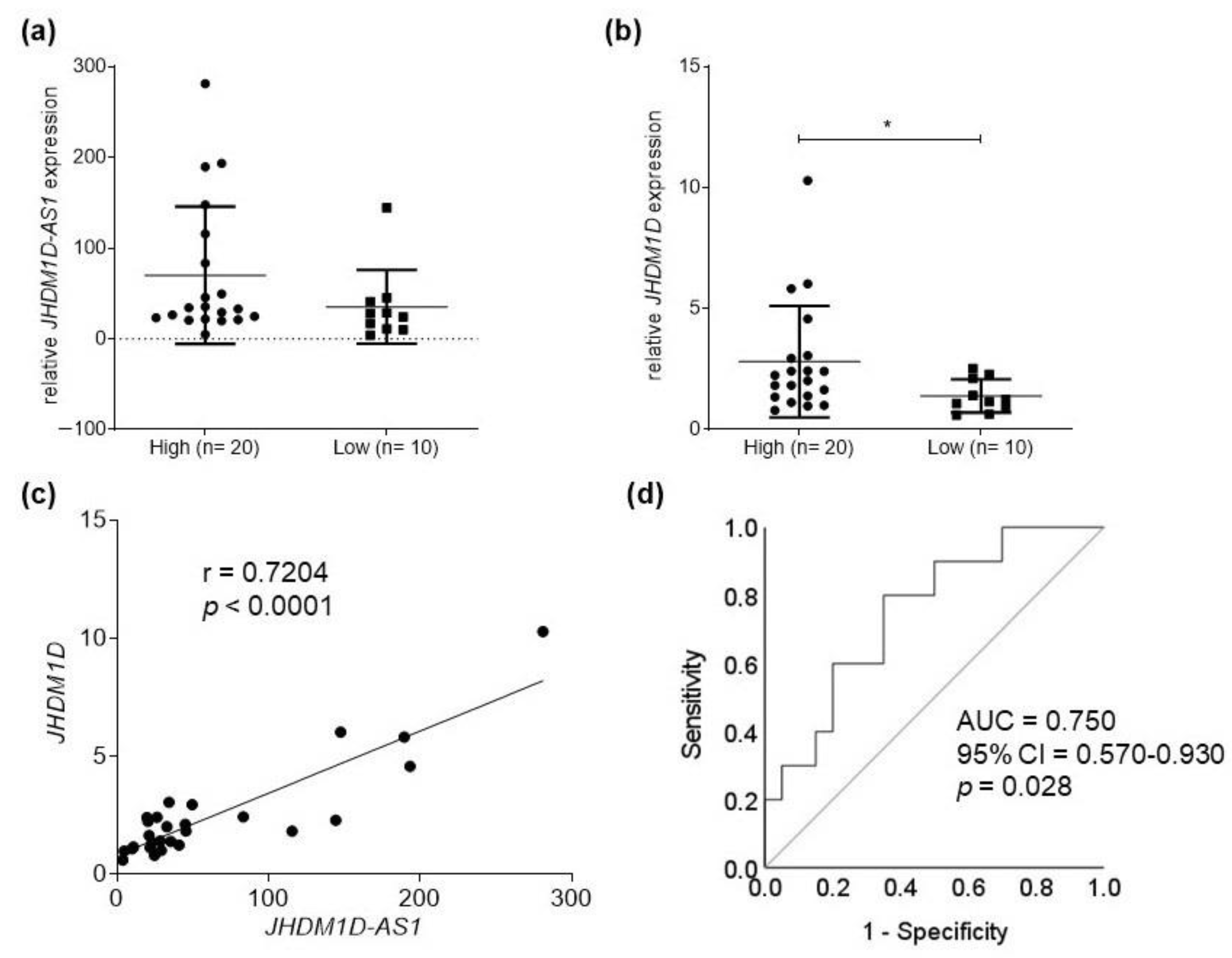
2.1. JHDM1D-AS1 and JHDM1D Expression in Bladder Tumor Samples

Despite the higher expression of lncRNA JHDM1D-AS1 in high-grade tumor samples, no significant difference was observed (Figure 1a). Nevertheless, JHDM1D gene expression was 2.01 times greater in high-grade tumors than in low-grade tumors (p = 0.0392) (Figure 1b). The analysis of the correlation between JHDM1D and JHDM1D-AS1 expression revealed a moderately positive correlation (r = 0.7204, p < 0.0001) (Figure 1c). In addition, analysis of ROC curves showed that the combination of JHDM1D and JHDM1D-AS1 had potential prognostic value (p = 0.028), demonstrating their ability to distinguish between low- and high-grade tumors (Figure 1d).

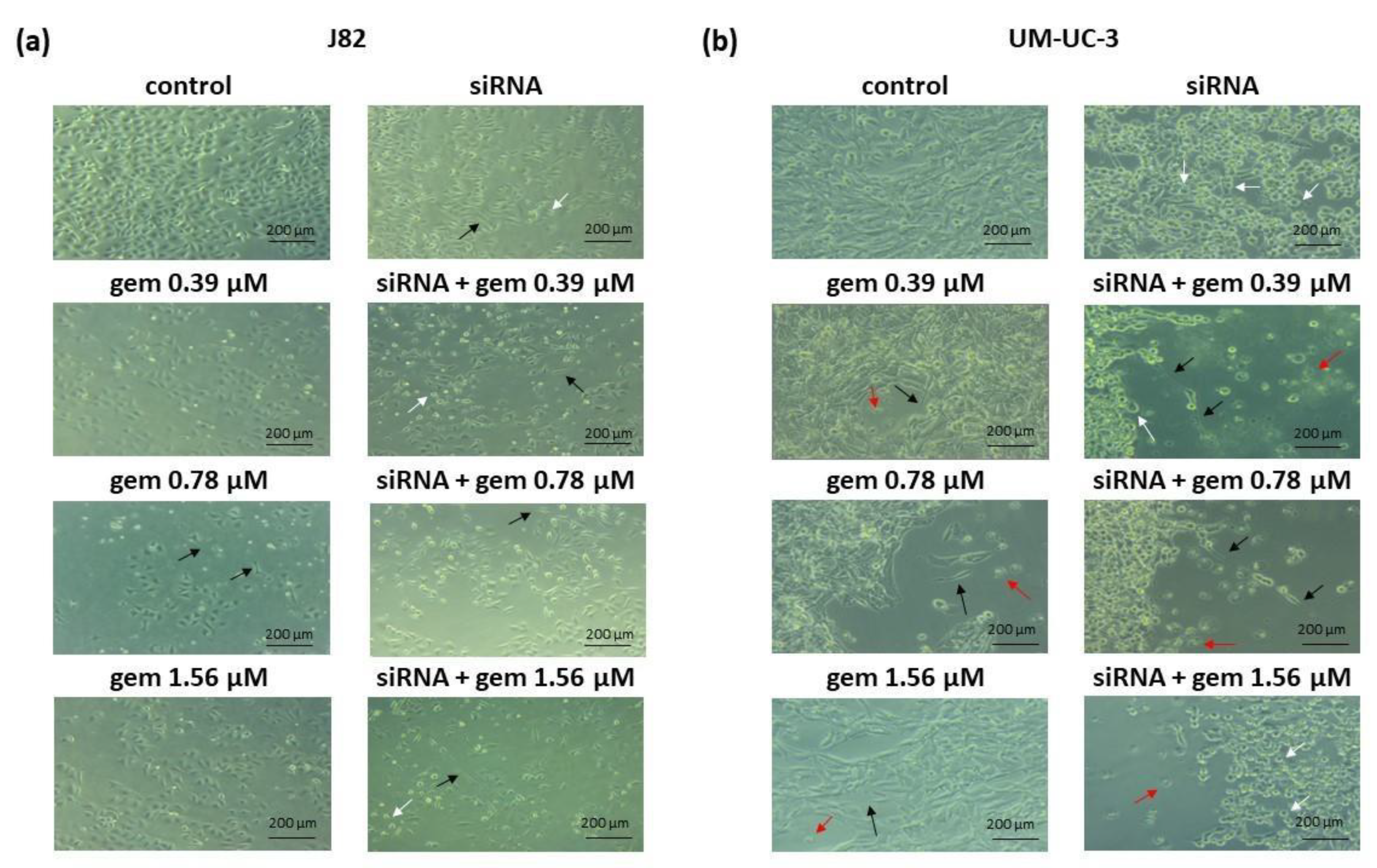
2.2. Cell Viability and Morphology

Compared to untreated and siRNA-JHDM1D-AS1 treated cells, J82 cells treated with 1.56 µM of gemcitabine exhibited a significant reduction in viability. In addition, in cells treated with siRNA-JHDM1D-AS1 combined with 0.39 and 0.78 µM gemcitabine, there was a significant reduction compared to the respective gemcitabine concentration alone (Figure 2a). Compared to untreated cells, cell viability decreased significantly in all treated UM-UC-3 cells (Figure 2b).
Figure 3 shows that treatments with siRNA-JHDM1D-AS1 alone or in combination with gemcitabine significantly reduced the cell density of J82 and UM-UC-3 lines compared to untreated cells. Furthermore, the treatments induced alterations in cell morphology, including long cell extensions, irregular appearance, rounding, and death.

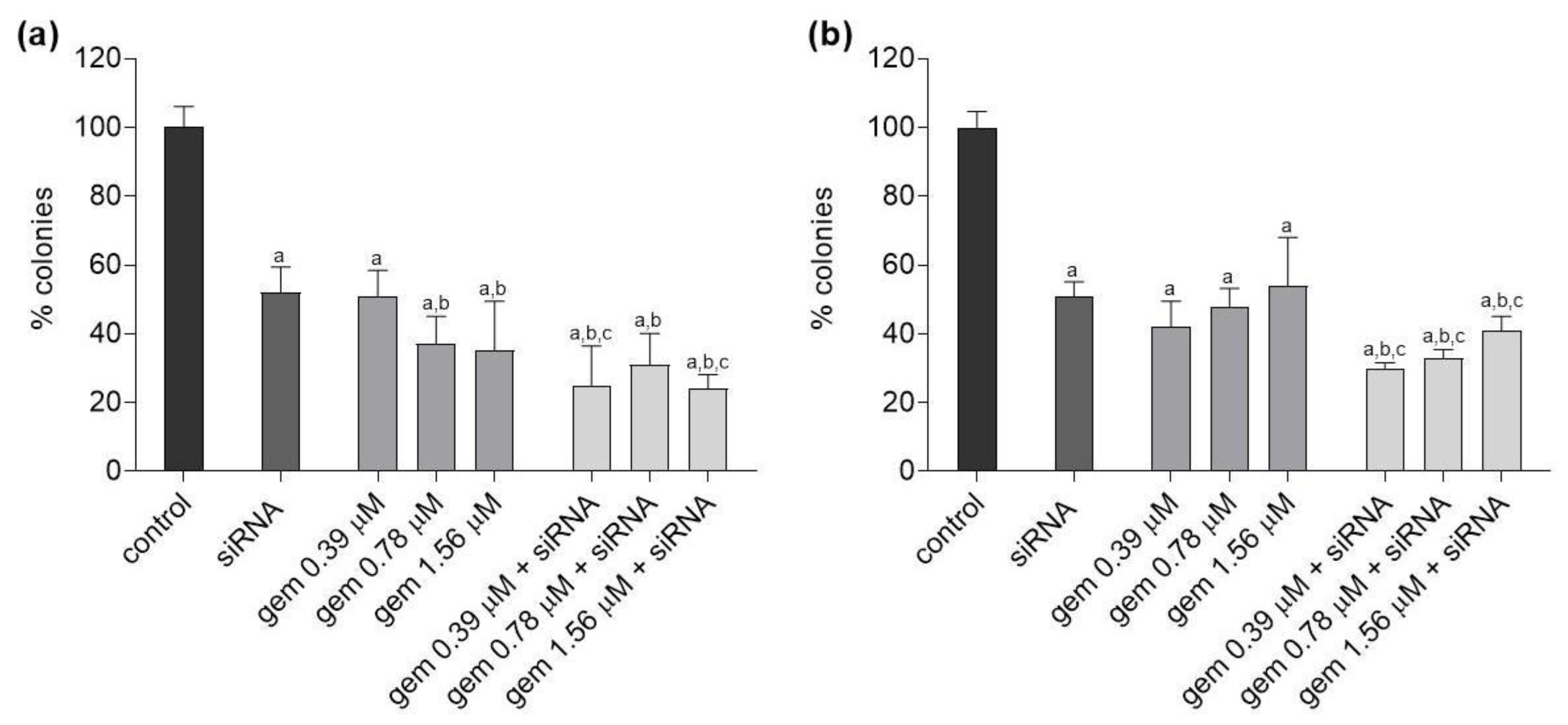
2.3. Clonogenic Survival

All treatments significantly decreased the clonogenic survival of J82 cells relative to untreated cells. Compared to cells treated with siRNA-JHDM1D-AS1, clonogenic survival was significantly reduced in this cell line after treatment with gemcitabine at 0.78 and 1.56 µM and after treatment with all gemcitabine and siRNA-JHDM1D-AS1 combinations. In addition, the combinations of gemcitabine 0.39 and 1.56 µM with siRNA-JHDM1D-AS1 caused a significant reduction in the cell colony formation compared to the respective concentration of gemcitabine alone (Figure 4a).
In UM-UC-3 cells, all treatments led to a significant decrease in clonogenic survival compared to untreated cells. In all combination treatments, clonogenic survival was reduced compared to siRNA-JHDM1D-AS1 alone and the respective concentration of gemcitabine alone (Figure 4b).

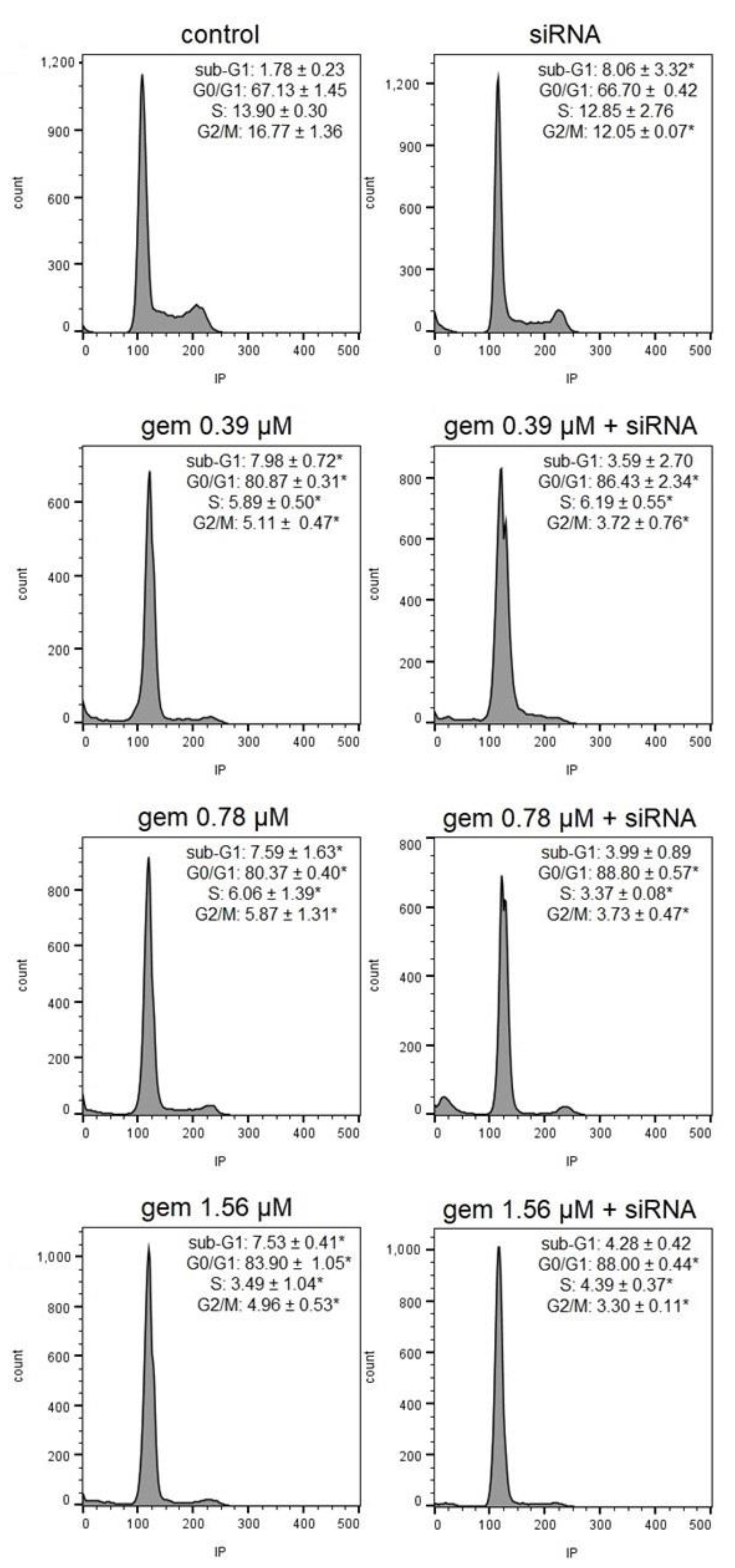
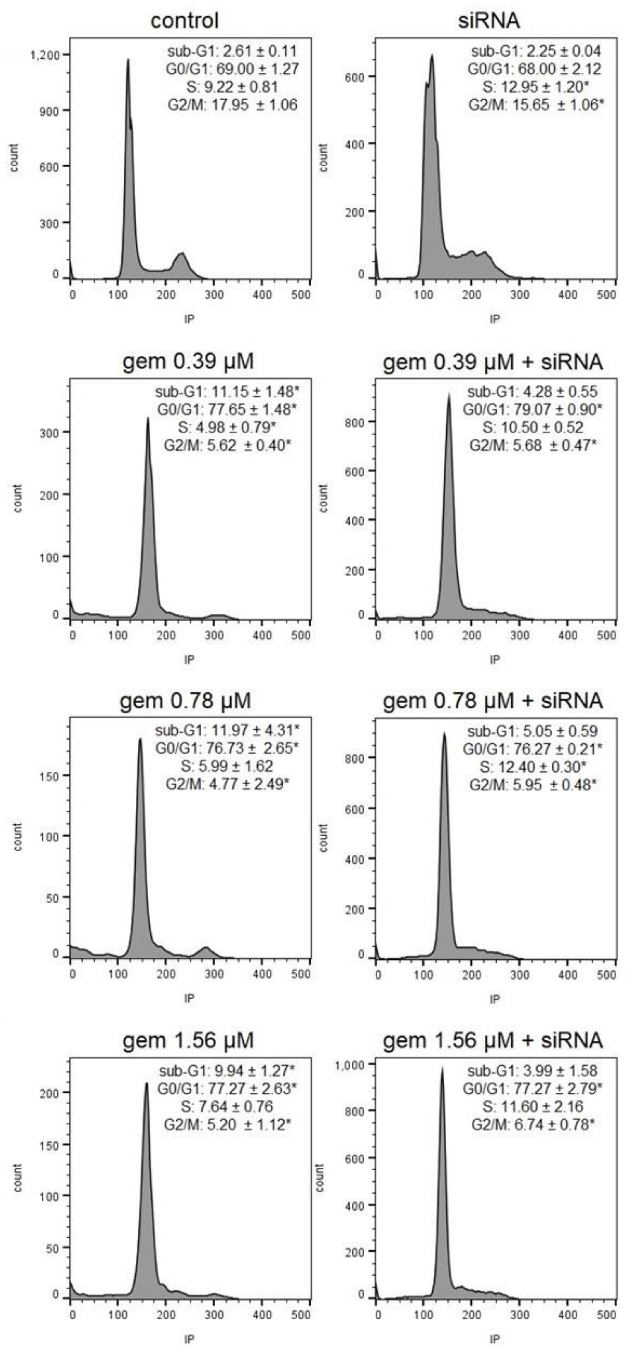
2.4. Cell Cycle Progression
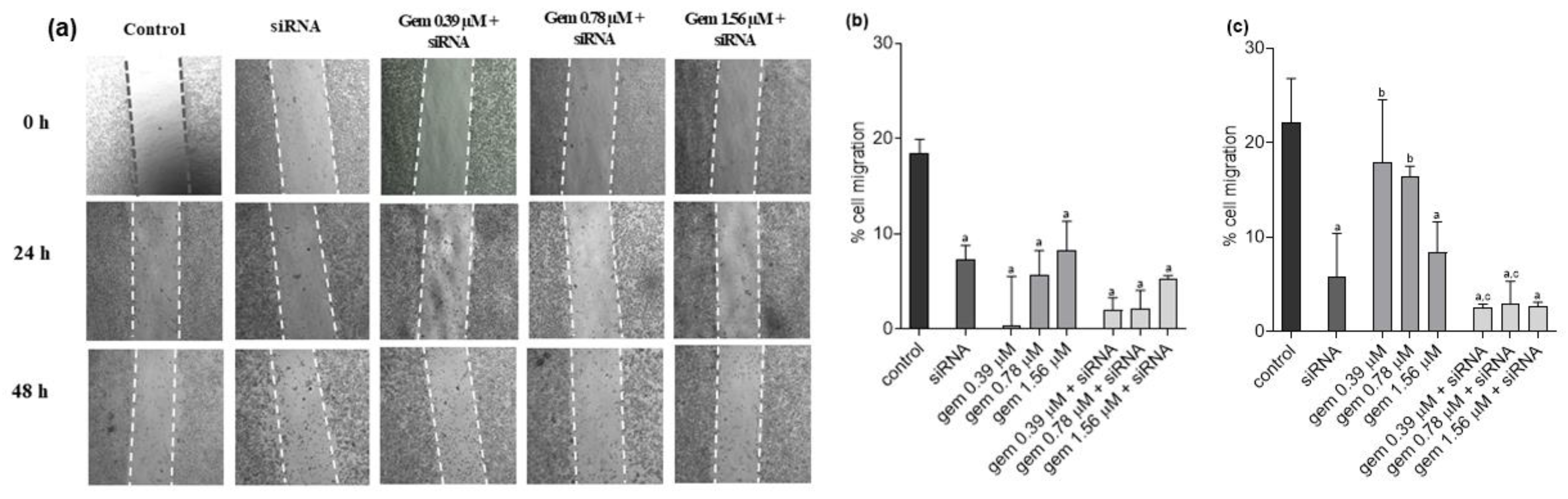
In the J82 cell cycle analysis, cell cycle arrest was observed at the G0/G1 phase in the cells treated with all concentrations of gemcitabine and with gemcitabine and siRNA-JHDM1D-AS1. Cell cycle arrest at the G0/G1 phase was significantly higher in the groups submitted to the combined treatment with gemcitabine and siRNA-JHDM1D-AS1, when compared to the groups treated solely with the respective gemcitabine concentrations. This interference was accompanied by a diminished population of cells in S and G2/M phases. In addition, there was an increase in the sub-G1 content in the cells treated with gemcitabine and siRNA-JHDM1D-AS1 alone (Figure 5 and Figure S3).
Similarly, in the UM-UC-3 cell line, there was mild cell cycle arrest at the G0/G1 phase in the cells treated with all concentrations of gemcitabine and with gemcitabine and siRNA JHDM1D-AS1. The combined treatment led to a significant increase in cell populations in the S phase, when compared to cells treated solely with the respective gemcitabine concentrations and to the untreated control. Additionally, an increase in the sub-G1 content was observed in the cells treated with gemcitabine alone. In the UM-UC-3 cells treated with siRNA JHDM1D-AS1 alone, there was cell cycle arrest at the S phase (Figure 6 and Figure S4).

2.5. Cell Migration

In J82 cells, we could observe that all treatments reduced the cell migration compared to untreated cells, in both analyzed times. In 24 h, there was also a significant reduction in the migration process after the treatment with gemcitabine at 0.39 and 0.78 µM and with the two highest combination treatments when compared to siRNA-JHDM1D-AS1 alone. The association of gemcitabine 1.56 µM and siRNA-JHDM1D-AS1 also reduced the J82 cell migration significantly compared with the same concentration of gemcitabine alone (Figure 7a,b). After 48 h of treatment, there was a consistent reduction in cell migration compared to untreated cells, for all treatments. In addition, all the combined treatments reduced cell migration significantly when compared to the groups treated solely with gemcitabine (Figure 7a,c).
For UM-UC-3 cells, a reduction in the migration process was observed after all 24 h treatments compared to the untreated cells (Figure 8a,b). After 48 h, this effect was maintained in the cells treated with siRNA-JHDM1D-AS1, with gemcitabine at 1.56 µM, and with all combinations of gemcitabine and siRNA-JHDM1D-AS1. In addition, there was a significant reduction in the number of migrated cells after treatment with 0.39 or 0.78 µM of gemcitabine combined with siRNA-JHDM1D-AS1 compared with the respective concentration of the chemotherapy compound alone (Figure 8a,c). Importantly, although there was no inhibition of migration after treatment with gemcitabine, it is possible to observe a reduction in cell density, which is consistent with the results of cell viability (Figures S5 and S6).

3. Discussion

Non-coding long RNAs play an important role in carcinogenesis [24,27]. Therefore, a better understanding of the functional role of lncRNAs could help in cancer diagnosis, prognosis, and treatment. Based on this, first, we conducted a marker lesion study using JHDM1D and lncRNA JHDM1D-AS1 expression to distinguish between low-and high-grade bladder tumors. The results showed different expression levels of JHDM1D in low- and high-grade tumors, suggesting a possible role of this gene in bladder tumor progression. This gene regulates many biological processes, including differentiation, development, and the growth of several cancer cells [23,28]. JHDM1D was found to be upregulated in prostate cancer tissue, and its chemical inhibition reduced proliferation and induced apoptosis of prostate cancer cells [22]. Nevertheless, Osawa et al. [29] demonstrated that this gene is overexpressed in tumor cells under nutritional stress and is associated with the suppression of tumor growth via down-regulation of pro-angiogenic factors in cancer cells and xenograft mouse models. This discrepancy may indicate a different role of JHDM1D in different tumor conditions and stages [23].
Antisense lncRNAs are transcribed from the promoter regions of the coding gene and can affect not only the expression of sense genes but also the expression of distant genes [30]. Similarly to Kondo et al. [31], we also identified a long noncoding antisense transcript, JHDM1D-AS1, whose expression increased proportionally to JHDM1D levels. The expression JHDM1D-AS1 did not differ between low- and high-grade samples, even though higher expression levels of the referred lncRNA are observed in high-grade tumors. The number of tissue samples analyzed may have been a limitation of the present study. A greater number of bladder tumor specimens could enable a clearer demonstration of the different expression levels of lncRNA JHDM1D-AS1. Indeed, data available from The Cancer Genome Atlas (TCGA), comprising a total of 252 tumors and 19 normal samples, show higher expression of this lncRNA in high-grade tumors compared to low-grade tumors. However, when JHDM1D and JHDM1D-AS1 expression levels were used in combination, potential prognostic value for the progression of bladder tumors was indicated. Thus, JHDM1D-AS1 silencing in high-grade cell lines, subsequently resulting in changes in biological behavior, should be considered.
Nowadays, gemcitabine is present in several standard protocols for muscle-invasive bladder cancer chemotherapy due to its efficacy and better toxicity profile [32]. Chemotherapy resistance is a significant barrier to cancer treatment, as it drastically reduces the efficacy of treatment and is strongly associated with tumor progression and recurrence. Several studies have demonstrated the influences of lncRNAs in the modulation of cellular pathways involved in chemoresistance, including in bladder cancer [13,19,33,34,35]. Despite the increased levels of lncRNA JHDM1D-AS1 in bladder cancer cells, there were no studies investigating the functional relevance of this lncRNA in high-grade bladder cancer cells or the connection of this lncRNA to gemcitabine treatment in these tumors. Consequently, in the second part of this study, we examined the functional role of JHDM1D-AS1 and the effects of combining various gemcitabine concentrations with JHDM1D-AS1 inhibition on high-grade bladder-tumor cell lines and their chemosensitivity.
Initially, the effect of silencing JHDM1D-AS1 expression was examined. Cytotoxicity related to the knockdown of this lncRNA in bladder carcinoma cells was observed. Some authors have observed that this lncRNA can be targeted to inhibit other tumor types. Wu et al. suggested that lncRNA JHDM1D-AS1 promote gastric cancer progression by upregulating oncogenic PRAF2 level by trapping miR-450a-2-3p [24]. Moreover, Yao et al. showed that, in small cell lung cancer, JHDM1D-AS1 binding attenuates proteasome-mediated degradation of DHX15, an ATP-dependent RNA helicase involved in ribosome biogenesis, enhancing growth and metastasis [25]. In comparison to the isolated chemical, it was observed that gemcitabine combined with siRNA-JHDM1D-AS1 decreased the viability of J82 cells, but not of UM-UC-3 cells. Additionally, the clonogenic survival assay was performed to establish the long-term effects of the treatment. Evaluation of the clone formation is one of the most important tests for determining the effect of a chemotherapy drug on tumor cells, as it indicates if the cells lost the ability to divide and proliferate [36]. In the two investigated cell lines, the combinations of gemcitabine and the siRNA for JHDM1D-AS1 decreased the proliferative capacity of the cells relative to the drug alone and the treatment with the siRNA alone. These findings indicate that silencing JHDM1D-AS1 improved the sensitivity of tumor cells to chemotherapy. It should be noted that, in both cells, treatment with siRNA alone or in combination led to a reduction in colony formation that was greater than the XTT-observed reduction in cell viability. These results also indicate that the treatment’s impact is mediated by DNA-damaging processes that comprise the reproductive integrity of cells. In particular, Da Silva et al. [37] showed that, in bladder cancer cell lines, gemcitabine induces cytostatic effects that, according to the findings of the present investigation, seem to be more apparent following lncRNA suppression. Consistently with previous findings, morphological changes indicative of loss of adhesion and cell death, such as cells with irregular and rounded shapes and the presence of cell debris, were more pronounced after the combined treatment of gemcitabine and siRNA-JHDM1D-AS1, along with a decrease in cell density, compared to treatments with the isolated compounds.
Cell migration plays an essential role in tumor invasion and metastasis, and can be assessed by observing cell migration, after treatment, through a gap in the cell monolayer [38]. The relationship between long non-coding RNAs and tumor cell metastasis has already been reported by Dhamija et al. [39]. In addition to modulating the regulation of genes related to metastasis, these molecules contribute to the regulation of the epithelial–mesenchymal transition (EMT) process and act on in vitro cell invasiveness and migration [39]. Furthermore, lncRNA JHDM1D-AS1 has been linked with the development and metastasis of non-small cell lung cancer and the proliferation and migration of gastric cancer cells [24,25]. In the present study, JHDM1D-AS1 silencing reduced cell migration in both cell lines when compared to the untreated control. Moreover, gemcitabine concentrations combined with JHDM1D-AS1 silencing led to a decrease in cell migration when compared to treatment with gemcitabine alone or with siRNA in both cell types, and the effects become more pronounced after 48 h. Thus, it appears that the lncRNA JHDM1D-AS1 is associated with the metastatic process and enhances the anti-migratory effects of gemcitabine in high-grade bladder-carcinoma cells.
Studies on the impact of gemcitabine on the cell cycle have yielded apparently inconsistent results about cell cycle kinetics [37,40]. G1/G0-phase arrest was detected in the two TP53-mutant cell lines treated with gemcitabine, and it was slightly but significantly increased in J82 cells when gemcitabine was combined with JHDM1D-AS1 silencing. Despite a slight increase in UM-UC-3 cell population in the G1/G0 phase, the most pronounced cell cycle perturbations detected following the combined treatment were cell cycle arrests in the S and G2/M phases. Thus, the more pronounced alterations in cell cycle kinetics were observed after treatment with gemcitabine and lncRNA inhibition.
In conclusion, our findings indicate that the long non-coding RNA JHDM1D-AS1 may be related to high-grade bladder cancers. In J82 and UM-UC-3 high-grade bladder-cancer cell lines, the silencing of this lncRNA produced decreased in cell viability and clone formation, morphological alterations, and decreased cell migration. In addition, the silencing of JHDM1D-AS1 combined with gemcitabine treatment resulted in enhanced cytotoxicity, higher rates of cell death, lower cell migration capacity, and cytostatic effects when compared to treatment with the isolated compound. Therefore, it appears that JHDM1D-AS1 plays an important role in the progression of bladder cancer, and its silencing increases the gemcitabine sensitivity of high-grade tumor cells. In future investigations, inhibiting this lncRNA may be considered as a strategy for achieving a therapeutic response with a lower concentration of gemcitabine, hence decreasing side effects.

4. Materials and Methods

4.1. Clinical Samples

A total of 30 fresh bladder cancer tissue samples, 20 histologically diagnosed as high-grade tumors and 10 as low-grade tumors, were collected at the University of Sao Paulo Biorepository (Sao Paulo, Brazil) and Amaral Carvalho Hospital’s tumor repository (Jau, Sao Paulo, Brazil). All tumor samples were collected via transurethral resection and histopathologically classified by a pathologist. The grading and stage were determined according to the World Health Organization (WHO) systems and Tumor-Node-Metastasis (TNM) 2017. All patients were male.
The study was approved by the Ethics Committee of the Sao Paulo State University (protocol 48193715.6.0000.5411), and all methods were performed following the approved guidelines.

4.2. Cell Lines, Culture Conditions, and Reagents

Two urothelial bladder cancer cell lines, acquired from the Cell Bank of Rio de Janeiro, Brazil, were used in the in vitro experiments: i. J82, J82, derived from high-grade (grade III) tumor with three point mutations in the TP53 gene (two in exon 8 (codon 271: Glu (GAG) → Lys (AAG); codon 274 Val (GTT) → Phe (TTT)) and one in exon 9 (codon 320 Lys (AAG) → Asn (AAC)) [41,42,43]; ii. UM-UC-3, derived from a high-grade tumor with a point mutation at TP53 (exon 4, codon 113: Phe (TTG) → Cys (TCG)) [41,42,43].
Both cell lines were cultivated in monolayers, using DMEM culture medium supplemented with 10% bovine serum and 100 U/mL of penicillin G, 100 U/mL of streptomycin (Sigma-Aldrich, Saint Louis, EUA), and 2.5 µg/mL of amphotericin B (Cristália, Itapira, Brazil), and maintained at 37 °C in an atmosphere of 5% CO2. All experiments were performed with exponentially growing cells [37]. Gemcitabine (Gemzar) was obtained from Eli Lilly do Brazil Ltd.a. Treatment with gemcitabine was set to 24 h. Dilutions were performed in nuclease-free water.

4.3. Expression and Knockdown Assays

Tissue biopsies were snap-frozen and stored at −80 °C. Total RNA was isolated using the RNeasy Mini Kit® (Qiagen, Hilden, Germany) according to the manufacturer’s protocol. RNA concentration and purity were determined using a NanoDrop spectrophotometer (Thermo Scientific, Waltham, MA, USA). RNA quality was analyzed using a 2100 Bioanalyzer (Agilent, Santa Clara, CA, USA), and only samples with an RNA integrity number (RIN) ≥ 6.0 were used. Complementary DNA (cDNA) was synthesized using the High Capacity Kit (Applied Biosystems, Waltham, MA, USA) with random priming according to the manufacturer’s instructions. Expression levels of the JHDM1D/KDM7A gene and JHDM1D-AS1 lncRNAs were analyzed using RT-qPCR. Endogenous reference genes (HSPCB and ACTB) were selected using the NormFinder software, version 5 [44].
For lncRNA JHDM1D-AS1 knockdown, J82 and UM-UC-3 cells were transfected with SMARTpool Lincode Human JHDM1D-AS1 siRNA (Horizon Discovery Ltd., Cambridge, UK), specific to human JHDM1D-AS1. Lipofectamine RNAiMAX (Invitrogen Life Technologies) was used as transfection agent for the siRNA-JHDM1D-AS1, according to the manufacturer’s protocol. Cells transfected with lincode non-targeting control siRNAs were used as a negative control. JHDM1D-AS1 knockdown in the two cell lines was confirmed 72 h post-transfection, by RT-qPCR (Figure S2). Total RNA was extracted with Quick-RNA™ MicroPrep ZymoSpin™ IC Columns kit and dosed in NanoDrop®. cDNA was confectioned using High Capacity® kit (Applied Biosystems), according to the manufacturer’s protocol. RTq-PCR was performed using SYBR Green (Thermo Fisher Scientific). GAPDH gene was used as endogenous control for normalization. Treatment time with siRNA-JHDM1D-AS1 was set to 48 h.

4.4. Cytotoxicity Assay (XTT)

The XTT assay (Cell Proliferation Kit II, ROCHE Diagnostics, Mannheim, Germany) was first used to define three concentrations of gemcitabine to be used in later experiments with J82 and UM-UC-3 cells. Gemcitabine concentrations were defined as 0.39, 0.78, and 1.56 µM. To evaluate the cytotoxic effects after treatment with gemcitabine + si-JHDM1D-AS1, 1 × 10⁴ cells were seeded into 24-well culture plates, and 24 h later, treated with siRNA-JHDM1D-AS1 (10 pmol). Forty-eight hours later, cells were treated with three different concentrations of gemcitabine (0.39 µM, 0.78 µM, and 1.56 µM) for 24 h. Untreated cells; cells treated with non-targeting siRNA, used as controls; and cells treated only with si-JHDM1D-AS1 or only with gemcitabine, were cultured in parallel. After treatment, cells were washed with Hank’s solution (0.4 g KCl, 0.06 g KH2PO4, 0.04 g Na2HPO4, 0.35 g NaHCO3, 1g glucose, and 8 g NaCl in 1000 mL H2O) and incubated at 37 °C with tetrazolium salt XTT (ROCHE Diagnostics, Mannheim, Germany) for 5 h. Immediately after incubation, absorbance was read in a spectrophotometer at a wavelength of 450 nm. Experiments were performed in technical triplicate.

4.5. Clonogenic Survival Assay

In order to evaluate the long-term effects of the treatment on J82 and UM-UC-3 cells, a clonogenic survival assay was performed. First, 3 × 10⁵ cells were seeded into 12-well culture plates and incubated for 24 h at 37 °C in an atmosphere of 5% CO2. After 24 h, cells were treated with siRNA-JHDM1D-AS1 (10 pmol) for 48 h and then treated with 0.39, 0.78, or 1.56 µM of gemcitabine for 24 h. Untreated cells and cells treated with non-targeting siRNA, used as controls, along with cells treated only with si-JHDM1D-AS1 or only with gemcitabine, were cultured in parallel. Past treatment time, the cells were washed with Hank’s solution, trypsinized, re-plated (1 × 10³ cells) into 12-well culture plates, and incubated at 37 °C in an atmosphere of 5% CO2. After 10 days (when untreated controls reach maximum confluence for colony formation experiences), the culture medium was removed, and cells were washed with Hank’s solution, fixed with 4% formaldehyde solution for 20 min, hydrated with 100% methanol for 20 min, and stained with crystal 0.5% violet solution dissolved in 25% methanol. A 33% acetic acid solution was used to remove the dying solution, and the contents of the plates were transferred to a 96-well plate. Absorbance was measured in a spectrophotometer at a wavelength of 570 nm [45] and was used to assess the percentage of colonies formed, that is, the reproductive capacity of the cells. Experiments were performed in technical triplicate.

4.6. Cell Morphology

To observe the effects of gemcitabine + si-JHDM1D-AS1 treatment on the morphologies of J82 and UM-UC-3 cells, 2 × 10⁵ cells of each cell line were seeded into 12-well culture plates, incubated for 24 h, and treated as described above. After treatment, the cells were observed and photographed under a phase-contrast optical microscope, at 200× magnification [46]. Experiments were performed in technical triplicate.

4.7. Wound Healing Assay

The wound healing assay was used to appraise cell migration after the combined treatment of gemcitabine and lncRNA JHDM1D-AS1 knockdown. For that, 4 × 10⁵ J82 and UM-UC-3 cells were seeded into 12-well culture plates and incubated at 37 °C and 5% of CO2; for 24 h. After incubation time, cells were treated with siRNA-JHDM1D-AS1 (10 pmol) for 48 h and then treated with 0.39, 0.78, or 1.56 µM of gemcitabine for 24 h. Immediately after gemcitabine treatment, a smooth and linear scratch was made in the middle of the cell monolayer, using a 200 µL pipette tip. Untreated cells and cells treated with non-targeting siRNA were used as controls, as cells treated only with siRNA-JHDM1D-AS1 or only gemcitabine were cultured in parallel. Cell migration over the scratch area was observed 24 and 48 h after gemcitabine treatment, at 40× magnification in an optical microscope with inverted light. Cell migration was evaluated and quantified using ImageJ® software (Adapted from Lima et al., 2020 [45]). Experiments were performed in technical triplicate.

4.8. Cell Cycle Progression

To analyze cell cycle alterations after the treatment with siRNA-JHDM1D-AS1 and gemcitabine, 4 × 10⁵ J82 and UM-UC-3 cells were seeded into 12-well culture plates for 24 h. Then, cells were treated with siRNA-JHDM1D-AS1 (10 pmol) for 48 h and afterward treated with 0.39, 0.78, or 1.56 µM of gemcitabine for 24 h. After treatment time, cells were washed with Hank’s solution, trypsinized, and centrifuged at 1000 rpm for 10 min. The supernatant was discarded, and the sediment was fixed with 70% ethanol and maintained at −20 °C for at least 12 h. Then, cells were washed with Hank’s solution, centrifuged, resuspended with 200 μL of labeling solution containing propidium iodide, and maintained in ice, protected from light, for 30 min [47]. Cell cycle kinetics were obtained by flow cytometry (BD FACSCalibur), and 20,000 events were detected. The percentages of cells in the Sub G1, G0/G1, S, and G2/M phases were analyzed using FlowJo® software. Experiments were performed in technical triplicate.

4.9. Statistical Analysis

The nonparametric Mann–Whitney test was used for differential gene expression analyses in the clinical samples, and the values are expressed as the mean ± SD. The relationships between the differentiated values were examined using Spearman’s rank correlation test (r = correlation coefficient), according to Akoglu (2018) [48]. A receiver operating characteristic (ROC) curve was constructed, and the area under the curve (AUC) was calculated to assess the specificity and sensitivity of the JHDM1D gene/lncRNA JHDM1D-AS1 in differentiating high- and low-grade tumors. Statistical significance was set to p < 0.05. Data from the cytotoxicity, clonogenic, cell cycle, and migration assays were analyzed using one-way ANOVA followed by Tukey’s multiple comparison test. Statistical significance was set to p < 0.05. All statistical analyses were performed using GraphPad Prism® 6.

Supplementary Materials

The following supporting information can be downloaded at: https://www.mdpi.com/article/10.3390/molecules28052412/s1. Figure S1. Chromosome location, structural organization, and length of JHDM1D and JHDM1D-AS1 genes. Figure S2. Relative expression of JHDM1D-AS1 in J82 and UM-UC-3 cells after 72h of siRNA-JHDM1D-AS1. Figure S3. Percentage of J82 cells in each phase of cell cycle after treatment with gemcitabine, siRNA JHDM1D-AS1, or gemcitabine combined with siRNA JHDM1D-AS1. Control: untreated cells; gem: gemcitabine. a: p < 0.05 compared to control; b: p < 0.05 compared to siRNA; c: p < 0.05 compared to respective gemcitabine concentration alone. Figure S4. Percentage of UM-UC-3 cells in each phase of cell cycle after treatment with gemcitabine, siRNA JHDM1D-AS1, or gemcitabine combined with siRNA JHDM1D-AS1. Control: untreated cells; gem: gemcitabine. a: p < 0.05 compared to control; b: p < 0.05 compared to siRNA; c: p < 0.05 compared to respective gemcitabine concentration alone. Figure S5. Photographs of cell migration in J82 cells after treatment with gemcitabine. Control: untreated cells; Gem: gemcitabine. Figure S6. Photographs of cell migration in J82 cells after treatment with gemcitabine. Control: untreated cells; Gem: gemcitabine.

Author Contributions

Conceptualization, D.M.F.S., A.L.V.S., K.R.M.L. and G.N.d.S.; Methodology, I.O.A.P., A.L.V.S. and A.P.B.L.; Formal Analysis, I.O.A.P., T.C.A., K.R.M.L. and G.N.d.S.; Investigation, I.O.A.P., T.C.A. and G.N.d.S.; Resources, D.M.F.S. and G.N.d.S.; Writing—Original Draft Preparation, I.O.A.P., G.N.d.S., T.C.A., A.L.V.S., K.R.M.L. and D.M.F.S.; Writing—Review and Editing, I.O.A.P., G.N.d.S., T.C.A. and D.M.F.S.; Supervision, D.M.F.S. and G.N.d.S.; Project Administration, D.M.F.S. and G.N.d.S.; Funding Acquisition, D.M.F.S. and G.N.d.S. All authors have read and agreed to the published version of the manuscript.

Funding

This study was supported by CAPES—Coordenação de Aperfeiçoamento de Pessoal de Nível Superior (Grants Finance Code 001 and PROPPI/UFOP Nº 03/2023), Universidade Federal de Ouro Preto (Grant PROPPI/UFOP Nº 03/2023) and CNPq—Conselho Nacional de Desenvolvimento Científico e Tecnológico (Grants 310905/2020-6 and 307201/2021-0).

Institutional Review Board Statement

The study was approved by the Ethics Committee of São Paulo State University (protocol 48193715.6.0000.5411).

Informed Consent Statement

Not applicable.

Data Availability Statement

Not applicable.

Acknowledgments

We appreciate the participation of all patients in this study. We are grateful for the assistance provided by the Laboratory of Epidemiology of the School of Medicine, Laboratory of Biochemistry and Clinical Research Laboratory of the Federal University of Ouro Preto.

Conflicts of Interest

The authors declare no conflict of interest.

References

  1. Sung, H.; Ferlay, J.; Siegel, R.L.; Laversanne, M.; Soerjomataram, I.; Jemal, A.; Bray, F. Global Cancer Statistics 2020: GLOBOCAN Estimates of Incidence and Mortality Worldwide for 36 Cancers in 185 Countries. CA Cancer J. Clin. 2021, 71, 209–249. [Google Scholar] [CrossRef] [PubMed]
  2. Mushtaq, J.; Thurairaja, R.; Nair, R. Bladder cancer. Surgery 2019, 37, 529–537. [Google Scholar] [CrossRef]
  3. Sanli, O.; Dobruch, J.; Knowles, M.A.; Burger, M.; Alemozaffar, M.; Nielsen, M.E.; Lotan, Y. Bladder cancer. Nat. Rev. Dis. Prim. 2017, 3, 17022. [Google Scholar] [CrossRef] [PubMed] [Green Version]
  4. Saginala, K.; Barsouk, A.; Aluru, J.S.; Rawla, P.; Padala, S.A.; Barsouk, A. Epidemiology of Bladder Cancer. Med. Sci. 2020, 8, 15. [Google Scholar] [CrossRef] [Green Version]
  5. Chang, S.S.; Boorjian, S.A.; Chou, R.; Clark, P.E.; Daneshmand, S.; Konety, B.R.; Pruthi, R.; Quale, D.Z.; Ritch, C.R.; Seigne, J.D.; et al. Diagnosis and Treatment of Non-Muscle Invasive Bladder Cancer: AUA/SUO Guideline. J. Urol. 2016, 196, 1021–1029. [Google Scholar] [CrossRef]
  6. Yu, C.; Hequn, C.; Jinbo, C.; Feng, Z.; Xiongbing, Z.; Jian, D. Gemcitabine/cisplatin versus methotrexate/vinblastine/doxorubicin/cisplatin for muscle-invasive bladder cancer: A systematic review and meta-analysis. J. Cancer Res. Ther. 2018, 14, 1260–1265. [Google Scholar] [CrossRef]
  7. Schlack, K.; Boegemann, M.; Steinestel, J.; Schrader, A.J.; Krabbe, L.M. The safety and efficacy of gemcitabine for the treatment of bladder cancer. Expert Rev. Anticancer Ther. 2016, 16, 255–271. [Google Scholar] [CrossRef]
  8. Brunton, L.L.; Hilal-Dandan, R.; Knollmann, B.C. As Bases Farmacológicas da Terapêutica de Goodman e Gilman, 13th ed.; AMGH: Goderich, ON, Cananda, 2018. [Google Scholar]
  9. Gutschner, T.; Diederichs, S. The hallmarks of cancer. RNA Biol. 2012, 9, 703–719. [Google Scholar] [CrossRef] [Green Version]
  10. Jarroux, J.; Morillon, A.; Pinskaya, M. History, Discovery, and Classification of lncRNAs. Adv. Exp. Med. Biol. 2017, 1008, 1–46. [Google Scholar] [CrossRef]
  11. Li, C.H.; Chen, Y. Targeting long non-coding RNAs in cancers: Progress and prospects. Int. J. Biochem. Cell Biol. 2013, 45, 1895–1910. [Google Scholar] [CrossRef]
  12. Forrest, M.E.; Khalil, A.M. Review: Regulation of the cancer epigenome by long non-coding RNAs. Cancer Lett. 2017, 407, 106–112. [Google Scholar] [CrossRef]
  13. Lima, A.P.B.; da Silva, G.N. Long Non-Coding RNA and Chemoresistance in Bladder Cancer—A Mini Review. Cancer Investig. 2023, 41, 164–172. [Google Scholar] [CrossRef]
  14. Flippot, R.; Beinse, G.; Boilève, A.; Vibert, J.; Malouf, G.G. Long non-coding RNAs in genitourinary malignancies: A whole new world. Nat. Rev. Urol. 2019, 16, 484–504. [Google Scholar] [CrossRef]
  15. Wang, F.; Li, X.; Xie, X.; Zhao, L.; Chen, W. UCA1, a non-protein-coding RNA up-regulated in bladder carcinoma and embryo, influencing cell growth and promoting invasion. FEBS Lett. 2008, 582, 1919–1927. [Google Scholar] [CrossRef] [Green Version]
  16. Luo, J.; Chen, J.; Li, H.; Yang, Y.; Yun, H.; Yang, S.; Mao, X. LncRNA UCA1 promotes the invasion and EMT of bladder cancer cells by regulating the miR-143/HMGB1 pathway. Oncol. Lett. 2017, 14, 5556–5562. [Google Scholar] [CrossRef] [Green Version]
  17. Fan, Y.; Shen, B.; Tan, M.; Mu, X.; Qin, Y.; Zhang, F.; Liu, Y. Long non-coding RNA UCA1 increases chemoresistance of bladder cancer cells by regulating Wnt signaling. FEBS J. 2014, 281, 1750–1758. [Google Scholar] [CrossRef]
  18. Ying, L.; Chen, Q.; Wang, Y.; Zhou, Z.; Huang, Y.; Qiu, F. Upregulated MALAT-1 contributes to bladder cancer cell migration by inducing epithelial-to-mesenchymal transition. Mol. Biosyst. 2012, 8, 2289–2294. [Google Scholar] [CrossRef]
  19. Liu, P.; Li, X.; Cui, Y.; Chen, J.; Li, C.; Li, Q.; Li, H.; Zhang, X.; Zu, X. LncRNA-MALAT1 mediates cisplatin resistance via miR-101-3p/VEGF-C pathway in bladder cancer. Acta Biochim. Biophys. Sin. 2019, 51, 1148–1157. [Google Scholar] [CrossRef]
  20. Li, C.; Cui, Y.; Liu, L.F.; Ren, W.B.; Li, Q.Q.; Zhou, X.; Li, Y.L.; Li, Y.; Bai, X.Y.; Zu, X.B. High Expression of Long Noncoding RNA MALAT1 Indicates a Poor Prognosis and Promotes Clinical Progression and Metastasis in Bladder Cancer. Clin. Genitourin. Cancer 2017, 15, 570–576. [Google Scholar] [CrossRef]
  21. Lee, K.-H.; Kim, B.-C.; Jeong, S.-H.; Jeong, C.W.; Ku, J.H.; Kim, H.H.; Kwak, C. Histone Demethylase KDM7A Regulates Androgen Receptor Activity, and Its Chemical Inhibitor TC-E 5002 Overcomes Cisplatin-Resistance in Bladder Cancer Cells. Int. J. Mol. Sci. 2020, 21, 5658. [Google Scholar] [CrossRef]
  22. Lee, K.H.; Hong, S.; Kang, M.; Jeong, C.W.; Ku, J.H.; Kim, H.H.; Kwak, C. Histone demethylase KDM7A controls androgen receptor activity and tumor growth in prostate cancer. Int. J. Cancer 2018, 143, 2849–2861. [Google Scholar] [CrossRef] [Green Version]
  23. Meng, Z.; Liu, Y.; Wang, J.; Fan, H.; Fang, H.; Li, S.; Yuan, L.; Liu, C.; Peng, Y.; Zhao, W.; et al. Histone demethylase KDM7A is required for stem cell maintenance and apoptosis inhibition in breast cancer. J. Cell Physiol. 2020, 235, 932–943. [Google Scholar] [CrossRef] [PubMed]
  24. Wu, M.; Liu, Y.; Pu, Y.-S.; Ma, Y.; Wang, J.-H.; Liu, E.-Q. JHDM1D-AS1 aggravates the development of gastric cancer through miR-450a-2-3p-PRAF2 axis. Life Sci. 2021, 265, 118805. [Google Scholar] [CrossRef] [PubMed]
  25. Yao, G.; Chen, K.; Qin, Y.; Niu, Y.; Zhang, X.; Xu, S.; Zhang, C.; Feng, M.; Wang, K. Long Non-coding RNA JHDM1D-AS1 Interacts with DHX15 Protein to Enhance Non-Small-Cell Lung Cancer Growth and Metastasis. Mol. Ther. Nucleic Acids 2019, 18, 831–840. [Google Scholar] [CrossRef] [Green Version]
  26. Li, J.; Han, L.; Roebuck, P.; Diao, L.; Liu, L.; Yuan, Y.; Weinstein, J.N.; Liang, H. TANRIC: An Interactive Open Platform to Explore the Function of lncRNAs in Cancer. Cancer Res. 2015, 75, 3728–3737. [Google Scholar] [CrossRef] [PubMed] [Green Version]
  27. Gao, X.; Liu, Q.; Chen, X.; Chen, S.; Yang, J.; Liu, Q.; Cheng, Y. Screening of tumor grade-related mRNAs and lncRNAs for Esophagus Squamous Cell Carcinoma. J. Clin. Lab Anal. 2021, 35, e23797. [Google Scholar] [CrossRef]
  28. Yang, X.; Wang, G.; Wang, Y.; Zhou, J.; Yuan, H.; Li, X.; Liu, Y.; Wang, B. Histone demethylase KDM7A reciprocally regulates adipogenic and osteogenic differentiation via regulation of C/EBPalpha and canonical Wnt signalling. J. Cell Mol. Med. 2019, 23, 2149–2162. [Google Scholar] [CrossRef]
  29. Osawa, T.; Muramatsu, M.; Wang, F.; Tsuchida, R.; Kodama, T.; Minami, T.; Shibuya, M. Increased expression of histone demethylase JHDM1D under nutrient starvation suppresses tumor growth via down-regulating angiogenesis. Proc. Natl. Acad. Sci. USA 2011, 108, 20725–20729. [Google Scholar] [CrossRef] [Green Version]
  30. Tee, A.E.; Liu, B.; Song, R.; Li, J.; Pasquier, E.; Cheung, B.B.; Jiang, C.; Marshall, G.M.; Haber, M.; Norris, M.D.; et al. The long noncoding RNA MALAT1 promotes tumor-driven angiogenesis by up-regulating pro-angiogenic gene expression. Oncotarget 2016, 7, 8663–8675. [Google Scholar] [CrossRef]
  31. Kondo, A.; Nonaka, A.; Shimamura, T.; Yamamoto, S.; Yoshida, T.; Kodama, T.; Aburatani, H.; Osawa, T. Long Noncoding RNA JHDM1D-AS1 Promotes Tumor Growth by Regulating Angiogenesis in Response to Nutrient Starvation. Mol. Cell. Biol. 2017, 37, e00125-17. [Google Scholar] [CrossRef] [Green Version]
  32. Lobo, N.; Mount, C.; Omar, K.; Nair, R.; Thurairaja, R.; Khan, M.S. Landmarks in the treatment of muscle-invasive bladder cancer. Nat. Rev. Urol. 2017, 14, 565–574. [Google Scholar] [CrossRef]
  33. Malek, E.; Jagannathan, S.; Driscoll, J.J. Correlation of long non-coding RNA expression with metastasis, drug resistance and clinical outcome in cancer. Oncotarget 2014, 5, 8027. [Google Scholar] [CrossRef] [Green Version]
  34. Pan, J.; Li, X.; Wu, W.; Xue, M.; Hou, H.; Zhai, W.; Chen, W. Long non-coding RNA UCA1 promotes cisplatin/gemcitabine resistance through CREB modulating miR-196a-5p in bladder cancer cells. Cancer Lett. 2016, 382, 64–76. [Google Scholar] [CrossRef]
  35. An, Q.; Zhou, L.; Xu, N. Long noncoding RNA FOXD2-AS1 accelerates the gemcitabine-resistance of bladder cancer by sponging miR-143. Biomed. Pharmacother. 2018, 103, 415–420. [Google Scholar] [CrossRef]
  36. Munshi, A.; Hobbs, M.; Meyn, R.E. Clonogenic cell survival assay. Methods Mol. Med. 2005, 110, 21–28. [Google Scholar] [CrossRef]
  37. da Silva, G.N.; de Camargo, E.A.; Salvadori, D.M. Toxicogenomic activity of gemcitabine in two TP53-mutated bladder cancer cell lines: Special focus on cell cycle-related genes. Mol. Biol. Rep. 2012, 39, 10373–10382. [Google Scholar] [CrossRef]
  38. Riahi, R.; Yang, Y.; Zhang, D.D.; Wong, P.K. Advances in Wound-Healing Assays for Probing Collective Cell Migration. SLAS Technol. 2012, 17, 59–65. [Google Scholar] [CrossRef]
  39. Dhamija, S.; Diederichs, S. From junk to master regulators of invasion: lncRNA functions in migration, EMT and metastasis. Int. J. Cancer 2016, 139, 269–280. [Google Scholar] [CrossRef]
  40. Montano, R.; Thompson, R.; Chung, I.; Hou, H.; Khan, N.; Eastman, A. Sensitization of human cancer cells to gemcitabine by the Chk1 inhibitor MK-8776: Cell cycle perturbation and impact of administration schedule in vitro and in vivo. BMC Cancer 2013, 13, 604. [Google Scholar] [CrossRef] [Green Version]
  41. Pandey, S.; Bourn, J.; Cekanova, M. Mutations of p53 decrease sensitivity to the anthracycline treatments in bladder cancer cells. Oncotarget 2018, 9, 28514. [Google Scholar] [CrossRef]
  42. Earl, J.; Rico, D.; Carrillo-de-Santa-Pau, E.; Rodriguez-Santiago, B.; Mendez-Pertuz, M.; Auer, H.; Gomez, G.; Grossman, H.B.; Pisano, D.G.; Schulz, W.A.; et al. The UBC-40 Urothelial Bladder Cancer cell line index: A genomic resource for functional studies. BMC Genom. 2015, 16, 403. [Google Scholar] [CrossRef] [Green Version]
  43. Rieger, K.M.; Little, A.F.; Swart, J.M.; Kastrinakis, W.V.; Fitzgerald, J.M.; Hess, D.T.; Libertino, J.A.; Summerhayes, I.C. Human bladder carcinoma cell lines as indicators of oncogenic change relevant to urothelial neoplastic progression. Br. J. Cancer 1995, 72, 683–690. [Google Scholar] [CrossRef] [PubMed] [Green Version]
  44. Andersen, C.L.; Jensen, J.L.; Orntoft, T.F. Normalization of real-time quantitative reverse transcription-PCR data: A model-based variance estimation approach to identify genes suited for normalization, applied to bladder and colon cancer data sets. Cancer Res. 2004, 64, 5245–5250. [Google Scholar] [CrossRef] [PubMed] [Green Version]
  45. Lima, A.P.B.; Almeida, T.C.; Barros, T.M.B.; Rocha, L.C.M.; Garcia, C.C.M.; da Silva, G.N. Toxicogenetic and antiproliferative effects of chrysin in urinary bladder cancer cells. Mutagenesis 2020, 35, 361–371. [Google Scholar] [CrossRef]
  46. da Silva, G.N.; de Castro Marcondes, J.P.; de Camargo, E.A.; da Silva Passos Junior, G.A.; Sakamoto-Hojo, E.T.; Salvadori, D.M. Cell cycle arrest and apoptosis in TP53 subtypes of bladder carcinoma cell lines treated with cisplatin and gemcitabine. Exp. Biol. Med. 2010, 235, 814–824. [Google Scholar] [CrossRef]
  47. Almeida, T.C.; Guerra, C.C.C.; De Assis, B.L.G.; de Oliveira Aguiar Soares, R.D.; Garcia, C.C.M.; Lima, A.A.; da Silva, G.N. Antiproliferative and toxicogenomic effects of resveratrol in bladder cancer cells with different TP53 status. Environ. Mol. Mutagen. 2019, 60, 740–751. [Google Scholar] [CrossRef]
  48. Akoglu, H. User’s guide to correlation coefficients. Turk. J. Emerg. Med. 2018, 18, 91–93. [Google Scholar] [CrossRef]
Figure 1. Expression of the JHDM1D gene (a) and JHDM1D-AS1 lncRNA (b) in patients with low- and high-grade bladder tumors. (c) Moderately positive correlation between JHDM1D and JHDM1D-AS1 expression (r = 0.7204) in low- and high-grade bladder tumor samples (Spearman correlation analysis). (d) Receiver operating characteristics (ROC) curves using the combination between JHDM1-AS1 and JHDM1D to distinguish between low- and high-grade bladder tumors. * p < 0.05.
Molecules 28 02412 g001
Figure 2. Percentage of viability in J82 (a) and UM-UC-3 (b) cell lines after treatment with gemcitabine, siRNA-JHDM1D-AS1, or gemcitabine combined with siRNA-JHDM1D-AS1. Control: untreated cells; gem: gemcitabine. a: p < 0.05 compared to control; b: p < 0.05 compared to siRNA; c: p < 0.05 compared to respective gemcitabine concentration alone.
Molecules 28 02412 g002
Figure 3. Morphologies of J82 (a) and UM-UC-3 (b) cells after treatment with gemcitabine, siRNA-JHDM1D-AS1, or gemcitabine combined with siRNA-JHDM1D-AS1. Control: untreated cells; gem: gemcitabine. Black arrows: elongated cells; white arrows: round cells; red arrows: dead cells. Phase-contrast microscope, ×200.
Molecules 28 02412 g003
Figure 4. Percentages of J82 (a) and UM-UC-3 (b) colonies after treatment with gemcitabine, siRNA JHDM1D-AS1, or gemcitabine combined with siRNA JHDM1D-AS1. Control: untreated cells; gem: gemcitabine. a: p < 0.05 compared to control; b: p < 0.05 compared to siRNA; c: p < 0.05 compared to respective gemcitabine concentration alone.
Molecules 28 02412 g004
Figure 5. Representative histograms and percentages of J82 cells in each phase after treatment with gemcitabine, siRNA JHDM1D-AS1, or gemcitabine combined with siRNA JHDM1D-AS1. Control: untreated cells; gem: gemcitabine; IP: propidium iodide. * p < 0.05 compared to control.
Molecules 28 02412 g005
Figure 6. Representative histograms and percentages of UM-UC-3 cells in each phase after treatment with gemcitabine, siRNA JHDM1D-AS1, or gemcitabine combined with siRNA JHDM1D-AS1. Control: untreated cells; gem: gemcitabine; IP: propidium iodide. * p < 0.05 compared to control.
Molecules 28 02412 g006
Figure 7. Photographs (a) and quantification (b,c) of cell migration in J82 cells after treatment with gemcitabine, siRNA JHDM1D-AS1, or gemcitabine combined with siRNA JHDM1D-AS1. Control: untreated cells; gem: gemcitabine. a: p < 0.05 compared to control; b: p < 0.05 compared to siRNA; c: p < 0.05 compared to respective gemcitabine concentration alone.
Molecules 28 02412 g007
Figure 8. Photographs (a) and quantification (b,c) of cell migration in UM-UC-3 cells after treatment with gemcitabine, siRNA JHDM1D-AS1, or gemcitabine combined with siRNA JHDM1D-AS1. Control: untreated cells; gem: gemcitabine. a: p < 0.05 compared to control; b: p < 0.05 compared to siRNA; c: p < 0.05 compared to respective gemcitabine concentration alone.
Molecules 28 02412 g008
Disclaimer/Publisher’s Note: The statements, opinions and data contained in all publications are solely those of the individual author(s) and contributor(s) and not of MDPI and/or the editor(s). MDPI and/or the editor(s) disclaim responsibility for any injury to people or property resulting from any ideas, methods, instructions or products referred to in the content.

