
Advanced Nano-Drug Delivery Systems in the Treatment of Ischemic Stroke

1
Key Laboratory of Neuropharmacology and Translational Medicine of Zhejiang Province, School of Pharmaceutical Sciences, Zhejiang Chinese Medical University, Hangzhou 310053, China
2
Interdisciplinary Institute for Medical Engineering, Fuzhou University, Fuzhou 350108, China
*
Author to whom correspondence should be addressed.
Molecules 2024, 29(8), 1848; https://doi.org/10.3390/molecules29081848
Submission received: 4 March 2024
/
Revised: 12 April 2024
/
Accepted: 15 April 2024
/
Published: 18 April 2024
(This article belongs to the Special Issue Advances in Nanomedicine and Nano-Based Drug Delivery System)
Abstract
:In recent years, the frequency of strokes has been on the rise year by year and has become the second leading cause of death around the world, which is characterized by a high mortality rate, high recurrence rate, and high disability rate. Ischemic strokes account for a large percentage of strokes. A reperfusion injury in ischemic strokes is a complex cascade of oxidative stress, neuroinflammation, immune infiltration, and mitochondrial damage. Conventional treatments are ineffective, and the presence of the blood–brain barrier (BBB) leads to inefficient drug delivery utilization, so researchers are turning their attention to nano-drug delivery systems. Functionalized nano-drug delivery systems have been widely studied and applied to the study of cerebral ischemic diseases due to their favorable biocompatibility, high efficiency, strong specificity, and specific targeting ability. In this paper, we briefly describe the pathological process of reperfusion injuries in strokes and focus on the therapeutic research progress of nano-drug delivery systems in ischemic strokes, aiming to provide certain references to understand the progress of research on nano-drug delivery systems (NDDSs).
1. Introduction
With the improvement of living standards, people’s diets have become more refined, often high in oil, fat, and sugar. This has increased the burden on cardiovascular health, leading to a rise in conditions such as hypertension, diabetes, and atherosclerosis [1,2,3,4]. These diseases are major contributors to the increasing frequency of strokes year by year. Strokes have consequently become the second leading cause of death worldwide, attributed to its narrow therapeutic window, high mortality and disability rates, and high recurrence rate [5]. Strokes are divided into ischemic and hemorrhagic strokes, with ischemic strokes (ISs) accounting for 71% of all strokes [6]. ISs are caused by vascular embolism, which leads to ischemia and hypoxia in brain tissue. The only FDA-approved treatment for an IS is tissue plasminogen activator (tPA), which is used to treat vascular embolisms by restoring blood flow through intravenous thrombolysis. However, a reperfusion injury has greater damage to cerebral neurons, triggering a cascade of events, mainly including, energy failure, loss of cellular ionic homeostasis, excitotoxicity, mitochondrial function impairment, reactive oxygen species (ROS) generation, and immune cell infiltration, resulting in neural inflammation [7,8,9].
As the world’s most populous country, China accounts for approximately one-third of global stroke deaths, posing a significant obstacle to people’s lives and health. Consequently, research on stroke treatment has become a prominent focus in China [10]. Currently, the main clinical modalities used for stroke treatment include surgical thrombolysis and pharmacological thrombolysis [11]. The intravenous thrombolysis of tPA within 4.5 h is effective in clinical treatment, and a thrombectomy performed within 24 h also yields benefits. However, these interventions carry a narrow therapeutic window and entail a certain risk of hemorrhage. Effective recovery is achieved in only a small percentage of patients due to time constraints. The reopening of blood flow can result in a reperfusion injury, substantial ROS production, and an inflammatory response that damages neurons [12,13,14]. To date, many studies report that the inhibition of neuroinflammation is effective in alleviating secondary injury after IS reperfusion [15,16]. The infiltration of immune cells, such as microglia, astrocytes, and NK cells, is the main cause of inflammation resulting from a reperfusion injury in ISs [17,18]. Among them, microglia mainly consist of M1 and M2 types, and it has been indicated that the transition from pro-inflammatory M1 microglia to anti-inflammatory M2 type is conducive to the recovery of stroke prognosis [19]. Moreover, the suppression of neuroinflammation and scavenging of ROS are key factors in enhancing prognostic recovery from strokes. Simultaneously, the salvage of ischemic penumbra neurons can be accomplished through the use of neuroprotective agents [20]. Edaravone, a free-radical scavenger commonly used in clinical treatment, can reduce BBB damage and inhibit inflammation. However, it faces significant challenges, including difficulty in crossing the BBB, poor bioavailability, and rapid clearance from the blood, all of which contribute to low drug efficacy [21]. To address these challenges, researchers have initiated studies in nanotechnology, aiming to convert drugs into nanoforms to enhance drug utilization and reduce side effects, thereby achieving the therapeutic goal [22].
For the past few decades, nanotechnology has been developing rapidly, and nano-drug delivery systems have shown great advantages [23]. Nano-drug delivery systems can improve the solubility of insoluble drugs, enhance the stabilization, and prolong the half-life in the body. The precise targeting and control of drug release could also be realized through additional targeting fragments or surface modifications in nano-drug delivery systems, ultimately achieving the purpose of increasing efficiency and reducing toxicity [24,25,26,27,28]. The BBB after an IS is somewhat compromised, with an increase in permeability, but not enough to fail completely and still have a strong ability to insulate against external substances. Fortunately, the application of nanotechnology shows large potential and a bright future for nano-drug delivery, facilitating improved drug utilization across the BBB and into the brain [29,30]. Interestingly, carriers can also treat diseases, for example, Prussian blue (PB), which has an ROS-scavenging effect [31]. This review briefly introduces the pathologic process of reperfusion in ISs and highlights recent studies of NDDSs in treating brain ischemic reperfusion injuries, aiming to offer references that could help bridge the gap between laboratory research and clinical application.
2. The Pathophysiology of ISs
Multiple complex pathological mechanisms are involved in brain damage caused by ISs [32,33,34]. Understanding these complex mechanisms not only deepens our understanding of the disorders but also aids researchers in facilitating the translation from the laboratory to clinical practice. At the onset of an IS, local ischemia and brain hypoxia lead to metabolic disorders in the intracerebral microenvironment, which then trigger a series of cascading injury processes, as shown in Figure 1 [35]. IS pathology begins with cerebral ischemia and hypoxia, culminating in neuronal apoptosis [36]. Energy failure is due to ischemia and hypoxia, which stimulate neuronal depolarization to release large amounts of glutamate. This process causes calcium influx and results in mitochondrial dysfunction [37]. At the same time, the accumulation of glutamate induces cellular neuro-excitotoxicity, the massive release of reactive oxygen and nitrogen species (RONS), and the penetration of peripheral immune cells to the brain [17,38,39]. After vascular reperfusion, oxygen and energy supply are restored, but mitochondrial dysfunction and the inability to process excess oxygen disrupt the balance between RONS production and clearance [40,41]. Excessive RONS would induce oxidative stress, which damages endothelial cells, and causes platelet aggregation and adhesion in microvessels, leading to the reformation of the thrombus and ultimately resulting in cerebral edema and bleeding [42,43,44]. This in turn further promotes immune cell infiltration, exacerbating the inflammatory response and causing neuroinflammation [45]. These reactions interact with each other and do not exist singularly. When an IS occurs, thrombolysis within a few hours can alleviate brain damage; however, secondary damage from cerebral ischemia–reperfusion poses a greater challenge [46,47].
Although cerebral neuronal damage in the core area of ischemia is irreversible, the penumbral area of cerebral ischemia affected by the spread of ROS and immune cell infiltration is recoverable [48,49]. Therefore, the semi-dark band in cerebral ischemic is an important target area for improving prognosis and restoring neurological function [5,50,51]. The cerebral ischemic penumbra can be rescued by suppressing the immune–inflammatory response and attenuating oxidative stress [52,53,54]. Some studies have found that administering exogenous mitochondria to the ischemic area can alleviate mitochondrial dysfunction. Exogenous mitochondria can use endogenous mechanisms to repair cellular damage, potentially resulting in therapeutic effects on the nerves after an IS [55,56,57].
In this section, we mainly focus on excitotoxicity, oxidative stress, and neural inflammation. Other mechanisms of injury will not be discussed further.
2.1. Excitotoxicity
Excitatory amino acids, such as glutamate and aspartate, serving as central neurotransmitters, play critical roles in transmitting messages in the nervous system [58,59]. Among them, glutamate is widely distributed throughout the brain and exerts a significant excitatory effect on central neurons [37]. Under physiological conditions, the brain microenvironment maintains homeostasis without excessive glutamate production, which can be metabolically regulated. However, when an ischemic stroke occurs, because the blood supply is interrupted, neurons are deprived of energy and oxygen, resulting in decreased ATP production. This leads to the abnormal functioning of the NA+-K+ ion pump in the cell membrane [60]. Meanwhile, N-methyl-D-aspartate receptors (NMDAR) regulate ion-gated pathways by binding glutamate, thereby exerting excitatory effects on neuronal cells [42,61]. Unfortunately, when substantial amounts of glutamate binding to aspartate receptors, it triggers a significant inward flow of Ca2+, resulting in calcium overload and NO− production [62]. NO− reacts with O2− to produce the destructive OONO−, exacerbating BBB damage and brain injury [63,64,65]. In conclusion, excitotoxicity arises from uncontrolled glutamate release and calcium overload, triggering a series of effects that ultimately result in the death of neurons [66,67]. Thus, excitotoxicity represents an important molecular mechanism contributing to IS damage [68].
2.2. Oxidative Stress
As a downstream consequence of an excitatory injury, oxidative stress emerges as another major molecular mechanism contributing to ISs [23]. Oxidative stress occurs when the oxidative homeostasis system is disrupted due to the massive release of free radicals [69]. Excessive free radicals cause cellular damage, DNA damage, skeleton disruption, and lipid peroxidation, ultimately resulting in neuronal damage and brain death [70,71]. In particular, the free radical blowout after reperfusion exposes neurons to even more severe challenges [72,73]. In addition, free radicals can stimulate the secretion of cytokines as well as the expression of adhesion molecules, which mediate inflammatory and immune responses, thereby exacerbating a brain tissue reperfusion injury [74].
2.3. Neural Inflammation
The inflammatory response plays a pivotal role in the pathological events associated with brain damage and repair processes in ischemic strokes [75]. Various inflammatory cells participate in the inflammatory reaction. Microglia, astrocytes, neutrophils, NK cells, and other immune cells constitute the major effector cells during an IS [76,77]. Here, we take microglia and NK cells as examples. Microglia are intrinsic immune cells residing in the brain that play an important role in CNS diseases and that can be classified into M1 and M2 phenotypes [78,79]. M1 microglia secrete massive ROS, pro-inflammatory factors, and protein hydrolases to promote inflammation and exacerbate neuronal injuries, whereas M2 microglia secrete anti-inflammatory factors, which attenuate inflammatory injuries and improve the recovery of brain injuries [75,80]. While the activation of microglia is initially intended to protect neuronal cells, their overactivation can lead to harmful inflammation and neuronal death [81]. Numerous studies have shown that facilitating the conversion of microglia from M1 to M2 types can reduce inflammation and improve stroke prognosis [52,82,83,84,85]. For NK cells, they exacerbate stroke injuries by increasing local inflammation and neuronal hyperactivation, ultimately resulting in neuronal cell death [86,87]. The reason for this is that NK cells, which are common immune cells in the body, are actively recruited into the site of injury when an IS occurs. Simultaneously, NK cells secrete a large number of cytokines that promote inflammatory damage, ultimately leading to irreversible brain damage. Although immune cells that initially infiltrate into brain tissue exacerbate brain damage and neurological dysfunction, they also later play beneficial roles, such as promoting glial scarring and the phagocytosis of debris, which are essential for wound healing [88].
3. Advanced NDDSs for ISs
Prolonging the time window of treatment and mitigating a secondary reperfusion injury are ideal for addressing ISs [89]. Fortunately, recent advances in NDDSs have emerged, offering promising possibilities for enhancing IS reperfusion therapy [90]. NDDSs are known for their small size, large surface area, controlled release, targeted modification, and high ability to penetrate the BBB [91,92]. In addition, NDDSs can be utilized to load hydrophobic, hydrophilic, and gene-based drugs while also functioning as labeling probes for tracer imaging of ISs [93,94]. In the realm of advanced NDDSs for an ischemic stroke (IS), common types include polymers, inorganic nanoparticles, liposomes, and cell membrane-coated nanoparticles [95,96].
3.1. Polymers
Polymers offer broad prospects for addressing drug delivery for ISs [29]. They excel as NDDSs for treating CNS disorders due to their exceptional qualities, including excellent biodegradability, high biocompatibility, and minimal toxicity [97,98,99]. There are numerous polymers, among which PLGA, poly(ethylene glycol) (PEG), and poly(lactic acid) (PLA) are common and the most widely studied [100]. The appropriate modification of polymeric nanoparticles can enhance brain targeting and achieve drug enrichment in the target region [100]. PLGA is synthesized from the polymerization of two monomers: lactic acid and hydroxyacetic acid. It is an FDA-approved drug excipient known for its high encapsulation efficiency and excellent biocompatibility [101,102]. Previous studies by Wang et al. have demonstrated that epidermal growth factor (EGF) and erythropoietin (EPO) have a stimulatory effect on stem cells, further promoting tissue repair [103,104]. Based on this premise, Wang et al. proceeded to explore the encapsulation of pegylated EGF in PLGA nanoparticles as well as the formulation of hydrogels through the encapsulation of EPO within two-phase particles composed of PLGA and poly(sebacic acid). This system, through the epidermis to the ischemic lesion area, was observed to effectively traverse the BBB and stimulate the differentiation of endogenous neural stem cells. This ultimately facilitated the notable restoration of tissue and nerve function in the mice [105]. A prospective approach to treating ISs could involve preventing neutrophil infiltration. Song et al. developed a rod-shaped PLGA nanoparticle loaded with piceatannol for targeted intervention in neutrophil–endothelial cell interactions. They drew inspiration from the fact that neutrophils exhibit a preference for phagocytosing elongated particles and that rod-shaped PLGA is readily phagocytosed by neutrophils [106] (Figure 2a). This strategy can protect neurons and attenuate stroke damage to the brain by reducing neutrophil adhesion to endothelial cells and reducing immune infiltration (Figure 2b). In addition, PLGA can be used to form nanoparticles containing superparamagnetic iron oxide and Cy7.5, serving a pivotal role in MRI and fluorescence imaging [107].
Surface modification with PEG prolongs in vivo circulation and decreases the immunogenicity of nanoparticles [108]. Curcumin is known as an anti-inflammatory and antioxidant, but weak hydrophilicity and chemical instability make its application challenging [109]. Wang et al. used an amphiphilic copolymer consisting of PEG and PLA to prepare NDDS-encapsulating curcumin, which improves the stability of curcumin in blood circulation. The NDDS could protect the BBB by inhibiting the decrease in tight junction proteins [110]. The results showed that this NDDS inhibited M1 microglia polarization and inflammatory damage, thereby further promoting the functional recovery of brain tissue in mice. In another interesting study, PEG was used for modification to prepare pH-sensitive polymers loaded with rapamycin (RAPA), named RAPA@NPs. A pH-sensitive link was employed to achieve acid-triggered drug release, Ce6 was selected as the near-infrared imaging agent, and Gd3+ chelator was chosen for implementing bimodal imaging [111]. The results showed that the prepared RAPA@NPs not only overcame the drawbacks, such as the poor solubility of RAPA itself, thereby improving the therapeutic efficiency of RAPA, but also exhibited good biocompatibility and acid-enhanced bimodal imaging capabilities. RAPA@NPs preferentially aggregated at the ischemic site of the brain and achieved significant neuroprotective effects. The study provides a promising NDDS for drug tracking, treatment, and the early diagnosis of an IS as well as a reference towards the accurate imaging and treatment of other diseases [111]. Similarly, Ding et al. developed PEG coupled with urokinase (UK), wherein UK: PEG-UK was mixed in a 1:1 ratio [112]. The results demonstrated that this NDDS provided dual targeting of the macro-vasculature and the microcirculation, showing excellent neurologic function scores and smaller infarcts in the MCAO model area.
Dendritic macromolecules are excellent candidates for biological and pharmaceutical applications [113,114,115]. Among them, polyamidoamine dendrimer (PAMAM) is the most widely studied dendrimer [116]. This can be attributed to their controllable size structure, good water solubility, extensive internal drug-carrying space, and the potential for external modification [117,118]. However, PAMAM suffers from susceptibility to clearance by the reticuloendothelial system (RES) and a lack of targeting ability [119]. Despite these limitations, PAMAM can acquire enhanced capabilities through modifying and transforming into more desirable carriers. Examples include the conjugation of ligands such as PEG, folate analogs, protein analogs (transferrin, lactoferrin), amino acids, and peptides [120,121,122,123]. Successful NDDSs based on PAMAM for the treatment of neuroinflammation and ISs have been reported, yielding satisfactory therapeutic outcomes [124,125,126,127].
3.2. Inorganic Nanoparticles
Inorganic nanoparticles possess unique physicochemical properties [90]. They are characterized by a controllable structure, modifiable surface, and high loading efficiency, showing great research value in disease treatment and diagnosis [131,132,133]. For instance, gold nanoparticles are easy to synthesize and functionalize for versatile applications, possessing specific physical, electrical, magnetic, and optical properties [134,135]. Silica and iron have natural properties that allow them to work for MRI tracking [136,137]. Fe3O4 and CeO2 have enzyme-like activities that contribute positively to the removal of excess ROS [138,139,140].
Expanding on last paragraph, given the capability of gold nanoclusters to penetrate the BBB and the anti-inflammatory and antioxidant properties of dihydrolipoic acid, Xiao et al. synthesized functionalized gold nanoclusters carrying dihydrolipoic acid [141,142,143,144]. After the gold nanoclusters carried the drug through the BBB and into the brain, the dihydrolipoic acid underwent reduction and exerted its scavenging effects [144]. By regulating microglia and polarizing them to the M2 type, this NDDS can reduce the inflammatory response and improve the neuronal survival rate. In addition, the clinical application of gold nanoparticles for the photothermal therapy of prostate cancer has been carried out [145,146].
Magnetic iron oxide nanoparticles have garnered increasing attention due to their unique properties [94]. Liu et al. developed co-doped Fe3O4 nanoenzymes to ameliorate the RONS overload injury caused by an IS [147]. In vitro cellular experiments demonstrated that Fe3O4 nanoenzymes were effective in ameliorating neuroinflammation caused by ISs, as well as significantly reducing the infarct volume in both transient and permanent stroke models. Wang et al. developed an MRI visualization technique to track and visualize transplanted stem cells, offering valuable insights to enhance the effectiveness of IS treatment [148]. Similarly, some studies used superparamagnetic iron oxide nanoparticles (SPIO) to label stem cells for imaging tracking in IS therapy [107,149,150]. In addition, iron oxide nanoparticles can be loaded with dexamethasone and L-carnosine peptide for targeted delivery in ISs, which already yield satisfactory experimental results. It demonstrated the feasibility that iron oxide nanoparticles can be used as an NDDS for the treatment of ISs [151]. Clinical approval of iron oxide-based MRI contrast agents has been obtained in Europe and the United States; however, challenges persist in their clinical translation [152]. Overall, iron oxide-based nanoparticles hold excellent potential for application and clinical translation [153,154,155,156,157].
CeO2 nanoparticles are effective free radical scavengers [158,159]. Li et al. developed CeO2 nanoparticles loaded with butylphthalide (NBP-CeO2 NPs), which have neuroprotective effects for ISs [160]. The results of long-term experiments demonstrated that NBP-CeO2 NPs can promote vascular repair and improve the behavioral functions of mice. The combination of free radical scavenging and neurovascular repair can significantly reduce reperfusion injury, and this treatment approach holds promise for applications in ISs. He et al. developed CeO2 nanoparticles encapsulated with zeolitic imidazolate framework-8-capped (CeO2@ZIF-8). Compared to bare CeO2, the CeO2@ZIF-8 exhibited higher stability and longer blood circulation time under physiological conditions [161]. Moreover, due to its peroxidase-like properties, ZIF-8 enhances the free radical scavenging ability of CeO2@ZIF-8 compared to free CeO2, showing excellent preventive and therapeutic effects in neuroprotective therapy for ISs. Liao et al. introduced the concept of mitochondrial microenvironmental regulation and developed an NDDS based on CeO2-targeting mitochondria [162] (Figure 3). Mitigating oxidative stress injury can be achieved by regulating the mitochondrial microenvironment to promote ischemic recovery.
In addition, manganese-based nanoparticles, including MnO2 and Mn3O4, have also received much attention in ISs [52,163,164]. In the news from 2023, during the European Congress of Radiology (ECR) in Vienna, Austria, the GE Healthcare Group announced that the completion of Phase I subject recruitment for its pioneering manganese-based macrocyclic magnetic resonance imaging (MRI) contrast agent in early clinical development program. To sum up, the integration of inorganic nanoparticles into ischemic treatment approaches holds great potential for enhancing therapeutic outcomes through the modulation of cellular processes and microenvironmental factors.
3.3. Liposomes
Liposomes, discovered by Bangham in 1965, are enclosed vesicles composed of ordered lipid bilayers [165]. The bilayers consist mainly of amphiphilic phospholipids with aqueous spaces inside. Thus, components with hydrophilic properties can be encapsulated in the aqueous cavity of the liposome, while components with hydrophobic properties can be encapsulated in the lipid bilayer [166] (Figure 4). Compared to other NDDSs, studies on liposomes are relatively well established. Due to their excellent biocompatibility, high safety profile, and noteworthy therapeutic efficacy, numerous liposome formulations have received FDA-approval, signifying the progression from laboratory investigation to clinical application [167,168]. Undoubtedly, the successful translation of liposomes from the laboratory to the clinic has garnered considerable attention from researchers across diverse fields, fostering further in-depth investigations into liposomes [169,170].
Plain liposomes are easily cleared by the reticuloendothelial system (RES), while PEGylated liposomes extend the drug’s circulation time in the bloodstream and mitigate the risk of recognition and clearance by immune cells [171]. Thomas et al. prepared a PEGylated liposome loaded with atorvastatin, which could accumulated at the ischemic lesion area effectively, reducing infarct volume and promoting neurological recovery [172]. The use of PEGylated liposomes for delivering neuroprotective agents also improves drug bioavailability in vivo. Notable examples include citicoline [173], lycopene [174], FK506 [175], and plasminogen activators [176]. In addition, the addition of some FC fragments, transferrin, and other kinds of ligands or stimuli-responsive fragments to liposomes can achieve active targeting and responsive release [177,178,179]. To address the phenomenon of neutrophil infiltration into the ischemic zone during an IS, resulting in the release of neutrophil extracellular traps (NETs) and subsequent neuronal damage, Sun et al. developed a smart liposome with ischemic lesion targeting and ROS-responsive release [180]. The results showed that the smart liposome could regulate NETs and promote microglia transformation to the M2 type to treat ISs, resulting in a cerebral infarction area approximately one-fourth that of the saline group. Li et al. investigated methods to enhance the delivery efficiency of the commercially available drug ginkgolide B (GB) by developing a liposomal formulation that binds GB to high-lipophilic docosahexaenoic acid (DHA) (Lipo@GB-DHA) [181]. The results indicated that when compared with the free GB group, the amount of GB in the target ischemic hemisphere in the Lipo@GB-DHA group was 2.2 times higher than that in the free GB group. Additionally, the Lipo@GB-DHA group showed a smaller infarct area, better inhibition of neuronal apoptosis, and improved recovery of neurological function. Yao et al. developed a pH-responsive fluorescent liposome probe and established a correlation between fluorescent imaging and neurologic deficiency scores. The approach provided a novel approach for assessing the extent of ISs in different acidic microenvironments [182]. Noteworthly, the transnasal administration of liposomes have achieved good therapeutic results in rat IS models [183,184].
In conclusion, substantial advancements have been achieved in utilizing liposomes as an NDDS. However, problems such as instability, leakage or poor targeting, and the sudden release of drugs remain to be solved in liposomes [185]. These issues highlight the critical need for the development of advanced technologies aimed at enhancing the properties of liposomes.
3.4. Cell Membrane-Coated Nanoparticles
Targeted nanomedicines hold promising potential in stroke therapy. However, some of them are intrinsically foreign and face the risk of being removed by the RES [186]. In order to address this obstacle, the researchers proposed a cell membrane-coated strategy. Cell membrane-coated nanoparticles possess the unique advantage of imparting biological characteristics by hiding traditional nanoparticles in natural cell membranes [187]. By selecting different kinds of cell membranes to modify the outer layers of NDDSs, NDDSs with enhanced surface functionality can be created to achieve diverse goals [188] (Figure 5). Cell membranes from different sources have different functional characteristics, as shown in Table 1. However, cell membranes still have some limitations [189]. Fortunately, it is possible to modify some targeting proteins or peptides on the membrane surface to further improve the targeting performance and ultimately achieve better therapeutic effects [190,191].
3.4.1. RBC Membrane-Coated Nanoparticles (RBC-NPs)
It is well known that RBCs are abundantly present within the blood and lack nucleus and mitochondria [189]. Consequently, the RBC membrane is easily extracted. The lifespan of RBCs in the body is about four months. During this time, the surface expression of the CD47 protein, which serves as a “do-not-eat-me” signal, prevents RBCs from being removed by the RES [203]. These characteristics of RBCs offer potential prerequisites for the development of RBC-NPs [204].
RBC-NPs were first reported by Zhang et al. in 2011 [205]. Compared with NPs coated with hydrophilic polymer PEG, which showed long circulation in vivo, the RBC-NPs retained RBCs’ biological properties, showing a longer circulation time. After that, researchers proposed diverse approaches to modify targeting peptides or ligands on the RBC membrane, which could not disrupt the RBC membrane but improve the brain targeting ability [206,207,208].
Shi et al. developed an engineered RBC-NP named Mn3O4@nanoerythrocyte-T7 (MNET) with smart oxygen regulation and free radical scavenging [209] (Figure 6a). Hemoglobin (HB) in RBCs underwent oxygen uptake and release, functioning like an oxygen sponge. Meanwhile, Mn3O4 NPs showed high biocompatibility and multiple antioxidant enzyme activities. By combining the advantages of RBCs, Mn3O4, and T7 peptides, the rescue of the ischemic microenvironment was finally realized using MNET. More excitingly, MNET could be used for continuous treatment via the oxygen spongy effect of HB. During an IS, the oxygen spongy function of HB can provide oxygen to the hypoxic area and reduce ischemic injury (Figure 6b). Oxygen reperfusion after thrombolysis causes the production of free radicals, and HB could play an important role in absorbing excess oxygen and reducing oxidative stress. Liu et al. also developed an integrated approach for acute ISs inspired by the oxygen spongy properties of HB [210]. They corrected abnormalities in glucose metabolism and provided energy to neurons by releasing methoxatin, which acts to activate the cellular Akt/GSK-3β pathway. It can be observed that the oxygen balance of the microenvironment and glucose metabolism are important for neuronal recovery. Lv et al. designed an erythrocyte membrane delivery system (SHp-RBC-NP/NR2B9C) with cerebral ischemic region-targeting and ROS-responsive release capabilities [211]. Based on the fact that ROS are released in large quantities from the ischemic region during the pathogenesis of an IS, phenylboronic acid with ROS-responsive release capabilities was added to the SHp-RBC-NP/NR2B9C. It was verified in the rat MCAO model that the SHp-RBC-NP/NR2B9C could successfully reach the ischemic lesion, release the neuroprotectant NR2B9C in the ischemic ROS microenvironment responsively, and reduce the volume of cerebral infarcts.
In conclusion, RBC-NPs, as the earliest membrane-coated NDDS studied, have tremendous possibilities through modification [212]. They have aroused researchers’ interest, and more researchers have begun to exploit the feasibility of fabricating hybrid RBC-NPs.
3.4.2. Platelet Membrane-Coated Nanoparticles
Platelets have a shorter lifespan compared to erythrocytes and are present in the blood in smaller numbers than erythrocytes [213]. Platelets, in addition to CD47, exhibit other expression clusters, such as CD55 and CD59, on their membrane surface, preventing phagocytosis and clearance by immune cells, consequently enhancing immune evasion [214]. In addition, platelets are known for their role in recognizing and repairing vascular damage and responding to inflammation. Consequently, nanoparticles encapsulated in platelet membranes can be directed toward the site of the injury for targeted delivery [215].
A precedent for biomimetic platelet membrane-coated nanoparticles, which mimic platelets and evade the immune system, was established by Zhang et al. in 2015 [193]. Since then, there has been a growing focus on platelet membrane-coated nanoparticles. For example, Cui et al. selected GB, a neuroprotective agent with anti-inflammatory and antioxidant properties, and coated GB with platelet membranes (PM-GB) [216]. Because platelet membranes could target inflammatory injuries, the concentration of GB at the injury site increased, resulting in improved drug delivery to the lesion. The results demonstrated that PM-GB was suitable for the treatment of ISs by inhibiting oxidative stress and reducing iron-related cell death. Zhao et al. designed a platelet membrane-coated nanoparticle with multi-function for the therapy of ISs [217] (Figure 7a). To be specific, when a platelet membrane adheres to inflamed neutrophils, platelet membrane-coated nanoparticles can then follow the neutrophils into the inflamed area. The platelet membrane-coated nanoparticles consisted of T7 peptide, PHis (an acid-responsive fragment), and MiRNA-Let-7c drugs. Based on the multiple functional fragments, this NDDS could be BBB targeting and swell to release miRNA to inhibit M1 cell polarization. Li et al. developed platelet-derived bio-nanobubbles with integrated diagnostic capabilities using platelet vesicles [218]. In addition to platelet vesicles, the formulation also includes γ-Fe2O3 and the NO precursor (L-arginine). The results showed that it could increase the flow of blood to the lesion site, prolong the treatment window through the vasodilatory effect of NO, and provide auxiliary diagnostic imaging [219] (Figure 7b). After that, Li further conducted the therapeutic mechanism of PAMNs. The results illustrated that it can rapidly dilate vessels and improve vascular flow, which is beneficial for the early-stage therapy of ISs (Figure 7c).
3.4.3. WBC Membrane-Coated Nanoparticles
WBCs are important blood cells of the body’s immune system, encompassing various types, such as NK cells, neutrophils, macrophages, and lymphocytes. As the body’s guardians, WBCs protect the body from disease and arrive at the injury site immediately when an IS occurs [227]. In contrast to RBCs and platelets, WBCs have nuclear structures and are less abundant, rendering their membrane isolation relatively challenging. To be noted, natural WBC membranes without modification have the capability of targeting inflammatory sites and tumor tissue [228,229].
Here, we mainly discuss membranes from neutrophils and macrophages to fabricate NDDSs. Neutrophils are virtually absent from the brain, but after an IS occurs, neutrophils rapidly increase in numbers and enter the ischemic lesion area in a short time [230]. The utilization of magnetic probes coated with neutrophil membranes holds promise for imaging neuroinflammation in ISs [231]. Neutrophil membranes or extracellular vesicles can be used to intracerebrally target, both of which have satisfactory effects in ISs [232,233]. Feng et al. treated ISs with neutrophil membrane-encapsulated Prussian blue nanoenzyme (MPBzyme@NCM) [234] (Figure 8a). The results showed that MPBzyme@NCM could polarize microglia from the M1 phenotype to the M2 phenotype, reduce inflammatory responses, and protect injured brain tissue (Figure 8b). This strategy showed promise in extending its potential application to other CNS illnesses.
Macrophages are immune cells with the capabilities of pathogen recognition and phagocytic clearance. At the onset of an inflammatory response, macrophages exhibit a tendency towards inflammation and migrate to the inflammatory site to phagocytose and eliminate pathogens [235,236]. Based on these remarkable features, macrophage membrane-coated nanoparticles were widely applied in delivery studies [237]. Li et al. developed mesoporous SiO2 nanoparticles loaded with the neuroprotectant FTY-720 (MnO2 + FTY) and then wrapped MnO2 + FTY with macrophage membrane vesicles to treat ISs [52] (Figure 9a). In this system, macrophage membranes confer nanoparticles with the ability to target inflammatory lesions. MnO2 nanoparticles have broad surface and CAT properties, which can effectively scavenge excess ROS and promote O2 conversion, thereby reducing inflammatory responses and rescuing dying neurons. FTY-720 reverses the pro-inflammatory microenvironment (Figure 9b). Su et al. used macrophage membranes to encapsulate curcumin for treating ISs, yielding promising therapeutic outcomes. This study offers valuable insights in combining the traditional Chinese medicine and modern technology [238].
In conclusion, the utilization of WBC membrane-coated nanoparticles emerges as a highly promising avenue for the treatment of both inflammation and cancer. Despite the significant advancements made in this field, further exploration is warranted to fully harness the capabilities of WBC membrane-coated nanoparticles.
3.4.4. Cancer Cell Membrane-Coated Nanoparticles
Cancer cells exhibit immune evasion and homology-targeting capabilities; thus, nanoparticles encapsulated within cancer cell membranes can be used for targeted therapy [197,239,240]. This strategy has been widely used in cancer homologous-targeted therapy and cancer vaccine development [241,242,243,244,245]. Apart from cancer, He et al. found that cancer cell membrane-coated nanoparticles could be applied to IS treatment. As shown in Figure 10, they developed a novel biomimetic nanoplatform, termed MPP/SCB, by cloaking a succinobucol-loaded pH-sensitive polymeric nanovehicle with a 4T1 cell membrane. They drew inspiration from the BBB-penetrating ability of 4T1 cancer cells during brain metastasis. The primary factors contributing to this phenomenon include the heightened affinity of certain adhesion molecules highly expressed on the membrane of 4T1 cells, enabling adhesion to leukocytes, endothelial cells, and platelets [246]. MPP/SCB significantly improved microvascular reperfusion in the ischemic hemisphere, leading to a remarkable 69.9% reduction in infarct volume and demonstrating superior neuroprotective effects compared to uncamouflaged PP/SCB. Although MPP/SCB show negligible biotoxicity, the potential presence of numerous tumor antigens on cancer cell membranes poses an unknown risk that warrants further investigation and validation. Their findings highlight the potential of cancer cell membrane-coated nanoparticles for the targeted therapy of cerebral ischemic lesions in ISs and inspire us to explore additional functions of cancer cell membranes beyond their interaction with cancer cell homologous targets.
3.4.5. Other Cell Membrane-Coated Nanoparticles
Stem cells, characterized by their capacity for self-renewal and differentiation, exhibit the proficient recognition and repair of damaged tissues, homing ability, inflammation suppression, and tumor-targeting abilities [247]. Stem cell studies in treating ISs are extensive, with a predominant focus on the transplantation of stem cells or the administration of stem cell-derived vesicles and trophic cytokines [248,249,250,251,252,253]. At present, fewer studies have been conducted on stem cell membrane-coated nanoparticles for IS therapy [254]. We notice that there was a study using stem cell membrane-coated nanoparticles as a delivery platform, which leveraged the SDF-1/CXCR4 pathway to enhance targeted drug delivery in ISs [255]. By encapsulating glyburide-loaded PLGA within stem cell membranes, Ma et al. significantly improved stroke treatment efficacy [256]. This innovative approach not only underscores the importance of the SDF-1/CXCR4 axis in cell migration and homing but also offers a promising strategy for enhancing intracerebral drug delivery.
Bacterial membrane-coated nanoparticles have been broadly used for targeted tumor therapy, antimicrobial therapy, vaccine development, and so on [257,258,259,260,261]. Although it has been reported that anaerobic bacteria can cross the BBB to treat gliomas [262], we did not find any reports of treating ISs with nanoparticles coated with bacterial membranes.
4. Summary and Perspective
ISs are the second leading cause of death in the world, which is attributed to the narrow therapeutic window and the complexity of the disease progression involving multiple mechanisms, including neuro-excitotoxicity, oxidative stress, neuroinflammation, mitochondrial damage, and so on. Understanding how an IS occurs is a necessary prerequisite for attempting to resolve the disease. Thrombolysis is the preferred treatment to prevent neuronal damage. The prognosis after thrombolysis is key to restoring normal limb function.
With the rapid development of nanotechnology, polymer nanoparticles, inorganic nanoparticles, and liposomes, membrane-coated nanoparticles have gradually emerged in diagnosing and treating numerous diseases, including ISs [263,264,265,266]. Some have successfully transitioned from the laboratory to clinical applications. However, there are still some problems that limit their application. In general, polymers increase drug stability and can be surface modified to enhance targeting or improve biocompatibility. However, the relatively high cost and complex preparation process limit their mass production. Some inorganic nanoparticles exhibit exceptional photothermal and imaging properties, but as exogenous materials, they still encounter challenges related to unknown cytotoxicity and complex degradation issues. Among them, magnetic nanoparticles show promising prospects, and functionalized magnetic nanoparticles are the trend of future development. Additionally, optimizing their multifunctional imaging capabilities holds significant potential for advancing the clinical diagnosis and treatment of diseases [267,268]. Liposomes have notably progressed compared to other nanoparticles, boasting advantages such as low toxicity and high biocompatibility. Various drugs have been successfully delivered using liposomes. However, liposomes are expensive, which can exacerbate the financial burden on patients. Membrane-coated nanoparticles have emerged as a novel approach for targeted drug delivery in recent years. It is characterized by the use of biomimetic membranes, where the drug is camouflaged as an endogenous substance. This strategy helps evade the RES clearance, thereby extending the drug’s half-life. Moreover, membrane-coated nanoparticles tend to exhibit targeting capabilities, enhancing the efficacy of precise therapy. The experimental results have yielded promising outcomes, suggesting a bright future ahead. However, these findings are still in their infancy, necessitating further investments. The translation from the laboratory to clinical settings necessitates the careful consideration of immune rejection, particularly regarding proteins or genes carried by biomembranes sourced from different origins. In addition, the fusion of multiple membranes is emerging as a trend. Challenges such as difficulty in scale-up production, uncertainty in variant proteins in membranes, and ensuring the controllable preparations necessitate ongoing research. Nevertheless, it is undeniable that membrane-coated nanoparticles have inherent advantages and hold broad potential for application.
Overall, an IS is a complex pathophysiological process, in which the interference of the BBB and other factors significantly impacts drug delivery. NDDSs struggle to deliver drugs effectively within the brain. Most studies involving NDDSs are still in the laboratory stage, and the translation of NDDSs into clinical practice remains a daunting challenge.
Author Contributions
Conception: J.Z.; supervision: Q.C. and Z.C.; drafting of the manuscript: J.Z.; revision: Q.C. and Z.C. All authors have read and agreed to the published version of the manuscript.
Funding
This work was supported by the National Natural Science Foundation of China (82301093), and the Research Project of Zhejiang Chinese Medical University (2023JKZKTS25).
Acknowledgments
The authors are grateful for grants received from the National Natural Science Foundation.
Conflicts of Interest
The authors declare no conflicts of interest.
References
- Sarfo, F.S.; Mobula, L.M.; Plange-Rhule, J.; Ansong, D.; Ofori-Adjei, D. Incident stroke among Ghanaians with hypertension and diabetes: A multicenter, prospective cohort study. J. Neurol. Sci. 2018, 395, 17–24. [Google Scholar] [CrossRef]
- Leys, D.; Deplanque, D.; Mounier-Vehier, C.; Mackowiak-Cordoliani, M.A.; Lucas, C.; Bordet, R. Stroke prevention: Management of modifiable vascular risk factors. J. Neurol. 2002, 249, 507–517. [Google Scholar] [CrossRef] [PubMed]
- Pandey, A.; Patel, M.R.; Willis, B.; Gao, A.; Leonard, D.; Das, S.R.; Defina, L.; Berry, J.D. Association between midlife cardiorespiratory fitness and risk of stroke: The cooper center longitudinal study. Stroke 2016, 47, 1720–1726. [Google Scholar] [CrossRef] [PubMed]
- Alloubani, A.; Saleh, A.; Abdelhafiz, I. Hypertension and diabetes mellitus as a predictive risk factors for stroke. Diabetes. Metab. Syndr. 2018, 12, 577–584. [Google Scholar] [CrossRef] [PubMed]
- Iadecola, C.; Buckwalter, M.S.; Anrather, J. Immune responses to stroke: Mechanisms, modulation, and therapeutic potential. J. Clin. Investig. 2020, 130, 2777–2788. [Google Scholar] [CrossRef] [PubMed]
- Campbell, B.C.V.; De Silva, D.A.; Macleod, M.R.; Coutts, S.B.; Schwamm, L.H.; Davis, S.M.; Donnan, G.A. Ischaemic stroke. Nat. Rev. Dis. Primers. 2019, 5, 70. [Google Scholar] [CrossRef] [PubMed]
- Bautista-Perez, S.M.; Silva-Islas, C.A.; Sandoval-Marquez, O.U.; Toledo-Toledo, J.; Bello-Martínez, J.M.; Barrera-Oviedo, D.; Maldonado, P.D. Antioxidant and Anti-Inflammatory Effects of Garlic in Ischemic Stroke: Proposal of a New Mechanism of Protection through Regulation of Neuroplasticity. Antioxidants 2023, 12, 2126. [Google Scholar] [CrossRef]
- Karsy, M.; Brock, A.; Guan, J.; Taussky, P.; Kalani, M.Y.; Park, M.S. Neuroprotective strategies and the underlying molecular basis of cerebrovascular stroke. Neurosurg. Focus. 2017, 42, E3. [Google Scholar] [CrossRef]
- Jayaraj, R.L.; Azimullah, S.; Beiram, R.; Jalal, F.Y.; Rosenberg, G.A. Neuroinflammation: Friend and foe for ischemic stroke. J. Neuroinflamm. 2019, 16, 142. [Google Scholar] [CrossRef]
- Wu, S.; Wu, B.; Liu, M.; Chen, Z.; Wang, W.; Anderson, C.S.; Sandercock, P.; Wang, Y.; Huang, Y.; Cui, L.; et al. Stroke in China: Advances and challenges in epidemiology, prevention, and management. Lancet Neurol. 2019, 18, 394–405. [Google Scholar] [CrossRef]
- Campbell, B.C.V.; Khatri, P. Stroke. Lancet 2020, 396, 129–142. [Google Scholar] [CrossRef] [PubMed]
- Campbell, B.C. Advances in stroke medicine. Med. J. Aust. 2019, 210, 367–374. [Google Scholar] [CrossRef] [PubMed]
- Nogueira, R.G.; Jadhav, A.P.; Haussen, D.C.; Bonafe, A.; Budzik, R.F.; Bhuva, P.; Yavagal, D.R.; Ribo, M.; Cognard, C.; Hanel, R.A.; et al. Thrombectomy 6 to 24 hours after stroke with a mismatch between deficit and infarct. N. Engl. J. Med. 2018, 378, 11–21. [Google Scholar] [CrossRef] [PubMed]
- Albers, G.W.; Marks, M.P.; Kemp, S.; Christensen, S.; Tsai, J.P.; Ortega-Gutierrez, S.; McTaggart, R.A.; Torbey, M.T.; Kim-Tenser, M.; Leslie-Mazwi, T.; et al. Thrombectomy for stroke at 6 to 16 hours with selection by perfusion imaging. N. Engl. J. Med. 2018, 378, 708–718. [Google Scholar] [CrossRef] [PubMed]
- Zhang, Y.; Gao, Z.; Wang, D.; Zhang, T.; Sun, B.; Mu, L.; Wang, J.; Liu, Y.; Kong, Q.; Liu, X.; et al. Accumulation of natural killer cells in ischemic brain tissues and the chemotactic effect of IP-10. J. Neuroinflamm. 2014, 11, 79. [Google Scholar] [CrossRef] [PubMed]
- Lattanzi, S.; Brigo, F.; Trinka, E.; Cagnetti, C.; Di Napoli, M.; Silvestrini, M. Neutrophil-to-Lymphocyte ratio in acute cerebral hemorrhage: A system review. Transl. Stroke. Res. 2019, 10, 137–145. [Google Scholar] [CrossRef] [PubMed]
- Anttila, J.E.; Whitaker, K.W.; Wires, E.S.; Harvey, B.K.; Airavaara, M. Role of microglia in ischemic focal stroke and recovery: Focus on Toll-like receptors. Prog. Neuropsychopharmacol. Biol. Psychiatry. 2017, 79, 3–14. [Google Scholar] [CrossRef] [PubMed]
- Liu, X.; Liu, J.; Zhao, S.; Zhang, H.; Cai, W.; Cai, M.; Ji, X.; Leak, R.K.; Gao, Y.; Chen, J.; et al. Interleukin-4 is essential for microglia/macrophage M2 polarization and long-term recovery after cerebral ischemia. Stroke 2016, 47, 498–504. [Google Scholar] [CrossRef]
- Cherry, J.D.; Olschowka, J.A.; O’Banion, M.K. Neuroinflammation and M2 microglia: The good, the bad, and the inflamed. J. Neuroinflamm. 2014, 11, 98. [Google Scholar] [CrossRef] [PubMed]
- Chen, J.; Jin, J.; Li, K.; Shi, L.; Wen, X.; Fang, F. Progresses and prospects of neuroprotective agents-loaded nanoparticles and biomimetic material in ischemic stroke. Front. Cell. Neurosci. 2022, 16, 868323. [Google Scholar] [CrossRef]
- Lee, X.R.; Xiang, G.L. Effects of edaravone, the free radical scavenger, on outcomes in acute cerebral infarction patients treated with ultra-early thrombolysis of recombinant tissue plasminogen activator. Clin. Neurol. Neurosurg. 2018, 167, 157–161. [Google Scholar] [CrossRef] [PubMed]
- Xu, Y.; Chen, A.; Wu, J.; Wan, Y.; You, M.; Gu, X.; Guo, H.; Tan, S.; He, Q.; Hu, B. Nanomedicine: An Emerging Novel Therapeutic Strategy for Hemorrhagic Stroke. Int. J. Nanomed. 2022, 17, 1927–1950. [Google Scholar] [CrossRef] [PubMed]
- Parvez, S.; Kaushik, M.; Ali, M.; Alam, M.M.; Ali, J.; Tabassum, H.; Kaushik, P. Dodging blood brain barrier with “nano” warriors: Novel strategy against ischemic stroke. Theranostics 2022, 12, 689–719. [Google Scholar] [CrossRef] [PubMed]
- Bhaskar, S.; Tian, F.; Stoeger, T.; Kreyling, W.; de la Fuente, J.M.; Grazú, V.; Borm, P.; Estrada, G.; Ntziachristos, V.; Razansky, D. Multifunctional nanocarriers for diagnostics, drug delivery and targeted treatment across blood-brain barrier: Perspectives on tracking and neuroimaging. Part Fibre Toxicol. 2010, 7, 3. [Google Scholar] [CrossRef] [PubMed]
- Santra, S.; Das, S.; Sengupta, A.; Molla, M.R. Tumor acidity-induced surface charge modulation in covalent nanonetworks for activated cellular uptake: Targeted delivery of anticancer drugs and selective cancer cell death. Biomater. Sci. 2023, 11, 5549–5559. [Google Scholar] [CrossRef] [PubMed]
- Peng, L.; Gineste, S.; Coudret, C.; Ciuculescu-Pradines, D.; Benoît-Marquié, F.; Mingotaud, C.; Marty, J.-D. Iron-based hybrid polyionic complexes as chemical reservoirs for the pH-triggered synthesis of Prussian blue nanoparticles. J. Colloid. Interface Sci. 2023, 649, 900–908. [Google Scholar] [CrossRef]
- Motta, S.; Siani, P.; Donadoni, E.; Frigerio, G.; Bonati, L.; Di Valentin, C. Metadynamics simulations for the investigation of drug loading on functionalized inorganic nanoparticles. Nanoscale 2023, 15, 7909–7919. [Google Scholar] [CrossRef] [PubMed]
- Grebinyk, A.; Prylutska, S.; Grebinyk, S.; Ponomarenko, S.; Virych, P.; Chumachenko, V.; Kutsevol, N.; Prylutskyy, Y.; Ritter, U.; Frohme, M. Drug delivery with a pH-sensitive star-like dextran-graft polyacrylamide copolymer. Nanoscale. Adv. 2022, 4, 5077–5088. [Google Scholar] [CrossRef] [PubMed]
- Cupaioli, F.A.; Zucca, F.A.; Boraschi, D.; Zecca, L. Engineered nanoparticles. How brain friendly is this new guest? Prog. Neurobiol. 2014, 119–120, 20–38. [Google Scholar] [CrossRef] [PubMed]
- Dong, X.; Gao, J.; Su, Y.; Wang, Z. Nanomedicine for ischemic stroke. Int. J. Mol. Sci. 2020, 21, 7600. [Google Scholar] [CrossRef]
- Ye, C.; Zhang, W.; Zhao, Y.; Zhang, K.; Hou, W.; Chen, M.; Lu, J.; Wu, J.; He, R.; Gao, W.; et al. Prussian blue nanozyme normalizes microenvironment to delay osteoporosis. Adv. Healthc. Mater. 2022, 11, e2200787. [Google Scholar] [CrossRef] [PubMed]
- Jin, R.; Yang, G.; Li, G. Inflammatory mechanisms in ischemic stroke: Role of inflammatory cells. J. Leukoc. Biol. 2010, 87, 779–789. [Google Scholar] [CrossRef]
- Grønberg, N.V.; Johansen, F.F.; Kristiansen, U.; Hasseldam, H. Leukocyte infiltration in experimental stroke. J. Neuroinflamm. 2013, 10, 115. [Google Scholar] [CrossRef] [PubMed]
- Kim, J.Y.; Kawabori, M.; Yenari, M.A. Innate inflammatory responses in stroke: Mechanisms and potential therapeutic targets. Curr. Med. Chem. 2014, 21, 2076–2097. [Google Scholar] [CrossRef]
- Xie, C.; Liao, J.; Zhang, N.; Sun, Y.; Li, Y.; Xiong, L.; Zhang, Y.; Liu, X.; Su, W.; Chen, H.; et al. Advanced nano drug delivery systems for neuroprotection against ischemic stroke. Chin. Chem. Lett. 2024, 35, 109149. [Google Scholar] [CrossRef]
- Gong, Z.; Guo, J.; Liu, B.; Guo, Y.; Cheng, C.; Jiang, Y.; Liang, N.; Hu, M.; Song, T.; Yang, L.; et al. Mechanisms of immune response and cell death in ischemic stroke and their regulation by natural compounds. Front. Immunol. 2023, 14, 1287857. [Google Scholar] [CrossRef] [PubMed]
- Andrabi, S.S.; Parvez, S.; Tabassum, H. Ischemic stroke and mitochondria: Mechanisms and targets. Protoplasma 2020, 257, 335–343. [Google Scholar] [CrossRef] [PubMed]
- Chamorro, Á.; Dirnagl, U.; Urra, X.; Planas, A.M. Neuroprotection in acute stroke: Targeting excitotoxicity, oxidative and nitrosative stress, and inflammation. Lancet Neurol. 2016, 15, 869–881. [Google Scholar] [CrossRef]
- Anrather, J.; Iadecola, C. Inflammation and stroke: An overview. Neurotherapeutics 2016, 13, 661–670. [Google Scholar] [CrossRef]
- Tuo, Q.Z.; Zhang, S.T.; Lei, P. Mechanisms of neuronal cell death in ischemic stroke and their therapeutic implications. Med. Res. Rev. 2022, 42, 259–305. [Google Scholar] [CrossRef]
- Jurcau, A.; Simion, A. Neuroinflammation in cerebral ischemia and ischemia/reperfusion injuries: From pathophysiology to therapeutic strategies. Int. J. Mol. Sci. 2021, 23, 14. [Google Scholar] [CrossRef] [PubMed]
- Lai, T.W.; Zhang, S.; Wang, Y.T. Excitotoxicity and stroke: Identifying novel targets for neuroprotection. Prog. Neurobiol. 2014, 115, 157–188. [Google Scholar] [CrossRef] [PubMed]
- Mittal, M.; Siddiqui, M.R.; Tran, K.; Reddy, S.P.; Malik, A.B. Reactive oxygen species in inflammation and tissue injury. Antioxid. Redox Signal. 2014, 20, 1126–1167. [Google Scholar] [CrossRef] [PubMed]
- Allen, C.L.; Bayraktutan, U. Oxidative stress and its role in the pathogenesis of ischaemic stroke. Int. J. Stroke 2009, 4, 461–470. [Google Scholar] [CrossRef] [PubMed]
- He, W.; Zhang, Z.; Sha, X. Nanoparticles-mediated emerging approaches for effective treatment of ischemic stroke. Biomaterials 2021, 277, 121111. [Google Scholar] [CrossRef] [PubMed]
- Akbik, F.; Xu, H.; Xian, Y.; Shah, S.; Smith, E.E.; Bhatt, D.L.; Matsouaka, R.A.; Fonarow, G.C.; Schwamm, L.H. Trends in Reperfusion Therapy for In-Hospital Ischemic Stroke in the Endovascular Therapy Era. JAMA Neurol. 2020, 77, 1486–1495. [Google Scholar] [CrossRef]
- Bai, J.; Lyden, P.D. Revisiting cerebral postischemic reperfusion injury: New insights in understanding reperfusion failure, hemorrhage, and edema. Int. J. Stroke 2015, 10, 143–152. [Google Scholar] [CrossRef]
- Deng, Y.H.; He, H.Y.; Yang, L.Q.; Zhang, P.Y. Dynamic changes in neuronal autophagy and apoptosis in the ischemic penumbra following permanent ischemic stroke. Neural Regen. Res. 2016, 11, 1108–1114. [Google Scholar] [CrossRef]
- Wang, X.; Fang, Y.; Huang, Q.; Xu, P.; Lenahan, C.; Lu, J.; Zheng, J.; Dong, X.; Shao, A.; Zhang, J. An updated review of autophagy in ischemic stroke: From mechanisms to therapies. Exp. Neurol. 2021, 340, 113684. [Google Scholar] [CrossRef]
- Seners, P.; Baron, J.C.; Olivot, J.M.; Albers, G.W. Does imaging of the ischemic penumbra have value in acute ischemic stroke with large vessel occlusion? Curr. Opin. Neurol. 2024, 37, 1–7. [Google Scholar] [CrossRef]
- Ya, J.; Pellumbaj, J.; Hashmat, A.; Bayraktutan, U. The role of stem cells as therapeutics for ischaemic stroke. Cells 2024, 13, 112. [Google Scholar] [CrossRef] [PubMed]
- Li, C.; Zhao, Z.; Luo, Y.; Ning, T.; Liu, P.; Chen, Q.; Chu, Y.; Guo, Q.; Zhang, Y.; Zhou, W.; et al. Macrophage-disguised manganese dioxide nanoparticles for neuroprotection by reducing oxidative stress and modulating inflammatory microenvironment in acute ischemic stroke. Adv. Sci. 2021, 8, e2101526. [Google Scholar] [CrossRef] [PubMed]
- Chen, H.; He, Y.; Chen, S.; Qi, S.; Shen, J. Therapeutic targets of oxidative/nitrosative stress and neuroinflammation in ischemic stroke: Applications for natural product efficacy with omics and systemic biology. Pharmacol. Res. 2020, 158, 104877. [Google Scholar] [CrossRef] [PubMed]
- Zhang, H.; Feng, Y.; Si, Y.; Lu, C.; Wang, J.; Wang, S.; Li, L.; Xie, W.; Yue, Z.; Yong, J.; et al. Shank3 ameliorates neuronal injury after cerebral ischemia/reperfusion via inhibiting oxidative stress and inflammation. Redox. Biol. 2024, 69, 102983. [Google Scholar] [CrossRef] [PubMed]
- Li, Y.; Wang, Y.; Yang, W.; Wu, Z.; Ma, D.; Sun, J.; Tao, H.; Ye, Q.; Liu, J.; Ma, Z.; et al. ROS-responsive exogenous functional mitochondria can rescue neural cells post-ischemic stroke. Front. Cell. Dev. Biol. 2023, 11, 1207748. [Google Scholar] [CrossRef] [PubMed]
- Manickam, D.S. Delivery of mitochondria via extracellular vesicles—A new horizon in drug delivery. J. Control. Release 2022, 343, 400–407. [Google Scholar] [CrossRef] [PubMed]
- Dave, K.M.; Stolz, D.B.; Manickam, D.S. Delivery of mitochondria-containing extracellular vesicles to the BBB for ischemic stroke therapy. Expert. Opin. Drug. Deliv. 2023, 20, 1769–1788. [Google Scholar] [CrossRef] [PubMed]
- Kostandy, B.B. The role of glutamate in neuronal ischemic injury: The role of spark in fire. Neurol. Sci. 2012, 33, 223–237. [Google Scholar] [CrossRef]
- Mongin, A.A. Disruption of ionic and cell volume homeostasis in cerebral ischemia: The perfect storm. Pathophysiology 2007, 14, 183–193. [Google Scholar] [CrossRef]
- Hu, H.J.; Song, M. Disrupted Ionic homeostasis in ischemic stroke and new therapeutic targets. J. Stroke Cerebrovasc. Dis. 2017, 26, 2706–2719. [Google Scholar] [CrossRef]
- Bading, H. Therapeutic targeting of the pathological triad of extrasynaptic NMDA receptor signaling in neurodegenerations. J. Exp. Med. 2017, 214, 569–578. [Google Scholar] [CrossRef] [PubMed]
- Huo, Y.; Feng, X.; Niu, M.; Wang, L.; Xie, Y.; Wang, L.; Ha, J.; Cheng, X.; Gao, Z.; Sun, Y. Therapeutic time windows of compounds against NMDA receptors signaling pathways for ischemic stroke. J. Neurosci. Res. 2021, 99, 3204–3221. [Google Scholar] [CrossRef] [PubMed]
- Winterbourn, C.C. Reconciling the chemistry and biology of reactive oxygen species. Nat. Chem. Biol. 2008, 4, 278–286. [Google Scholar] [CrossRef] [PubMed]
- Liu, Y.; Ai, K.; Ji, X.; Askhatova, D.; Du, R.; Lu, L.; Shi, J. Comprehensive insights into the multi-antioxidative mechanisms of melanin nanoparticles and their application to protect brain from injury in ischemic stroke. J. Am. Chem. Soc. 2017, 139, 856–862. [Google Scholar] [CrossRef] [PubMed]
- Zhang, K.; Tu, M.; Gao, W.; Cai, X.; Song, F.; Chen, Z.; Zhang, Q.; Wang, J.; Jin, C.; Shi, J.; et al. Hollow prussian blue nanozymes drive neuroprotection against ischemic stroke via attenuating oxidative stress, counteracting inflammation, and suppressing cell apoptosis. Nano Lett. 2019, 19, 2812–2823. [Google Scholar] [CrossRef] [PubMed]
- Faiz, M.; Sachewsky, N.; Gascón, S.; Bang, K.W.A.; Morshead, C.M.; Nagy, A. Adult neural stem cells from the subventricular zone give rise to reactive astrocytes in the cortex after stroke. Cell Stem Cell 2015, 17, 624–634. [Google Scholar] [CrossRef] [PubMed]
- Mayor, D.; Tymianski, M. Neurotransmitters in the mediation of cerebral ischemic injury. Neuropharmacology 2018, 134, 178–188. [Google Scholar] [CrossRef] [PubMed]
- Wang, F.; Xie, X.; Xing, X.; Sun, X. Excitatory Synaptic Transmission in Ischemic Stroke: A New Outlet for Classical Neuroprotective Strategies. Int. J. Mol. Sci. 2022, 23, 9381. [Google Scholar] [CrossRef] [PubMed]
- Liu, H.; Wu, X.; Luo, J.; Zhao, L.; Li, X.; Guo, H.; Bai, H.; Cui, W.; Guo, W.; Feng, D.; et al. Adiponectin peptide alleviates oxidative stress and NLRP3 inflammasome activation after cerebral ischemia-reperfusion injury by regulating AMPK/GSK-3β. Exp. Neurol. 2020, 329, 113302. [Google Scholar] [CrossRef]
- Orellana-Urzúa, S.; Rojas, I.; Líbano, L.; Rodrigo, R. Pathophysiology of Ischemic Stroke: Role of Oxidative Stress. Curr. Pharm. Des. 2020, 26, 4246–4260. [Google Scholar] [CrossRef]
- Rodrigo, R.; Fernández-Gajardo, R.; Gutiérrez, R.; Matamala, J.M.; Carrasco, R.; Miranda-Merchak, A.; Feuerhake, W. Oxidative stress and pathophysiology of ischemic stroke: Novel therapeutic opportunities. CNS Neurol. Disord-Drug Targets 2013, 12, 698–714. [Google Scholar] [CrossRef] [PubMed]
- Orellana-Urzúa, S.; Briones-Valdivieso, C.; Chichiarelli, S.; Saso, L.; Rodrigo, R. Potential role of natural antioxidants in countering reperfusion injury in acute myocardial infarction and ischemic stroke. Antioxidants 2023, 12, 1760. [Google Scholar] [CrossRef] [PubMed]
- Salatin, S.; Farhoudi, M.; Farjami, A.; Maleki Dizaj, S.; Sharifi, S.; Shahi, S. Nanoparticle formulations of antioxidants for the management of oxidative stress in stroke: A review. Biomedicines 2023, 11, 3010. [Google Scholar] [CrossRef] [PubMed]
- Lakhan, S.E.; Kirchgessner, A.; Hofer, M. Inflammatory mechanisms in ischemic stroke: Therapeutic approaches. J. Transl. Med. 2009, 7, 97. [Google Scholar] [CrossRef] [PubMed]
- Zhang, W.; Tian, T.; Gong, S.X.; Huang, W.Q.; Zhou, Q.Y.; Wang, A.P.; Tian, Y. Microglia-associated neuroinflammation is a potential therapeutic target for ischemic stroke. Neural Regen. Res. 2021, 16, 6–11. [Google Scholar] [CrossRef] [PubMed]
- Greenhalgh, A.D.; David, S.; Bennett, F.C. Immune cell regulation of glia during CNS injury and disease. Nat. Rev. Neurosci. 2020, 21, 139–152. [Google Scholar] [CrossRef] [PubMed]
- Maida, C.D.; Norrito, R.L.; Daidone, M.; Tuttolomondo, A.; Pinto, A. Neuroinflammatory mechanisms in ischemic stroke: Focus on cardioembolic stroke, background, and therapeutic approaches. Int. J. Mol. Sci. 2020, 21, 6454. [Google Scholar] [CrossRef] [PubMed]
- Ma, Y.; Wang, J.; Wang, Y.; Yang, G.Y. The biphasic function of microglia in ischemic stroke. Prog. Neurobiol. 2017, 157, 247–272. [Google Scholar] [CrossRef] [PubMed]
- Li, F.; Ma, Q.; Zhao, H.; Wang, R.; Tao, Z.; Fan, Z.; Zhang, S.; Li, G.; Luo, Y. L-3-n-Butylphthalide reduces ischemic stroke injury and increases M2 microglial polarization. Metab. Brain. Dis. 2018, 33, 1995–2003. [Google Scholar] [CrossRef]
- Kronenberg, G.; Uhlemann, R.; Richter, N.; Klempin, F.; Wegner, S.; Staerck, L.; Wolf, S.; Uckert, W.; Kettenmann, H.; Endres, M.; et al. Distinguishing features of microglia- and monocyte-derived macrophages after stroke. Acta Neuropathol. 2018, 135, 551–568. [Google Scholar] [CrossRef]
- Guruswamy, R.; ElAli, A. Complex roles of microglial cells in ischemic stroke pathobiology: New insights and future directions. Int. J. Mol. Sci. 2017, 18, 496. [Google Scholar] [CrossRef] [PubMed]
- Vay, S.U.; Flitsch, L.J.; Rabenstein, M.; Rogall, R.; Blaschke, S.; Kleinhaus, J.; Reinert, N.; Bach, A.; Fink, G.R.; Schroeter, M.; et al. The plasticity of primary microglia and their multifaceted effects on endogenous neural stem cells in vitro and in vivo. J. Neuroinflamm. 2018, 15, 226. [Google Scholar] [CrossRef]
- Ye, Y.; Jin, T.; Zhang, X.; Zeng, Z.; Ye, B.; Wang, J.; Zhong, Y.; Xiong, X.; Gu, L. Meisoindigo protects against focal cerebral ischemia-reperfusion injury by inhibiting NLRP3 inflammasome activation and regulating microglia/macrophage polarization via TLR4/NF-κB signaling pathway. Front. Cell. Neurosci. 2019, 13, 553. [Google Scholar] [CrossRef] [PubMed]
- Kanazawa, M.; Miura, M.; Toriyabe, M.; Koyama, M.; Hatakeyama, M.; Ishikawa, M.; Nakajima, T.; Onodera, O.; Takahashi, T.; Nishizawa, M.; et al. Microglia preconditioned by oxygen-glucose deprivation promote functional recovery in ischemic rats. Sci. Rep. 2017, 7, 42582. [Google Scholar] [CrossRef] [PubMed]
- Tang, Y.; Le, W. Differential roles of M1 and M2 microglia in neurodegenerative diseases. Mol. Neurobiol. 2016, 53, 1181–1194. [Google Scholar] [CrossRef] [PubMed]
- Fu, Y.; Liu, Q.; Anrather, J.; Shi, F.D. Immune interventions in stroke. Nat. Rev. Neurol. 2015, 11, 524–535. [Google Scholar] [CrossRef]
- Gan, Y.; Liu, Q.; Wu, W.; Yin, J.X.; Bai, X.F.; Shen, R.; Wang, Y.; Chen, J.; La Cava, A.; Poursine-Laurent, J.; et al. Ischemic neurons recruit natural killer cells that accelerate brain infarction. Proc. Natl. Acad. Sci. USA 2014, 111, 2704–2709. [Google Scholar] [CrossRef] [PubMed]
- Doyle, K.P.; Simon, R.P.; Stenzel-Poore, M.P. Mechanisms of ischemic brain damage. Neuropharmacology 2008, 55, 310–318. [Google Scholar] [CrossRef] [PubMed]
- Phipps, M.S.; Cronin, C.A. Management of acute ischemic stroke. Brit. Med. J. 2020, 368, l6983. [Google Scholar] [CrossRef]
- Mitchell, M.J.; Billingsley, M.M.; Haley, R.M.; Wechsler, M.E.; Peppas, N.A.; Langer, R. Engineering precision nanoparticles for drug delivery. Nat. Rev. Drug. Discov. 2021, 20, 101–124. [Google Scholar] [CrossRef]
- Furtado, D.; Björnmalm, M.; Ayton, S.; Bush, A.I.; Kempe, K.; Caruso, F. Overcoming the Blood-Brain Barrier: The Role of Nanomaterials in Treating Neurological Diseases. Adv. Mater. 2018, 30, e1801362. [Google Scholar] [CrossRef] [PubMed]
- Tian, X.; Fan, T.; Zhao, W.; Abbas, G.; Han, B.; Zhang, K.; Li, N.; Liu, N.; Liang, W.; Huang, H.; et al. Recent advances in the development of nanomedicines for the treatment of ischemic stroke. Bioact. Mater. 2021, 6, 2854–2869. [Google Scholar] [CrossRef] [PubMed]
- Stoll, G.; Bendszus, M. New approaches to neuroimaging of central nervous system inflammation. Curr. Opin. Neurol. 2010, 23, 282–286. [Google Scholar] [CrossRef] [PubMed]
- Qiao, R.; Fu, C.; Forgham, H.; Javed, I.; Huang, X.; Zhu, J.; Whittaker, A.K.; Davis, T.P. Magnetic iron oxide nanoparticles for brain imaging and drug delivery. Adv. Drug. Deliv. Rev. 2023, 197, 114822. [Google Scholar] [CrossRef] [PubMed]
- Tang, L.; Fu, C.; Zhang, A.; Li, X.; Cao, Y.; Feng, J.; Liu, H.; Dong, H.; Wang, W. Harnessing nanobiotechnology for cerebral ischemic stroke management. Biomater. Sci. 2023, 11, 791–812. [Google Scholar] [CrossRef]
- Chen, W.; Jiang, L.; Hu, Y.; Fang, G.; Yang, B.; Li, J.; Liang, N.; Wu, L.; Hussain, Z. Nanomedicines, an emerging therapeutic regimen for treatment of ischemic cerebral stroke: A review. J. Control. Release 2021, 340, 342–360. [Google Scholar] [CrossRef] [PubMed]
- Fournier, E.; Passirani, C.; Montero-Menei, C.N.; Benoit, J.P. Biocompatibility of implantable synthetic polymeric drug carriers: Focus on brain biocompatibility. Biomaterials 2003, 24, 3311–3331. [Google Scholar] [CrossRef] [PubMed]
- Miura, Y.; Takenaka, T.; Toh, K.; Wu, S.; Nishihara, H.; Kano, M.R.; Ino, Y.; Nomoto, T.; Matsumoto, Y.; Koyama, H.; et al. Cyclic RGD-linked polymeric micelles for targeted delivery of platinum anticancer drugs to glioblastoma through the blood-brain tumor barrier. ACS Nano 2013, 7, 8583–8592. [Google Scholar] [CrossRef]
- Kassem, S.; Piletsky, S.S.; Yesilkaya, H.; Gazioglu, O.; Habtom, M.; Canfarotta, F.; Piletska, E.; Spivey, A.C.; Aboagye, E.O.; Piletsky, S.A. Assessing the in vivo biocompatibility of molecularly imprinted polymer nanoparticles. Polymers 2022, 14, 4582. [Google Scholar] [CrossRef] [PubMed]
- Saraiva, C.; Praça, C.; Ferreira, R.; Santos, T.; Ferreira, L.; Bernardino, L. Nanoparticle-mediated brain drug delivery: Overcoming blood-brain barrier to treat neurodegenerative diseases. J. Control. Release 2016, 235, 34–47. [Google Scholar] [CrossRef]
- Chen, L.; Gao, X. The application of nanoparticles for neuroprotection in acute ischemic stroke. Ther. Deliv. 2017, 8, 915–928. [Google Scholar] [CrossRef] [PubMed]
- Zhou, J.; Patel, T.R.; Sirianni, R.W.; Strohbehn, G.; Zheng, M.Q.; Duong, N.; Schafbauer, T.; Huttner, A.J.; Huang, Y.; Carson, R.E.; et al. Highly penetrative, drug-loaded nanocarriers improve treatment of glioblastoma. Proc. Natl. Acad. Sci. USA 2013, 110, 11751–11756. [Google Scholar] [CrossRef] [PubMed]
- Cooke, M.J.; Wang, Y.; Morshead, C.M.; Shoichet, M.S. Controlled epi-cortical delivery of epidermal growth factor for the stimulation of endogenous neural stem cell proliferation in stroke-injured brain. Biomaterials 2011, 32, 5688–5697. [Google Scholar] [CrossRef]
- Wang, Y.; Cooke, M.J.; Morshead, C.M.; Shoichet, M.S. Hydrogel delivery of erythropoietin to the brain for endogenous stem cell stimulation after stroke injury. Biomaterials 2012, 33, 2681–2692. [Google Scholar] [CrossRef] [PubMed]
- Wang, Y.; Cooke, M.J.; Sachewsky, N.; Morshead, C.M.; Shoichet, M.S. Bioengineered sequential growth factor delivery stimulates brain tissue regeneration after stroke. J. Control. Release 2013, 172, 1–11. [Google Scholar] [CrossRef] [PubMed]
- Song, Z.; Fang, J.-h.; Wang, Z.; Xiao, R.; Guo, X.; Zhou, S. Rod-shaped polymeric nanoparticles intervene neutrophils for efficient ischemic stroke therapy. Adv. Funct. Mater. 2023, 33, 2212326. [Google Scholar] [CrossRef]
- Grayston, A.; Zhang, Y.; Garcia-Gabilondo, M.; Arrúe, M.; Martin, A.; Kopcansky, P.; Timko, M.; Kovac, J.; Strbak, O.; Castellote, L.; et al. Endovascular administration of magnetized nanocarriers targeting brain delivery after stroke. J. Cereb. Blood Flow Metab. 2021, 42, 237–252. [Google Scholar] [CrossRef]
- Suk, J.S.; Xu, Q.; Kim, N.; Hanes, J.; Ensign, L.M. PEGylation as a strategy for improving nanoparticle-based drug and gene delivery. Adv. Drug Deliv. Rev. 2016, 99, 28–51. [Google Scholar] [CrossRef]
- Zhang, L.; Han, Y.; Wu, X.; Chen, B.; Liu, S.; Huang, J.; Kong, L.; Wang, G.; Ye, Z. Research progress on the mechanism of curcumin in cerebral ischemia/reperfusion injury: A narrative review. Apoptosis 2023, 28, 1285–1303. [Google Scholar] [CrossRef]
- Wang, Y.; Luo, J.; Li, S.Y. Nano-Curcumin simultaneously protects the Blood-Brain Barrier and reduces M1 microglial activation during cerebral ischemia-reperfusion injury. ACS Appl. Mater. Interfaces 2019, 11, 3763–3770. [Google Scholar] [CrossRef]
- Cheng, Y.; Cheng, A.; Jia, Y.; Yang, L.; Ning, Y.; Xu, L.; Zhong, Y.; Zhuang, Z.; Guan, J.; Zhang, X.; et al. pH-Responsive multifunctional theranostic rapamycin-loaded nanoparticles for imaging and treatment of acute ischemic stroke. ACS Appl. Mater. Interfaces 2021, 13, 56909–56922. [Google Scholar] [CrossRef] [PubMed]
- Nan, D.; Jin, H.; Yang, D.; Yu, W.; Jia, J.; Yu, Z.; Tan, H.; Sun, Y.; Hao, H.; Qu, X.; et al. Combination of polyethylene glycol-conjugated urokinase nanogels and urokinase for acute ischemic stroke therapeutic implications. Transl. Stroke. Res. 2021, 12, 844–857. [Google Scholar] [CrossRef] [PubMed]
- Kalomiraki, M.; Thermos, K.; Chaniotakis, N.A. Dendrimers as tunable vectors of drug delivery systems and biomedical and ocular applications. Int. J. Nanomed. 2016, 11, 1–12. [Google Scholar] [CrossRef]
- Wang, J.; Li, B.; Qiu, L.; Qiao, X.; Yang, H. Dendrimer-based drug delivery systems: History, challenges, and latest developments. J. Biol. Eng. 2022, 16, 18. [Google Scholar] [CrossRef] [PubMed]
- Shaikh, A.; Kesharwani, P.; Gajbhiye, V. Dendrimer as a momentous tool in tissue engineering and regenerative medicine. J. Control. Release 2022, 346, 328–354. [Google Scholar] [CrossRef] [PubMed]
- Arkas, M.; Vardavoulias, M.; Kythreoti, G.; Giannakoudakis, D.A. Dendritic polymers in tissue engineering: Contributions of PAMAM, PPI PEG and PEI to injury restoration and bioactive scaffold evolution. Pharmaceutics 2023, 15, 524. [Google Scholar] [CrossRef] [PubMed]
- Zhang, F.; Trent Magruder, J.; Lin, Y.A.; Crawford, T.C.; Grimm, J.C.; Sciortino, C.M.; Wilson, M.A.; Blue, M.E.; Kannan, S.; Johnston, M.V.; et al. Generation-6 hydroxyl PAMAM dendrimers improve CNS penetration from intravenous administration in a large animal brain injury model. J. Control. Release 2017, 249, 173–182. [Google Scholar] [CrossRef] [PubMed]
- Sharma, A.; Porterfield, J.E.; Smith, E.; Sharma, R.; Kannan, S.; Kannan, R.M. Effect of mannose targeting of hydroxyl PAMAM dendrimers on cellular and organ biodistribution in a neonatal brain injury model. J. Control. Release 2018, 283, 175–189. [Google Scholar] [CrossRef] [PubMed]
- Li, X.; Naeem, A.; Xiao, S.; Hu, L.; Zhang, J.; Zheng, Q. Safety challenges and application strategies for the use of Dendrimers in medicine. Pharmaceutics 2022, 14, 1292. [Google Scholar] [CrossRef]
- Luong, D.; Kesharwani, P.; Deshmukh, R.; Mohd Amin, M.C.I.; Gupta, U.; Greish, K.; Iyer, A.K. PEGylated PAMAM dendrimers: Enhancing efficacy and mitigating toxicity for effective anticancer drug and gene delivery. Acta Biomater. 2016, 43, 14–29. [Google Scholar] [CrossRef]
- Luong, D.; Sau, S.; Kesharwani, P.; Iyer, A.K. Polyvalent Folate-Dendrimer-Coated Iron Oxide Theranostic Nanoparticles for Simultaneous Magnetic Resonance Imaging and Precise Cancer Cell Targeting. Biomacromolecules 2017, 18, 1197–1209. [Google Scholar] [CrossRef] [PubMed]
- Li, X.; Ta, W.; Hua, R.; Song, J.; Lu, W. A Review on Increasing the Targeting of PAMAM as Carriers in Glioma Therapy. Biomedicines 2022, 10, 2455. [Google Scholar] [CrossRef] [PubMed]
- Amreddy, N.; Babu, A.; Panneerselvam, J.; Srivastava, A.; Muralidharan, R.; Chen, A.; Zhao, Y.D.; Munshi, A.; Ramesh, R. Chemo-biologic combinatorial drug delivery using folate receptor-targeted dendrimer nanoparticles for lung cancer treatment. Nanomedicine 2018, 14, 373–384. [Google Scholar] [CrossRef] [PubMed]
- Sharma, R.; Kim, S.Y.; Sharma, A.; Zhang, Z.; Kambhampati, S.P.; Kannan, S.; Kannan, R.M. Activated Microglia Targeting Dendrimer-Minocycline Conjugate as Therapeutics for Neuroinflammation. Bioconjug Chem. 2017, 28, 2874–2886. [Google Scholar] [CrossRef] [PubMed]
- Sharma, R.; Kambhampati, S.P.; Zhang, Z.; Sharma, A.; Chen, S.; Duh, E.I.; Kannan, S.; Tso, M.O.M.; Kannan, R.M. Dendrimer mediated targeted delivery of sinomenine for the treatment of acute neuroinflammation in traumatic brain injury. J. Control. Release 2020, 323, 361–375. [Google Scholar] [CrossRef] [PubMed]
- Jeon, P.; Choi, M.; Oh, J.; Lee, M. Dexamethasone-Conjugated Polyamidoamine Dendrimer for Delivery of the Heme Oxygenase-1 Gene into the Ischemic Brain. Macromol. Biosci. 2015, 15, 1021–1028. [Google Scholar] [CrossRef]
- Santos, S.D.; Xavier, M.; Leite, D.M.; Moreira, D.A.; Custódio, B.; Torrado, M.; Castro, R.; Leiro, V.; Rodrigues, J.; Tomás, H.; et al. PAMAM dendrimers: Blood-brain barrier transport and neuronal uptake after focal brain ischemia. J. Control. Release 2018, 291, 65–79. [Google Scholar] [CrossRef] [PubMed]
- Zhang, S.; Peng, B.; Chen, Z.; Yu, J.; Deng, G.; Bao, Y.; Ma, C.; Du, F.; Sheu, W.C.; Kimberly, W.T.; et al. Brain-targeting, acid-responsive antioxidant nanoparticles for stroke treatment and drug delivery. Bioact. Mater. 2022, 16, 57–65. [Google Scholar] [CrossRef] [PubMed]
- Yu, Y.; Cheng, Y.; Tong, J.; Zhang, L.; Wei, Y.; Tian, M. Recent advances in thermo-sensitive hydrogels for drug delivery. J. Mater. Chem. B 2021, 9, 2979–2992. [Google Scholar] [CrossRef]
- Chen, C.Y.; Kim, T.H.; Wu, W.C.; Huang, C.M.; Wei, H.; Mount, C.W.; Tian, Y.; Jang, S.H.; Pun, S.H.; Jen, A.K. pH-dependent, thermosensitive polymeric nanocarriers for drug delivery to solid tumors. Biomaterials 2013, 34, 4501–4509. [Google Scholar] [CrossRef]
- Zhang, X.; Zhou, J.; Gu, Z.; Zhang, H.; Gong, Q.; Luo, K. Advances in nanomedicines for diagnosis of central nervous system disorders. Biomaterials 2021, 269, 120492. [Google Scholar] [CrossRef] [PubMed]
- Giner-Casares, J.J.; Henriksen-Lacey, M.; Coronado-Puchau, M.; Liz-Marzán, L.M. Inorganic nanoparticles for biomedicine: Where materials scientists meet medical research. Mater. Today 2016, 19, 19–28. [Google Scholar] [CrossRef]
- Bigham, N.P.; Wilson, J.J. Metal coordination complexes as therapeutic agents for ischemia-reperfusion injury. J. Am. Chem. Soc. 2023, 145, 9389–9409. [Google Scholar] [CrossRef]
- Bansal, S.A.; Kumar, V.; Karimi, J.; Singh, A.P.; Kumar, S. Role of gold nanoparticles in advanced biomedical applications. Nanoscale Adv. 2020, 2, 3764–3787. [Google Scholar] [CrossRef]
- Chiang, M.C.; Yang, Y.P.; Nicol, C.J.B.; Wang, C.J. Gold nanoparticles in neurological diseases: A review of neuroprotection. Int. J. Mol. Sci. 2024, 25, 2360. [Google Scholar] [CrossRef]
- Wang, C.; Lin, G.; Luan, Y.; Ding, J.; Li, P.C.; Zhao, Z.; Qian, C.; Liu, G.; Ju, S.; Teng, G.J. HIF-prolyl hydroxylase 2 silencing using siRNA delivered by MRI-visible nanoparticles improves therapy efficacy of transplanted EPCs for ischemic stroke. Biomaterials 2019, 197, 229–243. [Google Scholar] [CrossRef]
- Cha, B.G.; Jeong, H.-G.; Kang, D.-W.; Nam, M.-J.; Kim, C.K.; Kim, D.Y.; Choi, I.-Y.; Ki, S.K.; Kim, S.I.; Han, J.h.; et al. Customized lipid-coated magnetic mesoporous silica nanoparticle doped with ceria nanoparticles for theragnosis of intracerebral hemorrhage. Nano Res. 2018, 11, 3582–3592. [Google Scholar] [CrossRef]
- Dong, H.; Du, W.; Dong, J.; Che, R.; Kong, F.; Cheng, W.; Ma, M.; Gu, N.; Zhang, Y. Depletable peroxidase-like activity of Fe3O4 nanozymes accompanied with separate migration of electrons and iron ions. Nat. Commun. 2022, 13, 5365. [Google Scholar] [CrossRef] [PubMed]
- Ai, Y.; Hu, Z.N.; Liang, X.T.; Sun, H.B.; Xin, H.; Liang, Q.-L. Recent advances in nanozymes: From matters to bioapplications. Adv. Funct. Mater. 2021, 32, 2110432. [Google Scholar] [CrossRef]
- Liu, Q.; Zhang, A.; Wang, R.; Zhang, Q.; Cui, D. A review on metal- and metal oxide-based nanozymes: Properties, mechanisms, and applications. Nanomicro Lett. 2021, 13, 154. [Google Scholar] [CrossRef]
- Sang, D.; Luo, X.; Liu, J. Biological interaction and imaging of ultrasmall gold nanoparticles. Nanomicro Lett. 2023, 16, 44. [Google Scholar] [CrossRef] [PubMed]
- Nair, L.V.; Nair, R.V.; Shenoy, S.J.; Thekkuveettil, A.; Jayasree, R.S. Blood brain barrier permeable gold nanocluster for targeted brain imaging and therapy: An in vitro and in vivo study. J. Mater. Chem. B 2017, 5, 8314–8321. [Google Scholar] [CrossRef] [PubMed]
- Zhou, K.; Enkhjargal, B.; Xie, Z.; Sun, C.; Wu, L.; Malaguit, J.; Chen, S.; Tang, J.; Zhang, J.; Zhang, J.H. Dihydrolipoic acid inhibits lysosomal rupture and NLRP3 through lysosome-associated membrane protein-1/calcium/calmodulin-dependent protein Kinase II/TAK1 pathways after subarachnoid hemorrhage in rat. Stroke 2018, 49, 175–183. [Google Scholar] [CrossRef] [PubMed]
- Xiao, L.; Wei, F.; Zhou, Y.; Anderson, G.J.; Frazer, D.M.; Lim, Y.C.; Liu, T.; Xiao, Y. Dihydrolipoic acid-gold nanoclusters regulate microglial polarization and have the potential to alter neurogenesis. Nano Lett. 2020, 20, 478–495. [Google Scholar] [CrossRef]
- Riley, R.S.; Day, E.S. Gold nanoparticle-mediated photothermal therapy: Applications and opportunities for multimodal cancer treatment. Wires. Nanomed. Nanobi. 2017, 9, e1449. [Google Scholar] [CrossRef] [PubMed]
- Rastinehad, A.R.; Anastos, H.; Wajswol, E.; Winoker, J.S.; Sfakianos, J.P.; Doppalapudi, S.K.; Carrick, M.R.; Knauer, C.J.; Taouli, B.; Lewis, S.C.; et al. Gold nanoshell-localized photothermal ablation of prostate tumors in a clinical pilot device study. Proc. Natl. Acad. Sci. USA 2019, 116, 18590–18596. [Google Scholar] [CrossRef] [PubMed]
- Liu, Y.; Wang, X.; Li, X.; Qiao, S.; Huang, G.; Hermann, D.M.; Doeppner, T.R.; Zeng, M.; Liu, W.; Xu, G.; et al. A co-doped Fe3O4 nanozyme shows enhanced reactive oxygen and nitrogen species scavenging activity and ameliorates the deleterious effects of ischemic stroke. ACS Appl. Mater. Interfaces 2021, 13, 46213–46224. [Google Scholar] [CrossRef]
- Wang, Z.; Cai, Y.; Zhang, Q.; Li, J.; Lin, B.; Zhao, J.; Zhang, F.; Li, Y.; Yang, X.; Lu, L.; et al. Upregulating HIF-1α to boost the survival of neural stem cells via functional peptides-complexed MRI-Visible nanomedicine for stroke therapy. Adv. Healthc. Mater. 2022, 11, e2201630. [Google Scholar] [CrossRef] [PubMed]
- García-Belda, P.; Prima-García, H.; Aliena-Valero, A.; Castelló-Ruiz, M.; Ulloa-Navas, M.J.; Ten-Esteve, A.; Martí-Bonmatí, L.; Salom, J.B.; García-Verdugo, J.M.; Gil-Perotín, S. Intravenous SPION-labeled adipocyte-derived stem cells targeted to the brain by magnetic attraction in a rat stroke model: An ultrastructural insight into cell fate within the brain. Nanomedicine 2022, 39, 102464. [Google Scholar] [CrossRef]
- Lin, B.; Lu, L.; Wang, Y.; Zhang, Q.; Wang, Z.; Cheng, G.; Duan, X.; Zhang, F.; Xie, M.; Le, H.; et al. Nanomedicine Directs Neuronal Differentiation of Neural Stem Cells via Silencing Long Noncoding RNA for Stroke Therapy. Nano Lett. 2021, 21, 806–815. [Google Scholar] [CrossRef]
- Lu, X.; Zhang, Y.; Wang, L.; Li, G.; Gao, J.; Wang, Y. Development of L-carnosine functionalized iron oxide nanoparticles loaded with dexamethasone for simultaneous therapeutic potential of blood brain barrier crossing and ischemic stroke treatment. Drug Deliv. 2021, 28, 380–389. [Google Scholar] [CrossRef] [PubMed]
- Lu, Y.; Xu, Y.J.; Zhang, G.B.; Ling, D.; Wang, M.Q.; Zhou, Y.; Wu, Y.D.; Wu, T.; Hackett, M.J.; Hyo Kim, B.; et al. Iron oxide nanoclusters for T1 magnetic resonance imaging of non-human primates. Nat. Biomed. Eng. 2017, 1, 637–643. [Google Scholar] [CrossRef] [PubMed]
- Dadfar, S.M.; Camozzi, D.; Darguzyte, M.; Roemhild, K.; Varvarà, P.; Metselaar, J.; Banala, S.; Straub, M.; Güvener, N.; Engelmann, U.; et al. Size-isolation of superparamagnetic iron oxide nanoparticles improves MRI, MPI and hyperthermia performance. J. Nanobiotechnol. 2020, 18, 22. [Google Scholar] [CrossRef] [PubMed]
- Nowak-Jary, J.; Machnicka, B. In vivo biodistribution and clearance of magnetic iron oxide nanoparticles for medical applications. Int. J. Nanomed. 2023, 18, 4067–4100. [Google Scholar] [CrossRef] [PubMed]
- Yan, Y.; Liu, Y.; Li, T.; Liang, Q.; Thakur, A.; Zhang, K.; Liu, W.; Xu, Z.; Xu, Y. Functional roles of magnetic nanoparticles for the identification of metastatic lymph nodes in cancer patients. J. Nanobiotechnol. 2023, 21, 337. [Google Scholar] [CrossRef] [PubMed]
- Wang, S.; He, H.; Mao, Y.; Zhang, Y.; Gu, N. Advances in atherosclerosis theranostics harnessing iron oxide-based nanoparticles. Adv. Sci. 2024, e2308298. [Google Scholar] [CrossRef] [PubMed]
- Si, G.; Du, Y.; Tang, P.; Ma, G.; Jia, Z.; Zhou, X.; Mu, D.; Shen, Y.; Lu, Y.; Mao, Y.; et al. Unveiling the next generation of MRI contrast agents: Current insights and perspectives on ferumoxytol-enhanced MRI. Natl. Sci. Rev. 2024, 11, nwae057. [Google Scholar] [CrossRef] [PubMed]
- Kang, D.-W.; Kim, C.K.; Jeong, H.-G.; Soh, M.; Kim, T.; Choi, I.-Y.; Ki, S.-K.; Kim, D.Y.; Yang, W.; Hyeon, T.; et al. Biocompatible custom ceria nanoparticles against reactive oxygen species resolve acute inflammatory reaction after intracerebral hemorrhage. Nano Res. 2017, 10, 2743–2760. [Google Scholar] [CrossRef]
- Goujon, G.; Baldim, V.; Roques, C.; Bia, N.; Seguin, J.; Palmier, B.; Graillot, A.; Loubat, C.; Mignet, N.; Margaill, I.; et al. Antioxidant Activity and Toxicity Study of Cerium Oxide Nanoparticles Stabilized with Innovative Functional Copolymers. Adv. Healthc. Mater. 2021, 10, e2100059. [Google Scholar] [CrossRef]
- Li, X.; Han, Z.; Wang, T.; Ma, C.; Li, H.; Lei, H.; Yang, Y.; Wang, Y.; Pei, Z.; Liu, Z.; et al. Cerium oxide nanoparticles with antioxidative neurorestoration for ischemic stroke. Biomaterials 2022, 291, 121904. [Google Scholar] [CrossRef]
- He, L.; Huang, G.; Liu, H.; Sang, C.; Liu, X.; Chen, T. Highly bioactive zeolitic imidazolate framework-8-capped nanotherapeutics for efficient reversal of reperfusion-induced injury in ischemic stroke. Sci. Adv. 2020, 6, eaay9751. [Google Scholar] [CrossRef] [PubMed]
- Liao, J.; Li, Y.; Fan, L.; Sun, Y.; Gu, Z.; Xu, Q.Q.; Wang, Y.; Xiong, L.; Xiao, K.; Chen, Z.S.; et al. Bioactive ceria nanoenzymes target mitochondria in reperfusion injury to treat ischemic stroke. ACS Nano 2024, 18, 5510–5529. [Google Scholar] [CrossRef]
- Huang, G.; Zang, J.; He, L.; Zhu, H.; Huang, J.; Yuan, Z.; Chen, T.; Xu, A. Bioactive nanoenzyme reverses oxidative damage and endoplasmic reticulum stress in neurons under ischemic stroke. ACS Nano 2022, 16, 431–452. [Google Scholar] [CrossRef] [PubMed]
- Zhang, K.; Qi, C.; Cai, K. Manganese-based tumor immunotherapy. Adv. Mater. 2023, 35, e2205409. [Google Scholar] [CrossRef] [PubMed]
- Bangham, A.D.; Standish, M.M.; Watkins, J.C. Diffusion of univalent ions across the lamellae of swollen phospholipids. J. Mol. Biol. 1965, 13, 238–252. [Google Scholar] [CrossRef] [PubMed]
- Bruch, G.E.; Fernandes, L.F.; Bassi, B.L.T.; Alves, M.T.R.; Pereira, I.O.; Frézard, F.; Massensini, A.R. Liposomes for drug delivery in stroke. Brain Res. Bull. 2019, 152, 246–256. [Google Scholar] [CrossRef] [PubMed]
- Zylberberg, C.; Matosevic, S. Pharmaceutical liposomal drug delivery: A review of new delivery systems and a look at the regulatory landscape. Drug Deliv. 2016, 23, 3319–3329. [Google Scholar] [CrossRef] [PubMed]
- Liu, P.; Chen, G.; Zhang, J. A review of liposomes as a drug delivery system: Current status of approved products, regulatory environments, and future perspectives. Molecules 2022, 27, 1372. [Google Scholar] [CrossRef] [PubMed]
- So, P.W.; Ekonomou, A.; Galley, K.; Brody, L.; Sahuri-Arisoylu, M.; Rattray, I.; Cash, D.; Bell, J.D. Intraperitoneal delivery of acetate-encapsulated liposomal nanoparticles for neuroprotection of the penumbra in a rat model of ischemic stroke. Int. J. Nanomed. 2019, 14, 1979–1991. [Google Scholar] [CrossRef]
- Al-Ahmady, Z.S.; Jasim, D.; Ahmad, S.S.; Wong, R.; Haley, M.; Coutts, G.; Schiessl, I.; Allan, S.M.; Kostarelos, K. Selective liposomal transport through blood brain barrier disruption in ischemic stroke reveals two distinct therapeutic opportunities. ACS Nano 2019, 13, 12470–12486. [Google Scholar] [CrossRef]
- Gajbhiye, K.R.; Pawar, A.; Mahadik, K.R.; Gajbhiye, V. PEGylated nanocarriers: A promising tool for targeted delivery to the brain. Colloid. Surf. B. 2020, 187, 110770. [Google Scholar] [CrossRef]
- Thomas, R.G.; Kim, J.H.; Kim, J.H.; Yoon, J.; Choi, K.H.; Jeong, Y.Y. Treatment of ischemic stroke by atorvastatin-loaded PEGylated liposome. Transl. Stroke Res. 2023, 15, 388–398. [Google Scholar] [CrossRef]
- Ramos-Cabrer, P.; Agulla, J.; Argibay, B.; Pérez-Mato, M.; Castillo, J. Serial MRI study of the enhanced therapeutic effects of liposome-encapsulated citicoline in cerebral ischemia. Int. J. Pharm. 2011, 405, 228–233. [Google Scholar] [CrossRef]
- Zhao, Y.; Xin, Z.; Li, N.; Chang, S.; Chen, Y.; Geng, L.; Chang, H.; Shi, H.; Chang, Y.Z. Nano-liposomes of lycopene reduces ischemic brain damage in rodents by regulating iron metabolism. Free. Radic. Biol. Med. 2018, 124, 1–11. [Google Scholar] [CrossRef]
- Ishii, T.; Asai, T.; Oyama, D.; Agato, Y.; Yasuda, N.; Fukuta, T.; Shimizu, K.; Minamino, T.; Oku, N. Treatment of cerebral ischemia-reperfusion injury with PEGylated liposomes encapsulating FK506. FASEB J. 2013, 27, 1362–1370. [Google Scholar] [CrossRef]
- Koudelka, S.; Mikulik, R.; Mašek, J.; Raška, M.; Turánek Knotigová, P.; Miller, A.D.; Turánek, J. Liposomal nanocarriers for plasminogen activators. J. Control. Release 2016, 227, 45–57. [Google Scholar] [CrossRef]
- Chen, Z.L.; Huang, M.; Wang, X.R.; Fu, J.; Han, M.; Shen, Y.Q.; Xia, Z.; Gao, J.Q. Transferrin-modified liposome promotes α-mangostin to penetrate the blood-brain barrier. Nanomedicine 2016, 12, 421–430. [Google Scholar] [CrossRef]
- Kariolis, M.S.; Wells, R.C.; Getz, J.A.; Kwan, W.; Mahon, C.S.; Tong, R.; Kim, D.J.; Srivastava, A.; Bedard, C.; Henne, K.R.; et al. Brain delivery of therapeutic proteins using an Fc fragment blood-brain barrier transport vehicle in mice and monkeys. Sci. Transl. Med. 2020, 12, eaay1359. [Google Scholar] [CrossRef]
- Bozzuto, G.; Molinari, A. Liposomes as nanomedical devices. Int. J. Nanomed. 2015, 10, 975–999. [Google Scholar] [CrossRef]
- Sun, S.; Lv, W.; Li, S.; Zhang, Q.; He, W.; Min, Z.; Teng, C.; Chen, Y.; Liu, L.; Yin, J.; et al. Smart liposomal nanocarrier enhanced the treatment of ischemic stroke through neutrophil extracellular traps and cyclic guanosine monophosphate-adenosine monophosphate synthase-stimulator of interferon genes (cGAS-STING) pathway inhibition of ischemic penumbra. ACS Nano 2023, 17, 17845–17857. [Google Scholar] [CrossRef]
- Li, Y.; Zhang, M.; Li, S.; Zhang, L.; Kim, J.; Qiu, Q.; Lu, W.; Wang, J. Selective ischemic-hemisphere targeting Ginkgolide B liposomes with improved solubility and therapeutic efficacy for cerebral ischemia-reperfusion injury. Asian J. Pharm. Sci. 2023, 18, 100783. [Google Scholar] [CrossRef]
- Yao, S.; He, C.; Yuan, P.; Xu, X.; Zhou, X.; Shen, L.; Hu, Q.; Slater, N.K.H.; Sun, W.; Shen, Y.; et al. Real-time objective evaluation of the ischemic stroke through pH-responsive fluorescence imaging. Adv. Healthc. Mater. 2023, 12, e2201981. [Google Scholar] [CrossRef]
- Abou-Taleb, H.A.; Aldosari, B.N.; Zaki, R.M.; Afzal, O.; Tulbah, A.S.; Shahataa, M.G.; Abo El-Ela, F.I.; Salem, H.F.; Fouad, A.G. Formulation and therapeutic evaluation of isoxsuprine-loaded nanoparticles against diabetes-associated stroke. Pharmaceutics 2023, 15, 2242. [Google Scholar] [CrossRef]
- Yu, S.; Li, D.; Shi, A.; Long, Y.; Deng, J.; Ma, Y.; Li, X.; Wen, J.; Hu, Y.; He, X.; et al. Multidrug-loaded liposomes prevent ischemic stroke through intranasal administration. Biomed. Pharmacother. 2023, 162, 114542. [Google Scholar] [CrossRef]
- Jiang, Y.; Li, W.; Wang, Z.; Lu, J. Lipid-based nanotechnology: Liposome. Pharmaceutics 2023, 16, 34. [Google Scholar] [CrossRef]
- Han, X.; Gong, C.; Yang, Q.; Zheng, K.; Wang, Z.; Zhang, W. Biomimetic nano-drug delivery system: An emerging platform for promoting tumor treatment. Int. J. Nanomed. 2024, 19, 571–608. [Google Scholar] [CrossRef]
- Li, R.; He, Y.; Zhang, S.; Qin, J.; Wang, J. Cell membrane-based nanoparticles: A new biomimetic platform for tumor diagnosis and treatment. Acta Pharm. Sin. B 2018, 8, 14–22. [Google Scholar] [CrossRef]
- Fang, R.H.; Kroll, A.V.; Gao, W.; Zhang, L. Cell membrane coating nanotechnology. Adv. Mater. 2018, 30, e1706759. [Google Scholar] [CrossRef]
- Xia, Q.; Zhang, Y.; Li, Z.; Hou, X.; Feng, N. Red blood cell membrane-camouflaged nanoparticles: A novel drug delivery system for antitumor application. Acta Pharm. Sin. B 2019, 9, 675–689. [Google Scholar] [CrossRef]
- Velasco-Aguirre, C.; Morales, F.; Gallardo-Toledo, E.; Guerrero, S.; Giralt, E.; Araya, E.; Kogan, M.J. Peptides and proteins used to enhance gold nanoparticle delivery to the brain: Preclinical approaches. Int. J. Nanomed. 2015, 10, 4919–4936. [Google Scholar] [CrossRef]
- Su, J.; Sun, H.; Meng, Q.; Yin, Q.; Tang, S.; Zhang, P.; Chen, Y.; Zhang, Z.; Yu, H.; Li, Y. Long Circulation red-blood-cell-mimetic nanoparticles with peptide-enhanced tumor penetration for simultaneously inhibiting growth and lung metastasis of breast cancer. Adv. Funct. Mater. 2016, 26, 1243–1252. [Google Scholar] [CrossRef]
- Rao, L.; Bu, L.L.; Xu, J.H.; Cai, B.; Yu, G.T.; Yu, X.; He, Z.; Huang, Q.; Li, A.; Guo, S.S.; et al. Red blood cell membrane as a biomimetic nanocoating for prolonged circulation time and reduced accelerated blood clearance. Small 2015, 11, 6225–6236. [Google Scholar] [CrossRef]
- Hu, C.M.; Fang, R.H.; Wang, K.C.; Luk, B.T.; Thamphiwatana, S.; Dehaini, D.; Nguyen, P.; Angsantikul, P.; Wen, C.H.; Kroll, A.V.; et al. Nanoparticle biointerfacing by platelet membrane cloaking. Nature 2015, 526, 118–121. [Google Scholar] [CrossRef]
- Xiong, K.; Wei, W.; Jin, Y.; Wang, S.; Zhao, D.; Wang, S.; Gao, X.; Qiao, C.; Yue, H.; Ma, G.; et al. Biomimetic immuno-magnetosomes for high-performance enrichment of circulating tumor cells. Adv. Mater. 2016, 28, 7929–7935. [Google Scholar] [CrossRef]
- Xuan, M.; Shao, J.; Dai, L.; He, Q.; Li, J. Macrophage cell membrane camouflaged mesoporous silica nanocapsules for in vivo cancer therapy. Adv. Healthc. Mater. 2015, 4, 1645–1652. [Google Scholar] [CrossRef]
- Molinaro, R.; Corbo, C.; Martinez, J.O.; Taraballi, F.; Evangelopoulos, M.; Minardi, S.; Yazdi, I.K.; Zhao, P.; De Rosa, E.; Sherman, M.B.; et al. Biomimetic proteolipid vesicles for targeting inflamed tissues. Nat. Mater. 2016, 15, 1037–1046. [Google Scholar] [CrossRef]
- Zhu, J.Y.; Zheng, D.W.; Zhang, M.K.; Yu, W.Y.; Qiu, W.X.; Hu, J.J.; Feng, J.; Zhang, X.Z. Preferential cancer cell self-recognition and tumor self-targeting by coating nanoparticles with homotypic cancer cell membranes. Nano Lett. 2016, 16, 5895–5901. [Google Scholar] [CrossRef]
- Nitzsche, F.; Müller, C.; Lukomska, B.; Jolkkonen, J.; Deten, A.; Boltze, J. Concise review: MSC adhesion cascade-insights into homing and transendothelial migration. Stem Cells 2017, 35, 1446–1460. [Google Scholar] [CrossRef]
- Wu, H.H.; Zhou, Y.; Tabata, Y.; Gao, J.Q. Mesenchymal stem cell-based drug delivery strategy: From cells to biomimetic. J. Control. Release 2019, 294, 102–113. [Google Scholar] [CrossRef]
- Gao, W.; Fang, R.H.; Thamphiwatana, S.; Luk, B.T.; Li, J.; Angsantikul, P.; Zhang, Q.; Hu, C.M.; Zhang, L. Modulating antibacterial immunity via bacterial membrane-coated nanoparticles. Nano Lett. 2015, 15, 1403–1409. [Google Scholar] [CrossRef]
- Micoli, F.; MacLennan, C.A. Outer membrane vesicle vaccines. Semin. Immunol. 2020, 50, 101433. [Google Scholar] [CrossRef]
- Pan, H.; Zheng, M.; Ma, A.; Liu, L.; Cai, L. Cell/bacteria-based bioactive materials for cancer immune modulation and precision therapy. Adv. Mater. 2021, 33, e2100241. [Google Scholar] [CrossRef]
- Yang, J.; Wang, F.; Lu, Y.; Qi, J.; Deng, L.; Sousa, F.; Sarmento, B.; Xu, X.; Cui, W. Recent advance of erythrocyte-mimicking nanovehicles: From bench to bedside. J. Control. Release 2019, 314, 81–91. [Google Scholar] [CrossRef]
- Castro, F.; Martins, C.; Silveira, M.J.; Moura, R.P.; Pereira, C.L.; Sarmento, B. Advances on erythrocyte-mimicking nanovehicles to overcome barriers in biological microenvironments. Adv. Drug Deliv. Rev. 2021, 170, 312–339. [Google Scholar] [CrossRef]
- Hu, C.M.; Zhang, L.; Aryal, S.; Cheung, C.; Fang, R.H.; Zhang, L. Erythrocyte membrane-camouflaged polymeric nanoparticles as a biomimetic delivery platform. Proc. Natl. Acad. Sci. USA 2011, 108, 10980–10985. [Google Scholar] [CrossRef]
- Fang, R.H.; Hu, C.M.; Chen, K.N.; Luk, B.T.; Carpenter, C.W.; Gao, W.; Li, S.; Zhang, D.E.; Lu, W.; Zhang, L. Lipid-insertion enables targeting functionalization of erythrocyte membrane-cloaked nanoparticles. Nanoscale 2013, 5, 8884–8888. [Google Scholar] [CrossRef]
- Zou, Y.; Liu, Y.; Yang, Z.; Zhang, D.; Lu, Y.; Zheng, M.; Xue, X.; Geng, J.; Chung, R.; Shi, B. Effective and targeted human orthotopic glioblastoma xenograft therapy via a multifunctional biomimetic nanomedicine. Adv. Mater. 2018, 30, e1803717. [Google Scholar] [CrossRef]
- Lee, N.H.; You, S.; Taghizadeh, A.; Taghizadeh, M.; Kim, H.S. Cell membrane-cloaked nanotherapeutics for targeted drug delivery. Int. J. Mol. Sci. 2022, 23, 2223. [Google Scholar] [CrossRef]
- Shi, J.; Yu, W.; Xu, L.; Yin, N.; Liu, W.; Zhang, K.; Liu, J.; Zhang, Z. Bioinspired nanosponge for salvaging ischemic stroke via free radical scavenging and self-adapted oxygen regulating. Nano Lett. 2020, 20, 780–789. [Google Scholar] [CrossRef]
- Liu, P.; Zhang, T.; Li, C.; Zhang, Y.; Zhou, Z.; Zhao, Z.; Chen, Q.; Sun, T.; Jiang, C. Bioinspired nanoerythrocytes for metabolic microenvironment remodeling and long-term prognosis promoting of acute ischemic stroke. Nano Today 2023, 49, 101806. [Google Scholar] [CrossRef]
- Lv, W.; Xu, J.; Wang, X.; Li, X.; Xu, Q.; Xin, H. Bioengineered boronic ester modified dextran polymer nanoparticles as reactive oxygen species responsive nanocarrier for ischemic stroke treatment. ACS Nano 2018, 12, 5417–5426. [Google Scholar] [CrossRef]
- Glassman, P.M.; Hood, E.D.; Ferguson, L.T.; Zhao, Z.; Siegel, D.L.; Mitragotri, S.; Brenner, J.S.; Muzykantov, V.R. Red blood cells: The metamorphosis of a neglected carrier into the natural mothership for artificial nanocarriers. Adv. Drug Deliv. Rev. 2021, 178, 113992. [Google Scholar] [CrossRef]
- Thon, J.N.; Italiano, J.E. Platelets: Production, morphology and ultrastructure. Handb. Exp. Pharmacol. 2012, 3–22. [Google Scholar] [CrossRef]
- Lu, Y.; Hu, Q.; Jiang, C.; Gu, Z. Platelet for drug delivery. Curr. Opin. Biotechnol. 2019, 58, 81–91. [Google Scholar] [CrossRef]
- Wang, S.; Duan, Y.; Zhang, Q.; Komarla, A.; Gong, H.; Gao, W.; Zhang, L. Drug targeting via platelet membrane-coated nanoparticles. Small Struct. 2020, 1, 2000018. [Google Scholar] [CrossRef]
- Cui, J.-W.; Feng, H.-C.; Xu, C.; Jiang, D.-Y.; Zhang, K.-H.; Gao, N.N.; Wang, Y.; Tian, H.; Liu, C. Platelet membrane-encapsulated ginkgolide B biomimetic nanoparticles for the treatment of ischemic stroke. ACS Appl. Nano Mater. 2023, 6, 17560–17571. [Google Scholar] [CrossRef]
- Zhao, C.; Chen, Q.; Li, W.; Zhang, J.; Yang, C.; Chen, D. Multi-functional platelet membrane-camouflaged nanoparticles reduce neuronal apoptosis and regulate microglial phenotype during ischemic injury. Appl. Mater. Today 2022, 27, 101412. [Google Scholar] [CrossRef]
- Li, M.; Liu, Y.; Chen, J.; Liu, T.; Gu, Z.; Zhang, J.; Gu, X.; Teng, G.; Yang, F.; Gu, N. Platelet bio-nanobubbles as microvascular recanalization nanoformulation for acute ischemic stroke lesion theranostics. Theranostics 2018, 8, 4870–4883. [Google Scholar] [CrossRef]
- Li, M.; Li, J.; Chen, J.; Liu, Y.; Cheng, X.; Yang, F.; Gu, N. Platelet membrane biomimetic magnetic nanocarriers for targeted delivery and in situ generation of nitric oxide in early ischemic stroke. ACS Nano 2020, 14, 2024–2035. [Google Scholar] [CrossRef]
- Xu, J.; Wang, X.; Yin, H.; Cao, X.; Hu, Q.; Lv, W.; Xu, Q.; Gu, Z.; Xin, H. Sequentially site-specific delivery of thrombolytics and neuroprotectant for enhanced treatment of ischemic stroke. ACS Nano 2019, 13, 8577–8588. [Google Scholar] [CrossRef]
- Xu, J.; Zhang, Y.; Xu, J.; Liu, G.; Di, C.; Zhao, X.; Li, X.; Li, Y.; Pang, N.; Yang, C.; et al. Engineered nanoplatelets for targeted delivery of plasminogen activators to reverse thrombus in multiple mouse thrombosis models. Adv. Mater. 2020, 32, e1905145. [Google Scholar] [CrossRef]
- Wang, S.; Wang, R.; Meng, N.; Guo, H.; Wu, S.; Wang, X.; Li, J.; Wang, H.; Jiang, K.; Xie, C.; et al. Platelet membrane-functionalized nanoparticles with improved targeting ability and lower hemorrhagic risk for thrombolysis therapy. J. Control. Release 2020, 328, 78–86. [Google Scholar] [CrossRef]
- Wang, C.; Yang, X.; Jiang, Y.; Qi, L.; Zhuge, D.; Xu, T.; Guo, Y.; Deng, M.; Zhang, W.; Tian, D.; et al. Targeted delivery of fat extract by platelet membrane-cloaked nanocarriers for the treatment of ischemic stroke. J. Nanobiotechnol. 2022, 20, 249. [Google Scholar] [CrossRef]
- Chen, L.; Zhou, Z.; Hu, C.; Maitz, M.F.; Yang, L.; Luo, R.; Wang, Y. Platelet membrane-coated nanocarriers targeting plaques to deliver anti-CD47 antibody for atherosclerotic therapy. Research 2022, 2022, 9845459. [Google Scholar] [CrossRef]
- Kong, J.; Zou, R.; Chu, R.; Hu, N.; Liu, J.; Sun, Y.; Ge, X.; Mao, M.; Yu, H.; Wang, Y. An Ultrasmall Cu/Cu2O nanoparticle-based diselenide-bridged nanoplatform mediating reactive oxygen species scavenging and neuronal membrane enhancement for targeted therapy of ischemic stroke. ACS Nano 2024, 18, 4140–4158. [Google Scholar] [CrossRef]
- Quan, X.; Han, Y.; Lu, P.; Ding, Y.; Wang, Q.; Li, Y.; Wei, J.; Huang, Q.; Wang, R.; Zhao, Y. Annexin V-modified platelet-biomimetic nanomedicine for targeted therapy of acute ischemic stroke. Adv. Healthc. Mater. 2022, 11, e2200416. [Google Scholar] [CrossRef]
- Wang, D.; Wang, S.; Zhou, Z.; Bai, D.; Zhang, Q.; Ai, X.; Gao, W.; Zhang, L. White blood cell membrane-coated nanoparticles: Recent development and medical applications. Adv. Healthc. Mater. 2022, 11, e2101349. [Google Scholar] [CrossRef]
- Chu, D.; Dong, X.; Shi, X.; Zhang, C.; Wang, Z. Neutrophil-based drug delivery systems. Adv. Mater. 2018, 30, e1706245. [Google Scholar] [CrossRef]
- He, W.; Kapate, N.; Shields, C.W.t.; Mitragotri, S. Drug delivery to macrophages: A review of targeting drugs and drug carriers to macrophages for inflammatory diseases. Adv. Drug Deliv. Rev. 2020, 165–166, 15–40. [Google Scholar] [CrossRef]
- Perez-de-Puig, I.; Miró-Mur, F.; Ferrer-Ferrer, M.; Gelpi, E.; Pedragosa, J.; Justicia, C.; Urra, X.; Chamorro, A.; Planas, A.M. Neutrophil recruitment to the brain in mouse and human ischemic stroke. Acta Neuropathol. 2015, 129, 239–257. [Google Scholar] [CrossRef]
- Tang, C.; Wang, Q.; Li, K.; Li, X.; Wang, C.; Xue, L.; Ju, C.; Zhang, C. A neutrophil-mimetic magnetic nanoprobe for molecular magnetic resonance imaging of stroke-induced neuroinflammation. Biomater. Sci. 2021, 9, 5247–5258. [Google Scholar] [CrossRef]
- Dong, X.; Gao, J.; Zhang, C.Y.; Hayworth, C.; Frank, M.; Wang, Z. Neutrophil membrane-derived nanovesicles alleviate inflammation to protect mouse brain injury from ischemic stroke. ACS Nano 2019, 13, 1272–1283. [Google Scholar] [CrossRef]
- Liu, S.; Xu, J.; Liu, Y.; You, Y.; Xie, L.; Tong, S.; Chen, Y.; Liang, K.; Zhou, S.; Li, F.; et al. Neutrophil-biomimetic “Nanobuffer” for remodeling the microenvironment in the infarct core and protecting neurons in the penumbra via neutralization of detrimental factors to treat ischemic stroke. ACS Appl. Mater. Interfaces 2022, 14, 27743–27761. [Google Scholar] [CrossRef]
- Feng, L.; Dou, C.; Xia, Y.; Li, B.; Zhao, M.; Yu, P.; Zheng, Y.; El-Toni, A.M.; Atta, N.F.; Galal, A.; et al. Neutrophil-like cell-membrane-coated nanozyme therapy for ischemic brain damage and long-term neurological functional recovery. ACS Nano 2021, 15, 2263–2280. [Google Scholar] [CrossRef]
- Gao, C.; Huang, Q.; Liu, C.; Kwong, C.H.T.; Yue, L.; Wan, J.B.; Lee, S.M.Y.; Wang, R. Treatment of atherosclerosis by macrophage-biomimetic nanoparticles via targeted pharmacotherapy and sequestration of proinflammatory cytokines. Nat. Commun. 2020, 11, 2622. [Google Scholar] [CrossRef]
- Wu, Y.; Wan, S.; Yang, S.; Hu, H.; Zhang, C.; Lai, J.; Zhou, J.; Chen, W.; Tang, X.; Luo, J.; et al. Macrophage cell membrane-based nanoparticles: A new promising biomimetic platform for targeted delivery and treatment. J. Nanobiotechnol. 2022, 20, 542. [Google Scholar] [CrossRef]
- Qu, Y.; Chu, B.; Li, J.; Deng, H.; Niu, T.; Qian, Z. Macrophage-biomimetic nanoplatform-based therapy for inflammation-associated diseases. Small Methods 2023, e2301178. [Google Scholar] [CrossRef]
- Su, Y.; Guo, C.; Chen, Q.; Guo, H.; Wang, J.; Kaihang, M.; Chen, D. Novel multifunctional bionanoparticles modified with sialic acid for stroke treatment. Int. J. Biol. Macromol. 2022, 214, 278–289. [Google Scholar] [CrossRef]
- Kroll, A.V.; Fang, R.H.; Jiang, Y.; Zhou, J.; Wei, X.; Yu, C.L.; Gao, J.; Luk, B.T.; Dehaini, D.; Gao, W.; et al. Nanoparticulate delivery of cancer cell membrane elicits multiantigenic antitumor immunity. Adv. Mater. 2017, 29, 1703969. [Google Scholar] [CrossRef] [PubMed]
- Fang, R.H.; Gao, W.; Zhang, L. Targeting drugs to tumours using cell membrane-coated nanoparticles. Nat. Rev. Clin. Oncol. 2023, 20, 33–48. [Google Scholar] [CrossRef] [PubMed]
- Rao, L.; Yu, G.-T.; Meng, Q.-F.; Bu, L.; Tian, R.; Lin, L.-s.; Deng, H.; Yang, W.; Zan, M.; Ding, J.; et al. Cancer cell membrane-coated nanoparticles for personalized therapy in patient-derived xenograft models. Adv. Funct. Mater. 2019, 29, 1905671. [Google Scholar] [CrossRef]
- Yang, R.; Xu, J.; Xu, L.; Sun, X.; Chen, Q.; Zhao, Y.; Peng, R.; Liu, Z. Cancer cell membrane-coated adjuvant nanoparticles with mannose modification for effective anticancer vaccination. ACS Nano 2018, 12, 5121–5129. [Google Scholar] [CrossRef]
- Ochyl, L.J.; Bazzill, J.D.; Park, C.; Xu, Y.; Kuai, R.; Moon, J.J. PEGylated tumor cell membrane vesicles as a new vaccine platform for cancer immunotherapy. Biomaterials 2018, 182, 157–166. [Google Scholar] [CrossRef]
- Fang, R.H.; Hu, C.M.; Luk, B.T.; Gao, W.; Copp, J.A.; Tai, Y.; O’Connor, D.E.; Zhang, L. Cancer cell membrane-coated nanoparticles for anticancer vaccination and drug delivery. Nano Lett. 2014, 14, 2181–2188. [Google Scholar] [CrossRef]
- Xu, J.; Lv, J.; Zhuang, Q.; Yang, Z.; Cao, Z.; Xu, L.; Pei, P.; Wang, C.; Wu, H.; Dong, Z.; et al. A general strategy towards personalized nanovaccines based on fluoropolymers for post-surgical cancer immunotherapy. Nat. Nanotechnol. 2020, 15, 1043–1052. [Google Scholar] [CrossRef]
- He, W.; Mei, Q.; Li, J.; Zhai, Y.; Chen, Y.; Wang, R.; Lu, E.; Zhang, X.Y.; Zhang, Z.; Sha, X. Preferential targeting cerebral ischemic lesions with cancer cell-inspired nanovehicle for ischemic stroke treatment. Nano Lett. 2021, 21, 3033–3043. [Google Scholar] [CrossRef]
- Thanuja, M.Y.; Anupama, C.; Ranganath, S.H. Bioengineered cellular and cell membrane-derived vehicles for actively targeted drug delivery: So near and yet so far. Adv. Drug Deliv. Rev. 2018, 132, 57–80. [Google Scholar] [CrossRef]
- Wu, H.; Jiang, X.; Li, Y.; Zhou, Y.; Zhang, T.; Zhi, P.; Gao, J.-Q. Engineering stem cell derived biomimetic vesicles for versatility and effective targeted delivery. Adv. Funct. Mater. 2020, 30, 2006169. [Google Scholar] [CrossRef]
- Yang, H.; Han, M.; Li, J.; Ke, H.; Kong, Y.; Wang, W.; Wang, L.; Ma, W.; Qiu, J.; Wang, X.; et al. Delivery of miRNAs through metal-organic framework nanoparticles for assisting neural stem cell therapy for ischemic stroke. ACS Nano 2022, 16, 14503–14516. [Google Scholar] [CrossRef]
- Zhao, T.; Zhu, T.; Xie, L.; Li, Y.; Xie, R.; Xu, F.; Tang, H.; Zhu, J. Neural stem cells therapy for ischemic stroke: Progress and challenges. Transl. Stroke Res. 2022, 13, 665–675. [Google Scholar] [CrossRef]
- Sakata, H.; Niizuma, K.; Yoshioka, H.; Kim, G.S.; Jung, J.E.; Katsu, M.; Narasimhan, P.; Maier, C.M.; Nishiyama, Y.; Chan, P.H. Minocycline-preconditioned neural stem cells enhance neuroprotection after ischemic stroke in rats. J. Neurosci. 2012, 32, 3462–3473. [Google Scholar] [CrossRef] [PubMed]
- Wu, H.; Jiang, X.; Li, Y.; Dong, Y.; Zheng, J.; Li, L.; Li, Y.; Wang, J.; Lin, X.; Zhang, X.; et al. Hybrid stem cell-derived bioresponsive vesicles for effective inflamed blood-brain barrier targeting delivery. Nano Today 2023, 49, 101800. [Google Scholar] [CrossRef]
- Harrell, C.R.; Jovicic, N.; Djonov, V.; Arsenijevic, N.; Volarevic, V. Mesenchymal stem cell-derived exosomes and other extracellular vesicles as new remedies in the therapy of inflammatory diseases. Cells 2019, 8, 1605. [Google Scholar] [CrossRef]
- Sun, Y.; Jiang, X.; Gao, J. Stem cell-based ischemic stroke therapy: Novel modifications and clinical challenges. Asian J. Pharm. Sci. 2024, 19, 100867. [Google Scholar] [CrossRef]
- Cencioni, C.; Capogrossi, M.C.; Napolitano, M. The SDF-1/CXCR4 axis in stem cell preconditioning. Cardiovasc. Res. 2012, 94, 400–407. [Google Scholar] [CrossRef]
- Ma, J.; Zhang, S.; Liu, J.; Liu, F.; Du, F.; Li, M.; Chen, A.T.; Bao, Y.; Suh, H.W.; Avery, J.; et al. Targeted drug delivery to stroke via chemotactic recruitment of nanoparticles coated with membrane of engineered neural stem cells. Small 2019, 15, e1902011. [Google Scholar] [CrossRef] [PubMed]
- Chen, G.; Bai, Y.; Li, Z.; Wang, F.; Fan, X.; Zhou, X. Bacterial extracellular vesicle-coated multi-antigenic nanovaccines protect against drug-resistant Staphylococcus aureus infection by modulating antigen processing and presentation pathways. Theranostics 2020, 10, 7131–7149. [Google Scholar] [CrossRef] [PubMed]
- Zhang, H.; Wang, Y.; Li, M.; Cao, K.; Qi, Z.; Zhu, L.; Zhang, Z.; Hou, L. A self-guidance biological hybrid drug delivery system driven by anaerobes to inhibit the proliferation and metastasis of colon cancer. Asian J. Pharm. Sci. 2022, 17, 892–907. [Google Scholar] [CrossRef]
- Hu, K.; Palmieri, E.; Samnuan, K.; Ricchetti, B.; Oldrini, D.; McKay, P.F.; Wu, G.; Thorne, L.; Fooks, A.R.; McElhinney, L.M.; et al. Generalized modules for membrane antigens (GMMA), an outer membrane vesicle-based vaccine platform, for efficient viral antigen delivery. J. Extracell. Vesicles 2022, 11, e12247. [Google Scholar] [CrossRef]
- Gao, F.; Xu, L.; Yang, B.; Fan, F.; Yang, L. Kill the real with the fake: Eliminate intracellular staphylococcus aureus using nanoparticle coated with its extracellular vesicle membrane as active-targeting drug carrier. ACS Infect. Dis. 2019, 5, 218–227. [Google Scholar] [CrossRef]
- Li, Z.; Wang, Y.; Liu, J.; Rawding, P.; Bu, J.; Hong, S.; Hu, Q. Chemically and biologically engineered bacteria-based delivery systems for emerging diagnosis and advanced therapy. Adv. Mater. 2021, 33, e2102580. [Google Scholar] [CrossRef]
- Sun, R.; Liu, M.; Lu, J.; Chu, B.; Yang, Y.; Song, B.; Wang, H.; He, Y. Bacteria loaded with glucose polymer and photosensitive ICG silicon-nanoparticles for glioblastoma photothermal immunotherapy. Nat. Commun. 2022, 13, 5127. [Google Scholar] [CrossRef] [PubMed]
- Zhang, L.; Sun, H.; Zhao, J.; Lee, J.; Ee Low, L.; Gong, L.; Chen, Y.; Wang, N.; Zhu, C.; Lin, P.; et al. Dynamic nanoassemblies for imaging and therapy of neurological disorders. Adv. Drug Deliv. Rev. 2021, 175, 113832. [Google Scholar] [CrossRef] [PubMed]
- Zhao, N.; Yan, L.; Zhao, X.; Chen, X.; Li, A.; Zheng, D.; Zhou, X.; Dai, X.; Xu, F.J. Versatile types of organic/inorganic nanohybrids: From strategic design to biomedical applications. Chem. Rev. 2019, 119, 1666–1762. [Google Scholar] [CrossRef]
- Huang, Y.; Guo, X.; Wu, Y.; Chen, X.; Feng, L.; Xie, N.; Shen, G. Nanotechnology’s frontier in combatting infectious and inflammatory diseases: Prevention and treatment. Signal Transduct. Target. Ther. 2024, 9, 34. [Google Scholar] [CrossRef] [PubMed]
- Zhan, Y.; Dai, Y.; Ding, Z.; Lu, M.; He, Z.; Chen, Z.; Liu, Y.; Li, Z.; Cheng, G.; Peng, S.; et al. Application of stimuli-responsive nanomedicines for the treatment of ischemic stroke. Front. Bioeng. Biotechnol. 2023, 11, 1329959. [Google Scholar] [CrossRef] [PubMed]
- Dash, S.; Das, T.; Patel, P.; Panda, P.K.; Suar, M.; Verma, S.K. Emerging trends in the nanomedicine applications of functionalized magnetic nanoparticles as novel therapies for acute and chronic diseases. J. Nanobiotechnol. 2022, 20, 393. [Google Scholar] [CrossRef]
- Ostruszka, R.; Halili, A.; Pluháček, T.; Rárová, L.; Jirák, D.; Šišková, K. Advanced protein-embedded bimetallic nanocomposite optimized for in vivo fluorescence and magnetic resonance bimodal imaging. J. Colloid. Interface Sci. 2024, 663, 467–477. [Google Scholar] [CrossRef]
Figure 1.
Pathologic mechanisms and cascading injury processes in ISs [35]. (a) Calcium overload leads to neuroexcitotoxicity; (b) Inflammation; (c) Mitochondrial dysfunction leads to oxidative stress; (d) Apoptosis; Copyright 2023, Elsevier.
Figure 1.
Pathologic mechanisms and cascading injury processes in ISs [35]. (a) Calcium overload leads to neuroexcitotoxicity; (b) Inflammation; (c) Mitochondrial dysfunction leads to oxidative stress; (d) Apoptosis; Copyright 2023, Elsevier.
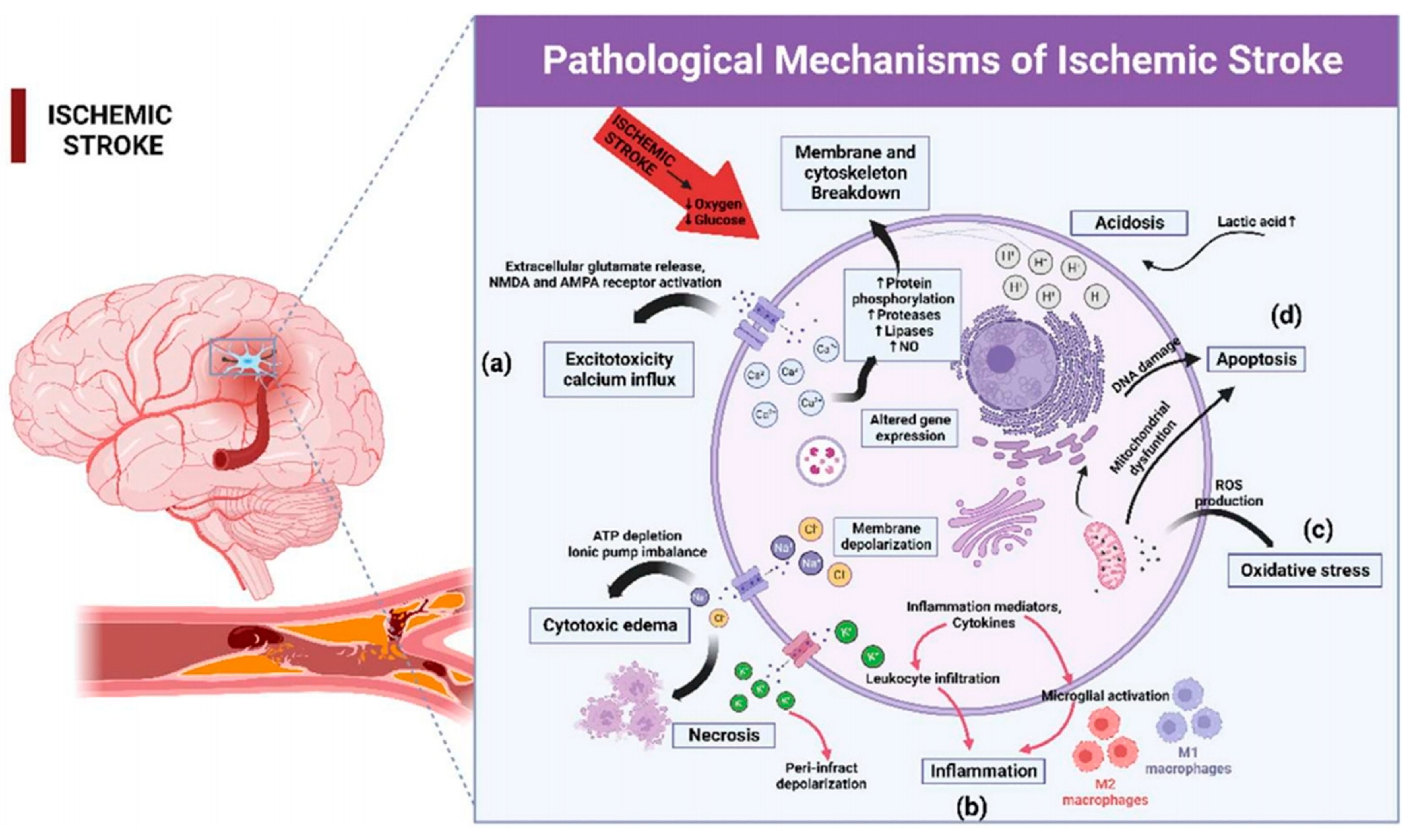
Figure 2.
(a) Preparation of rod-like nanoparticles by solvent evaporation and film stretching methods; (b) Rod-like nanoparticles (Pic@AR5) were specifically phagocytosed by neutrophils, followed by the release of pisaclitaxel which blocked the Syk pathway and reduced the expression of β2 integrins, thus preventing the adhesion of most neutrophils to endothelial cells. Meanwhile, a small number of Pic@AR5-carried neutrophils enter the BBB and release piceatannol in the ischemic zone, inhibiting the Syk pathway in microglia and attenuating microglia-mediated neuroinflammation [106]. Copyright 2023, Wiley-VCH.

Figure 3.
Schematic illustration of mitochondria-targeted cerium nanoenzymes for the therapy of ISs. Neuroprotective effects are achieved by attenuating oxidative stress and modulating microglia phenotype [162]. Copyright 2024, American Chemical Society.
Figure 3.
Schematic illustration of mitochondria-targeted cerium nanoenzymes for the therapy of ISs. Neuroprotective effects are achieved by attenuating oxidative stress and modulating microglia phenotype [162]. Copyright 2024, American Chemical Society.
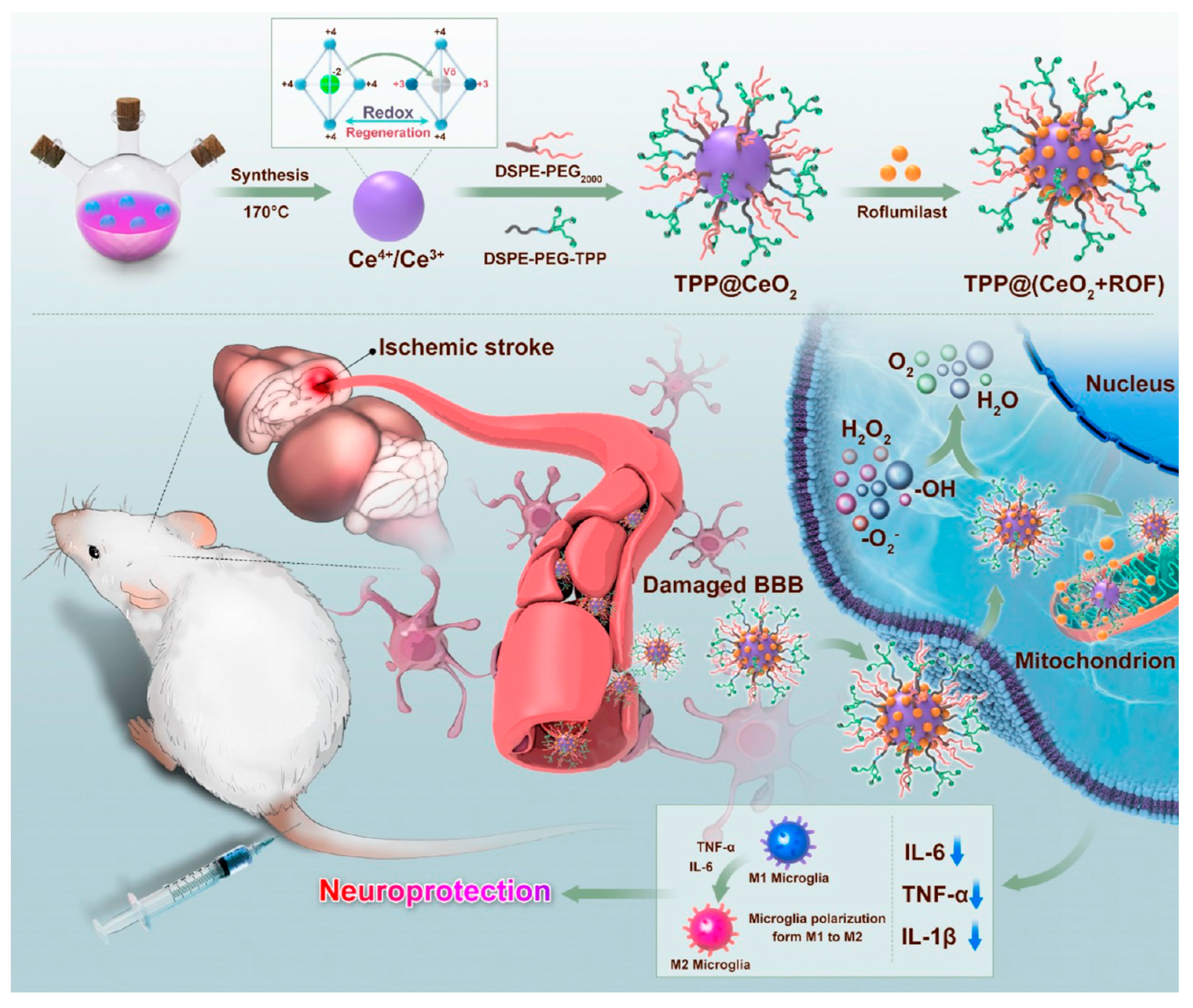
Figure 4.
Schematic illustration of the functionalization of liposomes [166]. Copyright 2019, Elsevier.
Figure 4.
Schematic illustration of the functionalization of liposomes [166]. Copyright 2019, Elsevier.
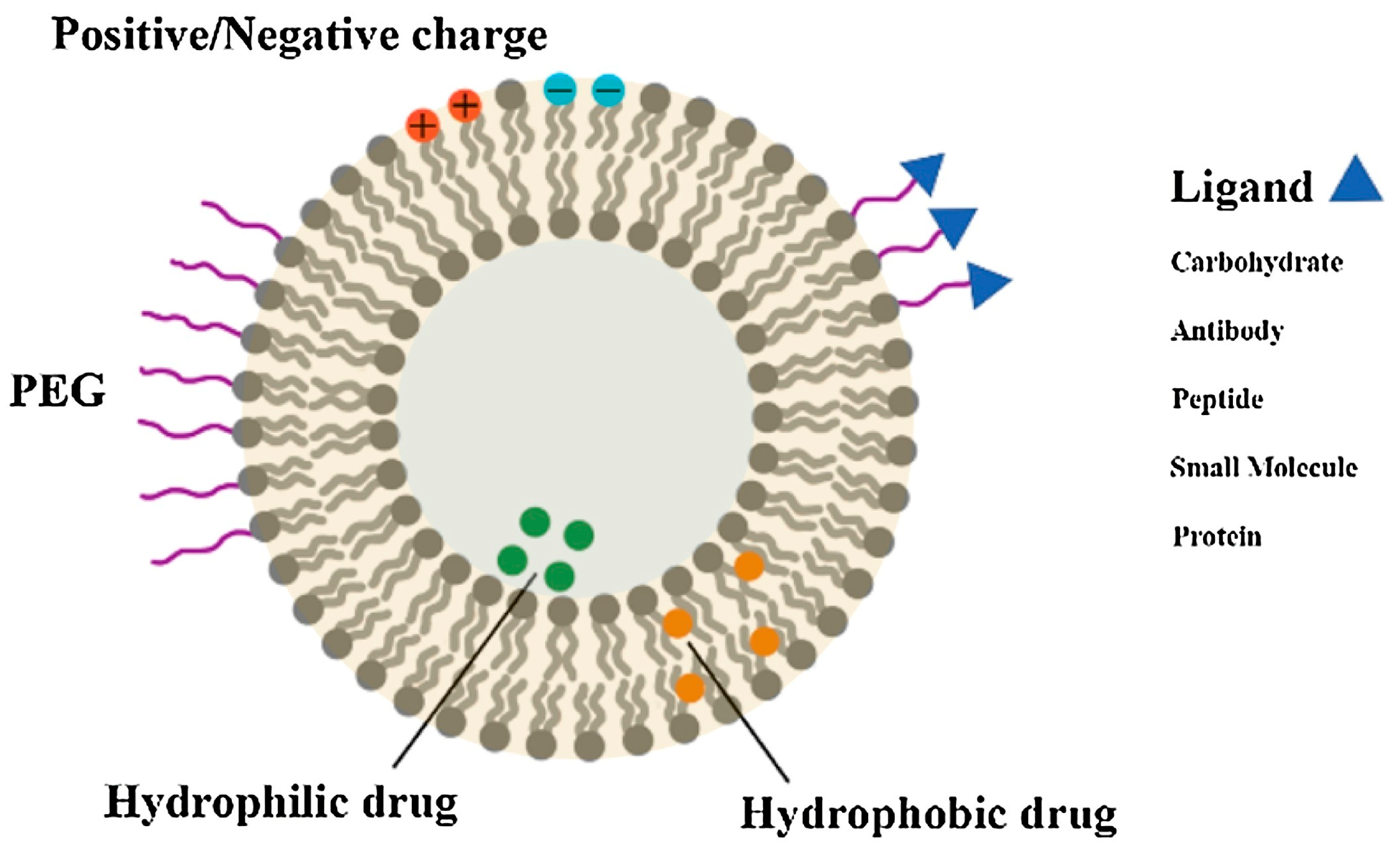
Figure 5.
Diverse cell membrane-coated nanoparticles [188]. Copyright 2018, WILEY-VCH.
Figure 5.
Diverse cell membrane-coated nanoparticles [188]. Copyright 2018, WILEY-VCH.
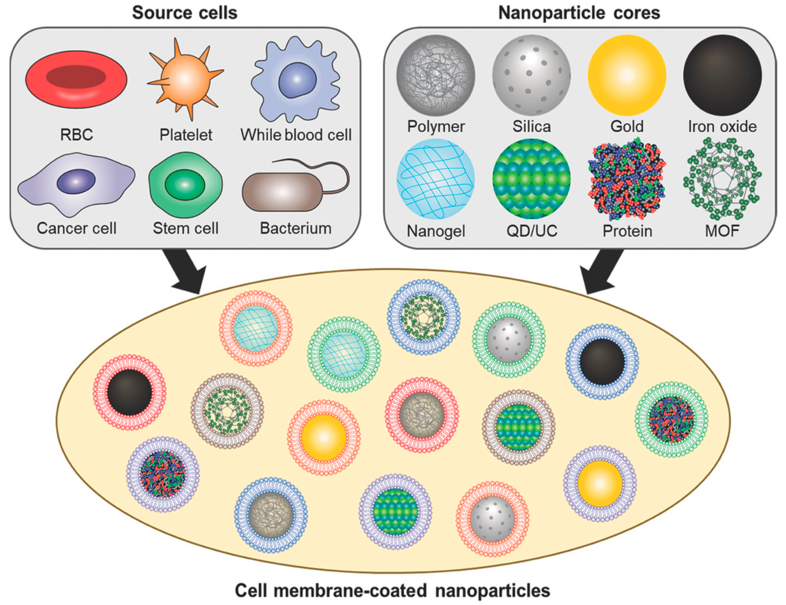
Figure 6.
(a) Flowchart of MNET preparation; (b) Schematic illustration of oxygen sponge function and free radical scavenging of MNET in vivo. MNET crosses the BBB and accumulates at the site of infarction, with effects of releasing oxygen to relieve hypoxia and storing oxygen in a pro-oxidant environment after thrombolysis [209]. Copyright 2020, American Chemical Society.
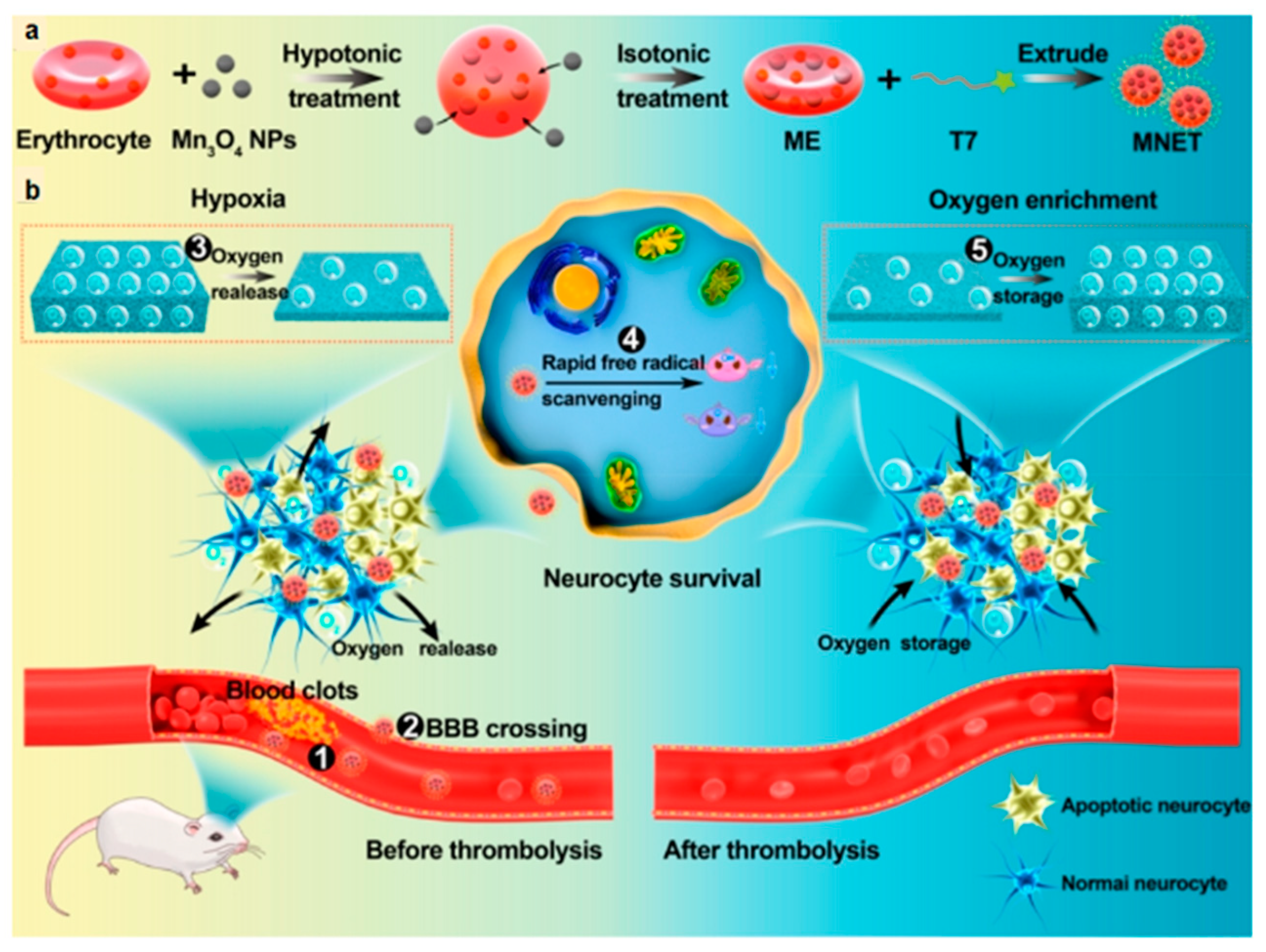
Figure 7.
(a) Flowchart of miRNA/TPHI@PM preparation and how it functions. Delivery of miRNA through platelet membrane-coated nanoparticles to sites of brain lesions aims to reduce neuronal apoptosis and regulate microglial cell phenotype [217]. Copyright 2022, Elsevier Ltd. (b) The structure of PAMNs [219]. Copyright 2021, American Chemical Society. (c) Schematic illustration of the therapeutic mechanism of PAMNs. Due to the endowed platelet membrane function and external magnetic targeting, PAMNs rapidly reaches the ischemic region of stroke and achieves rapid targeted delivery of l-arginine. Meanwhile, NO generation in situ allows the induction of vasodilation and the reduction of PLT aggregation [219]. Copyright 2021, American Chemical Society.
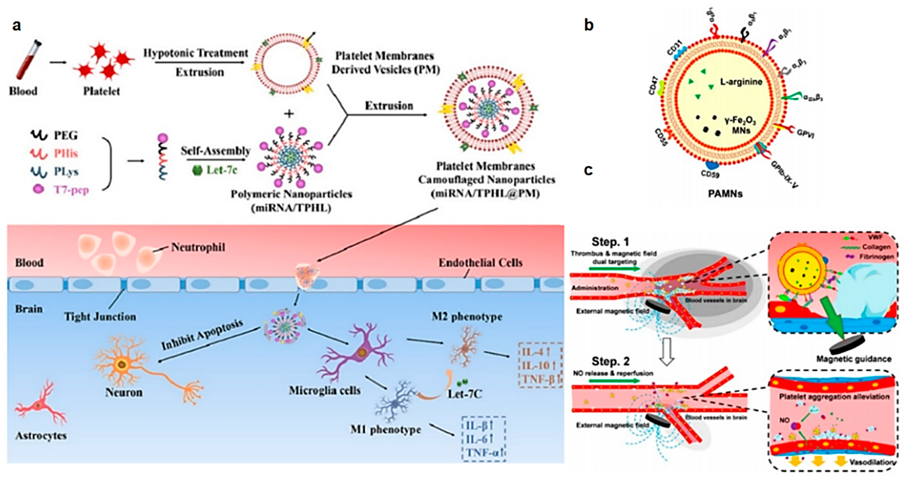
Figure 8.
(a) Preparation of MPBzyme@NCM; (b) The effect of MPBzyme@NCM in vivo. Long-term therapeutic effects of stroke were exerted through the ability to reduce neutrophil recruitment, regulate microglia polarization from M1 to M2, reduce neuronal apoptosis, and increase neural stem cell proliferation [234]. Copyright 2021, American Chemical Society.

Figure 9.
(a) Preparation of Ma@(MnO2 + FTY); (b) Schematic illustration of the effect of Ma@(MnO2 + FTY) [52]. Copyright 2021, Wiley-VCH.
Figure 9.
(a) Preparation of Ma@(MnO2 + FTY); (b) Schematic illustration of the effect of Ma@(MnO2 + FTY) [52]. Copyright 2021, Wiley-VCH.
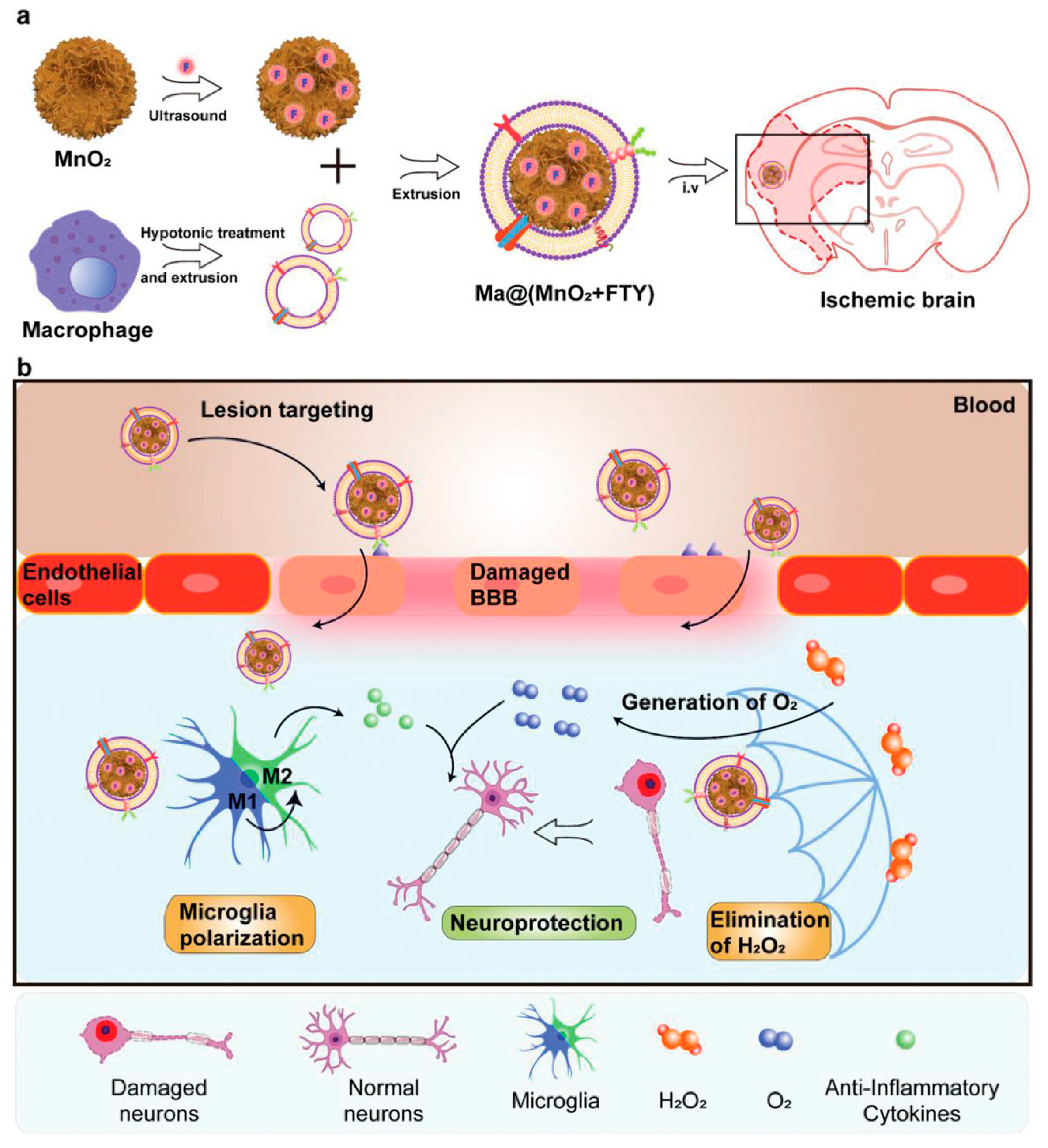
Figure 10.
Preparation of MPP/SCB and its effect on ISs [246]. Copyright 2021, American Chemical Society.
Figure 10.
Preparation of MPP/SCB and its effect on ISs [246]. Copyright 2021, American Chemical Society.
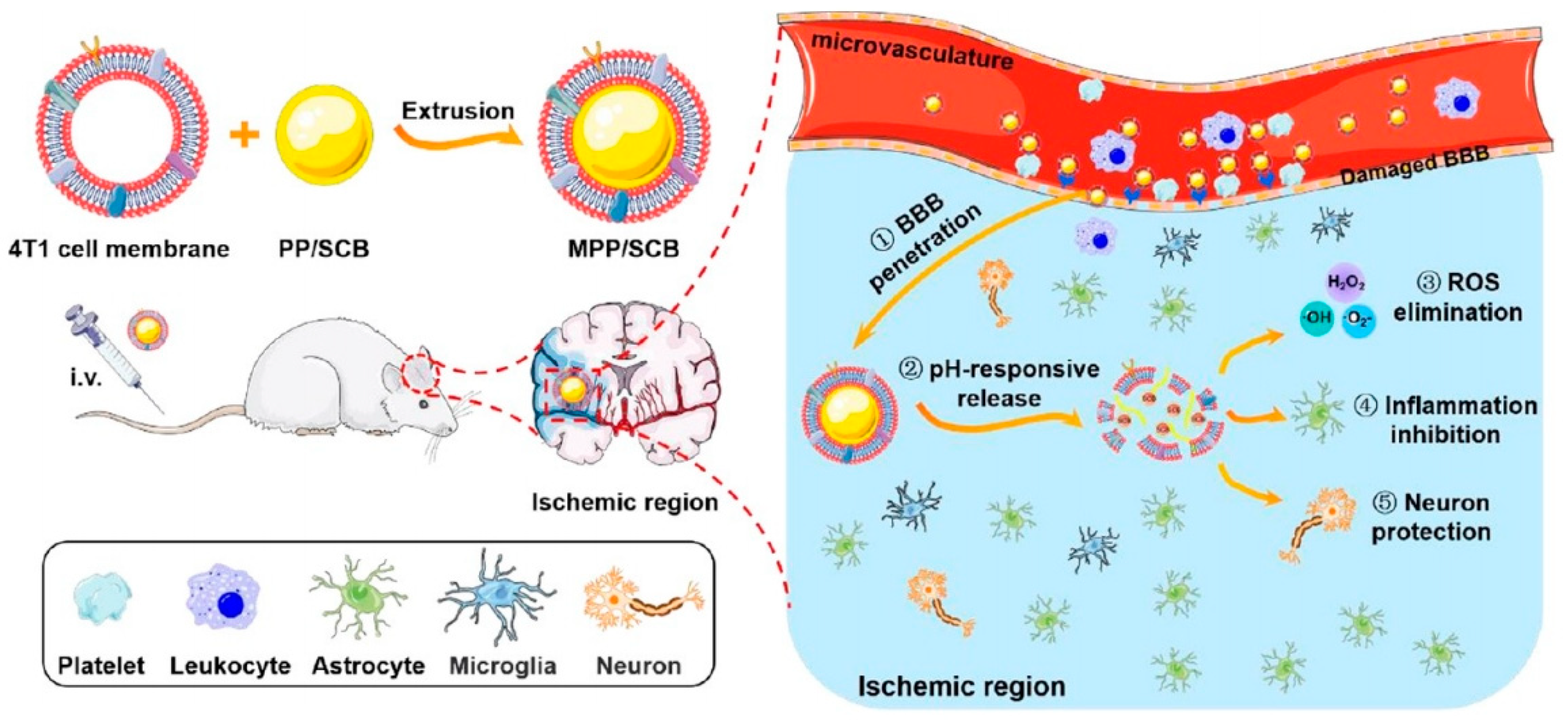

Table 1.
A summary of the key features of various cell membranes.
| Cell Membrane Source | Key Features | Function | Reference |
|---|---|---|---|
| Red blood cell (RBC) | Immune evasion Easy to extract Surface expression of CD47 | Prolonged circulation | [192] |
| Platelet | Specific targeting of damaged tissue Adherence to inflammatory neutrophil Surface expression of CD47, CD55 and CD59 | Injury sites targeting | [193] |
| White blood cell (WBC) | Specific targeting of inflammatory tissue Endothelial adherence Adhesion at tumor sites Penetration of the BBB | Tumor and inflammatory site targeting | [194,195,196] |
| Cancer cell | Immune evasion Homologous targeting Anti-tumor ability | Tumor targeting | [197] |
| Stem cell | Immune evasion Tumor-specific properties Homing ability | Tumor targeting Inflammatory damage targeting | [198,199] |
| Bacteria | Promoting adaptive immunity | Tumor anaerobic targeting | [200,201,202] |
Disclaimer/Publisher’s Note: The statements, opinions and data contained in all publications are solely those of the individual author(s) and contributor(s) and not of MDPI and/or the editor(s). MDPI and/or the editor(s) disclaim responsibility for any injury to people or property resulting from any ideas, methods, instructions or products referred to in the content. |
© 2024 by the authors. Licensee MDPI, Basel, Switzerland. This article is an open access article distributed under the terms and conditions of the Creative Commons Attribution (CC BY) license (https://creativecommons.org/licenses/by/4.0/).
Share and Cite
MDPI and ACS Style
Zhang, J.; Chen, Z.; Chen, Q. Advanced Nano-Drug Delivery Systems in the Treatment of Ischemic Stroke. Molecules 2024, 29, 1848. https://doi.org/10.3390/molecules29081848
AMA Style
Zhang J, Chen Z, Chen Q. Advanced Nano-Drug Delivery Systems in the Treatment of Ischemic Stroke. Molecules. 2024; 29(8):1848. https://doi.org/10.3390/molecules29081848
Chicago/Turabian StyleZhang, Jiajie, Zhong Chen, and Qi Chen. 2024. "Advanced Nano-Drug Delivery Systems in the Treatment of Ischemic Stroke" Molecules 29, no. 8: 1848. https://doi.org/10.3390/molecules29081848